Embed Size (px)
Citation preview
HUMAN COMPUTER
INTERACTION VIA
BRAINWAVES FOR DISABLED
PEOPLE LOURENÇO BARBOSA DE CASTRO
Faculty of Engineering – University of Porto
September 2013

HUMAN COMPUTER INTERACTION VIA BRAINWAVES FOR DISABLED PEOPLE
Thesis submitted in partial fulfillment of the requirements for the degree of Master of Sci-
ence in Biomedical Engineering.
Faculty of Engineering – University of Porto
DEVELOPED BY
Lourenço Barbosa de Castro
B a c h e l o r o f Sc i e n c e i n B i o l o g i ca l Sc i e n c e s
C at h o l i c U n i ve rs i t y o f Po r t u ga l
ORIENTATION
João Manuel R.S. Tavares
A s s o c i a te Pro fe s s o r
Fa c u l t y o f E n g i n e e r i n g – U n i ve rs i t y o f Po r to , D e p a r t m e nt o f M e c h a n i ca l E n g i n e e r i n g

ACKNOWLEDGEMENTS
To my family and friends, for all the support given throughout this project’s duration.
To Fraunhofer Portugal, Prof. João Manuel Tavares and Prof. Ana Maria Mendonça for giv-
ing me the opportunity to be a part of this project.
Additional acknowledgement to Fraunhofer Portugal for providing all the required re-
sources and funding.
To Luis Rosado, for all the help, support and availability during this last year.
ABSTRACT
The idea of interacting with computers using only the human brain quickly emerged after
the discovery of electroencephalography in 1929. Although plenty of research has been
done related to this subject, and several consumer based products are already being sold,
little importance has been given to mobile devices, which are rapidly dominating the per-
sonal computing scene.
With this in mind, an Android application was developed in this project, attempting to
facilitate the usage of mobile computers for disabled people. Using exclusively electric
activity acquired from a brain computer interface device, different types of interactions
were experimented, expecting to improve the mentioned people's autonomy and quality
of life. The users of the application were able to control several features just by winking,
which achieved over 87% success rate after a short calibration session. Additionally, the
application was able to discriminate the users' state of mind when subjected to different
conditions. Four situations were tested, all of them exhibiting unique neural oscillations
patterns, making the application accurate in understanding how the user was feeling.
To conclude, the main contribution of this project was creating a type of interaction that
was not previously available, and, although more limited, presented a higher success rate
than the chosen device's native application.

RESUMO
A ideia de estabelecer interação com computadores recorrendo apenas ao cérebro hu-
mano surgiu rapidamente após a descoberta da eletroencefalografia, em 1929. Apesar de
ter sido realizada bastante investigação sobre este tema, e de diversos produtos estarem
comercialmente disponíveis, pouca importância tem sido dada à sua adaptação a dispo-
sitivos móveis, dispositivos esses que estão rapidamente a dominar o mercado dos com-
putadores pessoais.
Com isto em mente, uma aplicação Android foi desenvolvida neste projeto, com o intuito
de facilitar a utilização de computadores móveis a pessoas incapacitadas. Utilizando ape-
nas sinais elétricos obtidos por um dispositivo de comunicação entre o cérebro e o com-
putador, diferentes tipos de interações foram testadas, na tentativa de melhorar a auto-
nomia e qualidade de vida das pessoas anteriormente referidas. Os utilizadores desta apli-
cação podem controlar várias funcionalidades apenas pestanejando, o que, após uma
curta sessão de calibração, atingiu uma taxa de sucesso superior a 87%. Adicionalmente,
a aplicação desenvolvida foi capaz de diferenciar o estado de espírito dos utilizadores,
quando sujeitos a diversas condições específicas. Neste caso, quatro situações foram tes-
tadas, em que todas elas apresentaram padrões de atividade cerebral distintos, tornando
esta aplicação eficaz a perceber como o seu utilizador se sente.
Concluindo, a principal contribuição deste projeto foi criar um tipo de comunicação que
não era possível anteriormente, entre um dispositivo de interação cérebro-computador e
um tablet Android. Apesar de mais limitado do que aplicação nativa desse mesmo dispo-
sitivo, apresentou uma maior taxa de sucesso nas funcionalidades implementadas.
II LOURENÇO BARBOSA DE CASTRO – 2013
INDEX
1. INTRODUCTION 1
1.1. MOTIVATION 2
1.2. OBJECTIVES 3
1.3. STRUCTURE 4
1.4. CONTRIBUTIONS 6
2. BRAIN-COMPUTER INTERFACES 7
2.1. INTRODUCTION 8
2.1.1.RESEARCH 8
2.1.2 INDUSTRY 10
2.2. BRAIN 12
2.2.1. LOBES 12
2.2.1.1. FRONTAL LOBE 12
2.2.1.2. PARIETAL LOBE 13
2.2.1.3. OCCIPITAL LOBE 13
2.2.1.4. TEMPORAL LOBE 13
2.2.2. NEURONS 14
2.3. ELECTROENCEPHALOGRAPHY 15
2.3.1. INTRODUCTION 15
2.3.2. ELECTRODES 15
2.3.3. POSITIONS 16
2.3.4. NOISE 17
2.3.5. NEURAL OSCILLATIONS 18
2.3.5.1. DELTA 18
2.3.5.2. THETA 18
2.3.5.3. ALPHA 19
2.3.5.4. MU 19
2.3.5.5. BETA 20
2.4. ELECTROMYOGRAPHY 20
2.5. SIGNALS DISCRIMINATION 21
2.5.1. EVENT-RELATED POTENTIALS 22
2.5.2. P300 POTENTIAL 22
2.5.2.1. METHODOLOGY 22
2.5.2.2. IMPLEMENTATION 23
2.5.3. COGNITION/EXPRESSIONS 24
HUMAN COMPUTER INTERACTION VIA BRAINWAVES FOR DISABLED PEOPLE III
2.6. TYPES OF BCIS 25
2.6.1. INVASIVE 25
2.6.2. NON-INVASIVE 26
2.7. SUMMARY 27
3. EMOTIV EPOC 29
3.1. INTRODUCTION 30
3.2. HARDWARE 31
3.3. TYPES OF INTERACTION 32
3.3.1. EXPRESSIV SUITE 32
3.3.2. COGNITIV SUITE 33
3.3.3. AFFECTIV SUITE 33
3.4. SUMMARY 33
4. DEVELOPED APPLICATION 35
4.1. TARGET USERS 36
4.2. ANDROID OPERATING SYSTEM 37
4.2.1. OPERATING SYSTEM 37
4.2.2. NEXUS 7 TABLET DEVICE 37
4.3. FEATURES 38
4.3.1. TIME DOMAIN FEATURES 38
4.3.2. FREQUENCY DOMAIN FEATURES 38
4.3.3. SPATIAL FEATURES 39
4.4. SOFTWARE 39
4.4.1. DATA ACQUISITION 39
4.4.2. EVENTS DETECTION 40
4.4.3. FUNCTIONALITIES 40
4.4.3.1 EYES NAVIGATION 41
4.4.3.2. KEYBOARD 41
4.4.3.3. CONTACTS 42
4.4.3.4. CALCULATION 43
4.4.3.5. BRAINWAVES 44
4.4.3.6. CONFIGURATION 45
4.5. APPLICABILITY 46
4.5.1. COMPUTER CONTROL 46
4.5.2. COGNITIVE ORIENTATION 47
4.5.3. BRAINWAVES MONITORING 48
4.5.3.1. ALERTS 48
4.5.3.2. FOCUS 48

IV LOURENÇO BARBOSA DE CASTRO – 2013
4.5.3.3. ENGAGEMENT 48
4.6. SUMMARY 49
5. RESULTS AND DISCUSSION 51
5.1. WINKS 52
5.2. BRAINWAVES 54
5.3. SUMMARY 60
6. CONCLUSION AND FUTURE IMPROVEMENTS 63
6.1. CONCLUSION 64
6.2. FUTURE IMPROVEMENTS 64
BIBLIOGRAPHY 67
HUMAN COMPUTER INTERACTION VIA BRAINWAVES FOR DISABLED PEOPLE V
TABLES
TABLE 1 32 EMOTIV EPOC'S HARDWARE SPECIFICATIONS (EMOTIV, EMOTIV SYSTEMS WEBSITE 2013) TABLE 4 54 ELECTRIC ACTIVITY MEASURED WHILE THE SUBJECT WAS WINKING THEIR LEFT EYE. TABLE 5 55 RESULTS FOR THE RIGHT WINKING TRIALS, SHOWING THE RATE OF SUCCESS FOR EACH CALIBRATIONS AMOUNT TABLE 6 55 RESULTS FOR THE LEFT WINKING TRIALS, SHOWING THE RATE OF SUCCESS FOR EACH CALIBRATIONS AMOUNT TABLE 7 63 RESULTS OF THE BRAINWAVES TRIALS, COMPARING THE TOP ROW WITH THE LEFT COLUMN STATE. BOTH HIGH AND LOW PEAKS ARE DENOTED, AS WELL AS OTHER INTERESTING FEATURES.
FIGURES
FIGURE 1 14 REPRESENTATION OF A NEURON. 1: DENDRITES; 2: CELL BODY (SOMA); 3: NUCLEUS; 4: AXON (FORESMAN 2009).
FIGURE 2 16 EXAMPLE OF THE INTERNATIONAL 10-20 SYSTEM, DISPLAYING THE LAYOUT OF THE ELECTRODES ON A SUBJECT'S HEAD (FIELDTRIP 2013).
FIGURE 3 18 REPRESENTATION OF 1 SECOND OF EEG ACTIVITY FROM THE OZ POSITION, FILTERED TO DISPLAY ONLY DELTA WAVES (GAMBOA 2005).
FIGURE 4 18 REPRESENTATION OF 1 SECOND OF EEG ACTIVITY FROM THE OZ POSITION, FILTERED TO DISPLAY ONLY THETA WAVES (GAMBOA 2005).
FIGURE 5 19 REPRESENTATION OF 1 SECOND OF EEG ACTIVITY FROM THE OZ POSITION, FILTERED TO DISPLAY ONLY ALPHA WAVES (GAMBOA 2005).
FIGURE 6 20 REPRESENTATION OF 1 SECOND OF EEG ACTIVITY FROM THE OZ POSITION, FILTERED TO DISPLAY ONLY BETA WAVES (GAMBOA 2005).
FIGURE 7 25 REPRESENTATION OF THE BRAINGATE TECHNOLOGY, A TYPE OF INVASIVE BCI (BRAINGATE 2009).
FIGURE 8 26 EMOTIV EPOC, A TYPE OF NON-INVASIVE BCI EQUIPMENT (EMOTIV, EMOTIV SYSTEMS WEBSITE 2013).

VI LOURENÇO BARBOSA DE CASTRO – 2013
FIGURE 9 32 EMOTIV EPOC'S ELECTRODES POSITIONING, ACCORDING TO THE INTERNATIONAL 10-20 SYSTEM (EMOTIV, EMOTIV SYSTEMS WEBSITE 2013).
FIGURE 10 44 KEYBOARD SECTION OF THE APPLICATION, SHOWING THE SELECTION OF THE LETTER L, WHILE THE WORD "HELLO" WAS PREVIOUSLY TYPED.
FIGURE 11 45 CONTACTS SECTION, EXEMPLIFYING THE SELECTION OF A CONTACT. IT IS ALSO DISPLAYED THE CURRENT POSITION OF THE GYROSCOPE.
FIGURE 12 46 CALCULATION SECTION, SHOWING A CORRECT SELECTION DURING THE SECOND LEVEL OF DIFFICULTY.
FIGURE 13 47 DETAILED OSCILLATION INFORMATION, DISPLAYED AFTER CLICKING ON A SENSOR OF INTEREST.
FIGURE 14 48 CONFIGURATION SECTION OF THE APPLICATION, DISPLAYING THE VALUES OF TWO SENSORS IN REAL-TIME.
FIGURE 15 57 COMPARING THE ACTIVITY OF A NORMAL AND RELAXED STATE WITH A NORMAL AND FOCUSED STATE.
FIGURE 16 58 COMPARING THE ACTIVITY OF A NORMAL AND RELAXED STATE WITH AN EYES CLOSED WITH SOFT AND RELAXING MUSIC STATE.
FIGURE 17 59 COMPARING THE ACTIVITY OF A NORMAL AND RELAXED STATE WITH AN EYES CLOSED WITH LOUD AND FAST MUSIC STATE.
FIGURE 18 59 COMPARING THE ACTIVITY OF A NORMAL AND FOCUSED STATE WITH A CLOSED EYES STATE WITH SOFT AND RELAXING MUSIC PLAYING.
FIGURE 19 60 COMPARING THE ACTIVITY OF A NORMAL AND FOCUSED STATE WITH A CLOSED EYES STATE WITH LOUD AND FAST MUSIC PLAYING.
FIGURE 20 61 COMPARING THE ACTIVITY OF TWO CLOSED EYES STATES, ONE WITH SOFT RELAXING MUSIC, OTHER WITH FAST LOUD MUSIC PLAYING.

HUMAN COMPUTER INTERACTION VIA BRAINWAVES FOR DISABLED PEOPLE VII
ACRONYMS
Analog-to-digital ADC
Attention deficit hyperactivity disorder ADHD
Amyotrophic lateral sclerosis ALS
Application programming interface API
Brain-computer interface BCI
Common mode sense CMS
Driven right leg DRL
Electroencephalography EEG
Electromyography EMG
Event-related potential ERP
Functional magnetic resonance imaging fMRI
Least significant bit LSB
Magnetoencephalography MEG
On-the-go OTG
Steady state visually evoked potential SSVEP


1
CHAPTER 1
INTRODUCTION In this first chapter, the subject of the thesis is presented, explaining its motivation and
objectives, ending with a comparison between the developed work and the current state of
the art regarding Brain-Computer Interfaces.
1.1. MOTIVATION 2
1.2. OBJECTIVES 3
1.3. STRUCTURE 4
1.4. CONTRIBUTIONS 6

CHAPTER 1 INTRODUCTION
2 LOURENÇO BARBOSA DE CASTRO – 2013
MOTIVATION 1.1 W HY C HO O SIN G T H IS SU B J E C T A N D W HAT M A K E S IT IM P O RTA N T
The main goal of this thesis is to study new forms of interaction between humans and com-
puters, more specifically using brain-computer interfaces.
The world we live in is in constant change. Often we reinvent the ways we see ourselves,
mysteries of yesterday are commonly solved, and we keep pushing our limitations beyond
the imaginable. However, the less fortunate, less capable, less healthy are seldom thought
of, frequently being left behind. We are making technology accelerate to a point it is sur-
passing most of these people, while only a select few are being carried.
Despite the mobile computing market growing stronger each day, creating new forms of
communication, of learning or even living, the ones who are unable or inept to perform
simple movements cannot keep up with the course our world is taking. Several medical
conditions or other health related accidents can lead to a person becoming incarcerated in
their own body, shutting down almost completely their interaction with the environment;
brain diseases can destroy most of one's motor functions while leaving their consciousness
entirely unaffected; aging related events can compromise skills that we assume as granted,
like learning, mental coordination or memory, whereas excitement and willingness remain
intact.
Technology should not be limited to a fraction of mankind; it should not close some gaps
at the expense of broadening others. The future should be for everyone. Brain-computer
interfaces (BCI) are helping to instill this mindset, focusing on who you are and what you
want, rather than what you currently can do.
Breakthroughs like brain controlled wheelchairs or prosthetic arms, successful brain-to-
brain communication attempts or possible mind reading techniques inspired this thesis,
with hopes of helping to develop even more elaborate and meaningful ideas.
Although this is still a fairly futuristic technology, frequently associated with skepticism, its
first steps started during the 1960s, making it a reasonably ancient science on our ex-
tremely fast paced present. Considering that during brain-computer interfaces’ conception

CHAPTER 1 INTRODUCTION
HUMAN COMPUTER INTERACTION VIA BRAINWAVES FOR DISABLED PEOPLE 3
not even general-purpose computing was well established, it is understandable that its ad-
aptation to mobile computers is not as intrinsic as one would think. However, devices like
smartphones or tablet computers are taking over our daily lives, being expected to domi-
nate the digital market in a near future.
Mobile technology increases computing accessibility, interactivity, and affordability, making
it a perfect ally for brain-computer interaction. Reduced computer sizes will help us take
this technology anywhere we go, increasing its potential exponentially. It will also, conse-
quentially, increase the range of users who can benefit from it, which will ideally include all
the types of people mentioned earlier, bringing them more autonomy, quality of life and
even happiness.
In conclusion, brain-computer interfaces might shape the world the same way the inven-
tion of computers or the Internet did: closing gaps, reducing life-limiting issues and revolu-
tionizing, once again, our views about ourselves.
OBJECTIVES 1.2 W HAT T HI S THE S IS A IM S TO A C HIE V E
Interaction with mobile devices
Given the importance of mobile computing and the noticeable lack of its integration with
brain-computer interfaces, the first objective of this project was to proof the possibility of
inter-connecting both technologies using currently available tools.
Accessibility features
Afterwards, the next step was to implement features which would improve the accessibility
and usability of portable computers when connected to brain-computer interfaces. For this
purpose, it was necessary to identify some of the main obstacles that disabilities or medical

CHAPTER 1 INTRODUCTION
4 LOURENÇO BARBOSA DE CASTRO – 2013
conditions can inflict, in order to present efficient solutions based on brain-computer inter-
action.
Trials and future prospects
Lastly, since this field is still heavily underdeveloped considering its already demonstrated
potential, this project aimed to study the future possibilities of interacting with mobile
computers employing exclusively bioelectric signals. Also, by developing and demonstrat-
ing some basic brain-computer interface functionalities, this project hopes to help opening
doors to more elaborate and life-changing creations.
STRUCTURE 1.3 HOW T HE T HE S I S I S O R G A N IZ E D
Chapter 1
This thesis is initiated with an introductory chapter, in which it is presented the motivation
behind the subject’s choice, as well as its objectives and contributions to the state of the
art. This chapter was created as succinct as possible, leaving most theoretical concepts to
be described on the subsequent chapters.
Chapter 2 Brain-Computer Interfaces
In chapter 2, the topic of brain-computer interfaces is introduced. The initial experiments
regarding this type of interaction are described, mentioning some of the most prominent
people involved. It is also given emphasis to the commercially focused devices, both avail-
able and in development. Then, the required precursor technologies and concepts are de-
scribed, including brain regions and corresponding functions, electrodes positioning and
neurophysiological signals and their adequate detection techniques.
CHAPTER 1 INTRODUCTION
HUMAN COMPUTER INTERACTION VIA BRAINWAVES FOR DISABLED PEOPLE 5
Chapter 3 Emotiv EPOC
Afterwards, the specific device used in this work is presented, in contrast with other devices
and projects. The main objective of this chapter is to situate the headset used in this project
along with the current state-of-the-art brain-computer interfaces technologies, describing
all of its features, both physical and digital, and arguing why it was selected among other
available products.
Chapter 4 Developed Application
The next chapter is dedicated to the developed application. Initially, the usability factors
are addressed, explaining for whom the application was designed and the types of disabil-
ities and diseases that should benefit from it. The different types of useful signal features
are introduced, along with their detection and classification procedures. Then, both the
Android platform and the device used in this project are briefly introduced, explaining their
specifications and why they were chosen. Lastly, the software characteristics are discussed,
explaining the data acquisition methods and how the electrophysiological signals were
used to control the application’s functions, finishing with practical examples of how this
technology could be used.
Chapter 5 Results and Discussion
Following is the results and discussion chapter, where firstly it is explained how the tech-
nology was tested and which results were obtained. The validity and accuracy of the results
are hereby discussed
Chapter 6 Future Improvements and Conclusions
The last chapter explains the conclusions of this project, and makes use of such assertions
to comprehensively prospect possible future improvements to the developed application
and, on a broader extent, to the brain-computer interfaces field.

CHAPTER 1 INTRODUCTION
6 LOURENÇO BARBOSA DE CASTRO – 2013
CONTRIBUTIONS 1.4 W HE R E D O ES T HI S P ROJ E C T S TA N D C O M PA R E D TO THE STATE O F THE A RT
Adaptation to mobile devices
The main contribution of this thesis is the adaptation of functional brain-computer inter-
facing to mobile systems, resulting in an interaction between an electroencephalography
(EEG) headset and an Android tablet computer. By contributing as a proof-of-concept of
brain-computer interface technology applied to mobile computers, this project hopes to
motivate the development of similar and more complex implementations using alternative
devices. Additionally, by experimenting with basic forms of BCI interaction, it is expected
that more elaborate and useful techniques can surface in this greatly promising field.
Rehabilitation features
Although some available devices already have tools to connect to mobile computers, they
are all considerably more limited than the equipment used in this project, making the in-
teraction consequentially less interesting. Even though very efficient and well-designed
software is available for said devices, the implemented features do not take rehabilitation
in consideration, leaving much to be desired. This project is directly related to that subject,
attempting to build functionalities that could help disabled people regain their autonomy.
Brain-Computer Interfaces efficiency
Furthermore, a major electronics manufacturer is testing the possibilities of using brain-
computer interfaces with their own mobile devices, which should vastly increase the public
interest in this subject. However, their study is still on very early stages of trials, and high
grade EEG headsets are being used. On the other hand, this project’s implementation is
already functional, using a cheaper device already available on the market for a few years.
7
CHAPTER 2
BRAIN-COMPUTER INTERFACES This chapter introduces the discovery of electroencephalography, which made interacting
with computers using brainwaves possible. Next, research and commercial approaches are
described, mentioning important scientists and companies that have been increasingly
developing the BCI field. The human brain is briefly described, with the most relevant
structures and physiological aspects being mentioned. Also, both EEG and
electromyography are specifically explained, as well as some other neurophysiological
signals and their interpretations.
2.1. INTRODUCTION 8
2.2. BRAIN 12
2.3. ELECTROENCEPHALOGRAPHY 15
2.4. ELECTROMYOGRAPHY 20
2.5. SIGNALS DISCRIMINATION 21
2.6. TYPES OF BCIS 25
2.7. SUMMARY 27

CHAPTER 2 BRAIN-COMPUTER INTERFACES
8 LOURENÇO BARBOSA DE CASTRO – 2013
INTRODUCTION 2.1 THE B EG IN N IN G O F B R A I N - CO M P U TE R IN TE R FA C E S . IM P O R TA N T P EO P L E A N D CO M PA N IE S .
After the discovery of electroencephalography in 1929, by Hans Berger (Berger 1929), the
possibility of using brainwaves to communicate or even interact with the environment be-
came a reality. The so called brain-computer interfaces are best described as communica-
tion systems which do not depend on the brain’s regular pathways, such as peripheral
nerves or muscles (Wolpaw, et al. 2000). This creation led to diverse research milestones,
as well as several, well established BCI companies. Both of these fields are described in this
chapter.
RESEARCH 2.1.1
The earliest publications directly related to brain-computer interfaces were developed by
José M. R. Delgado, during the decade of 1960. His work consisted in stimulating and re-
cording the electrical activity of the brain in unrestrained monkeys and chimpanzees, using
bilaterally implanted electrodes assemblies to read the electrophysiological signals and a
radio frequency system to induce stimulation. This allowed for a better understanding of
cerebral behavior and spontaneous activities while the brain was not suffering any kind of
disturbance to its typical functioning. The motivation behind this study was to better un-
derstand brain dysfunctions associated with behavioral abnormalities, which were diag-
nosed using restrictive methods that would not produce sufficiently realistic results
(Delgado, et al. 1968).
In 1980, focusing on the neurophysiology of paralyzed patients, a study elaborated by Ed-
ward M. Schmidt tried to implement long-term connections to the central nervous system,
using microelectrodes. This could allow for direct stimulation of paralyzed limbs from the
motor cortex, ideally restoring movement. Although it was concluded that additional im-
provements would be required for feasible implementations, the obtained results indicated
information transfer rates only moderately slower than in the regular physiological pathway
(Schmidt 1980).
CHAPTER 2 BRAIN-COMPUTER INTERFACES
HUMAN COMPUTER INTERACTION VIA BRAINWAVES FOR DISABLED PEOPLE 9
The first published demonstration of a brain-controlled device came in 1999, developed by
a group led by Miguel A. L. Nicolelis. Initially, brain activity of a group of mice was recorded
while they pressed a lever to position a robotic arm that allowed them to obtain water.
Next, this activity pattern was associated with the robotic arm control, triggering it when
the brain signals were recognized. Finally, a subgroup of the studied mice started to rou-
tinely and exclusively use this brain activity to reach their goal, and, as the training pro-
gressed, resorting to the initial lever diminished or stopped. This study’s findings raised the
possibility for paralyzed patients to control external devices or even their own muscles us-
ing only electrophysiological patterns (Chapin, et al. 1999).
During 2008, the first brain-computer interfaces mainstream report was presented by Co-
lumbia Broadcasting System (CBS), on an episode of their news program 60 Minutes. Sev-
eral interviews were conducted to people whose lives were changed by this technology. A
man diagnosed with amyotrophic lateral sclerosis (ALS), who lost all types of movement
control except for his eyes, managed to start communicating again. This was possible using
a non-invasive system developed by Jonathan Wolpaw, by selecting characters on a screen
using only EEG information. Another presented major development was the Braingate pro-
ject, created by John Donoghue’s team, consisting in implanting a grid of electrodes on the
patient’s motor cortex. This allowed for a stroke victim, who lost control of her body, to
move a computer cursor using brain activity readings, which was later adapted to the steer-
ing of an electric wheelchair (CBSNews 2008).
Recently, the already mentioned Braingate project presented another breakthrough: again
using a microelectrode array implanted in the motor cortex, two patients were able to con-
trol a robotic arm with three-dimensional reach and grasp functions. Such implementation
allowed one of them to autonomously drink coffee from a bottle, task which would be im-
possible otherwise. This was achieved by recording neural activity while the subject was
imagining the referred movements, and afterwards triggering the robotic arm control when
the same patterns were detected. John P. Donoghue’s team, once again, developed this
work (Hochberg, et al. 2012).
Lastly, and still only reproducible in mice, Miguel A. L. Nicolelis group published in 2013 the
results of an experiment where brain-to-brain information was shared. The cerebral activity
CHAPTER 2 BRAIN-COMPUTER INTERFACES
10 LOURENÇO BARBOSA DE CASTRO – 2013
of one rat was recorded while performing a simple visual identification that would produce
a reward. Then, this activity was transmitted, via intracortical microstimulation, to a second
rat, making it perform the correct identification without any visual cues. This study hoped
to help the development of new types of social interaction and biological computing de-
vices (Pais-Vieira, et al. 2013).
INDUSTRY 2.1.2
From a commercial perspective, several companies have been established on the brain-
computer interfaces field, mainly focusing on low cost devices for common users. The main
goal of this approach is to introduce BCI technology as a gaming controller option, which
can require less EEG quality and present low risk interactions. Even though said companies
exist since, at least, 2004, none of them have obtained a mainstream position, fact that
retains brain-computer interfaces in a “science-fiction” status.
However, the low price tags and increasingly decent quality of this grade of devices at-
tracted the attention of the research community, bringing us closer to a future of easily
accessible neural rehabilitation tools. Following are a few of those companies, worth men-
tioning for their initiative, impact and philosophy.
NeuroSky
As previously mentioned, in 2004, one of the first BCI dedicated companies joined the field:
NeuroSky. Although its primary focus was developing dry sensors for industry partners, this
company currently has a few devices of their own. Using a single dry sensor, both raw brain-
waves and neural oscillation bands can be accessed, and, by means of a dedicated online
store, several games and other applications can be used. This company provides both low
cost and medical grade EEG technology, making a wide range of partnerships possible, from
gaming and toys makers to academic research groups (NeuroSky 2012).
CHAPTER 2 BRAIN-COMPUTER INTERFACES
HUMAN COMPUTER INTERACTION VIA BRAINWAVES FOR DISABLED PEOPLE 11
InteraXon
In 2007, after working together on brainwave-controlled art installations, a small group of
people founded Interaxon, a company simply dedicated to commercialize thought con-
trolled computing, trying to make it a part of everyday life. Currently, they sell a relatively
discreet headband, featuring 6 dry sensors, which is mainly used as a neural coach to mon-
itor and to optimize the brain's electrical oscillations (InteraXon 2010).
Emotiv
Lastly, the Australian company Emotiv was founded in 2003 by a small group of scientists
and entrepreneurs. Their device was designed with gaming in mind, using 14 electrodes to
detect features such as emotional states, facial expressions and even conscious thoughts.
It is also the only consumer-grade BCI equipment that includes a two-axis gyroscope. Addi-
tionally, as referred on their official website, the company wants to “democratize” brain
research, providing more accessible tools to everyone, objective which they are slowly
achieving (Emotiv 2013).
Currently, more devices are being developed, which, soon, should be comparable to med-
ical grade equipment, helping computers understand more efficiently how we think and
who we really are. Also, major advances are taking place on the field of human brain emu-
lation, making the creation of even better and more complex BCI devices possible.
Another point worth noting is that major companies on this field are using an online appli-
cations store approach for their devices and making development kits available to the gen-
eral public. This should help boosting the technology even further, as people with little to
no BCI knowledge can easily gain interest or even take part in this subject (Emotiv 2013,
NeuroSky 2012).
CHAPTER 2 BRAIN-COMPUTER INTERFACES
12 LOURENÇO BARBOSA DE CASTRO – 2013
BRAIN 2.2
The brain is the most complex structure known to man. It is the organ where most of the
exterior stimuli are processed and translated into functions. Although apparently pre-
sented as a mostly homogeneous mass of neurons, the brain is divided in distinct regions,
each with considerably different purposes.
LOBES 2.2.1
Brain lobes designate paired structures, symmetrically appearing on each hemisphere.
Originally used exclusively as anatomic divisions, they are now also used to classify the
functional regions of the brain, with names given according to their location.
Therefore, the electric activity measured near each pair of lobes should be correlated with
their function, which helps the development of new types of brain-computer interaction.
Likewise, absent or lowered activity should help diagnosing medical disorders originated
on these anatomical structures (Yalçin 2000, Stuss 1992).
Frontal Lobe 2.2.1.1
The frontal lobe is a structure associated with higher cognitive functions, such as intelli-
gence and voluntary actions, located frontally in each hemisphere. This lobe suffered a very
noticeable enlargement on humans as a consequence of evolution, being usually corre-
lated to our cognitive superiority compared to lesser primates (Semendeferi, et al. 2002).
This is the region that manages attention, comprehension and language, which also implies
a connection to diseases like Attention Deficit Hyperactivity Disorder (ADHD), schizophrenia
and bipolarity. Moreover, damages to the frontal lobe result in various changes in emo-
tional response and personality, such as mood disorders, adversities planning the future
and reduced attention span (Stuss 1992).
CHAPTER 2 BRAIN-COMPUTER INTERFACES
HUMAN COMPUTER INTERACTION VIA BRAINWAVES FOR DISABLED PEOPLE 13
Parietal Lobe 2.2.1.2
Behind the frontal lobe, on the top section of the brain, is positioned the parietal lobe. This
region integrates different senses in order to build a coherent picture of the surrounding
world. Using the ventral and dorsal visual pathways, it can compute vision for perception
and vision for action, respectively, which, consequentially, makes it possible for human be-
ings to coordinate their movements with the environment. This is also the region that pro-
cesses somatosensory information, such as perceiving temperature or pain. Disorders re-
lated to this brain lobe include loss or deterioration of the previously mentioned functions
and also the ineptitude to tell from right or left and the inability to recognize faces or the
surrounding environment (Culham 2006).
Occipital Lobe 2.2.1.3
Located in the rear section of the human skull is the smallest of the paired lobes, the oc-
cipital lobe. This structure is the primary visual area of the brain, involved both in high and
low level visual processing, including recognition of objects and faces and their spatial lo-
calization. Different information, like color, orientation and motion, is processed separately
by different groups of neurons. This region is strongly correlated with photosensitive epi-
lepsy, which causes abnormal responses in brain activity to visual stimulation, like television
flickering (Yalçin 2000). This is also an important region to extract information related to
visual evoked potentials (Weinstein 1977), which is a powerful tool for brain-computer in-
teraction (Allisona, et al. 2008).
Temporal Lobe 2.2.1.4
Beneath the frontal and parietal lobes, appears the temporal lobe, which consists of a large
number of substructures with a wide range of functions. The most notable section is the
hippocampus and its adjacent regions, essential for long-term declarative memory (Squire
2001), although it is acknowledged that this is not the only brain structure related to
memory. Additionally, it is hypothesized that this region presents a strong involvement in
CHAPTER 2 BRAIN-COMPUTER INTERFACES
14 LOURENÇO BARBOSA DE CASTRO – 2013
perceptual functions, and that lesions in this location impair this type of activity (Baxter
2009). Another interesting feature of the temporal lobe is language processing, making dis-
orders in this area, like tumors, create strong language deficits (Haglund, et al. 1994).
NEURONS 2.2.2
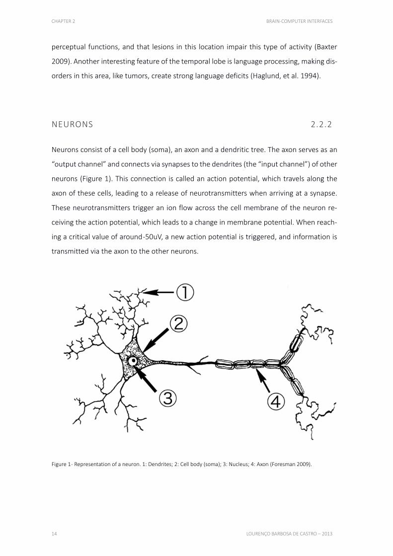
Neurons consist of a cell body (soma), an axon and a dendritic tree. The axon serves as an
“output channel” and connects via synapses to the dendrites (the “input channel”) of other
neurons (Figure 1). This connection is called an action potential, which travels along the
axon of these cells, leading to a release of neurotransmitters when arriving at a synapse.
These neurotransmitters trigger an ion flow across the cell membrane of the neuron re-
ceiving the action potential, which leads to a change in membrane potential. When reach-
ing a critical value of around -50uV, a new action potential is triggered, and information is
transmitted via the axon to the other neurons.
Figure 1 - Representation of a neuron. 1: Dendrites; 2: Cell body (soma); 3: Nucleus; 4: Axon (Foresman 2009).

CHAPTER 2 BRAIN-COMPUTER INTERFACES
HUMAN COMPUTER INTERACTION VIA BRAINWAVES FOR DISABLED PEOPLE 15
ELECTROENCEPHALOGRAPHY 2.3
INTRODUCTION 2.3.1
EEG is one of the most widely used noninvasive techniques for recording electrical brain
activity. After its discovery, this technique has been employed to answer many different
questions about the functioning of the human brain and has served as a diagnostic tool in
clinical practice.
Electroencephalographs, thus, measure membrane potential variations occurring in neu-
rons. The polarity of the signal changes according to the location of the synapses, being
excitatory and inhibitory synapses inversely correlated. For excitatory, the polarity is nega-
tive when located in superficial cortical layers and positive when close to the soma of a cell,
while the opposite happens for inhibitory synapses (Hoffmann 2007).
Although the cerebral activity is better detected over the region of interest, the volume
conduction in the cerebrospinal fluid, skull and scalp allows the signal to spread to distant
electrodes. Additionally, this barrier created between the neurons and the sensors makes
frequencies over 40 Hz almost invisible. Both these conditions generally restrict EEG to
global measurements of the brain activity (Hoffmann 2007).
ELECTRODES 2.3.2
The most common type of electrodes used in consumer-grade EEG devices are metal disk
and cup electrodes, generally built with tin, silver, gold or surgical steel, or even some of
these combined. Their size is important to ensure sufficient contact is made with the scalp,
and is generally within the 4-10 mm range. For a better conductivity, a special gel or saline
solution is usually required, although most recent devices are being developed with dry
sensor technology (Wachspress 2010). This alternative has proven to be relatively decent
in terms of daily usage, but recent research showed significant decreases when comparing
CHAPTER 2 BRAIN-COMPUTER INTERFACES
16 LOURENÇO BARBOSA DE CASTRO – 2013
dry to water-based electrodes. Also, brain activity is always recorded with respect to refer-
ence electrodes, which means EEG signals are small potential differences between elec-
trodes placed at different positions on the scalp (Hoffmann 2007).
Additionally, while low-cost EEG devices feature a low number of electrodes, research has
shown that, ideally, at least 12 electrodes were required to maintain a 90% accuracy in
terms of motor imagery detection (Tam, et al. 2011).
POSITIONS 2.3.3 D ESC R IP T IO N O F T HE M O S T U SE D E L EC TR O D E S P O S I T IO N IN G SYS TE M
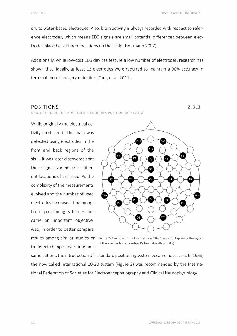
While originally the electrical ac-
tivity produced in the brain was
detected using electrodes in the
front and back regions of the
skull, it was later discovered that
these signals varied across differ-
ent locations of the head. As the
complexity of the measurements
evolved and the number of used
electrodes increased, finding op-
timal positioning schemes be-
came an important objective.
Also, in order to better compare
results among similar studies or
to detect changes over time on a
same patient, the introduction of a standard positioning system became necessary. In 1958,
the now called International 10-20 system (Figure 2) was recommended by the Interna-
tional Federation of Societies for Electroencephalography and Clinical Neurophysiology.
Figure 2 - Example of the International 10-20 system, displaying the layout
of the electrodes on a subject's head (Fieldtrip 2013).

CHAPTER 2 BRAIN-COMPUTER INTERFACES
HUMAN COMPUTER INTERACTION VIA BRAINWAVES FOR DISABLED PEOPLE 17
This system measured specific anatomic landmarks on the skull, the nasion (depression
between the eyes) and the inion (lowest point on the back of the skull), and then used 10%
or 20% of that distance to calculate the electrodes interval. Each electrode is designated by
the first letter of the lobe it is placed on top of, and a number, odd on the left and even on
the right hemisphere (Niedermeyer, et al. 2004).
NOISE 2.3.4 W HAT K IN D O F N O ISE I S R E L E VA N T A N D W HE R E D O ES I T O R IG IN ATE
In addition to the effects of volume conduction, the analysis of the EEG is further compli-
cated by the presence of artifacts. Mostly because of its low electrical amplitudes, the ver-
ified signal-to-noise ratio is much reduced, making artifacts a major concern.
Artifacts can be due to physiological or nonphysiological sources. Physiological sources for
artifacts include eye movement and blinking, skeletal muscles activity, heart activity and
slow potential drifts due to transpiration. Nonphysiological sources for artifacts include
power supply line noise (at 50 Hz or 60 Hz), noise generated by the EEG amplifier and noise
generated by sudden changes in the properties of the electrode-scalp interface. Artifacts
often have much larger amplitude than the signals of interest. Therefore, artifact removal
and filtering procedures have to be applied before an analysis of EEG signals can be at-
tempted.
Despite the above mentioned shortcomings, EEG remains one of the most interesting
methods for measuring electrical brain signals, being used on research of different sleep
stages, epilepsy monitoring, coma outcome prognosis and many other, more theoretical,
scientific purposes (Hoffman, et al. 2008).

CHAPTER 2 BRAIN-COMPUTER INTERFACES
18 LOURENÇO BARBOSA DE CASTRO – 2013
NEURAL OSCILLATIONS 2.3.5 D IF F E R E N TIATE N E U R A L O SC I L L AT IO N S A C CO R D I N G TO T HE IR F R EQ U E N C Y R A N G ES
Delta (0-4 Hz) 2.3.5.1
The delta range (Figure 3) consists in high amplitude brainwaves, usually associated with
the deepest stages of sleep, helping to characterize its depth. Disruptions in these waves
may be the result of physiological damage, intoxication, delirium and neurological disorders
like dementia or schizophrenia. Depression, anxiety and ADHD are also linked with dis-
rupted delta-wave activity. This activity is mostly found frontally in adults and posteriorly in
children (Clarke, et al. 2001).
Figure 3 - Representation of 1 second of EEG activity from the Oz position, filtered to display only delta waves (Gamboa
2005).
Theta (4-8 Hz) 2.3.5.2
Brainwaves frequently observed in young children (Figure 4), or in older children and adults
during drowsy, meditative or sleeping stages (except deep sleep). These are also manifested
during some short term memory tasks, when the hippocampus is active, and correlated to
voluntary behaviors, like exploration, and alert states. Theta waves are found in regions not
related to the task at hand (Bland, et al. 2001).
Figure 4 - Representation of 1 second of EEG activity from the Oz position, filtered to display only theta waves (Gamboa
2005).
CHAPTER 2 BRAIN-COMPUTER INTERFACES
HUMAN COMPUTER INTERACTION VIA BRAINWAVES FOR DISABLED PEOPLE 19
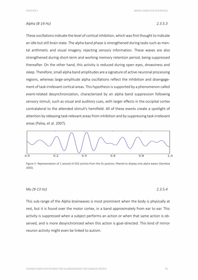
Alpha (8-14 Hz) 2.3.5.3
These oscillations indicate the level of cortical inhibition, which was first thought to indicate
an idle but still brain state. The alpha band phase is strengthened during tasks such as men-
tal arithmetic and visual imagery, rejecting sensory information. These waves are also
strengthened during short-term and working memory retention period, being suppressed
thereafter. On the other hand, this activity is reduced during open eyes, drowsiness and
sleep. Therefore, small alpha band amplitudes are a signature of active neuronal processing
regions, whereas large-amplitude alpha oscillations reflect the inhibition and disengage-
ment of task-irrelevant cortical areas. This hypothesis is supported by a phenomenon called
event-related desynchronization, characterized by an alpha band suppression following
sensory stimuli, such as visual and auditory cues, with larger effects in the occipital cortex
contralateral to the attended stimuli’s hemifield. All of these events create a spotlight of
attention by releasing task-relevant areas from inhibition and by suppressing task-irrelevant
areas (Palva, et al. 2007).
Figure 5 - Representation of 1 second of EEG activity from the Oz position, filtered to display only alpha waves (Gamboa
2005).
Mu (9-13 Hz) 2.3.5.4
This sub-range of the Alpha brainwaves is most prominent when the body is physically at
rest, but it is found over the motor cortex, in a band approximately from ear to ear. This
activity is suppressed when a subject performs an action or when that same action is ob-
served, and is more desynchronized when this action is goal-directed. This kind of mirror
neuron activity might even be linked to autism.
CHAPTER 2 BRAIN-COMPUTER INTERFACES
20 LOURENÇO BARBOSA DE CASTRO – 2013
The Event-related Desynchronization of this wave may be used in BCIs. Groups of neurons
at rest tend to fire in synchrony, so when a user is requested to perform a specific action,
the resulting desynchronization can be analyzed by a computer (Nyström, et al. 2010).
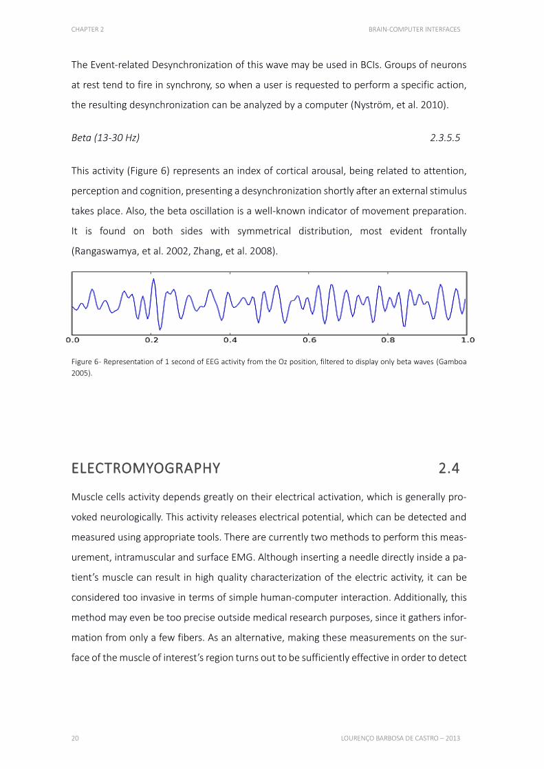
Beta (13-30 Hz) 2.3.5.5
This activity (Figure 6) represents an index of cortical arousal, being related to attention,
perception and cognition, presenting a desynchronization shortly after an external stimulus
takes place. Also, the beta oscillation is a well-known indicator of movement preparation.
It is found on both sides with symmetrical distribution, most evident frontally
(Rangaswamya, et al. 2002, Zhang, et al. 2008).
Figure 6 - Representation of 1 second of EEG activity from the Oz position, filtered to display only beta waves (Gamboa
2005).
ELECTROMYOGRAPHY 2.4
Muscle cells activity depends greatly on their electrical activation, which is generally pro-
voked neurologically. This activity releases electrical potential, which can be detected and
measured using appropriate tools. There are currently two methods to perform this meas-
urement, intramuscular and surface EMG. Although inserting a needle directly inside a pa-
tient’s muscle can result in high quality characterization of the electric activity, it can be
considered too invasive in terms of simple human-computer interaction. Additionally, this
method may even be too precise outside medical research purposes, since it gathers infor-
mation from only a few fibers. As an alternative, making these measurements on the sur-
face of the muscle of interest’s region turns out to be sufficiently effective in order to detect
CHAPTER 2 BRAIN-COMPUTER INTERFACES
HUMAN COMPUTER INTERACTION VIA BRAINWAVES FOR DISABLED PEOPLE 21
activity, even though the high power of the EMG signals tend to make them propagate far
from their origin (Gomez-Gil, et al. 2011).
Most EEG based systems pick up, inevitably, electrical myography signals, generated in the
facial and neck muscles. However, these are usually treated as artifacts, being carefully re-
moved from the final outcome. Systems such as the Emotiv EPOC are trying to make use of
that data, interpreting it in order to detect facial expressions such as blinks, winks, frowns
and smiles.
The usefulness of this type of detection is diverse. Combinations of facial muscles can be
translated into computer commands, both for disabled people with movement impair-
ments and for avid technology users, searching for novel forms of interaction. Specific facial
expressions, like smiling, can be associated with mental states, such as joy or happiness,
helping computers understand one’s behavior.
SIGNALS DISCRIMINATION 2.5
Ideally, a BCI system would directly detect every wish, intention and reaction of its user,
based on its brain activity. To allow for discrimination of different neurophysiologic signals
or to map such signals to movements, users have to acquire conscious control over their
brain activity. Two fundamentally different approaches exist to achieve this. In the first,
subjects perceive a set of stimuli displayed by the BCI system and can control their brain
activity by focusing onto one specific stimulus. The changes in neurophysiologic signals re-
sulting from perception and processing of stimuli are termed event-related potentials
(ERPs). In the second approach, users control their brain activity by concentrating on a spe-
cific mental task, e.g., the imagination of hand movement can be used to modify activity in
the motor cortex. In this approach, feedback signals are often used to help subjects learn-
ing the production of easily detectable patterns of neurophysiologic signals.
CHAPTER 2 BRAIN-COMPUTER INTERFACES
22 LOURENÇO BARBOSA DE CASTRO – 2013
EVENT-RELATED POTENTIALS 2.5.1
ERPs are stereotyped, spatio-temporal patterns of brain activity, occurring time-locked to
an event, e.g. after presentation of a stimulus, before execution of a movement or after the
detection of a novel stimulus. Traditionally, ERPs are recorded with EEG and have been used
in neuroscience for studying the different stages of perception, cognition and action. It is
worth noting that event-related changes can also be measured with other signal acquisition
techniques like the MEG or fMRI.
P300 POTENTIAL 2.5.2
The P300 is a positive deflection in the human EEG of about 2-5uV, appearing approxi-
mately 300ms after the presentation of a rare or surprising, task-relevant stimuli. This is an
endogenous event-related potential, which means it is caused by a late and conscious pro-
cessing of stimuli, and depends mainly on the stimulus content. It can be reliably measured,
generally observed over centro-parietal brain regions (Fz, Cz and Pz regions [Figure 2]), and
its waveform can be influenced by various factors. If the user is not concentrated enough,
though the P300 wave might disappear completely, and if the task is too difficult, the la-
tency of the wave increases while its amplitude decreases.
Although P300 can be evoked using all five basic human senses, the most used, for practical
reasons, are auditory and visual. It is also worth mentioning that this kind of potential can
be detected on nearly all healthy users, requiring almost no training.
Methodology 2.5.2.1
Generally, to evoke P300, subjects are asked to observe a random sequence of two kinds
of stimuli. One stimulus type (the oddball, or target stimulus) appears only rarely in the
sequence, while the other stimulus type (the normal, on non-target stimulus) appears more
often. In short, whenever the target stimulus happens, P300 can be observed in the EEG.
CHAPTER 2 BRAIN-COMPUTER INTERFACES
HUMAN COMPUTER INTERACTION VIA BRAINWAVES FOR DISABLED PEOPLE 23
Another method is to use three types of stimuli, in which one of them is a, so called, dis-
tracter stimulus, which also appears in a sequence with target and non-target stimuli, but
less frequently. Also, the subject is usually not informed about this stimulus, to increase the
element of surprise of a non-target element. After presentation of many distracter stimuli,
the related wave’s amplitude decreases, while the target P300 wave remains unaffected.
This stimuli interaction requirement presents as the main drawback of the P300 potential,
since the user must pay attention to a specific source to perform brain-computer commu-
nication.
Several negative and positive components precede the P300, being P300 the highest posi-
tive deflection of these components.
The P300 peak is inversely related to the probability of the evoking stimulus, being 10% a
good probability value to evoke this potential. Also, if many nontarget stimuli precede the
target stimulus, a higher amplitude is observable in this potential.
The amplitude of the P300 wave is positively correlated to the interstimulus interval, i.e.
the amount of time between two consecutive stimuli. Theoretically, longer intervals might
yield better results, because the P300 amplitudes are larger. On the other hand, this re-
quires longer duration of runs, requiring longer periods of concentration, which might
prove to be difficult for some disabled subjects. Overall, longer interstimulus intervals might
decrease P300 amplitude and classification accuracy. Moreover, higher bitrates are ob-
tained with shorter intervals, but if the stimuli are made too short, subjects with cognitive
deficits might have problems to detect all the targets. In conclusion, an optimal interstim-
ulus interval for P300-based BCIs can only be determined experimentally. Additionally, the
P300 system works in such way that the user doesn't have to perform any training at all,
besides being instructed to keep their focus on a specified stimulus.
Implementation 2.5.2.2
Farwell and Donchin were the first to employ the P300 as a control signal in a BCI (Farwell
and Donchin 1988). They described the P300 speller in which a matrix containing symbols
CHAPTER 2 BRAIN-COMPUTER INTERFACES
24 LOURENÇO BARBOSA DE CASTRO – 2013
from the alphabet is displayed on a screen. Rows and columns of the matrix are flashed in
random order, and flashes of the row or column containing the desired symbol constitute
the oddball stimulus, while all other flashes constitute nontarget stimuli. Since the seminal
paper of Farwell and Donchin, many studies about P300-based BCI systems have appeared,
usually associated with machine learning techniques such as support vector machines or
neural networks (Campbell, et al. 2010). This potential has also been linked to subject spe-
cific factors such as gender, age or brain diseases.
Systems using this potential are relatively slow forms of brain-computer interaction, since
it is usually required for a stimuli to occur several times after confirming the user's inten-
tion. Due to the low power of the resulting electric wave, signal to noise ratio is very low,
making its detection very difficult, resulting in the need of high resolution equipment. This
condition also increases the signal processing complexity required for a successful detec-
tion. Additionally, as previously mentioned, the user must focus their attention on a specific
stimuli, which can be difficult for some types of people. Finally, since this system is con-
stantly transmitting information and scanning for specific signals, it is complicated to make
pauses, either voluntary or not.
COGNITION/EXPRESSIONS 2.5.3 HOW C A N CO G N IT IO N A N D FA C IA L E X P R E SS IO N S B E U SE D TO IN TE R A C T W IT H CO M P U TE R S
Using cognitions or facial expressions is also a possibility in this field. EEG and EMG patterns
can be read and interpreted, and afterwards assigned to specific computer commands.
This methodology requires a solid and time-consuming calibration, in order for the user's
actions to be detected accurately. Also, from a daily usage perspective, performing several
facial expressions in public should make some people uncomfortable, and might even be
impossible for impaired patients.
However, after sufficient calibration is performed, using conscious thoughts and facial ex-
pressions should result in fast brain-computer interactions, although some currently avail-
able systems have some limitations in terms of using multiple types of thoughts. Addition-
CHAPTER 2 BRAIN-COMPUTER INTERFACES
HUMAN COMPUTER INTERACTION VIA BRAINWAVES FOR DISABLED PEOPLE 25
ally, when considering muscular electric activity, noise becomes less relevant, as facial ex-
pressions usually produce high amplitude signals. Another advantage of this approach, over
pure EEG signals, is that momentaneous interruptions are possible, since permanently
keeping focus is not essential, helping this system to become a better option for daily usage.
TYPES OF BCIS 2.6 D ESC R IP T IO N O F IN VA S IV E A N D N O N - IN VA S IV E M E T HO D O LO G I E S
INVASIVE 2.6.1
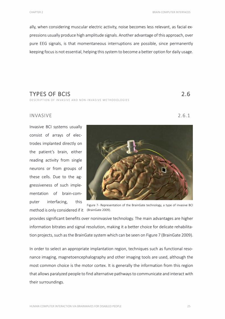
Invasive BCI systems usually
consist of arrays of elec-
trodes implanted directly on
the patient’s brain, either
reading activity from single
neurons or from groups of
these cells. Due to the ag-
gressiveness of such imple-
mentation of brain-com-
puter interfacing, this
method is only considered if it
provides significant benefits over noninvasive technology. The main advantages are higher
information bitrates and signal resolution, making it a better choice for delicate rehabilita-
tion projects, such as the BrainGate system which can be seen on Figure 7 (BrainGate 2009).
In order to select an appropriate implantation region, techniques such as functional reso-
nance imaging, magnetoencephalography and other imaging tools are used, although the
most common choice is the motor cortex. It is generally the information from this region
that allows paralyzed people to find alternative pathways to communicate and interact with
their surroundings.
Figure 7 - Representation of the BrainGate technology, a type of invasive BCI
(BrainGate 2009).
CHAPTER 2 BRAIN-COMPUTER INTERFACES
26 LOURENÇO BARBOSA DE CASTRO – 2013
Another important factor to ponder is the simultaneous amount of neurons needed to ob-
tain a meaningful signal, since this technology requires risky medical procedures that need
to be justified by a decent information transfer rate. The research community’s opinion is
inconclusive in this subject, with values ranging from 2 cells up to 100, leading to different
types of approaches, some of which previously described in section 2.1 (Wolpaw, et al.
2000).
The use of invasive technology on human beings is also conditioned by the period of time
that the specific electrodes arrays can be kept inside a patient’s brain, while transmitting
stable recordings. Recently, the BrainGate implant proved to be efficient even after a period
of nearly 3 years, making it the most viable option currently available (Simeral, et al. 2011).
Additionally, after long periods of time, this type of BCI can become so intrinsic to the pa-
tient that their brain completely maps its activity to the computer interface, creating a true
human-machine hybrid (Hockenberry 2001).
NON-INVASIVE 2.6.2
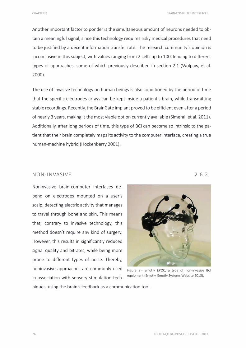
Noninvasive brain-computer interfaces de-
pend on electrodes mounted on a user’s
scalp, detecting electric activity that manages
to travel through bone and skin. This means
that, contrary to invasive technology, this
method doesn’t require any kind of surgery.
However, this results in significantly reduced
signal quality and bitrates, while being more
prone to different types of noise. Thereby,
noninvasive approaches are commonly used
in association with sensory stimulation tech-
niques, using the brain’s feedback as a communication tool.
Figure 8 - Emotiv EPOC, a type of non-invasive BCI
equipment (Emotiv, Emotiv Systems Website 2013).

CHAPTER 2 BRAIN-COMPUTER INTERFACES
HUMAN COMPUTER INTERACTION VIA BRAINWAVES FOR DISABLED PEOPLE 27
In order to improve signal conductivity, a specialized gel or water solution is used, although
the current tendency is to adopt dry-based sensors. The information transfer rate is gener-
ally slower, compensated by the fact that starting using such technology is as simple as
wearing a specialized cap or headband (Middendorf, et al. 2000).
The accessibility and affordability of this approach makes it the best target for entertain-
ment developments, like mind controlled games or personalized entertainment systems.
Every consumer-based BCI company develops noninvasive devices, usually offering rela-
tively simple features, such as detecting the user’s focus or mental stress levels. However,
recent advances in this field have improved its bitrate sufficiently for noninvasive BCIs being
considered as neural rehabilitation tools.
SUMMARY 2.7
This chapter explained the foundations of this project, mentioning the discovery of EEG by
Hans Berger in 1929 and the methodology behind this technique. The most used electrodes
types are metal disk and cup electrodes, usually built with tin, silver, gold or surgical steel,
with sizes ranging from 4 to 10 mm. Although most devices are trying to avoid this charac-
teristic, it is commonly required the use of a special gel or saline solution for better con-
ductivity. The 10-20 electrodes positioning system is widely adopted by EEG and BCI com-
munities, creating a standard for better results comparison and validation.
The low signal amplitude verified in EEG makes it highly prone to noise, mostly caused by
muscular artifacts or power supply line noise. This aspect makes noise removal a very im-
portant factor in BCI analysis.
Neural oscillations can be divided in different ranges, which can be associated with different
behaviors and mindsets. Although not precisely known, lowest frequencies are usually as-
sociated with relaxation and deep sleep, and disruptions in this range are seen in conditions
such as ADHD or depression. Higher frequencies can be correlated with attention, creativity
and active cognitive processing in general.
CHAPTER 2 BRAIN-COMPUTER INTERFACES
28 LOURENÇO BARBOSA DE CASTRO – 2013
Electromyography can be described as the detection and analysis of the electric activity
generated by muscular activation. This study is generally and more accurately elaborated
using needles inserted in the muscles of interest, although it can also be measured on the
surface of the skin with sufficient precision. These signals, otherwise seen as artifacts, are
being used as features by some BCI devices, creating associations with computer com-
mands or being studied for a better understanding of users’ emotions.
Following, several interpretations of neurophysiological signals are described. Event-re-
lated potentials are brainwave patterns associated to a specific event. The P300 potential
is an EEG deflection verified after a surprising stimuli, which is being used to help disabled
people select options on a computer screen. Cognitive patterns and facial expressions,
based both on EEG and EMG, are another source of electric signals that can be translated
on a computer, after sufficient calibration, which can make computers adapt to their users.
Finally, the two generic BCI types were explained, invasive and non-invasive interfaces.
While introducing electrodes directly on the brain’s surface provides great improvements
in signal quality and resolution, the risks involved make this approach only considerable
when no other viable options are available. On the other hand, non-invasive technology is
more likely to be affected by noise or low resolution effects, but it is much more affordable
and completely safe, measuring the brain’s activity over the scalp.
29
CHAPTER 3
EMOTIV EPOC This section focuses on the EEG device used in this project, the Emotiv EPOC. Describes its
creation, purpose and their current position on the market. Following is a comparison
between this device and the competition, explaining advantages and disadvantages,
justifying why this model was chosen. To finalize, Emotiv’s native software is presented,
along with its functionalities.
3.1. INTRODUCTION 30
3.2. HARDWARE 31
3.3. TYPES OF INTERACTION 32
3.4. SUMMARY 33
CHAPTER 3 EMOTIV EPOC
30 LOURENÇO BARBOSA DE CASTRO – 2013
INTRODUCTION 3.1
Introduced in 2007 by the Australian company Emotiv, the EPOC as a complex, low-cost
mobile EEG equipment. Its main purpose was to implement novel forms of gaming interac-
tions and more immersive entertainment experiences. The company is also supporting
third party research and development, by providing development kits and an active support
team. This resulted in an increasingly growing online application store, research papers
based on their device and in the most active brain-computer interfaces community.
Existing devices
The most noticeable advantage when compared to other low-cost mobile EEG devices is
the number of electrodes. While other devices feature, at most, 6 electrodes, the EPOC
more than doubles that amount, with 14 sensors and two references. This allows for a
greater spatial resolution and increased noise reduction, obtained from comparing and an-
alyzing data from different regions of the brain. It is also, currently, the only available BCI
device to feature a gyroscope, which, besides providing additional forms of interaction, also
helps reducing noise, specifically cause by head movements. The sampling rate of 128 Hz
is similar to most commercialized products, enabling the study of the most important brain-
waves bands.
When compared to medical grade equipment, the EPOC loses in every aspect but the price,
as expected. The number of electrodes used for research can reach almost 100, while sam-
pling rates can be as high as 1000 Hz. However, this type of devices can cost several tens of
thousands of dollars, while the EPOC’s costs a few hundred (Castermans 2011).
CHAPTER 3 EMOTIV EPOC
HUMAN COMPUTER INTERACTION VIA BRAINWAVES FOR DISABLED PEOPLE 31
HARDWARE 3.2
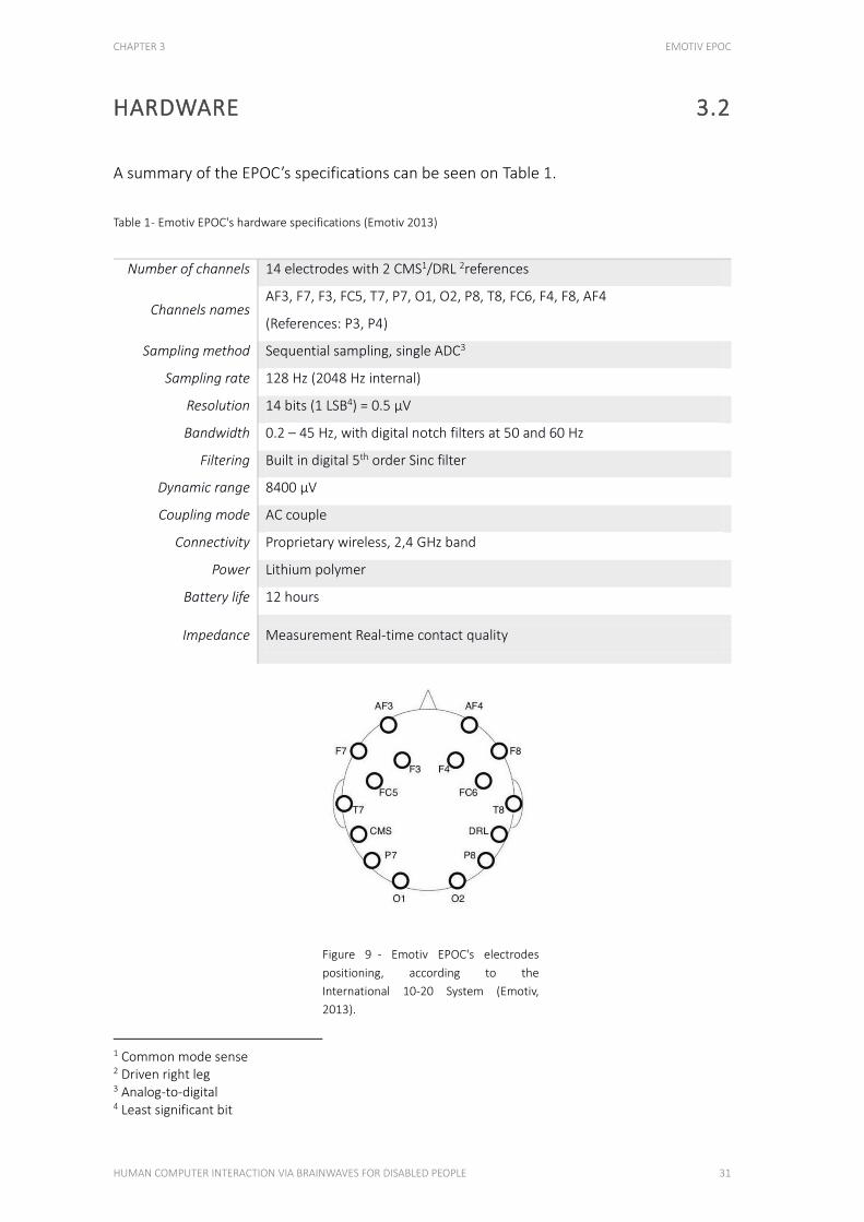
A summary of the EPOC’s specifications can be seen on Table 1.
Table 1 - Emotiv EPOC's hardware specifications (Emotiv 2013)
Number of channels 14 electrodes with 2 CMS1/DRL 2references
Channels names AF3, F7, F3, FC5, T7, P7, O1, O2, P8, T8, FC6, F4, F8, AF4
(References: P3, P4)
Sampling method Sequential sampling, single ADC3
Sampling rate 128 Hz (2048 Hz internal)
Resolution 14 bits (1 LSB4) = 0.5 μV
Bandwidth 0.2 – 45 Hz, with digital notch filters at 50 and 60 Hz
Filtering Built in digital 5th order Sinc filter
Dynamic range 8400 μV
Coupling mode AC couple
Connectivity Proprietary wireless, 2,4 GHz band
Power Lithium polymer
Battery life 12 hours
Impedance Measurement Real-time contact quality
1 Common mode sense 2 Driven right leg 3 Analog-to-digital 4 Least significant bit
Figure 9 - Emotiv EPOC's electrodes
positioning, according to the
International 10-20 System (Emotiv,
2013).
CHAPTER 3 EMOTIV EPOC
32 LOURENÇO BARBOSA DE CASTRO – 2013
The EPOC has gold-plated sensors fixed to flexible plastic arms. The two references are po-
sitioned on the mastoid region, one acting as a ground point (to which all other sensors are
compared) and another to reduce external electrical interference (Badcock, et al. 2013).
The exact positioning of each electrode can be seen on Figure 9.
TYPES OF INTERACTION 3.3 N ATI V E F U N C TIO N A L I T I ES O F THE D E V IC E
EXPRESSIV SUITE 3.3.1
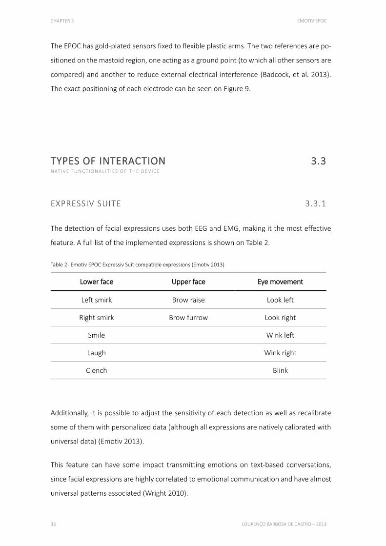
The detection of facial expressions uses both EEG and EMG, making it the most effective
feature. A full list of the implemented expressions is shown on Table 2.
Table 2 - Emotiv EPOC Expressiv Suit compatible expressions (Emotiv 2013)
Lower face Upper face Eye movement
Left smirk Brow raise Look left
Right smirk Brow furrow Look right
Smile Wink left
Laugh Wink right
Clench Blink
Additionally, it is possible to adjust the sensitivity of each detection as well as recalibrate
some of them with personalized data (although all expressions are natively calibrated with
universal data) (Emotiv 2013).
This feature can have some impact transmitting emotions on text-based conversations,
since facial expressions are highly correlated to emotional communication and have almost
universal patterns associated (Wright 2010).
CHAPTER 3 EMOTIV EPOC
HUMAN COMPUTER INTERACTION VIA BRAINWAVES FOR DISABLED PEOPLE 33
COGNITIV SUITE 3.3.2
In terms of cognitive detection, the EPOC identifies previously calibrated brainwave pat-
terns, which can be associated with a specific action. For instance, the user picturing the
action of pushing a cube produces electric activity that is recorded and saved as the action
“push”. When that specific activity is recaptured, the computer knows which action to per-
form. This means there is not actual “mind reading” involved, and that new users need to
calibrate the software for proper functioning, while being encouraged to recalibrate the
application occasionally (Breen 2008). The native software allows for up to 4 simultaneous
calibrated actions, although it is relatively hard to master more than 2. Also, this approach
is not considered absolutely viable for risk involving actions, as a consequence of our ina-
bility to perfectly control our thoughts (Wright 2010).
AFFECTIV SUITE 3.3.3
Emotions such as engagement, frustration or enthusiasm are supposedly detected using
the EPOC. This feature has a seemingly lower accuracy rate, with the detected emotions
not corresponding to the users’ reports. The major issue with this section is the ineffective-
ness of a possible calibration, since it would be almost impracticable to ask a user to simu-
late an emotion. Therefore, Emotiv has to rely on data mining, which has not yet proved to
be very successful. Additionally, the Affectiv Suite’s algorithms are not available to develop-
ers, meaning that Emotiv is the only source for improvements (Breen 2008, Wright 2010).
SUMMARY 3.4
The EPOC, presented in 2007, is currently the most advanced consumer-based EEG headset
available on the market. This was the main reason for it to be chosen on this project, even
though its price was slightly higher than the competition.
CHAPTER 3 EMOTIV EPOC
34 LOURENÇO BARBOSA DE CASTRO – 2013
Its native software allowed several facial expressions recognition, identifying cognitive pat-
terns and understand different emotional states. Most of these features were elaborated
using data from extended trials, acquired while the device was in development, although
facial expressions and cognitive detections can be calibrated for increased effectiveness.
35
CHAPTER 4
DEVELOPED APPLICATION This chapter starts by describing the foundations of the developed application, explaining
how it works and for whom it was designed, with emphasis on a detailed presentation of its
different sections and their specific functions. Next, results are shown and discussed, with
an explanation about how they were acquired. To conclude the chapter, the applicability of
the developed software is discussed, presenting several practical examples.
4.1. TARGET USERS 38
4.2. ANDROID 39
4.3. FEATURES 40
4.4. SOFTWARE 41
4.5. APPLICABILITY 48
4.6. SUMMARY 51

CHAPTER 4 DEVELOPED APPLICATION
36 LOURENÇO BARBOSA DE CASTRO – 2013
TARGET USERS 4.1 TO W HO M T HI S A P P L IC AT IO N WA S D E S IG N E D
One of the main purposes of this work is to implement features that can help overcoming
difficulties on common daily tasks. These difficulties can originate in several different
sources, such as diseases or accidents, which may cause severe muscular disabilities that
strongly reduce affected people’s capabilities and autonomy.
Locked in syndrome
One of the most frequently targeted disease on BCI research is amyotrophic lateral sclerosis
(also known as Lou Gehrig’s disease), which essentially locks patients inside their own bod-
ies, progressively deteriorating muscle functions. Affected people usually die of respiratory
failure, also due to muscular degeneration, after living a mean time of 43 months with this
disease (Turner, et al. 2003).
Strokes
Another cause for muscular paralysis are strokes, affecting 15 million people worldwide
every year, while killing 5 million of those and leaving another 5 permanently disabled
(World Health Organization 2002). These people have their lives greatly impaired, losing
most or all of their autonomy.
Spinal cord injury
Spinal cord injuries can also leave affected patients unable to move, with either paraplegia
or tetraplegia. Currently, there are no restorative treatments for such conditions, meaning
that developing alternatives for their muscular related activity is a desired option (King, et
al. 2013).
Ageing
Additionally, as people age, they tend to lose muscle control and strength, increasing their
dependency on family members or healthcare professionals.
CHAPTER 4 DEVELOPED APPLICATION
HUMAN COMPUTER INTERACTION VIA BRAINWAVES FOR DISABLED PEOPLE 37
Most of these conditions leave one’s ability to reason intact, making them a good target for
brain-computer interfaces. By providing an alternative method to interact with computers,
it might be possible to return some of these people’s autonomy, enabling them to com-
municate or perform basic tasks.
ANDROID OPERATING SYSTEM 4.2
OPERATING SYSTEM 4.2.1 B R IE F IN TRO D U C T IO N TO THE A N D RO ID P L ATFO R M
Android was started in 2003, as a Linux-based mobile operating system, making it open-
source and, thus, easily modifiable. The original company was bought by Google (U.S.A.) in
2005, preserving its name and philosophy, while keeping it away from the general public.
It was only 5 years after the initial developments, in 2008, that the first smartphone running
Android, version 1.0, was released. The biggest advantage of this operating system was its
unrestricted availability to manufacturers, allowing them to focus solely on hardware de-
velopment, which greatly helped Android growing exponentially (Kovach 2013). Currently,
this is the most widely used mobile operating system, with nearly 80% market share
(International Data Corporation 2013), making it an exceptional target for innovative acces-
sibility applications development.
NEXUS 7 TABLET DEVICE 4.2.2 TA B L E T D E V IC E U SE D I N T HI S P ROJ EC T
The Nexus 7 was chosen mainly for 3 aspects: USB Host, good price to quality relationship
and appropriate screen size. The USB Host application programming interface (API) is a
possibility first launched on Android 3.1 (Honeycomb), version released in May 2011. Not
all devices currently support this feature, in favor of better battery life, but an increasingly
number of modern devices is embracing this technology (Google, USB Host 2013). As of

CHAPTER 4 DEVELOPED APPLICATION
38 LOURENÇO BARBOSA DE CASTRO – 2013
the start of this project, this mobile computer was the only known tablet device to have
USB Host enabled, which was crucial to interact with the Emotiv EPOC. Considering all the
available options, this tablet showed the best price to quality ratio, featuring a quad-core
processor, 1280x800 screen resolution and 1GB of RAM, which was useful to quickly per-
form the required calculations and display the sensors data, all in real-time. The device’s
weight, 340g was slightly above the average of the same sized tablets, but was still light
enough to be easily carried, even with only one hand. Additionally, the screen size of 7
inches was sufficiently large to properly display keyboard characters and contacts pictures
(Asus 2012).
FEATURES 4.3
TIME DOMAIN FEATURES 4.3.1
These features are related to changes in the amplitude of the signals, over a period of time
after the presentation of a stimuli or after the actions of a user. P300 potentials can be
characterized using time domain features. The number of spikes occurring in a certain time
interval is also a time domain feature.
FREQUENCY DOMAIN FEATURES 4.3.2
These domain’s features are related to changes in oscillatory activity, which are useful be-
cause the phase of the oscillation is usually not related to the time of the stimuli presenta-
tion or the action of the user. Features like the synchronization of signals from different
brain regions are also of the frequency domain.
CHAPTER 4 DEVELOPED APPLICATION
HUMAN COMPUTER INTERACTION VIA BRAINWAVES FOR DISABLED PEOPLE 39
SPATIAL FEATURES 4.3.3
When data from more than one electrode is available, the extracted features have to be
combined in an efficient way. This efficiency is the goal of spatial features extraction.
One method is to use only electrodes carrying useful information for a specific set of tasks,
i.e., electrodes placed over regions of the brain related to specific cognitive functions or
event potentials. These electrodes can be selected manually or using algorithms that auto-
matically choose an optimal sensor subset. Algorithms like the common spatial patterns
algorithm or the independent component analysis can perform such tasks (Blankertz, et al.
2008, Li, et al. 2009).
SOFTWARE 4.4
DATA ACQUISITION 4.4.1 HOW T HE DATA I S A CQ U IR E D F RO M THE H E A D SE T
As previously mentioned, the developed application relies on the Android’s USB Host API.
Using a USB on-the-go (OTG) cable, the Android tablet could communicate with Emotiv
EPOC’s USB dongle, therefore establishing a connection between both devices (Google
2013).
The application actively detects the connection of new USB devices with characteristics
specific to the EPOC. When the equipment of interest is connected to the tablet, its serial
number is stored, for data decryption purposes, and the proper communication pathway,
the USB endpoint, is opened. This endpoint is the channel used to receive large amounts
of data from the headset, more precisely a 32 bytes packet containing the electric values
captured by all the sensors and their contact quality, gyroscope orientation and the device’s
battery level. The data packet is retrieved at the EPOC’s sampling rate, 128 Hz, followed by
pre-processing to remove the direct current (D.C.) component and round the values to a
single decimal place (this last step is done because, as previously mentioned, the EPOC’s
CHAPTER 4 DEVELOPED APPLICATION
40 LOURENÇO BARBOSA DE CASTRO – 2013
resolution has a least significant bit of 0.5 μV, making every following figure insignificant
(Emotiv 2013)). Finally, these corrected values are distributed to the appropriate active Java
class, using Android’s PropertyChangeListener API (Google 2013).
EVENTS DETECTION 4.4.2
After the acquisition and pre-processing, the application interprets the electric signal, look-
ing for specific patterns which are associated with user actions. The actions that the soft-
ware currently detects are winks and horizontal eye movements, being used to trigger com-
mands in the tablet device.
In order for this detection to occur, a calibration is required. This process consisted in de-
tecting the peak activity for an event-specific electrode, while also recording the output of
the sensor on the opposite side of the head. After several recordings, a range of values
could be established for both electrodes, creating thresholds for their actual values as well
as their ratios. This way, when a peak was detected in a channel of interest, the detection
process was initiated. Using this channel and its symmetrical sensor’s values, ratios were
calculated and compared with the saved data. If it fit inside the trained ranges, a positive
detection was triggered, otherwise nothing happened.
FUNCTIONALITIES 4.4.3
With accessibility in mind, several features were implemented to try to help disabled peo-
ple overcome their daily difficulties. The actual tasks targeted in this project were typing,
simple mathematical operations, selecting phone contacts and general application naviga-
tion. Additionally, there is a feature to monitor neural oscillations across the entire brain
and a configuration page where calibration takes place. This calibration is user-dependent,
saving the trained data on separate profiles, thus making the application personalized and
operational for different people.
CHAPTER 4 DEVELOPED APPLICATION
HUMAN COMPUTER INTERACTION VIA BRAINWAVES FOR DISABLED PEOPLE 41
Each section is accompanied by a help button, located on the top right part of the screen.
Essentially, the help provided is a short version of the following descriptions, therefore en-
abling the user to quickly learn how to operate the application.
Eyes navigation 4.4.3.1
This feature simulates horizontal swipes on the screen, replacing them with sideways looks,
which enables the user to switch the current section without touching the device. There is
an option to quickly toggle this function in order to avoid conflicts with other application
parts where looking sideways is detected.
Keyboard 4.4.3.2
In order to provide an alternative for paralyzed or movement impaired people to be able
to type messages on a computer screen, this section was developed. Using only winks, the
user can efficiently select characters of interest until a sentence is built, with option for
spaces and deleting mistakes (Figure 10).
After initiating this section of the application, the first right eye wink will activate the first
row, changing its color. Subsequent right winks will cycle through all the available rows, also
changing their color. When reaching the row containing the target character, a left wink will
select it, fixating the current color and changing the right wink command. Afterwards, right
winks cycle through all the columns, creating an intersection with the previously selected
row. The letter that falls in this intersection is selectable, again, using a left wink, which
types it on a specified field on the bottom of the screen. Additionally, when no characters
are selected, a left wink deletes the previous typed letter.
To summarize the controls, winking the right eye moves the cursor, while the left eye selects
the current character or deletes the previous letter.
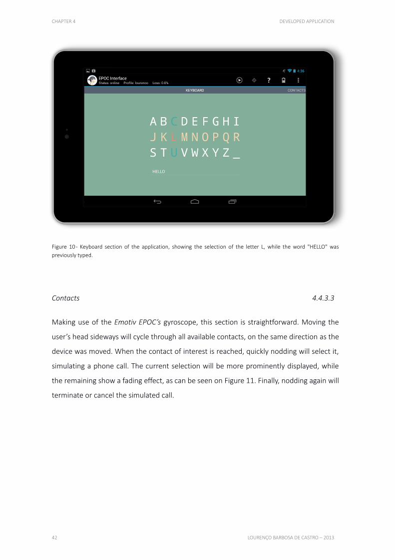
CHAPTER 4 DEVELOPED APPLICATION
42 LOURENÇO BARBOSA DE CASTRO – 2013
Figure 10 - Keyboard section of the application, showing the selection of the letter L, while the word "HELLO" was
previously typed.
Contacts 4.4.3.3
Making use of the Emotiv EPOC’s gyroscope, this section is straightforward. Moving the
user’s head sideways will cycle through all available contacts, on the same direction as the
device was moved. When the contact of interest is reached, quickly nodding will select it,
simulating a phone call. The current selection will be more prominently displayed, while
the remaining show a fading effect, as can be seen on Figure 11. Finally, nodding again will
terminate or cancel the simulated call.
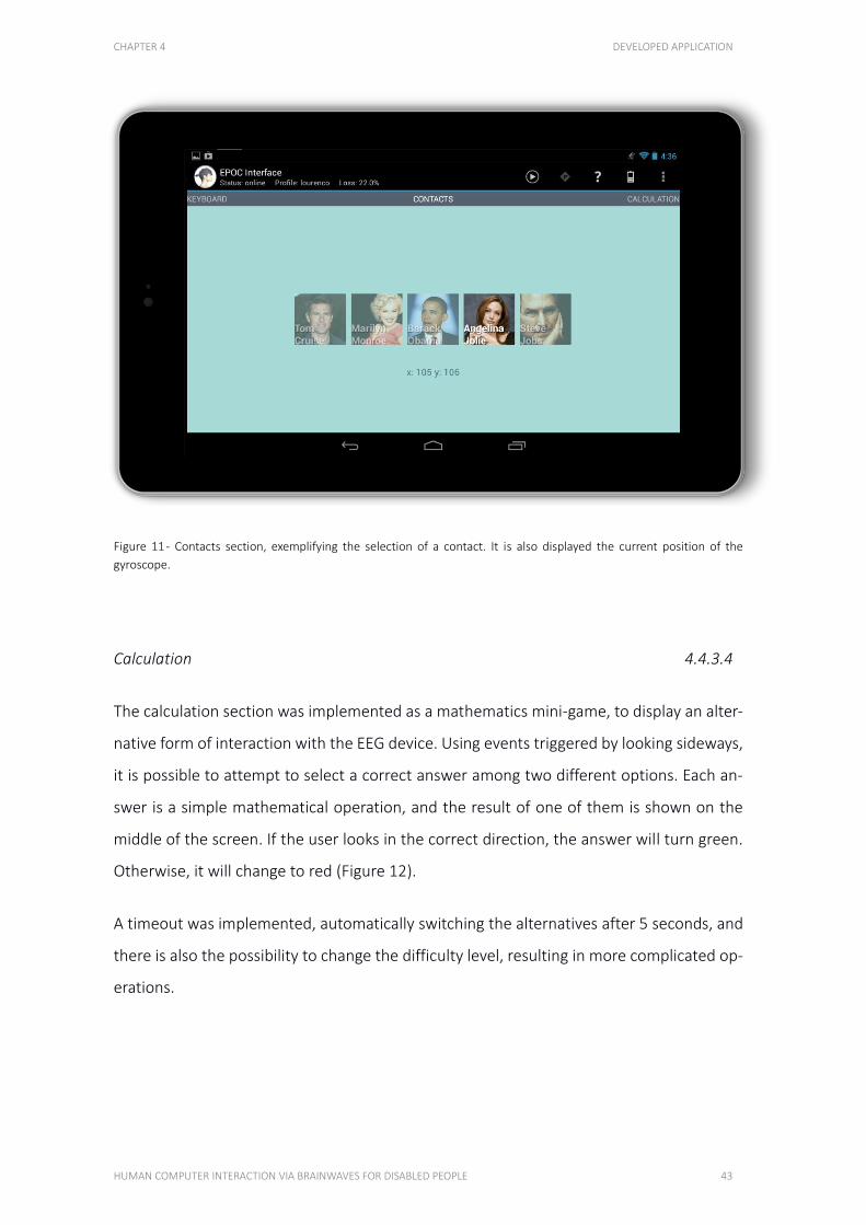
CHAPTER 4 DEVELOPED APPLICATION
HUMAN COMPUTER INTERACTION VIA BRAINWAVES FOR DISABLED PEOPLE 43
Figure 11 - Contacts section, exemplifying the selection of a contact. It is also displayed the current position of the
gyroscope.
Calculation 4.4.3.4
The calculation section was implemented as a mathematics mini-game, to display an alter-
native form of interaction with the EEG device. Using events triggered by looking sideways,
it is possible to attempt to select a correct answer among two different options. Each an-
swer is a simple mathematical operation, and the result of one of them is shown on the
middle of the screen. If the user looks in the correct direction, the answer will turn green.
Otherwise, it will change to red (Figure 12).
A timeout was implemented, automatically switching the alternatives after 5 seconds, and
there is also the possibility to change the difficulty level, resulting in more complicated op-
erations.

CHAPTER 4 DEVELOPED APPLICATION
44 LOURENÇO BARBOSA DE CASTRO – 2013
Figure 12 - Calculation section, showing a correct selection during the second level of difficulty.
Brainwaves 4.4.3.5
The brainwaves section is used to visualize EEG frequencies across the user’s brain, sepa-
rated by the 4 neural oscillation bands that the EPOC can capture, Delta, Theta, Alpha and
Beta. One chart for each sensor is used, positioned according to the location on the head-
set, simulating the comprised region of the human brain. Additionally, one can click on a
sensor of interest, alternating to a single, larger, chart which displays a more detailed fre-
quency information (Figure 13).
For a better frequency analysis, the charts are only refreshed every 8 seconds, providing a
larger sample pool, which enables the detection of broader ranges of frequencies.
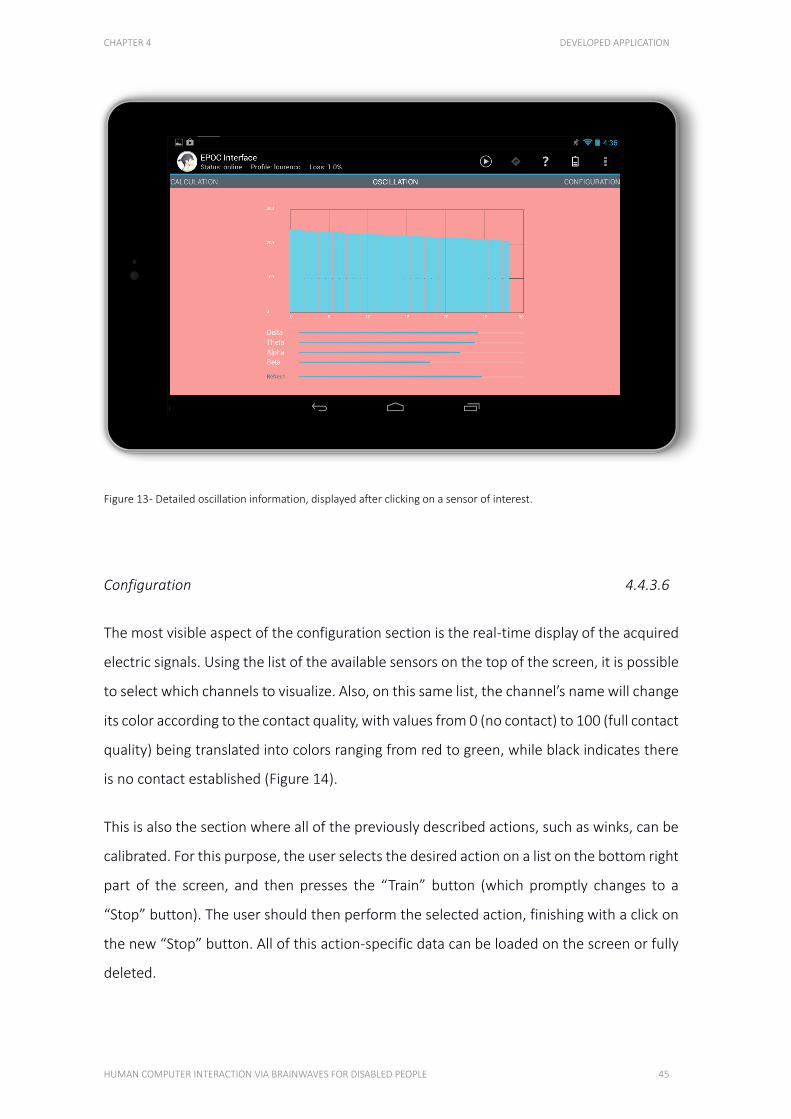
CHAPTER 4 DEVELOPED APPLICATION
HUMAN COMPUTER INTERACTION VIA BRAINWAVES FOR DISABLED PEOPLE 45
Figure 13 - Detailed oscillation information, displayed after clicking on a sensor of interest.
Configuration 4.4.3.6
The most visible aspect of the configuration section is the real-time display of the acquired
electric signals. Using the list of the available sensors on the top of the screen, it is possible
to select which channels to visualize. Also, on this same list, the channel’s name will change
its color according to the contact quality, with values from 0 (no contact) to 100 (full contact
quality) being translated into colors ranging from red to green, while black indicates there
is no contact established (Figure 14).
This is also the section where all of the previously described actions, such as winks, can be
calibrated. For this purpose, the user selects the desired action on a list on the bottom right
part of the screen, and then presses the “Train” button (which promptly changes to a
“Stop” button). The user should then perform the selected action, finishing with a click on
the new “Stop” button. All of this action-specific data can be loaded on the screen or fully
deleted.

CHAPTER 4 DEVELOPED APPLICATION
46 LOURENÇO BARBOSA DE CASTRO – 2013
Figure 14 - Configuration section of the application, displaying the values of two sensors in real-time.
APPLICABILITY 4.5
COMPUTER CONTROL 4.5.1 CO N TRO L L IN G A CO M P U T E R U S IN G O N LY TH I S A P P L IC AT IO N
Controlling a smartphone or tablet using only brainwaves or facial expressions can be ex-
tremely helpful to disabled people with different kinds of paralysis, for whom handling a
device such as the ones mentioned is very challenging or even impossible. This type of
control would also help reducing the learning curve when trying to use a new device, mak-
ing its usage easier and more intuitive. Most common smartphone and tablet functions
should become more accessible to physically disabled people, since this technology allows
them to perform selections on the screen using brainwaves and facial expressions. For in-
stance, the possibility of allowing someone without arms’ movement to call a friend to chat
or a family member in case of emergency, just by selecting a corresponding picture on the
screen.
CHAPTER 4 DEVELOPED APPLICATION
HUMAN COMPUTER INTERACTION VIA BRAINWAVES FOR DISABLED PEOPLE 47
Technology nowadays allows us to control some common home appliances and even switch
lighting on and off using only a smartphone or tablet (Cofre, et al. 2012). By controlling
such devices with brainwaves or facial expressions, it is technically possible to perform com-
mon household tasks without the required associated movements. This greatly improves
the autonomy and quality of life of people who are currently bound to depend on family
members or health professionals.
This technology can work as a framework for other applications to be developed, in which
a regular touch interface is included but also brainwave and facial expressions control is
possible. This will make smartphones and tablets more accessible to more people.
COGNITIVE ORIENTATION 4.5.2 G U ID E T HE U SE R BA SE D O N T HE IR B R A IN WAV ES
Guiding individuals to better career or occupational choices, by analyzing their EEG read-
ings when exposed to media or tasks related to specific professions or occupations, could
enable a better understanding of their skills and aptitudes.
This can help young individuals with decisions like making a better career choice or finding
a sport, and also provide a simple method for the elderly to discover an engaging new
hobby.
Since EEG can detect emotions like engagement, excitement and frustration, the brain ac-
tivity of individuals watching specific media or engaging in tasks related to a certain interest
might help them decide which path to follow.
This is particularly useful when considering professions or occupations which are compli-
cated or expensive to trial, like being a doctor or an astronaut, or when the subjects have
communication disorders or suffer from a psychopathology. Also, this would help making
the career guidance process much faster and motivational, which would also make it more
successful.
Readings of the classically defined frequency bands (alpha, beta, theta, delta, etc.) or the
event-related potential components (P300) can be used to detect and assess the referred
CHAPTER 4 DEVELOPED APPLICATION
48 LOURENÇO BARBOSA DE CASTRO – 2013
emotions and also subtle shifts in alertness, attention or workload, indicating the level of
interest and mental aptitude to specific tasks or subjects.
BRAINWAVES MONITORING 4.5.3 M O N ITO R A N D A P P RO P R I ATE LY R EA C T TO C E RTA IN TY P ES O F M IN D SE T S
Apart from the previous idea, brainwaves monitoring in general can be extremely useful for
disabled people. Different approaches are presented next.
Alerts 4.5.3.1
By actively detecting changes in the different neural oscillations bands, it is possible to trig-
ger alerts when the user is feeling too stressed or alarmed, sending notifications to pre-
determined people.
Focus 4.5.3.2
Analyzing focus levels can be very helpful to maintain a student’s attention to their work,
by sending visual or audible alerts when distraction starts to rise. It could also be used to
ensure a disabled person is paying enough attention to an assistive application, which often
requires almost constant focus on the screen.
Engagement 4.5.3.3
For entertainment purposes, measuring engagement or excitement levels could allow de-
velopers to deliver personalized gaming or media experiences, by altering gameplay or
even changing a movie’s content based on sections the user showed more interest in.

CHAPTER 4 DEVELOPED APPLICATION
HUMAN COMPUTER INTERACTION VIA BRAINWAVES FOR DISABLED PEOPLE 49
Additionally, this could improve targeted advertisements, by keeping track of topics that a
specific user found more exciting.
SUMMARY 4.6
This chapter showed that the developed application mainly targeted individuals with move-
ment impairments and reduced autonomy, such as people suffering from Locked in syn-
dromes, muscular paralysis caused by strokes or spinal cord injuries and also the elderly.
The application was developed on the Android platform using the Nexus 7 tablet computer,
which allowed for data acquisition from the EEG headset using the USB Host API. Each sen-
sor’s values were retrieved at 128 Hz and distributed to the appropriate Java classes, which
had particularly different properties.
The entire application could be navigated using only the user’s eye movements, which were
detected using EEG and EMG signals from the Emotiv EPOC. The first section allowed the
users to select characters in a keyboard by winking, enabling them to type simple messages.
Next, there was a page simulating a smartphone’s contacts list, in which the desired contact
could be selected using head movements, detected by a gyroscope. The following page
implemented a simple mathematics game, where the user had to look in the direction of
the correct answer, which would trigger a red or green colored text depending on the se-
lection. The last feature displayed the current state of the neural oscillations across the
brain, separated by the bands the EPOC could detect more precisely, with an option to
visualize a certain sensor with more detail. Additionally, a configuration section was availa-
ble, where the user could calibrate actions such as winking or looking and could also visu-
alize real-time sensor data.
The final section of this chapter presented a few applicability options, in which, using the
described features, this application could be useful. Fully controlling a computer without
limbs movement could be extremely useful for the people targeted by this project, and the
developed software could be a first step in that direction. By comparing and analyzing

CHAPTER 4 DEVELOPED APPLICATION
50 LOURENÇO BARBOSA DE CASTRO – 2013
brainwaves patterns, young people could be guided towards better career or occupational
choices. Lastly, also analyzing brainwaves, several states of mind could be detected and
understood, making computers more helpful in alarming situations, reminding the users to
maintain their focus or provide personalized entertainment experiences.
51
CHAPTER 5
RESULTS AND DISCUSSION This project's trials consisted in two phases: detecting winks and calculating brainwaves
frequencies during different states of mind. Each of these datasets were gathered using only
the Emotiv EPOC connected to an Android tablet, running the developed application, using
no additional software.
5.1. WINKS 54
5.2. BRAINWAVES 56
5.3. SUMMARY 63
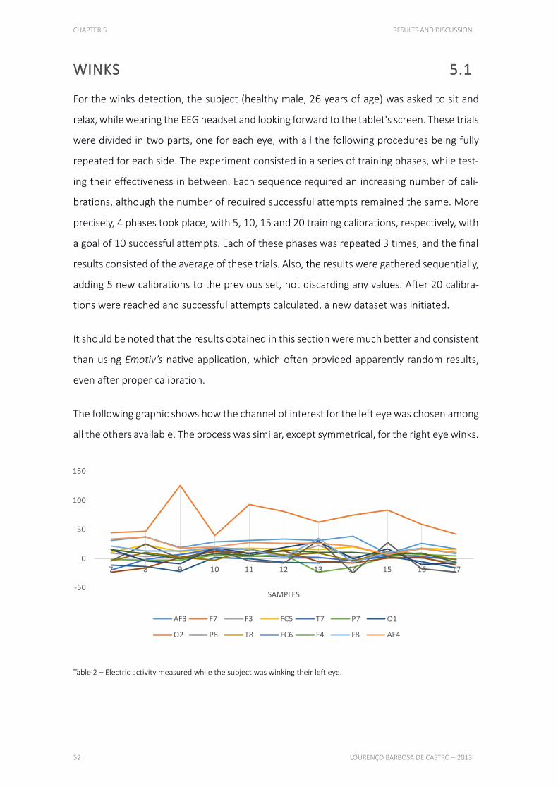
CHAPTER 5 RESULTS AND DISCUSSION
52 LOURENÇO BARBOSA DE CASTRO – 2013
WINKS 5.1
For the winks detection, the subject (healthy male, 26 years of age) was asked to sit and
relax, while wearing the EEG headset and looking forward to the tablet's screen. These trials
were divided in two parts, one for each eye, with all the following procedures being fully
repeated for each side. The experiment consisted in a series of training phases, while test-
ing their effectiveness in between. Each sequence required an increasing number of cali-
brations, although the number of required successful attempts remained the same. More
precisely, 4 phases took place, with 5, 10, 15 and 20 training calibrations, respectively, with
a goal of 10 successful attempts. Each of these phases was repeated 3 times, and the final
results consisted of the average of these trials. Also, the results were gathered sequentially,
adding 5 new calibrations to the previous set, not discarding any values. After 20 calibra-
tions were reached and successful attempts calculated, a new dataset was initiated.
It should be noted that the results obtained in this section were much better and consistent
than using Emotiv’s native application, which often provided apparently random results,
even after proper calibration.
The following graphic shows how the channel of interest for the left eye was chosen among
all the others available. The process was similar, except symmetrical, for the right eye winks.
Table 2 – Electric activity measured while the subject was winking their left eye.
-50
0
50
100
150
7 8 9 10 11 12 13 14 15 16 17
SAMPLES
AF3 F7 F3 FC5 T7 P7 O1
O2 P8 T8 FC6 F4 F8 AF4
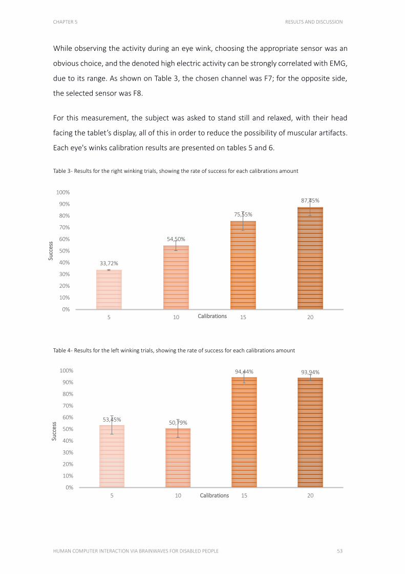
CHAPTER 5 RESULTS AND DISCUSSION
HUMAN COMPUTER INTERACTION VIA BRAINWAVES FOR DISABLED PEOPLE 53
While observing the activity during an eye wink, choosing the appropriate sensor was an
obvious choice, and the denoted high electric activity can be strongly correlated with EMG,
due to its range. As shown on Table 3, the chosen channel was F7; for the opposite side,
the selected sensor was F8.
For this measurement, the subject was asked to stand still and relaxed, with their head
facing the tablet’s display, all of this in order to reduce the possibility of muscular artifacts.
Each eye's winks calibration results are presented on tables 5 and 6.
Table 3 - Results for the right winking trials, showing the rate of success for each calibrations amount
Table 4 - Results for the left winking trials, showing the rate of success for each calibrations amount
33,72%
54,50%
75,55%
87,45%
0%
10%
20%
30%
40%
50%
60%
70%
80%
90%
100%
5 10 15 20
Succ
ess
Calibrations
53,45%50,79%
94,44% 93,94%
0%
10%
20%
30%
40%
50%
60%
70%
80%
90%
100%
5 10 15 20
Succ
ess
Calibrations
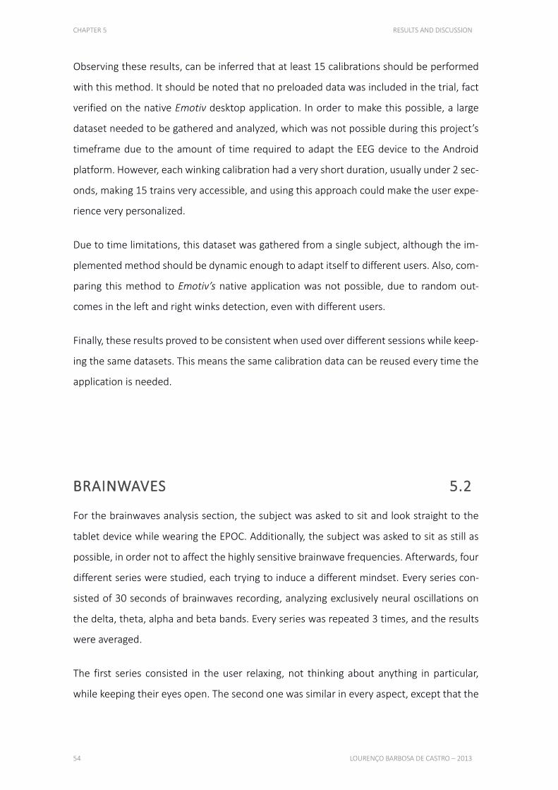
CHAPTER 5 RESULTS AND DISCUSSION
54 LOURENÇO BARBOSA DE CASTRO – 2013
Observing these results, can be inferred that at least 15 calibrations should be performed
with this method. It should be noted that no preloaded data was included in the trial, fact
verified on the native Emotiv desktop application. In order to make this possible, a large
dataset needed to be gathered and analyzed, which was not possible during this project’s
timeframe due to the amount of time required to adapt the EEG device to the Android
platform. However, each winking calibration had a very short duration, usually under 2 sec-
onds, making 15 trains very accessible, and using this approach could make the user expe-
rience very personalized.
Due to time limitations, this dataset was gathered from a single subject, although the im-
plemented method should be dynamic enough to adapt itself to different users. Also, com-
paring this method to Emotiv’s native application was not possible, due to random out-
comes in the left and right winks detection, even with different users.
Finally, these results proved to be consistent when used over different sessions while keep-
ing the same datasets. This means the same calibration data can be reused every time the
application is needed.
BRAINWAVES 5.2
For the brainwaves analysis section, the subject was asked to sit and look straight to the
tablet device while wearing the EPOC. Additionally, the subject was asked to sit as still as
possible, in order not to affect the highly sensitive brainwave frequencies. Afterwards, four
different series were studied, each trying to induce a different mindset. Every series con-
sisted of 30 seconds of brainwaves recording, analyzing exclusively neural oscillations on
the delta, theta, alpha and beta bands. Every series was repeated 3 times, and the results
were averaged.
The first series consisted in the user relaxing, not thinking about anything in particular,
while keeping their eyes open. The second one was similar in every aspect, except that the
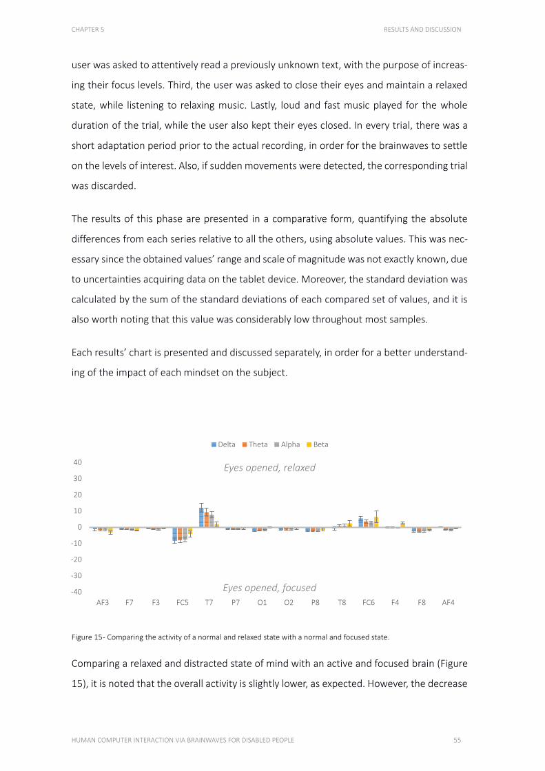
CHAPTER 5 RESULTS AND DISCUSSION
HUMAN COMPUTER INTERACTION VIA BRAINWAVES FOR DISABLED PEOPLE 55
user was asked to attentively read a previously unknown text, with the purpose of increas-
ing their focus levels. Third, the user was asked to close their eyes and maintain a relaxed
state, while listening to relaxing music. Lastly, loud and fast music played for the whole
duration of the trial, while the user also kept their eyes closed. In every trial, there was a
short adaptation period prior to the actual recording, in order for the brainwaves to settle
on the levels of interest. Also, if sudden movements were detected, the corresponding trial
was discarded.
The results of this phase are presented in a comparative form, quantifying the absolute
differences from each series relative to all the others, using absolute values. This was nec-
essary since the obtained values’ range and scale of magnitude was not exactly known, due
to uncertainties acquiring data on the tablet device. Moreover, the standard deviation was
calculated by the sum of the standard deviations of each compared set of values, and it is
also worth noting that this value was considerably low throughout most samples.
Each results’ chart is presented and discussed separately, in order for a better understand-
ing of the impact of each mindset on the subject.
Figure 15 - Comparing the activity of a normal and relaxed state with a normal and focused state.
Comparing a relaxed and distracted state of mind with an active and focused brain (Figure
15), it is noted that the overall activity is slightly lower, as expected. However, the decrease
-40
-30
-20
-10
0
10
20
30
40
AF3 F7 F3 FC5 T7 P7 O1 O2 P8 T8 FC6 F4 F8 AF4
Delta Theta Alpha Beta
Eyes opened, relaxed
Eyes opened, focused
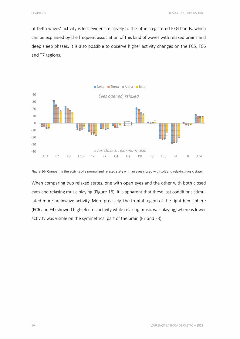
CHAPTER 5 RESULTS AND DISCUSSION
56 LOURENÇO BARBOSA DE CASTRO – 2013
of Delta waves’ activity is less evident relatively to the other registered EEG bands, which
can be explained by the frequent association of this kind of waves with relaxed brains and
deep sleep phases. It is also possible to observe higher activity changes on the FC5, FC6
and T7 regions.
Figure 16 - Comparing the activity of a normal and relaxed state with an eyes closed with soft and relaxing music state.
When comparing two relaxed states, one with open eyes and the other with both closed
eyes and relaxing music playing (Figure 16), it is apparent that these last conditions stimu-
lated more brainwave activity. More precisely, the frontal region of the right hemisphere
(FC6 and F4) showed high electric activity while relaxing music was playing, whereas lower
activity was visible on the symmetrical part of the brain (F7 and F3).
-40
-30
-20
-10
0
10
20
30
40
AF3 F7 F3 FC5 T7 P7 O1 O2 P8 T8 FC6 F4 F8 AF4
Delta Theta Alpha Beta
Eyes opened, relaxed
Eyes closed, relaxing music
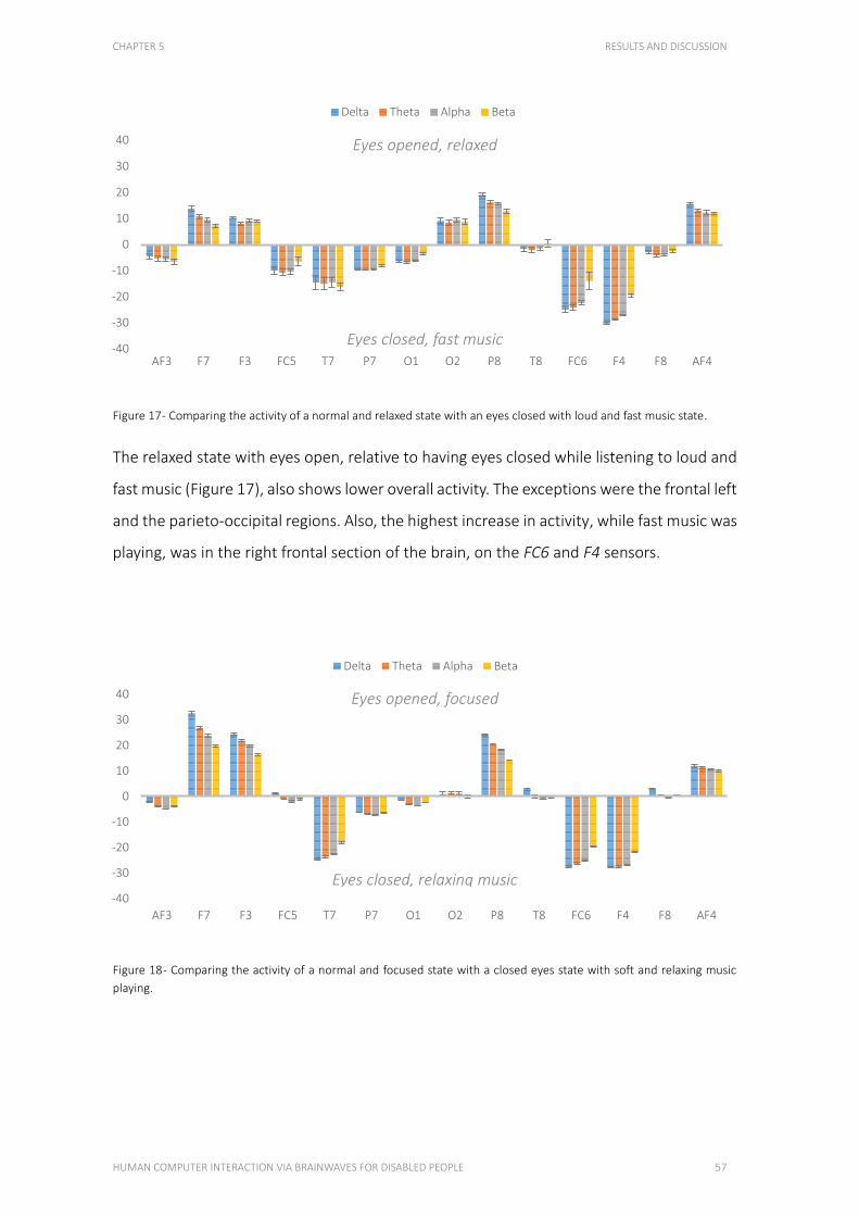
CHAPTER 5 RESULTS AND DISCUSSION
HUMAN COMPUTER INTERACTION VIA BRAINWAVES FOR DISABLED PEOPLE 57
Figure 17 - Comparing the activity of a normal and relaxed state with an eyes closed with loud and fast music state.
The relaxed state with eyes open, relative to having eyes closed while listening to loud and
fast music (Figure 17), also shows lower overall activity. The exceptions were the frontal left
and the parieto-occipital regions. Also, the highest increase in activity, while fast music was
playing, was in the right frontal section of the brain, on the FC6 and F4 sensors.
Figure 18 - Comparing the activity of a normal and focused state with a closed eyes state with soft and relaxing music
playing.
-40
-30
-20
-10
0
10
20
30
40
AF3 F7 F3 FC5 T7 P7 O1 O2 P8 T8 FC6 F4 F8 AF4
Delta Theta Alpha Beta
-40
-30
-20
-10
0
10
20
30
40
AF3 F7 F3 FC5 T7 P7 O1 O2 P8 T8 FC6 F4 F8 AF4
Delta Theta Alpha Beta
Eyes opened, relaxed
Eyes closed, fast music
Eyes opened, focused
Eyes closed, relaxing music
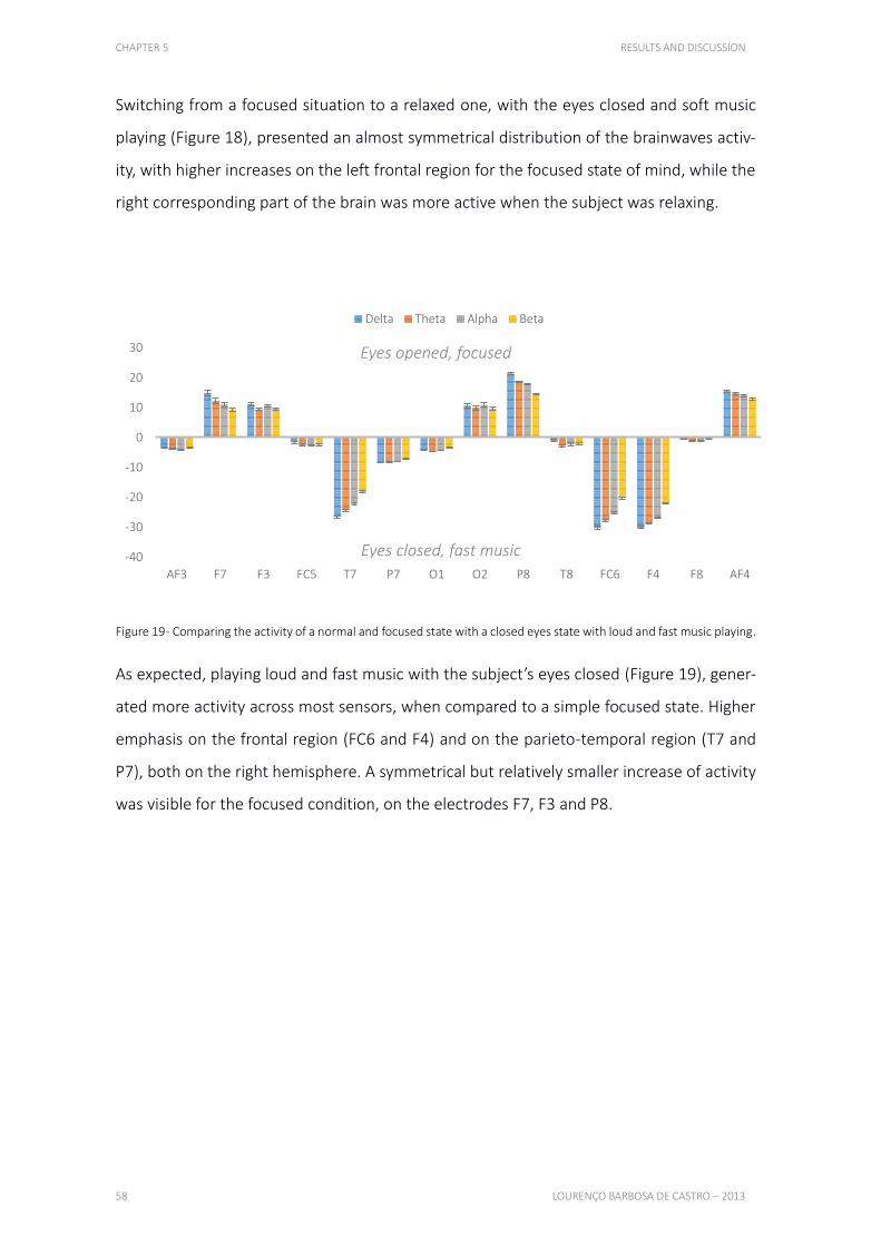
CHAPTER 5 RESULTS AND DISCUSSION
58 LOURENÇO BARBOSA DE CASTRO – 2013
Switching from a focused situation to a relaxed one, with the eyes closed and soft music
playing (Figure 18), presented an almost symmetrical distribution of the brainwaves activ-
ity, with higher increases on the left frontal region for the focused state of mind, while the
right corresponding part of the brain was more active when the subject was relaxing.
Figure 19 - Comparing the activity of a normal and focused state with a closed eyes state with loud and fast music playing.
As expected, playing loud and fast music with the subject’s eyes closed (Figure 19), gener-
ated more activity across most sensors, when compared to a simple focused state. Higher
emphasis on the frontal region (FC6 and F4) and on the parieto-temporal region (T7 and
P7), both on the right hemisphere. A symmetrical but relatively smaller increase of activity
was visible for the focused condition, on the electrodes F7, F3 and P8.
-40
-30
-20
-10
0
10
20
30
AF3 F7 F3 FC5 T7 P7 O1 O2 P8 T8 FC6 F4 F8 AF4
Delta Theta Alpha Beta
Eyes opened, focused
Eyes closed, fast music

CHAPTER 5 RESULTS AND DISCUSSION
HUMAN COMPUTER INTERACTION VIA BRAINWAVES FOR DISABLED PEOPLE 59
Figure 20 - Comparing the activity of two closed eyes states, one with soft relaxing music, other with fast loud music
playing.
Lastly, comparing both closed eyes states (Figure 20), the most likely outcome was visible,
with the loud and fast music producing higher activities, with the exception of the right
occipital area. The impact was more visible on the left frontal region, over the F7 and F3
electrodes.
The most important aspect learned from this experimental phase is that each tested mind
state produced a particular pattern of brainwaves activity, which can be effectively differ-
entiated. For an accessibility approach, detecting when the user switches from a calm and
relaxed situation to a more stressed and nervous condition might be an important alert
signal for family members or health professionals. Additionally, from an autonomy perspec-
tive, an impaired user could see their computer adapt to situational needs, such as display-
ing help topics when frustration is detected, or playing a warning sound when focus is being
lost. Table 5 summarizes these results.
-40
-30
-20
-10
0
10
20
30
40
AF3 F7 F3 FC5 T7 P7 O1 O2 P8 T8 FC6 F4 F8 AF4
Delta Theta Alpha Beta
Eyes closed, fast music
Eyes closed, relaxing music
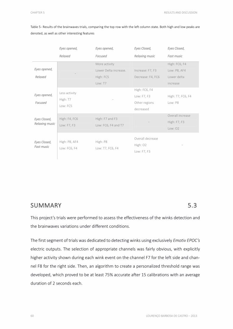
CHAPTER 5 RESULTS AND DISCUSSION
60 LOURENÇO BARBOSA DE CASTRO – 2013
Table 5 - Results of the brainwaves trials, comparing the top row with the left column state. Both high and low peaks are
denoted, as well as other interesting features
SUMMARY 5.3
This project’s trials were performed to assess the effectiveness of the winks detection and
the brainwaves variations under different conditions.
The first segment of trials was dedicated to detecting winks using exclusively Emotiv EPOC’s
electric outputs. The selection of appropriate channels was fairly obvious, with explicitly
higher activity shown during each wink event on the channel F7 for the left side and chan-
nel F8 for the right side. Then, an algorithm to create a personalized threshold range was
developed, which proved to be at least 75% accurate after 15 calibrations with an average
duration of 2 seconds each.
Eyes opened,
Relaxed
Eyes opened,
Focused
Eyes Closed,
Relaxing music
Eyes Closed,
Fast music
Eyes opened,
Relaxed -
More activity
Lower Delta increase.
High: FC5
Low: T7
Increase: F7, F3
Decrease: F4, FC6
High: FC6, F4
Low: P8, AF4
Lower delta
increase
Eyes opened,
Focused
Less activity
High: T7
Low: FC5
-
High: FC6, F4
Low: F7, F3
Other regions
decreased
High: T7, FC6, F4
Low: P8
Eyes Closed,
Relaxing music High: F4, FC6
Low: F7, F3
High: F7 and F3
Low: FC6, F4 and T7 -
Overall increase
High: F7, F3
Low: O2
Eyes Closed,
Fast music High: P8, AF4
Low: FC6, F4
High: P8
Low: T7, FC6, F4
Overall decrease
High: O2
Low: F7, F3
-
CHAPTER 5 RESULTS AND DISCUSSION
HUMAN COMPUTER INTERACTION VIA BRAINWAVES FOR DISABLED PEOPLE 61
For the second part of trials, brainwaves were measured under four circumstances: while
the user was relaxing, reading an unknown text, listening to slow and relaxing music and,
lastly, listening to loud and fast music. These results showed distinct brainwave patterns in
each condition, which could be used to differentiate states of mind and make computers
adapt to their users.

63
CHAPTER 6
CONCLUSION AND FUTURE IMPROVEMENTS This chapter presents a conclusion, in which this project’s developments and contributions
are evaluated and compared to the existing technology and software, specifically with the
device’s manufacturer’s native applications. The chapter ends describing which specific
improvements should be made to the developed application, focusing on accessibility
features. These improvements take in consideration the different sections of the application
and the different types of neurophysiological signals acquired.
6.1. CONCLUSION 64
6.2. FUTURE IMPROVEMENTS 64
CHAPTER 6 CONCLUSION AND FUTURE IMPROVEMENTS
64 LOURENÇO BARBOSA DE CASTRO – 2013
CONCLUSION 6.1
This project improved the way brain-computer interfaces interact with mobile devices by
introducing a functional application for a well-established BCI device. This development
could help portable EEG technology becoming more popular, more useful and more effi-
cient. Additionally, using Android’s open-source platform can bring better collaborative
ideas to this field, perfecting such applications faster.
Although more limited than the native Emotiv application, the software developed in this
project proved to be more efficient establishing interactions between a user and a mobile
computer. In order for brain-computer interfaces to become a part of our daily lives, im-
proved efficiency must be achieved, since several risk factors can arise when interacting
with the surrounding environment using only cerebral activity.
Using this application could improve certain types of people’s autonomy and quality of life,
as well as help computers understand and adapt to their users and to how they feel. This
could lead to inspiring and fascinating new technologies, which could help shape our fu-
ture.
FUTURE IMPROVEMENTS 6.2
Due to the wide range of tasks required for this project, several different types of improve-
ments are still possible.
More elaborate trials
The most important additional work that should be mentioned is further testing on differ-
ent types of subjects. Time limitations excluded this possibility, which, taking the wide va-
riety of EEG patterns in consideration, makes it a very important step in this kind of project.
CHAPTER 6 CONCLUSION AND FUTURE IMPROVEMENTS
HUMAN COMPUTER INTERACTION VIA BRAINWAVES FOR DISABLED PEOPLE 65
This type of tests could greatly improve the detection of muscular events, as well as the
interpretation of the different range of neural oscillations.
Usage of more electrodes
For the EMG analysis, which made wink detection possible, only two electrodes were used.
Although it proved to be fairly efficient for the implemented features, using a wider range
of electrodes could make more kinds of detections possible, and also improve the ones
already available on the application.
Better EEG understanding
Concerning the EEG analysis, a better understanding of the electric output provided by the
EPOC should be an important factor for a more accurate display of the voltage range, which
could make this application more interesting in terms of science research purposes.
Signal processing improvements
Better pre-processing techniques should be explored, in order to isolate the brain-related
bands from the myography-related signals. Direct signal features, specifically focused on
the detection of neural oscillations bands, should be used, making the application react to
the user’s current mental state. Also, suitable machine learning approaches should be im-
plemented, increasing the efficiency of the events’ detection.
Compatibility with other devices
Considering the amount of BCI devices currently available, and several others already an-
nounced, this project’s application was developed to be easily adaptable. However, no ad-
ditional devices are actually supported, which could be an interesting feature to imple-
ment, increasing its usability and market reach.

BIBLIOGRAPHY
HUMAN COMPUTER INTERACTION VIA BRAINWAVES FOR DISABLED PEOPLE 67
BIBLIOGRAPHY
Allisona, Brendan Z., Dennis J. McFarlandb, Gerwin Schalkb, Shi Dong Zhenga, Melody
Moore Jacksonc, and Jonathan R. Wolpaw. "Towards an independent brain–computer
interface using steady state visual evoked potentials." Clinical Neurophysiology, February
2008: 399–408.
Asus. Nexus 7. ASUSTeK Computer Inc. 2012.
http://www.asus.com/Tablets_Mobile/Nexus_7/ (accessed September 20, 2013).
Badcock, Nicholas A., Petroula Mousikou, Yatin Mahajan, Peter de Lissa, Johnson Thie,
and Genevieve McArthur. "Validation of the Emotiv EPOC® EEG gaming system for
measuring research quality auditory ERPs." PeerJ, February 19, 2013.
Baxter, Mark G. "Involvement of medial temporal lobe structures in memory and
perception." Neuron, March 2009: 667-677.
Berger, H. "Über das Elektrenkephalogramm des Menschen." Archiv für Psychiatrie und
Nervenkrankheiten, 1929: 527-570.
Bland, Brian H., and Scott D. Oddie. "Theta band oscillation and synchrony in the
hippocampal formation and associated structures: The case for its role in sensorimotor
integration." Behavioural Brain Research, December 2001: 119-136.
Blankertz, Benjamin, Motoaki Kawanabe Ryota Tomioka, Friederike U. Hohlefeld, Vadim
Nikulin, and Klaus-robert Müller. "Invariant Common Spatial Patterns: Alleviating
Nonstationarities in Brain-Computer Interfacing." In Advances in Neural Information
Processing Systems . Neural Information Processing Systems Foundation, 2008.
BrainGate. BrainGate official website. 2009. http://www.braingate.com/ (accessed
September 6, 2013).
BIBLIOGRAPHY
68 LOURENÇO BARBOSA DE CASTRO – 2013
Breen, Randy. "Demonstration of Brain Computer Interface using the Emotiv Epoc."
Computer Systems Colloquium. Stanford University. April 2, 2008.
Campbell, Andrew, Choudhury, Tanzeem; Hu, Shaohan; Lu, Hong; Mukerjee, Matthew K.;
Rabbi, Mashfiqui; Raizada, Rajeev D.S. "NeuroPhone: brain-mobile phone interface using
a wireless EEG headset." Proceedings of the second ACM SIGCOMM workshop on
Networking, systems, and applications on mobile handhelds. New York, NY: Association
for Computing Machinery, 2010. 3-8.
Castermans, Thierry. "Detecting biosignals with the Emotiv EPOC headset: a Review."
Tangible Feelings : a Symposium on EEG. Brussels: TCTS Lab, 2011.
CBSNews. "Harnessing The Power Of The Brain." CBSNews - 60 Minutes. Denise Schrier
Cetta. November 2, 2008.
http://www.cbsnews.com/stories/2008/10/31/60minutes/main4560940.shtml
(accessed July 25, 2013).
Chapin, JK, KA Moxon, RS Markowitz, and MA Nicolelis. "Real-time control of a robot arm
using simultaneously recorded neurons in the motor cortex." July 1999: 664-70.
Clarke, Adam R, Robert J Barry, Rory McCarthy, and Mark Selikowitz. "EEG-defined
subtypes of children with attention-deficit/hyperactivity disorder." Clinical
Neurophysiology, November 2001: 2098–2105.
Cofre, J.P., G. Moraga, C. Rusu, and I Mercado. "Developing a Touchscreen-based
Domotic Tool for Users with Motor Disabilities." Ninth International Conference on
Information Technology: New Generations. Las Vegas, NV: Conference Publishing
Services, 2012. 696 - 701.
Culham, Jody C, and Kenneth F Valyear. "Human parietal cortex in action." Current
Opinion in Neurobiology, April 2006: 205-212.
Delgado, JM; Mark, V; Sweet, W; Ervin, F; Weiss, G; Bach-Y-Rita, G; Hagiwara, R.
"Intracerebral radio stimulation and recording in completely free patients." October
1968: 329-40.
BIBLIOGRAPHY
HUMAN COMPUTER INTERACTION VIA BRAINWAVES FOR DISABLED PEOPLE 69
Emotiv. Emotiv Emerges from Stealth to Revolutionize. Press Release, San Francisco, CA:
Technology Venture Partners Pty., 2007.
Emotiv. Emotiv Systems Website. 2013. www.emotiv.com (accessed June 26, 2013).
Farwell, LA, and E Donchin. "Talking off the top of your head: toward a mental prosthesis
utilizing event-related brain potentials." Electroencephalography and clinical
neurophysiology, December 1988: 510-523.
Fetz, EE. "Operant Conditioning of Cortical Unit Activity." Science 163, no. 3870
(February 1969): 955-58.
Fieldtrip. Donders Centre for Cognitive Neuroimaging. January 23, 2013.
http://fieldtrip.fcdonders.nl/template/layout (accessed September 28, 2013).
Foresman, Pearson Scott. Wikimedia. September 13, 2009.
http://commons.wikimedia.org/wiki/File:Neuron_(PSF).png (accessed September 28,
2013).
Gamboa, Hugo. December 2005. http://en.wikipedia.org/wiki/File:Eeg_delta.svg
(accessed September 28, 2013).
Gamboa, Hugo. December 2005. http://en.wikipedia.org/wiki/File:Eeg_theta.svg
(accessed September 28, 2013).
Gamboa, Hugo. December 2005. http://en.wikipedia.org/wiki/File:Eeg_alpha.svg
(accessed September 28, 2013).
Gamboa, Hugo. December 2005. http://en.wikipedia.org/wiki/File:Eeg_beta.svg
(accessed September 28, 2013).
Ganapati, Priya. "Winter Olympics to demo lighting controlled by thoughts." Wired
Magazine. Condé Nast. March 2, 2010.
http://www.wired.com/gadgetlab/2010/02/thought-controlled-lights/ (accessed
September 10, 2013).
BIBLIOGRAPHY
70 LOURENÇO BARBOSA DE CASTRO – 2013
Gomez-Gil, Jaime, Israel San-Jose-Gonzalez, Luis Fernando Nicolas-Alonso, and Sergio
Alonso-Garcia. "Steering a Tractor by Means of an EMG-Based Human-Machine
Interface." Sensors, 2011: 7110–7126.
Google. "PropertyChangeListener." Android Developers Website. 2013.
http://developer.android.com/reference/java/beans/PropertyChangeListener.html
(accessed September 3, 2013).
Google. "USB Host." Android Developers Website. 2013.
http://developer.android.com/guide/topics/connectivity/usb/host.html (accessed
September 3, 2013).
Haglund, MM, MS Berger, M Shamseldin, E Lettich, and GA Ojemann. "Cortical
localization of temporal lobe language sites in patients with gliomas." Neurosurgery,
April 1994: 567-576.
Hochberg, Leigh R.; Bacher, Daniel; Jarosiewicz, Beata; Masse, Nicolas Y.; Simeral, John
D.; Vogel, Joern; Haddadin, Sami; Liu, Jie; Cash, Sydney S.; Smagt, Patrick van der;
Donoghue, John P. "Reach and grasp by people with tetraplegia using a neurally
controlled robotic arm." Nature, May 17, 2012: 372-375.
Hockenberry, John. "The Next Brainiacs." Wired Magazine. August 2001.
http://www.wired.com/wired/archive/9.08/assist_pr.html (accessed September 5,
2013).
Hoffmann, Ulrich. Bayesian machine learning applied in a brain-computer interface for
disabled users. Lausanne: École Polytechnique Fédérale de Lausanne, 2007.
Hoffmann, Ulrich, Jean-Marc Vesin, Touradj Ebrahimi, and Karin Diserens. "An efficient
P300-based brain-computer interface for disabled subjects." Journal of Neuroscience
Methods, January 15, 2008: 115-125.
InteraXon. InteraXon - Though Controlled Computing. 2010. http://interaxon.ca/
(accessed June 26, 2013).
BIBLIOGRAPHY
HUMAN COMPUTER INTERACTION VIA BRAINWAVES FOR DISABLED PEOPLE 71
International Data Corporation. "Apple Cedes Market Share in Smartphone Operating
System Market as Android Surges and Windows Phone Gains, According to IDC." IDC
Website. August 7, 2013. http://www.idc.com/getdoc.jsp?containerId=prUS24257413
(accessed September 15, 2013).
King, Christine E, Po T Wang, Luis A Chui, An H Do, and Zoran Nenadic. "Operation of a
brain-computer interface walking simulator for individuals with spinal cord injury."
Journal of Neuroengineering and Rehabilitation, 2013: 77.
Kovach, Steve. "How Android Grew To Be More Popular Than The iPhone." Business
Insider. August 13, 2013. http://www.businessinsider.com/history-of-android-2013-
8?op=1 (accessed September 15, 2013).
Li, Kun, Ravi Sankar, Y. Arbel, and E. Donchin. "Single trial independent component
analysis for P300 BCI system." Annual International Conference of the IEEE Engineering
in Medicine and Biology Society. Minneapolis, MN, 2009. 4035-4038.
Li, Shan. Mind reading is on the market. Los Angeles, CA: Los Angeles Times, 2010.
Middendorf, M, G McMillan, G Calhoun, and KS Jones. "Brain-computer interfaces based
on the steady-state visual-evoked response." Transactions on Rehabilitation Engineering,
June 2000: 211-214.
NeuroSky. NeuroSky official website. 2012. http://www.neurosky.com/ (accessed
September 2, 2013).
Niedermeyer, Ernst, and Fernando Lopes Silva. Electroencephalography: Basic Principles,
Clinical Applications and Related Fields. Fifth edition. Lippincott Williams and Wilkins,
2004.
Nyström, Pär, Therese Ljunghammar, Kerstin Rosander, and Claes von Hofsten. "Using
mu rhythm desynchronization to measure mirror neuron activity in infants."
Developmental Science, June 18, 2010: 327-335.

BIBLIOGRAPHY
72 LOURENÇO BARBOSA DE CASTRO – 2013
Pais-Vieira, M, M Lebedev, C Kunicki, J Wang, and MA Nicolelis. "A Brain-to-Brain
Interface for Real-Time Sharing of Sensorimotor Information." February 2013: 1-10.
Palva, Satu, and J. Matias Palva. "New vistas for alpha-frequency band oscillations."
Trends in Neurosciences, April 2007: 150-158.
Rangaswamya, Madhavi; Porjesz, Bernice; Chorliana, David B; Wanga, Kongming; Jonesa,
Kevin A; Bauerb, Lance O; Rohrbaughf, John; O’Connorc, Sean J; Kupermand, Samuel;
Reiche, Theodore; Begleitera, Henri. "Beta power in the EEG of alcoholics." Biological
Psychiatry, October 15, 2002: 831–842.
Schmidt, E.M. "Single neuron recording from motor cortex as a possible source of signals
for control of external devices." Annals of Biomedical Engineering, July 1, 1980: 339-349.
Schmidt, EM, JS McIntosh, L Durelli, and MJ Bak. "Fine control of operantly conditioned
firing patterns of cortical neurons." September 1978: 349-69.
Semendeferi, K, A Lu, N Schenker, and H Damasio. "Humans and great apes share a large
frontal cortex." Nature Neuroscience, March 2002: 272-276.
Simeral, J D, S-P Kim, M J Black, J P Donoghue, and L R Hochberg. "Neural control of
cursor trajectory and click by a human with tetraplegia 1000 days after implant of an
intracortical microelectrode array." Journal of Neural Engineering, April 2011.
Squire, L.R., and S. Zola-Morgan. "The medial temporal lobe memory system." Science,
September 20, 2001: 1380-1386.
Stuss, Donald T., Catherine A. Gow, and C. Ross Hetherington. ""No longer Gage": frontal
lobe dysfunction and emotional changes." Journal of Consulting and Clinical Psychology,
June 1992: 349-359.

BIBLIOGRAPHY
HUMAN COMPUTER INTERACTION VIA BRAINWAVES FOR DISABLED PEOPLE 73
Tam, Wing-Kin, Kai-yu Tong, Fei Meng, and Shangkai Gao. "A Minimal Set of Electrodes
for Motor Imagery BCI to Control an Assistive Device in Chronic Stroke Subjects: A Multi-
Session Study." Transactions on Neural Systems & Rehabilitation Engineering, December
2011: 617-627.
Turner, MR, A Al-Chalabi, MJ Parton, CE Shaw, and PN Leigh. "Prolonged survival in
motor neuron disease: a descriptive study of the King's database 1990-2002." Journal of
neurology, neurosurgery, and psychiatry, July 2003: 995-997.
Wachspress, Samuel. "EEG." University of Illinois - Engineering Wiki. December 15, 2010.
https://wiki.engr.illinois.edu/display/BIOE414/EEG (accessed September 7, 2013).
Weinstein, G W. "Clinical aspects of the visually evoked potential." Transcriptions of the
American Ophthalmological Society, 1977: 627–673.
Wolpaw, Jonathan R.; Birbaumer, Niels; Heetderks, William J.; McFarland, Dennis J.;
Peckham, P. Hunter; Schalk, Gerwin; Donchin, Emanuel; Quatrano, Louis A.; Robinson,
Charles J.; Vaughan, Theresa M. "Brain-computer interface technology: a review of the
first international meeting." Transactions on Rehabilitation Engineering, June 2000: 164
- 173.
World Health Organization. "The world health report 2002 - Reducing Risks, Promoting
Healthy Life." 2002.
Wright, Frankling Pierce. "Emochat: Emotional instant messaging with the EPOC
headset." Edited by University of Maryland. 2010.
http://search.proquest.com/docview/851703167 (accessed August 28, 2013).
Yalçin, AD, A Kaymaz, and H Forta. "Reflex occipital lobe epilepsy." Seizure, September
2000: 436-441.
Zhang, Yan, Yonghong Chen, Steven L. Bressler, and Mingzhou Ding. "Response
preparation and inhibition: The role of the cortical sensorimotor beta rhythm."
Neuroscience, September 22, 2008: 238-246.