Embed Size (px)
Citation preview
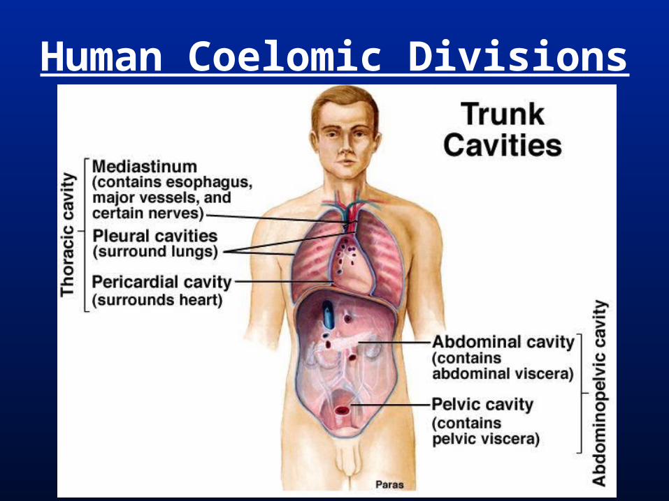
Human Coelomic Divisions
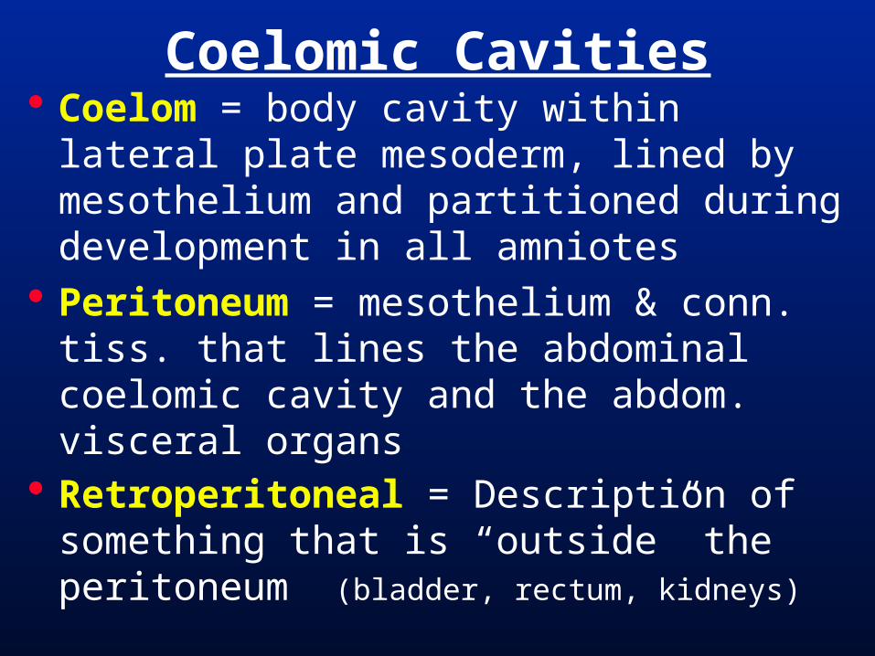
Coelom = body cavity within lateral plate mesoderm, lined by mesothelium and partitioned during development in all amniotes
Peritoneum = mesothelium & conn. tiss. that lines the abdominal coelomic cavity and the abdom. visceral organs
Retroperitoneal = Description of something that is “outside” the peritoneum (bladder, rectum, kidneys)
Coelomic Cavities
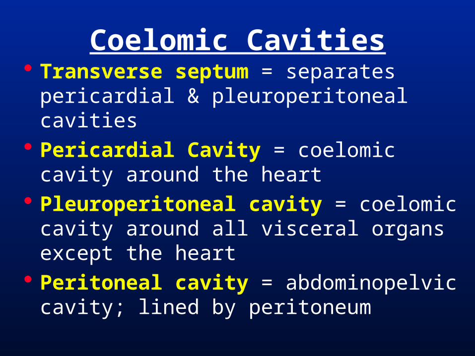
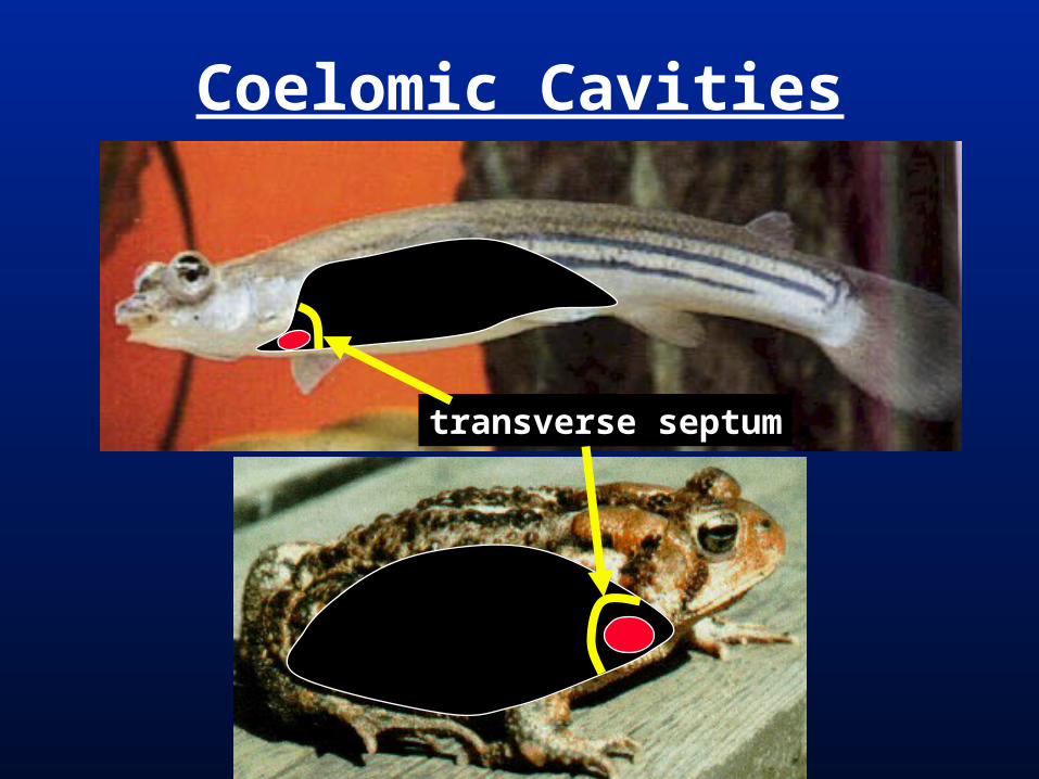
Transverse septum = separates pericardial & pleuroperitoneal cavities
Pericardial Cavity = coelomic cavity around the heart
Pleuroperitoneal cavity = coelomic cavity around all visceral organs except the heart
Peritoneal cavity = abdominopelvic cavity; lined by peritoneum
Coelomic Cavities
Coelomic Cavities
transverse septum

Pleural / Pulmonary Cavity = coelomic cavity around the lungs
evolved independently in reptiles and mammals
Pulmonary fold = (in reptiles) forms along the dorsal midline of the pleuroperitoneal cavity...
grows ventrolaterally around each lung. Each lung in separate pleural cavity.
Coelomic Cavities
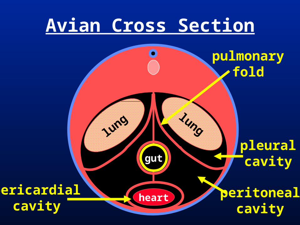
Avian Cross Section
gut
lunglung
heart
pulmonaryfold
pleuralcavity
peritonealcavity
pericardialcavity

Unique to CrocodyliansPost Hepatic “Diaphragm”

Coelomic (pleuroperitoneal) fold = in mammals, forms transversely across the dorsal pleuroperitoneal cavity, behind the lungs and...
grows ventrally to meet the transverse septum.
Diaphragm = mucularized coelomic fold separating pleural and peritoneal cavities (anterior to the liver)
Mammal Coelomic Cavities
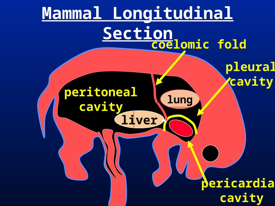
Mammal Longitudinal Section
lung
liver
pleuralcavity
peritonealcavity
pericardialcavity
coelomic fold
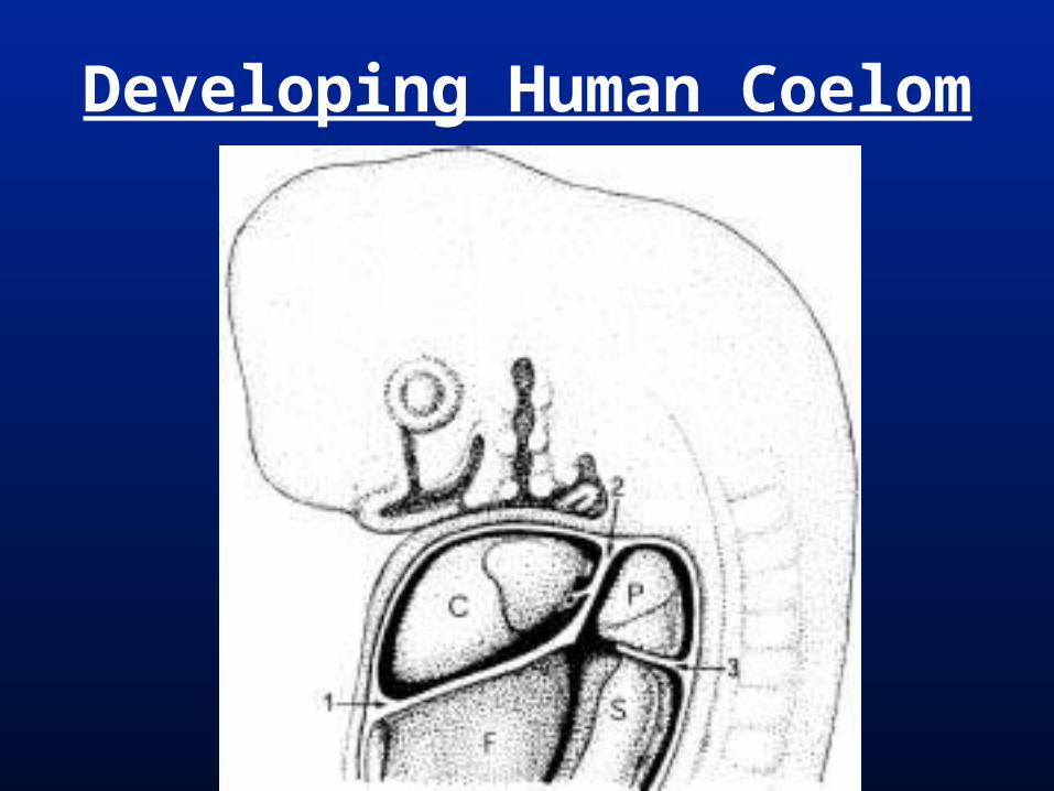
Developing Human Coelom

Coelomic fold component of diaphragm becomes muscularized.
Transverse septum component of diaphragm become the central tendon.
Mammalian Diaphragm

Human Coelomic Divisions
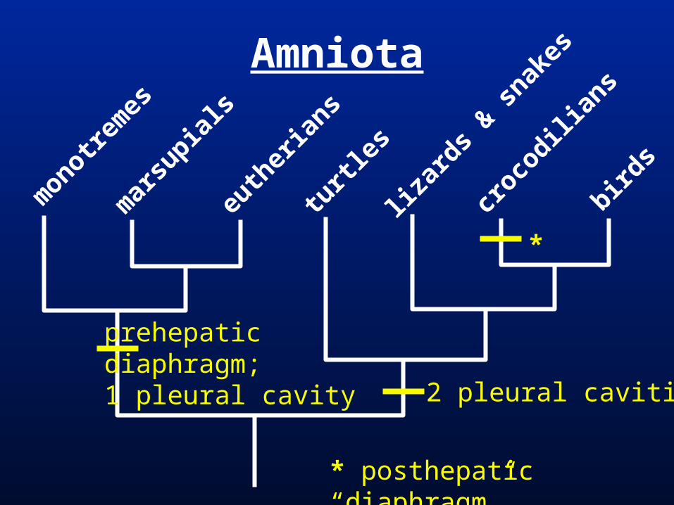
Amniota
monotre
mes
mar
supia
ls
euth
eria
ns
turtl
es
lizar
ds & s
nakes
croco
dilian
s
birds
prehepaticdiaphragm;1 pleural cavity 2 pleural cavities
* posthepatic “diaphragm”
*
Human Digestive System (and assoc. digestive organ) Development
General Digestive Terms
Human Digestive Tube Overview (Oral Cavity to Anus)
Human Associated Digestive Organ Development and Overview
Comparative Digestive Anatomy
Digestive System
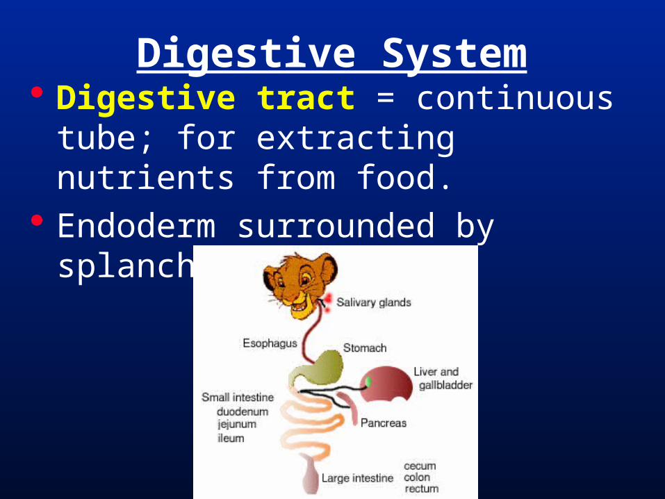
Digestive tract = continuous tube; for extracting nutrients from food.
Endoderm surrounded by splanchnic mesoderm.
Digestive System
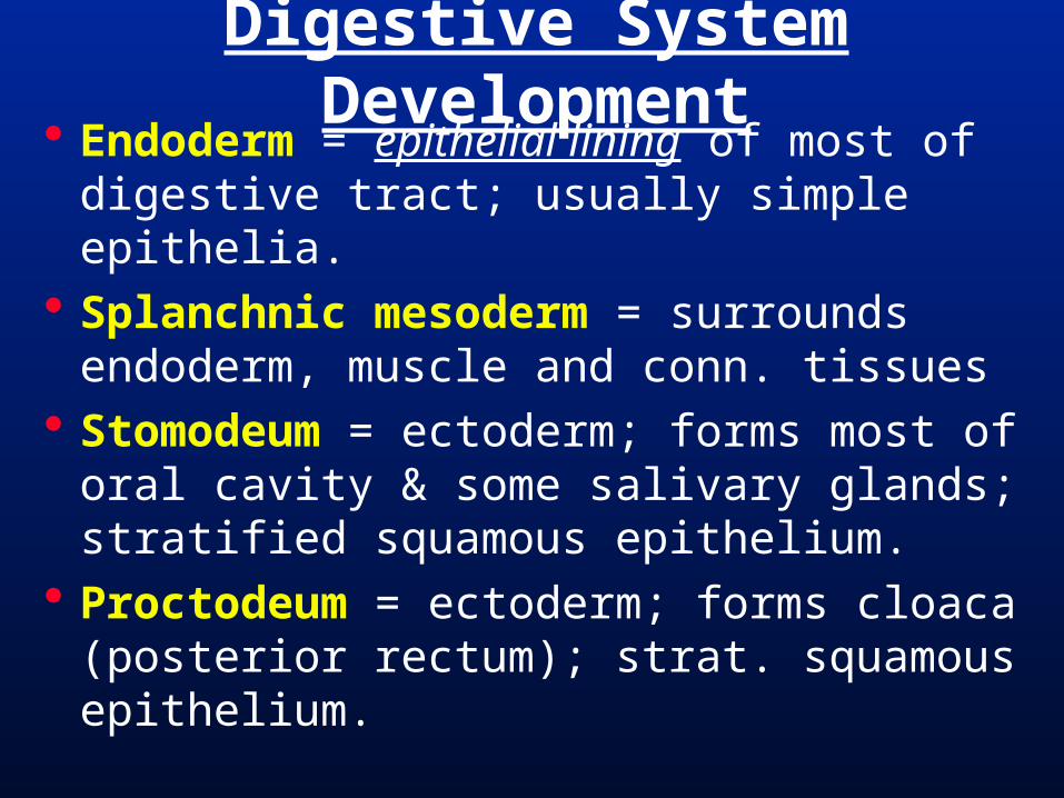
Endoderm = epithelial lining of most of digestive tract; usually simple epithelia.
Splanchnic mesoderm = surrounds endoderm, muscle and conn. tissues
Stomodeum = ectoderm; forms most of oral cavity & some salivary glands; stratified squamous epithelium.
Proctodeum = ectoderm; forms cloaca (posterior rectum); strat. squamous epithelium.
Digestive System Development
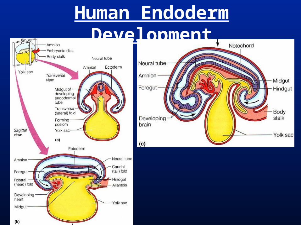
Human Endoderm Development

Human Endoderm Development
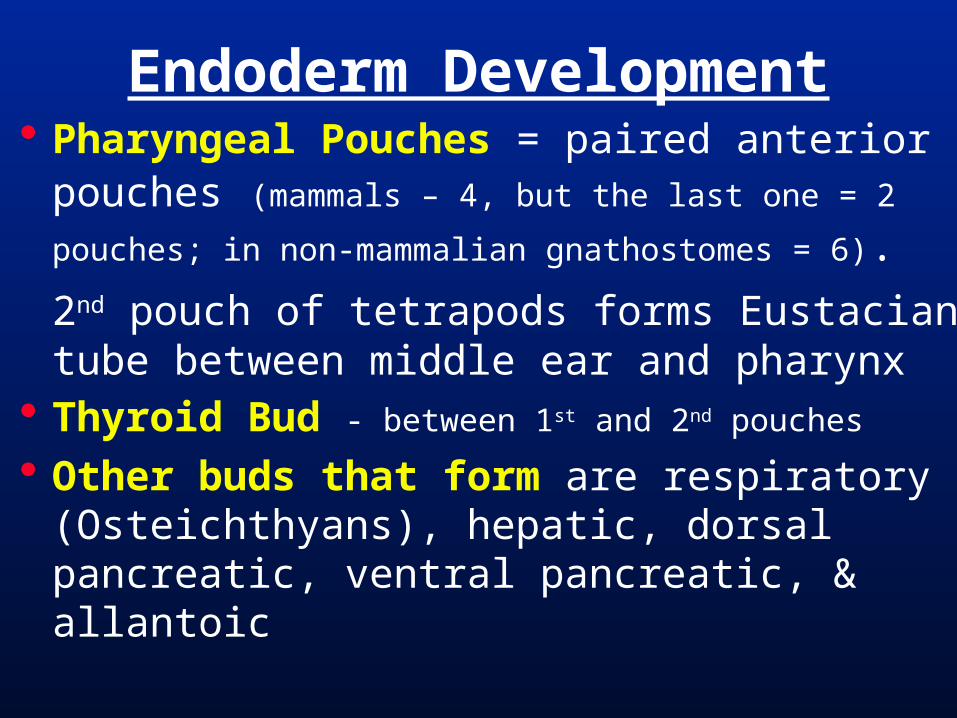
Pharyngeal Pouches = paired anterior pouches (mammals – 4, but the last one = 2
pouches; in non-mammalian gnathostomes = 6).
2nd pouch of tetrapods forms Eustacian tube between middle ear and pharynx
Thyroid Bud - between 1st and 2nd pouches
Other buds that form are respiratory (Osteichthyans), hepatic, dorsal pancreatic, ventral pancreatic, & allantoic
Endoderm Development
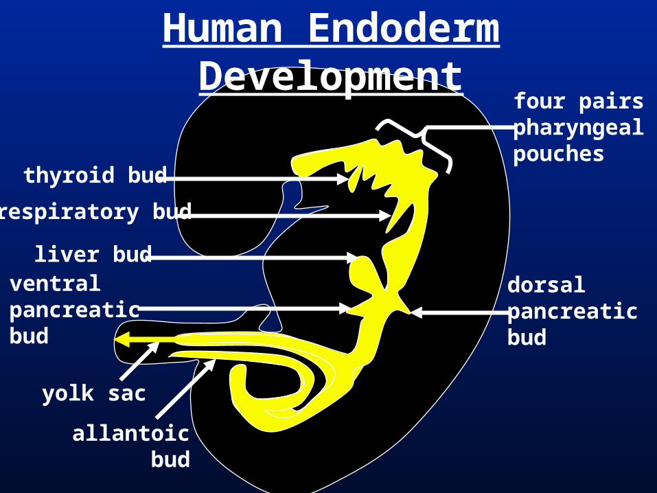
Human Endoderm Development
thyroid bud
respiratory bud
yolk sac
allantoicbud
liver budventralpancreaticbud
dorsalpancreaticbud
four pairspharyngealpouches

Liver = Largest coelomic organ; regulates chemical metabolism (detoxifies, glycogen storage, secretes serum albumin) & produces bile (emulsifies fats & contains RBC waste).
Develops from ventral hepatic bud from duodenum in ventral mesentary. Mostly endodermal; a little lateral plate mesoderm.
Bile released into duodenum via hepatic duct then common bile duct.
Gallbladder = Stores bile; connected to bile duct through cystic duct.
Human Liver
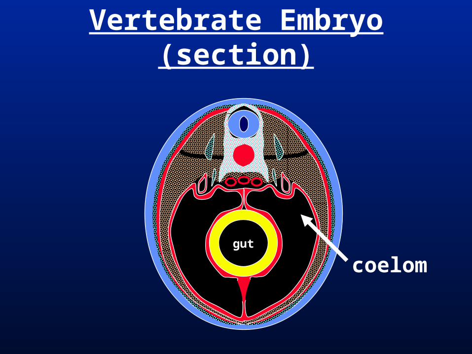
Vertebrate Embryo (section)
gut
coelom

Vertebrate Embryo (section)
gut
coelom
dorsalmesentery(splanchnic mesoderm)
hepatic bud
ventral mesentery(splanchnic mesoderm)
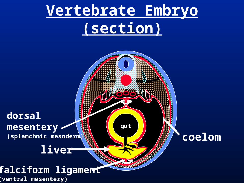
Vertebrate Embryo (section)
gut
coelom
dorsalmesentery(splanchnic mesoderm)
liver
falciform ligament(ventral mesentery)

Pancreas = A distinct gland located along the duodenum with endocrine (sugar metabolism regulation) and exocrine function (digestive enzyme secretion).
Endodermal.
Develops from dorsal and ventral pancreatic buds off of duodenum.
Human digestive enzymes enter intestine through vent. pancreatic duct connecting to common bile duct.
Human Pancreas

Pancreas Development

Human Digestive DevelopmentDevelop. Adult Human Epithelial Devel. Outpouchings
stomodeum anterior oral & nasal cavities
parotid salivary glands
pharynx (with openings)
oropharynx & nasopharynx(only Eustacean tube)
submandibular & sublingual salivary glands
thyroid from thyroid budtrachea & lungs from resp. bud
esophagus long -
stomach present -
intestine small intestine(duodenum, jejunoileum)
colon
liver + ducts from hepatic budpancreas from 2 pancreatic buds
cecum / appendixur. bladder in part from allantois
proctodeum rectum -
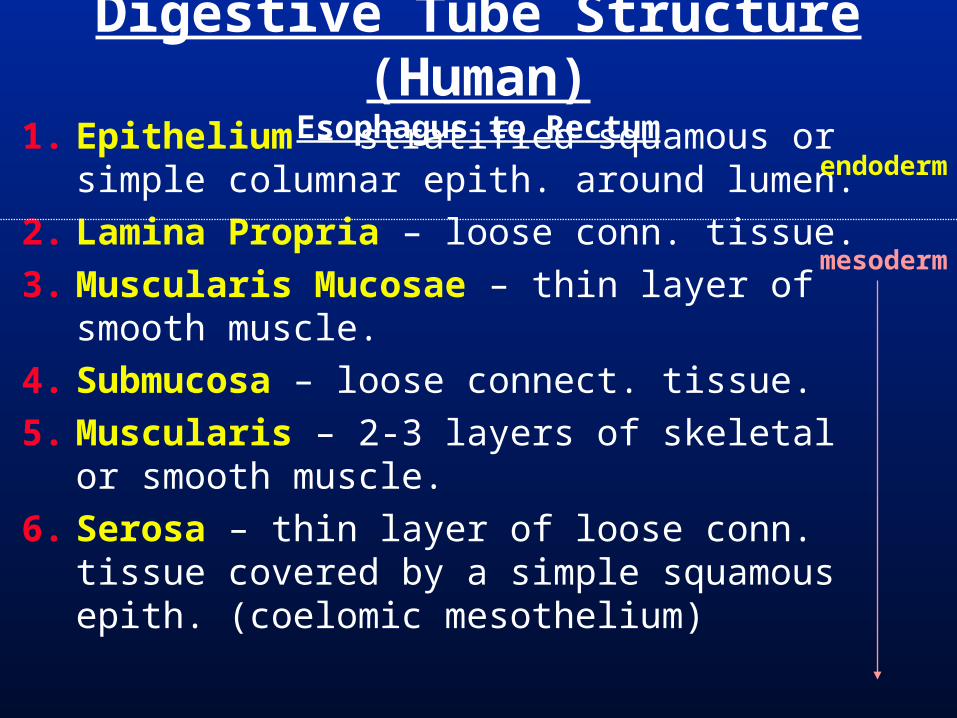
1. Epithelium – stratified squamous or simple columnar epith. around lumen.
2. Lamina Propria – loose conn. tissue.
3. Muscularis Mucosae – thin layer of smooth muscle.
4. Submucosa – loose connect. tissue.
5. Muscularis – 2-3 layers of skeletal or smooth muscle.
6. Serosa – thin layer of loose conn. tissue covered by a simple squamous epith. (coelomic mesothelium)
Digestive Tube Structure (Human)Esophagus to Rectum
endoderm
mesoderm
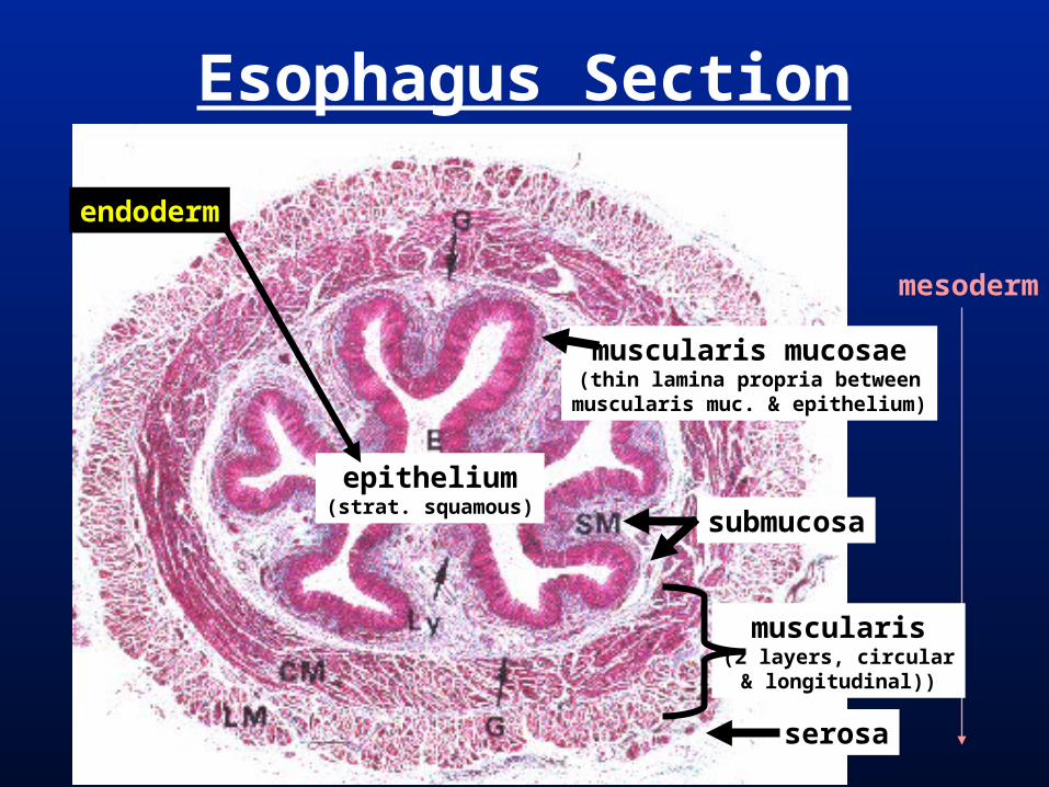
Esophagus Section
mesoderm
epithelium(strat. squamous)
muscularis mucosae(thin lamina propria between
muscularis muc. & epithelium)
submucosa
muscularis(2 layers, circular& longitudinal))
serosa
endoderm
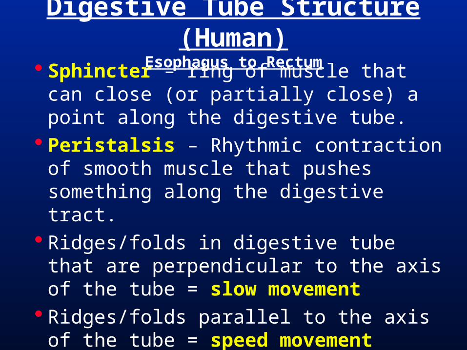
Sphincter – ring of muscle that can close (or partially close) a point along the digestive tube.
Peristalsis – Rhythmic contraction of smooth muscle that pushes something along the digestive tract.
Ridges/folds in digestive tube that are perpendicular to the axis of the tube = slow movement
Ridges/folds parallel to the axis of the tube = speed movement
Digestive Tube Structure (Human)Esophagus to Rectum


Cleft Palate Cleft Palate = Secondary palate doesn’t fuse
along midline during development.
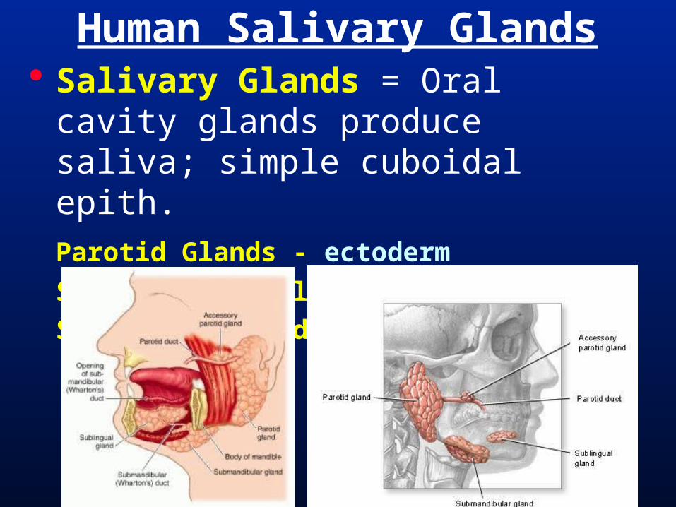
Salivary Glands = Oral cavity glands produce saliva; simple cuboidal epith. Parotid Glands - ectoderm
Submandibular Glands - endoderm
Sublingual Glands - endoderm
Human Salivary Glands
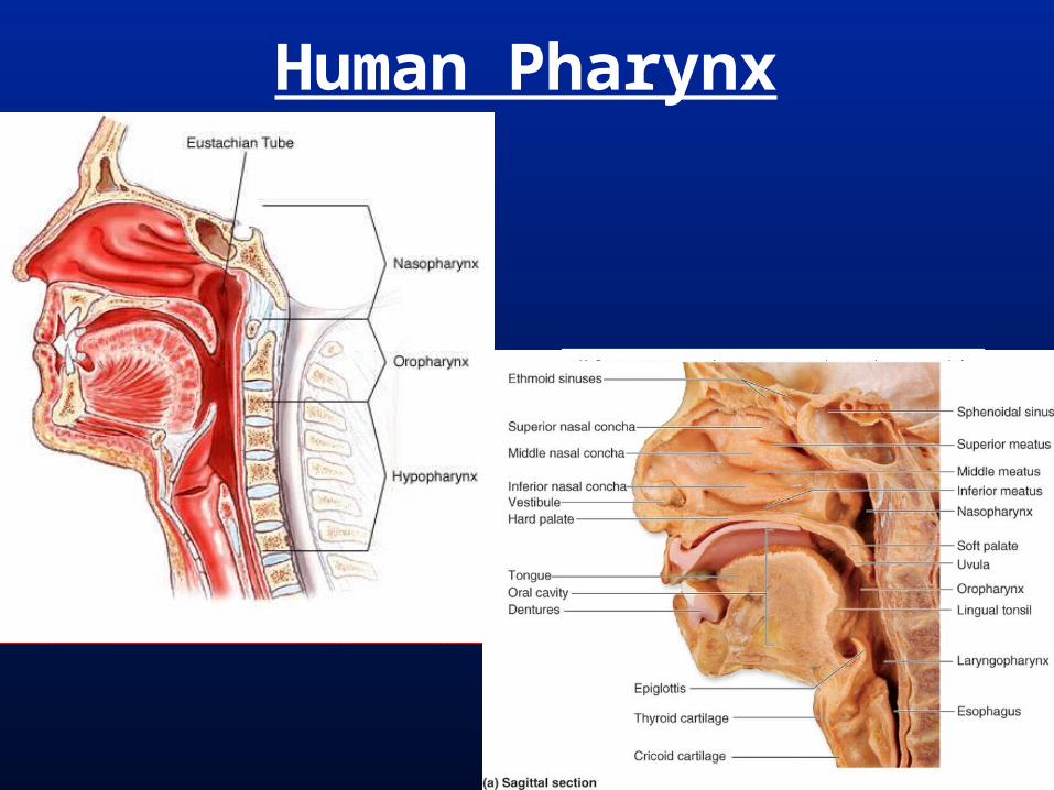
Human Pharynx

Human Oropharynx
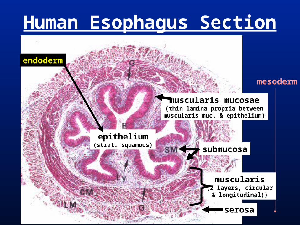
Human Esophagus Section
mesoderm
epithelium(strat. squamous)
muscularis mucosae(thin lamina propria between
muscularis muc. & epithelium)
submucosa
muscularis(2 layers, circular& longitudinal))
serosa
endoderm
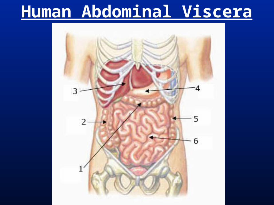
Human Abdominal Viscera
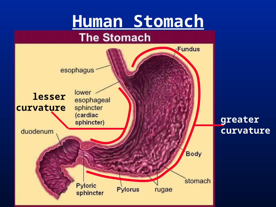
Human Stomach
lessercurvature
greatercurvature

Human Stomach SectionMuscularis has three layers, oblique, circular,
and longitudinal.

Human Small Intestine
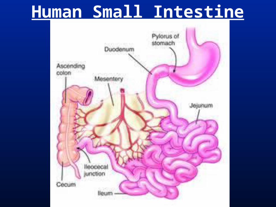
Human Small Intestine
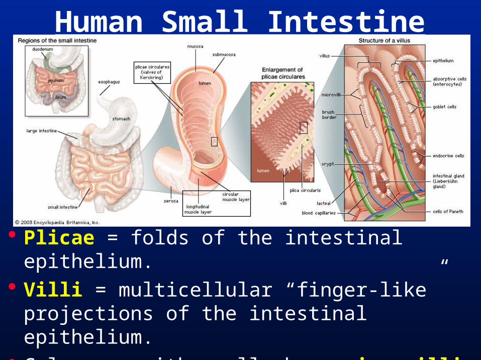
Plicae = folds of the intestinal epithelium. Villi = multicellular “finger-like” projections
of the intestinal epithelium. Columnar epith. cells have microvilli.
Human Small Intestine
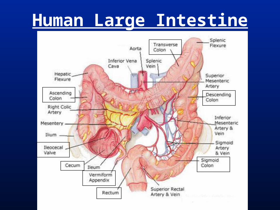
Human Large Intestine
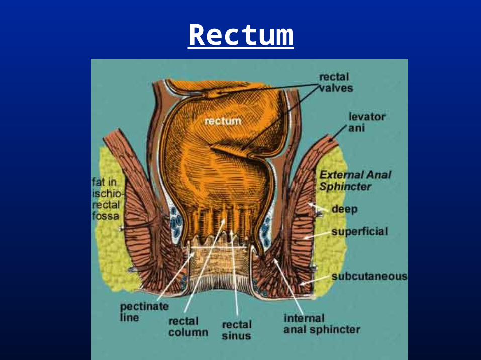
Rectum
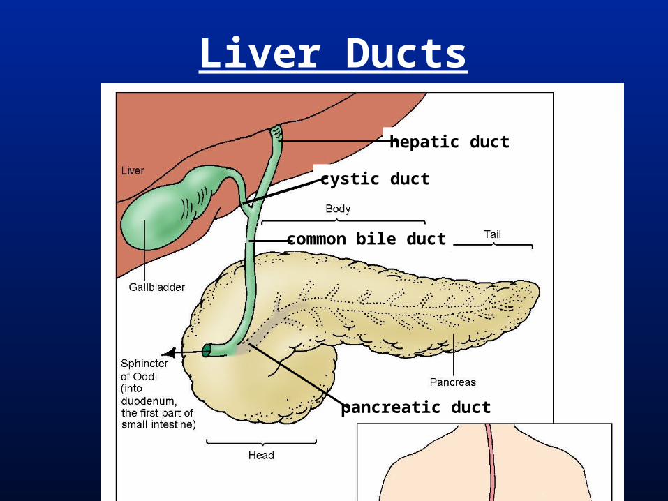
Liver Ducts
cystic duct
common bile duct
pancreatic duct
hepatic duct

Human LiverTwo big lobes, left & right separated by
falciform ligament. Right lobe has subsidiary caudate and quadrate lobes.
ventral view(anterior view)
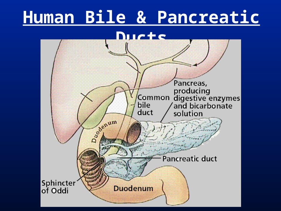
Human Bile & Pancreatic Ducts

Human Digestive System
Oral Cavity = mouth to pharynx, no separate nasal cavity in vertebrates without a secondary palate.
Mammals, crocodylians, & many turtles have a secondary palate, all other verts. lack this.
Salivary Glands = Present as multicellular, large glands only in Tetrapods, largest in amniotes.
Comparative Digestive Anatomy

Pharynx in non-amniotes also site or respiration (gills).
Esophagus is much longer in tetrapods than in non-tetrapods.
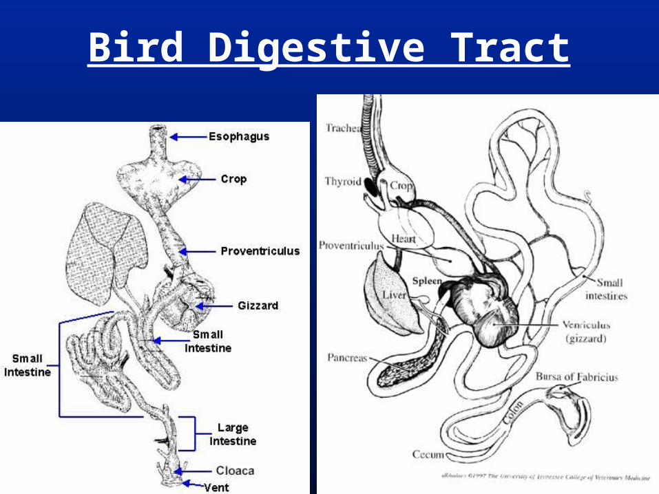
Crop = Distended region of the esophagus in birds for food storage
Pharynx- Esophagus
Stomach is ancestrally absent in vertebrates and its presence is a synapomorphy of gnathostomes.
Secondarily lost in lungfishes and a few groups of teleosts.
Gizzard = Highly muscularized stomach body of archosaurs, stratified squamous (keratinized) epithelium. (Gastroliths = “stomach stones”)
Stomach
Bird Digestive Tract

No lg. & sm. distinction, just intestine. Pyloric ceca = out-pocketings of ant.
intestine for digestion & absorption in some Actinopterygii.
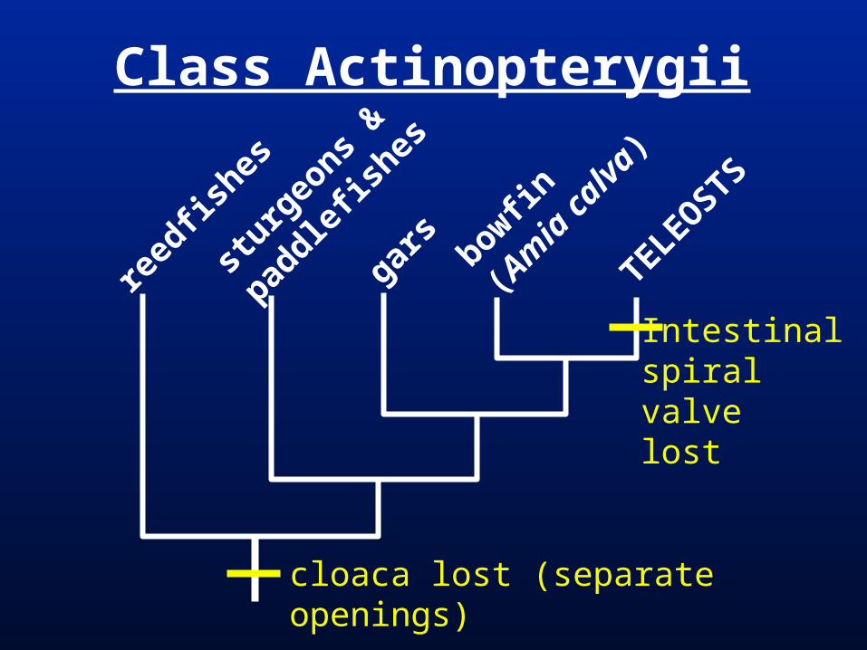
Spiral valve = Spiral flap in intestine, increases surface area. In all craniates except teleosts & tetrapods. Spiral valve ntestines with spiral valves usually shorter and wider.
Rectal gland = out-pocketing of posterior intestine; secretes excess salts in Chondrichthys.
Non-Tetrapod (Fish) Intestine
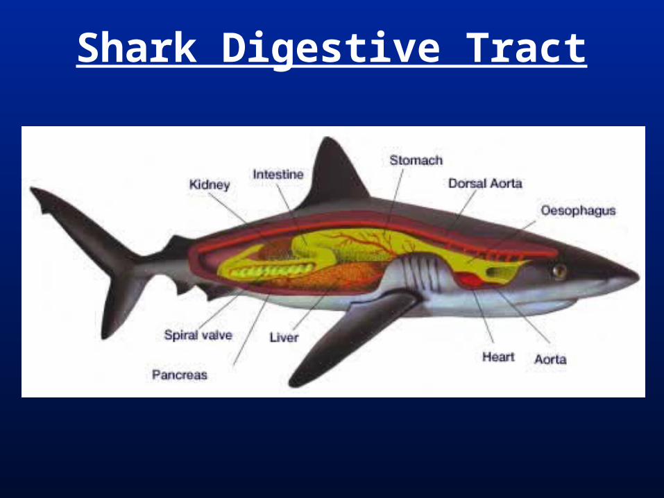
Shark Digestive Tract
Tetrapods have small & large intestines separated by the Ileocolic / Ileocecal valve.
Cecum = out-pocketing of anterior large intestine for fermentation of plant matter or other digestion. Synapomorphy of amniotes (ancestrally 1, birds have 2)
Tetrapod Small & Large Intestine

Cloaca = (sewer in Latin) ectodermal; intestine, reproductive system, & urinary system all empty into the cloaca.
Vent = external cloacal opening Cloaca lost in therian mammals and
Actinopterygiians.
Separate, external anal and urogenital openings.
Cloaca
Liver usually large in chondrichthyans because it stores fats for buoyancy.
Gallbladder in gnathostomes but lost in many groups.
Pancreas distinct in gnathostomes (may be incorporated into liver in some teleosts & lungfishes as a hepatopancreas).
Pancreas may be single (as in humans and most verts.) or double when the two buds do not fuse.
Associated Organs/Glands
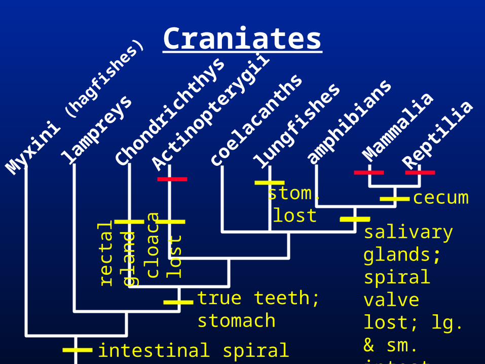
salivaryglands; spiral valve lost; lg. & sm. intest. (illeocecal sphincter)
Craniates
Myx
ini (
hagfis
hes)
lam
preys
Chondrichth
ys
Actin
optery
gii
coel
acan
ths
lungfis
hes
amphib
ians
Mam
mal
ia
Reptil
ia
true teeth; stomach
stom.lost
intestinal spiral valve; cloaca
rect
al g
land
cloa
ca lo
st
cecum
sturg
eons
&
paddle
fishes
gars bowfin
(Am
ia c
alva
)
TELEOSTS
reed
fishes
Class Actinopterygii
Intestinalspiralvalvelost
cloaca lost (separate openings)
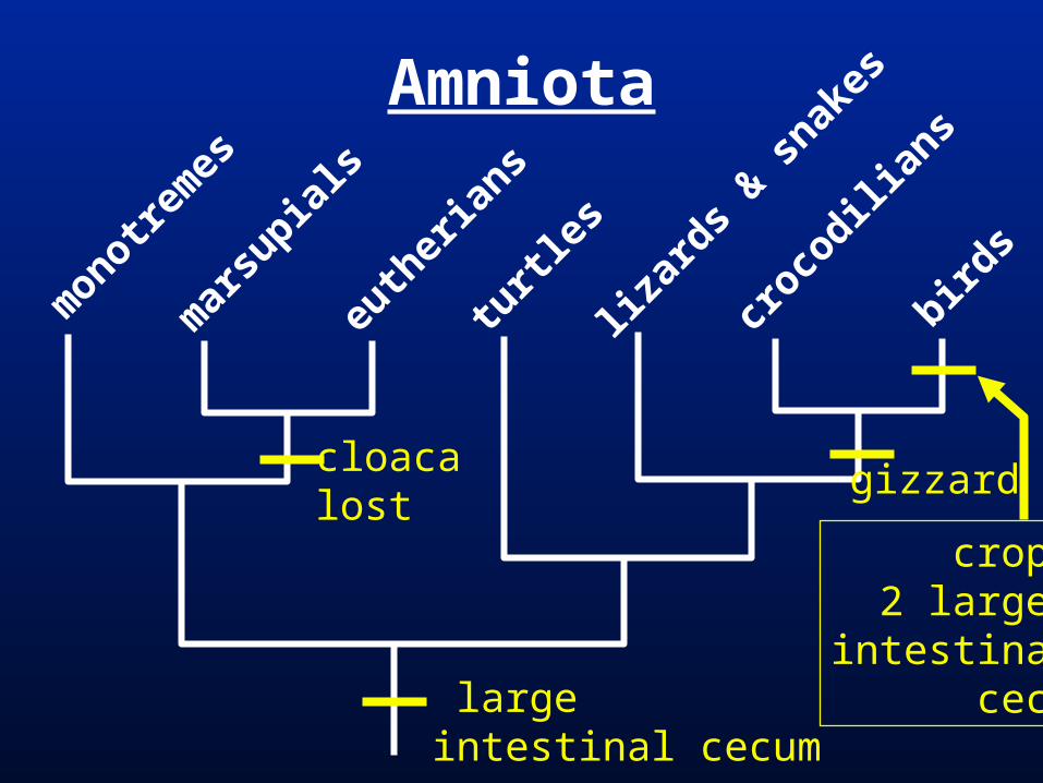
Amniota
monotre
mes
mar
supia
ls
euth
eria
ns
turtl
es
lizar
ds & s
nakes
croco
dilian
s
birds
crop;2 large
intestinalceca
gizzardcloacalost
large intestinal cecum

Subphylum Vertebrata
Myx
ini (
hagfis
hes)
lam
preys
Chondrichth
ys
Actin
optery
gii
coel
acan
ths
lungfis
hes
amphib
ians
Mam
mal
ia
Reptil
ia
liver; pancreatic cells (diffuse in hagfishes & lampreys)
gallbladder; distinct pancreas (from two developmental buds)
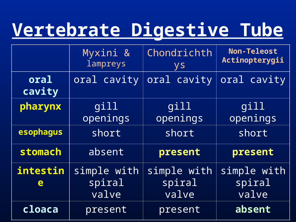
Vertebrate Digestive TubeMyxini & lampreys
Chondrichthys Non-TeleostActinopterygii
oral cavity oral cavity oral cavity oral cavity
pharynx gill openings gill openings gill openings
esophagus short short short
stomach absent present present
intestine simple with spiral valve
simple with spiral valve
simple with spiral valve
cloaca present present absent
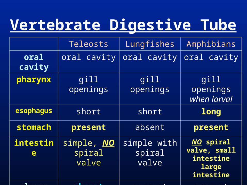
Vertebrate Digestive TubeTeleosts Lungfishes Amphibians
oral cavity oral cavity oral cavity oral cavity
pharynx gill openings gill openings gill openings when larval
esophagus short short long
stomach present absent present
intestine simple, NO spiral valve
simple with spiral valve
NO spiral valve, small intestinelarge intestine
cloaca absent present present
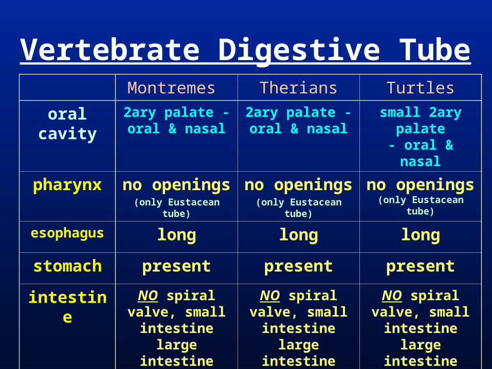
Vertebrate Digestive TubeMontremes Therians Turtles
oral cavity 2ary palate - oral & nasal
2ary palate -oral & nasal
small 2ary palate- oral & nasal
pharynx no openings(only Eustacean tube)
no openings(only Eustacean tube)
no openings (only Eustacean tube)
esophagus long long long
stomach present present present
intestine NO spiral valve, small intestinelarge intestine (with 1 cecum)
NO spiral valve, small intestinelarge intestine (with 1 cecum)
NO spiral valve, small intestinelarge intestine (with 1 cecum)
cloaca present absent present

Vertebrate Digestive Tubeliz. & snakes crocodilians birds
oral cavity oral cavity large 2ary palate- oral & nasal
oral cavity
pharynx no openings (only Eustacean tube)
no openings (only Eustacean tube)
no openings (only Eustacean tube)
esophagus long long long with crop
stomach present present with gizzard
present with gizzard
intestine NO spiral valve, small intestinelarge intestine (with 1 cecum)
NO spiral valve, small intestinelarge intestine
NO spiral valve, small intestinelarge intestine (with 2 ceca)
cloaca present present present

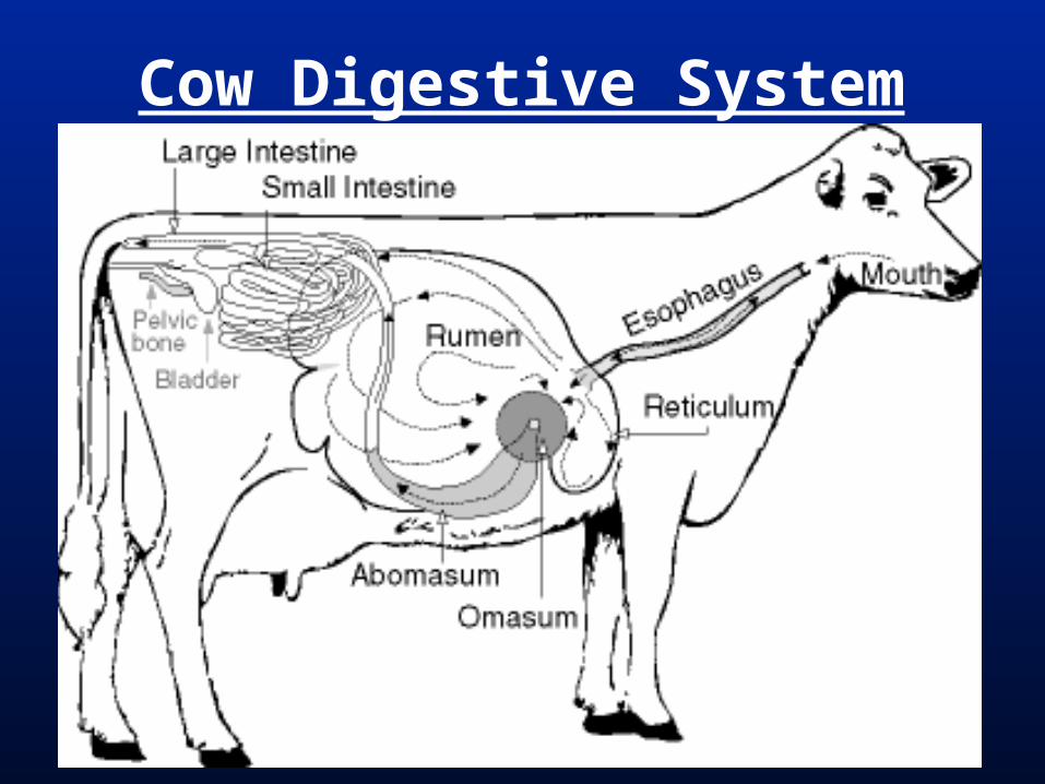
Cow Digestive System

Horse Digestive System
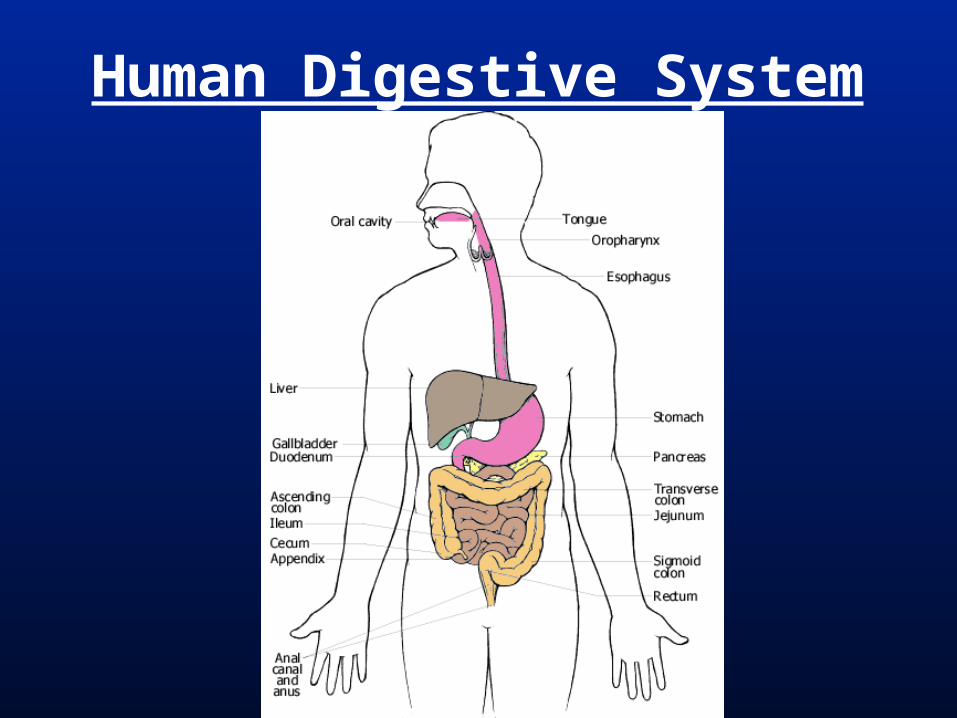
Human Digestive System