Embed Size (px)
Citation preview
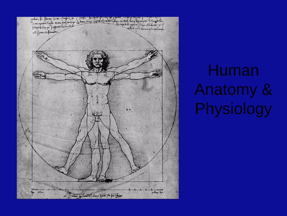
Human
Anatomy &
Physiology
Overview of Anatomy and
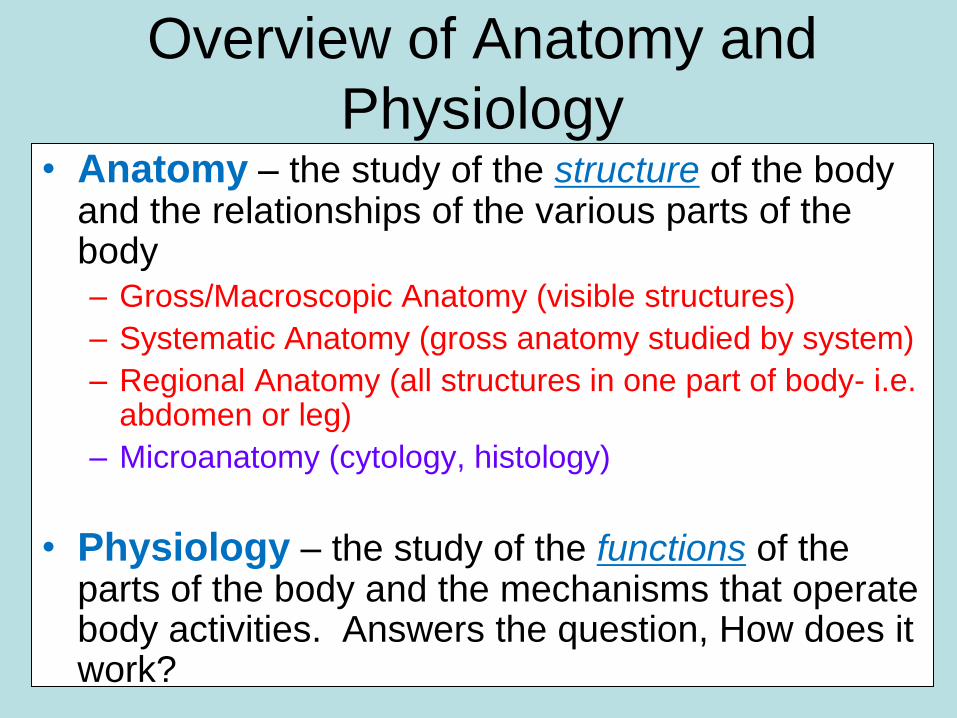
Physiology • Anatomy – the study of the structure of the body
and the relationships of the various parts of the body – Gross/Macroscopic Anatomy (visible structures)
– Systematic Anatomy (gross anatomy studied by system)
– Regional Anatomy (all structures in one part of body- i.e. abdomen or leg)
– Microanatomy (cytology, histology)
• Physiology – the study of the functions of the parts of the body and the mechanisms that operate body activities. Answers the question, How does it work?

Principle of Complementarity
• Function always reflects structure
• What a structure can do depends on its specific form
Basic Terminology
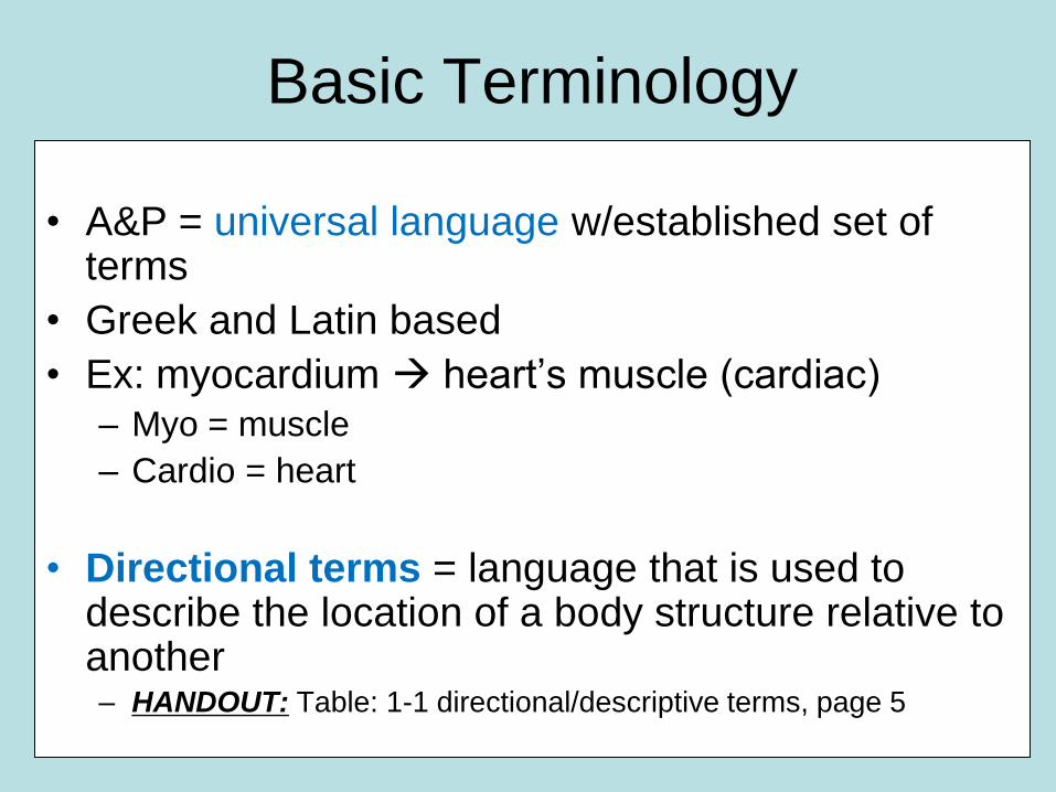
• A&P = universal language w/established set of terms
• Greek and Latin based
• Ex: myocardium heart’s muscle (cardiac) – Myo = muscle
– Cardio = heart
• Directional terms = language that is used to describe the location of a body structure relative to another – HANDOUT: Table: 1-1 directional/descriptive terms, page 5
Basic Anatomical Terminology:
Reference Positions
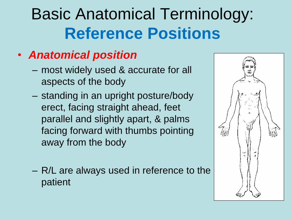
• Anatomical position
– most widely used & accurate for all
aspects of the body
– standing in an upright posture/body
erect, facing straight ahead, feet
parallel and slightly apart, & palms
facing forward with thumbs pointing
away from the body
– R/L are always used in reference to the
patient
Basic Anatomical Terminology:
Reference Positions
• Fundamental position (reclining)
– is essentially same as anatomical
position except arms are at the sides &
facing the body
– Face down = prone position;
– Face up = supine position
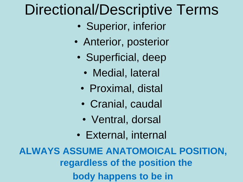
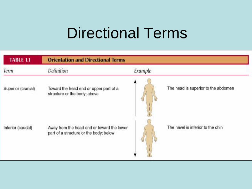
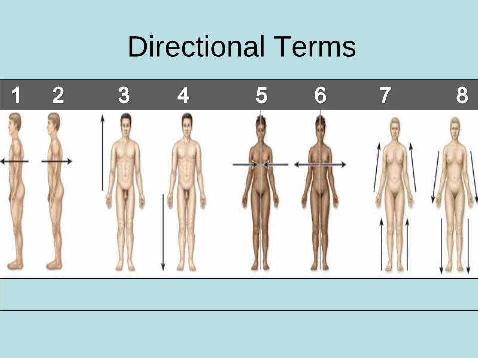
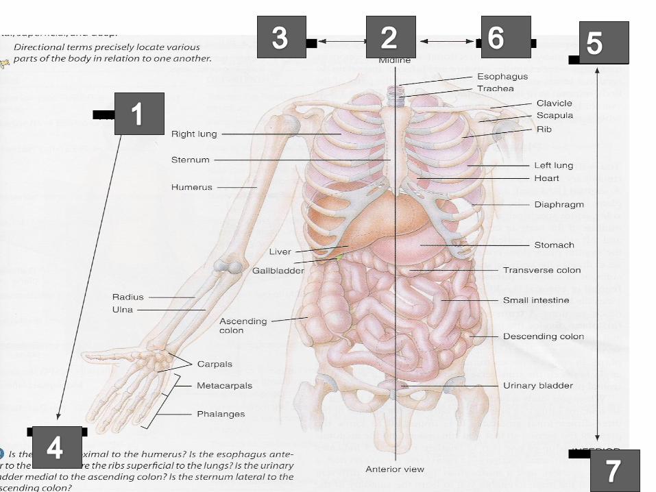
Directional/Descriptive Terms • Superior, inferior
• Anterior, posterior
• Superficial, deep
• Medial, lateral
• Proximal, distal
• Cranial, caudal
• Ventral, dorsal
• External, internal
ALWAYS ASSUME ANATOMOICAL POSITION,
regardless of the position the
body happens to be in
Directional Terms
Anatomical Directional
Terminology
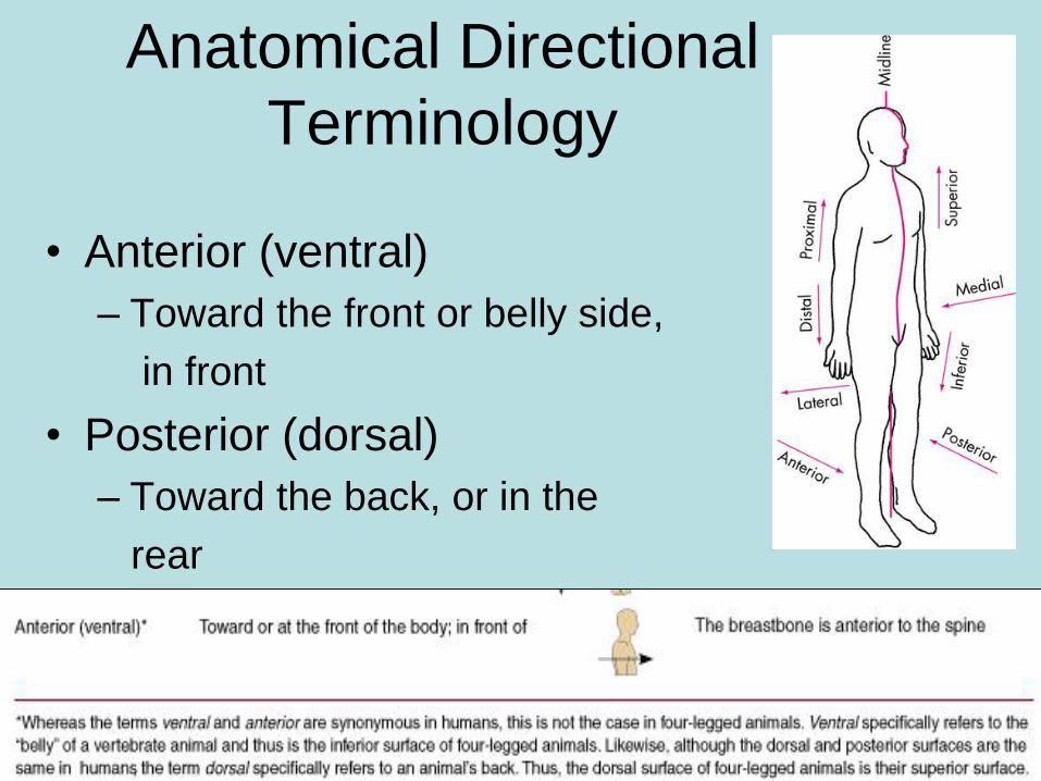
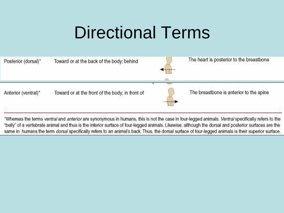
• Anterior (ventral)
– Toward the front or belly side,
in front
• Posterior (dorsal)
– Toward the back, or in the
rear
Directional Terms
Anatomical Directional
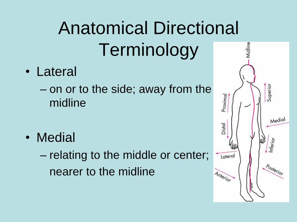
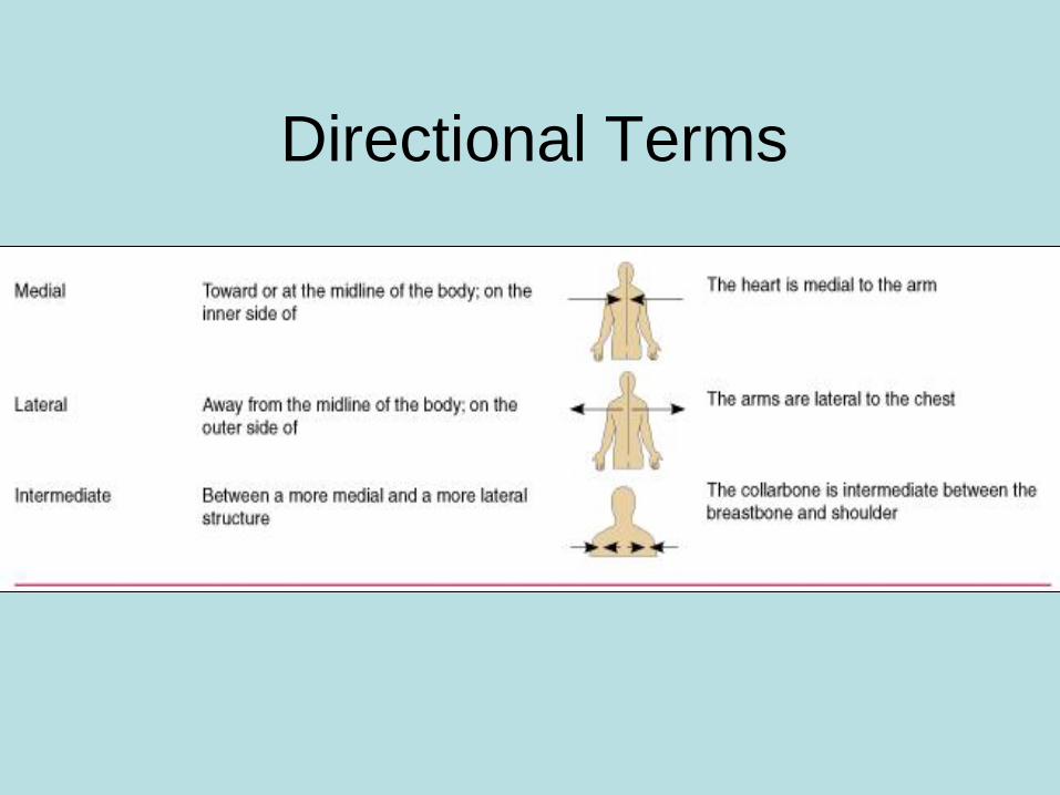
Terminology • Lateral
– on or to the side; away from the
midline
• Medial
– relating to the middle or center;
nearer to the midline
Directional Terms
Anatomical Directional
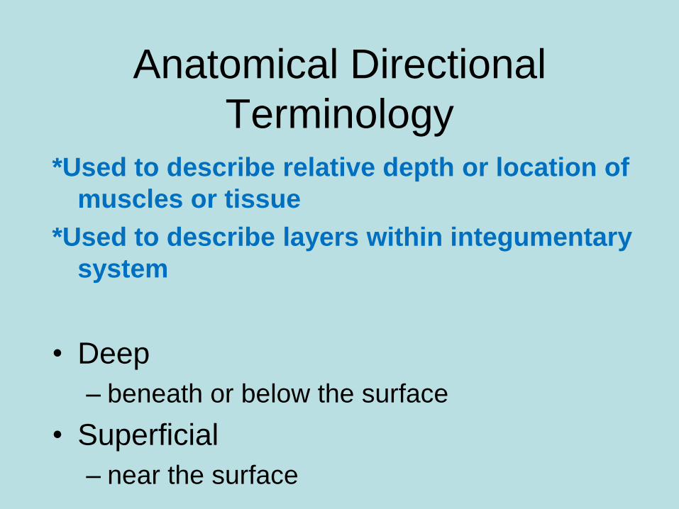
Terminology *Used to describe relative depth or location of
muscles or tissue
*Used to describe layers within integumentary
system
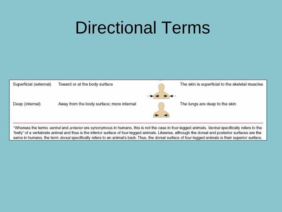
• Deep
– beneath or below the surface
• Superficial
– near the surface
Directional Terms
Anatomical Directional
Terminology
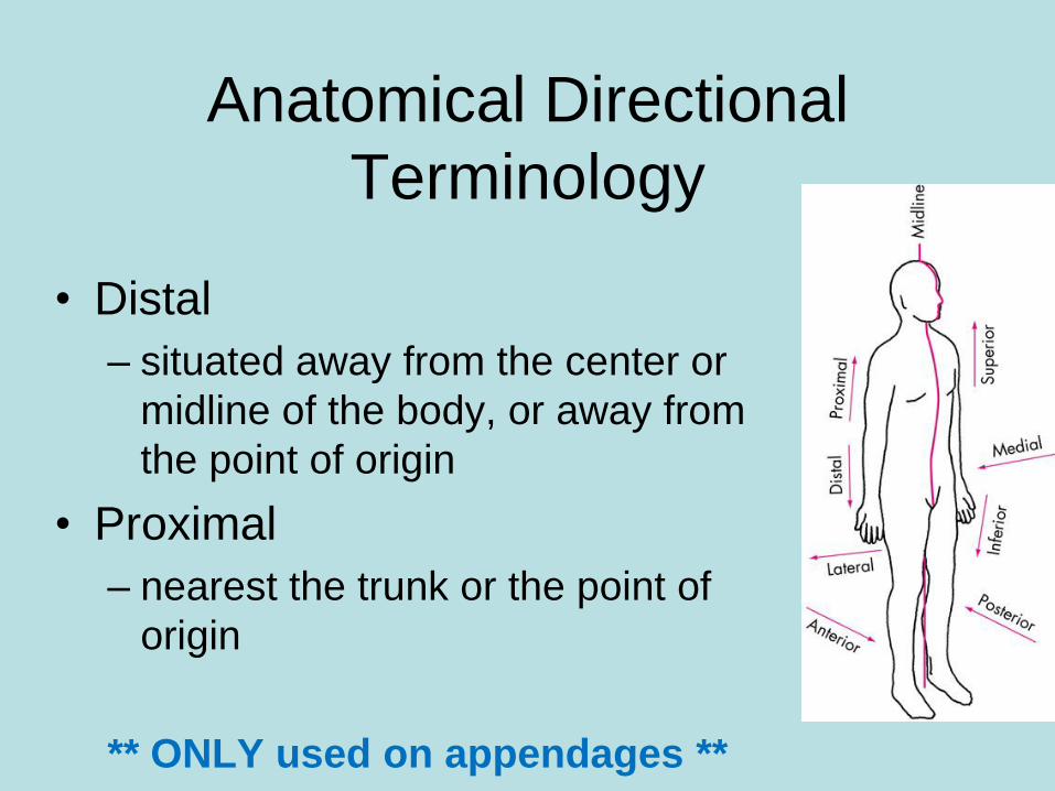
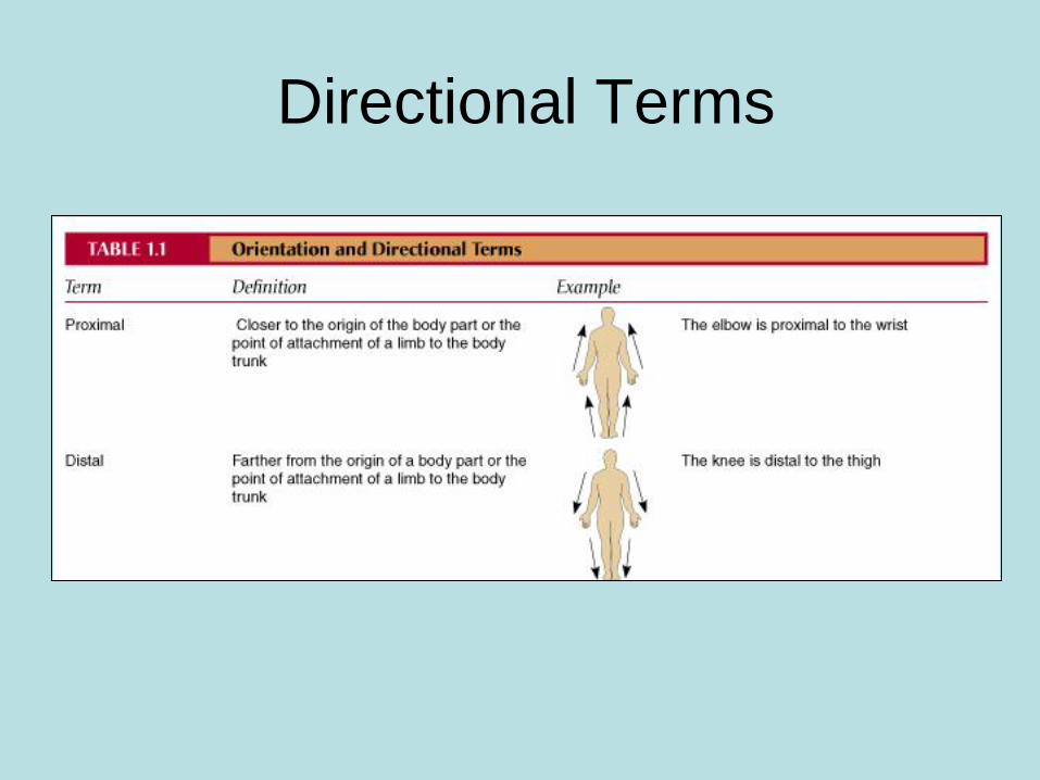
• Distal
– situated away from the center or
midline of the body, or away from
the point of origin
• Proximal
– nearest the trunk or the point of
origin
** ONLY used on appendages **
Directional Terms
Directional Terms
Directional Terms
• Complete questions 12-30 in Chapter
One packet
• http://www.wisc-
online.com/Objects/ViewObject.aspx?ID
=AP15305 (anatomical terminology)
Directional Terms
• Example:
– The nose is _____ to the eyes
– The wrist is _____ to elbow
– The lateral bone in the forearm is the
• At your table, make a list of 10
examples of directional terms – put on
whiteboard ; make answer key on loose
leaf paper

Homework
• Anatomical Terms Worksheet
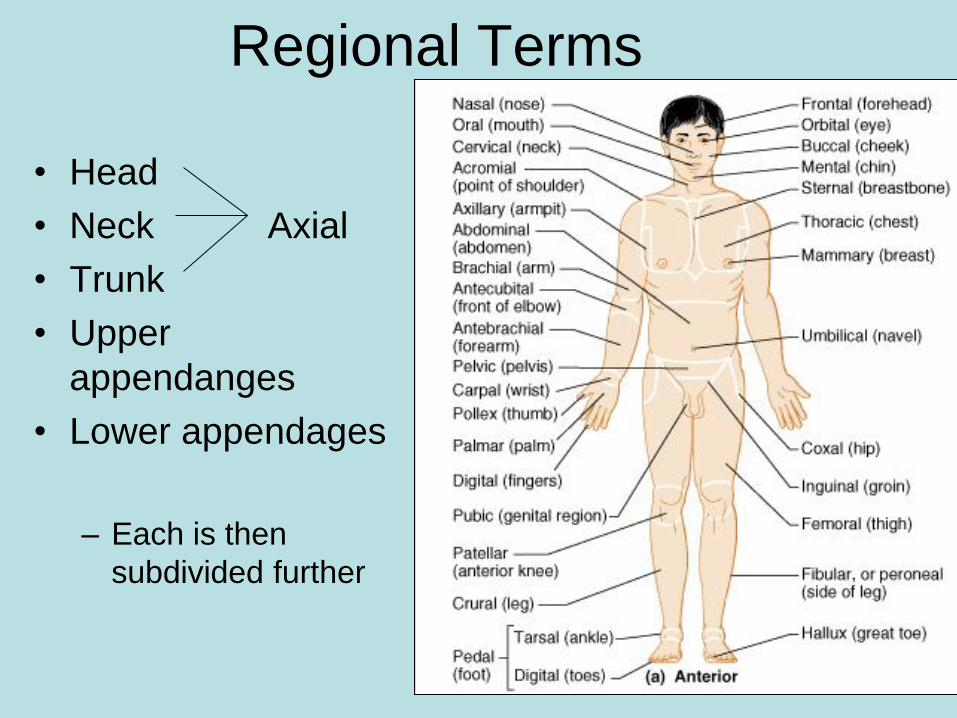
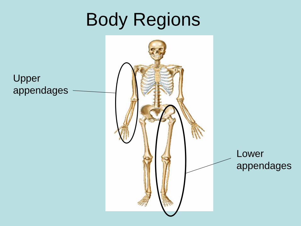
Regional Terms
• Head
• Neck Axial
• Trunk
• Upper
appendanges
• Lower appendages
– Each is then
subdivided further
Regional Terms
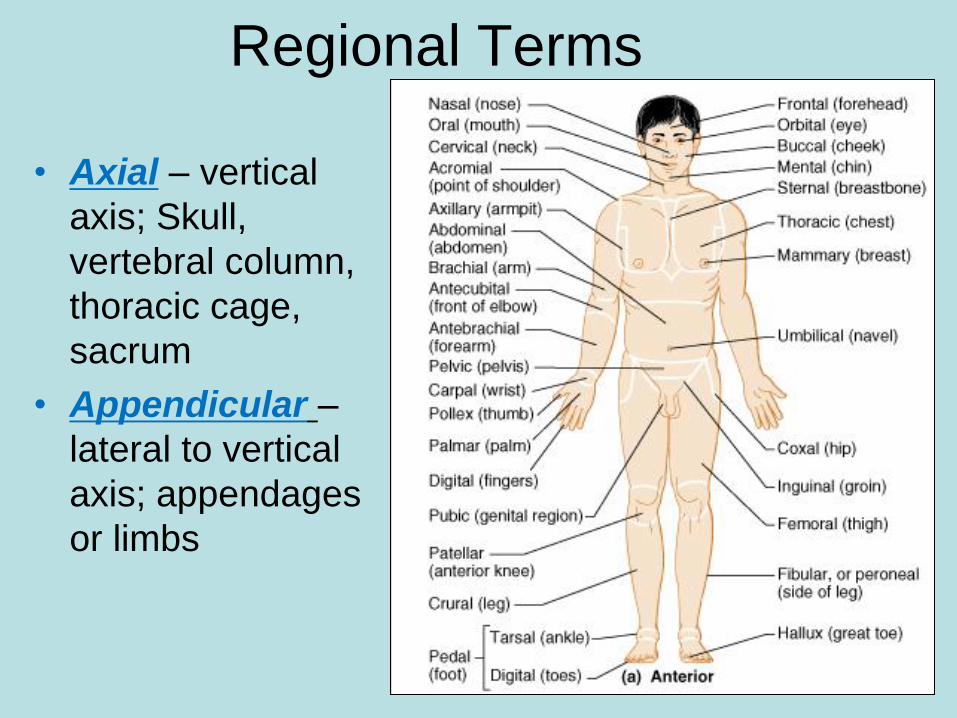
• Axial – vertical
axis; Skull,
vertebral column,
thoracic cage,
sacrum
• Appendicular –
lateral to vertical
axis; appendages
or limbs
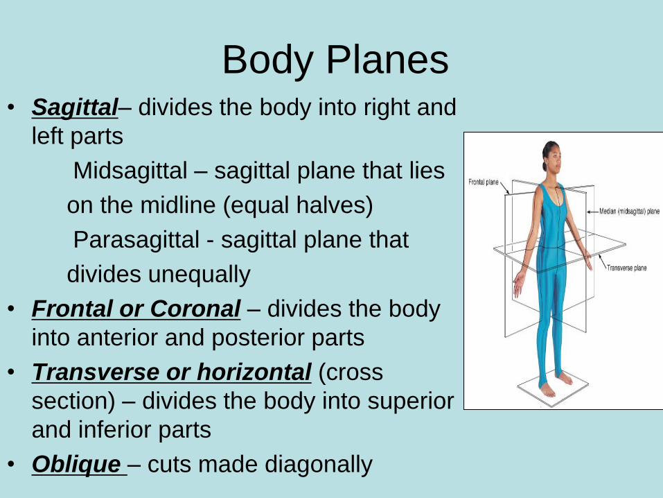
Body Planes • Sagittal– divides the body into right and
left parts
Midsagittal – sagittal plane that lies
on the midline (equal halves)
Parasagittal - sagittal plane that
divides unequally
• Frontal or Coronal – divides the body
into anterior and posterior parts
• Transverse or horizontal (cross
section) – divides the body into superior
and inferior parts
• Oblique – cuts made diagonally
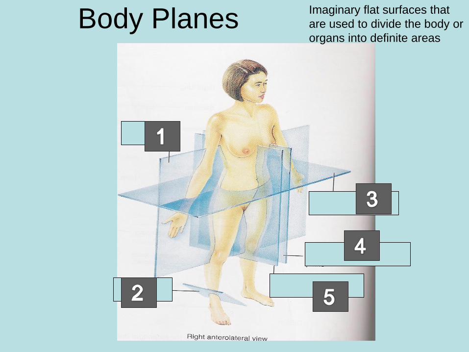
Body Planes
frontal
midsagittal
parasagittal
transverse
Imaginary flat surfaces that
are used to divide the body or
organs into definite areas
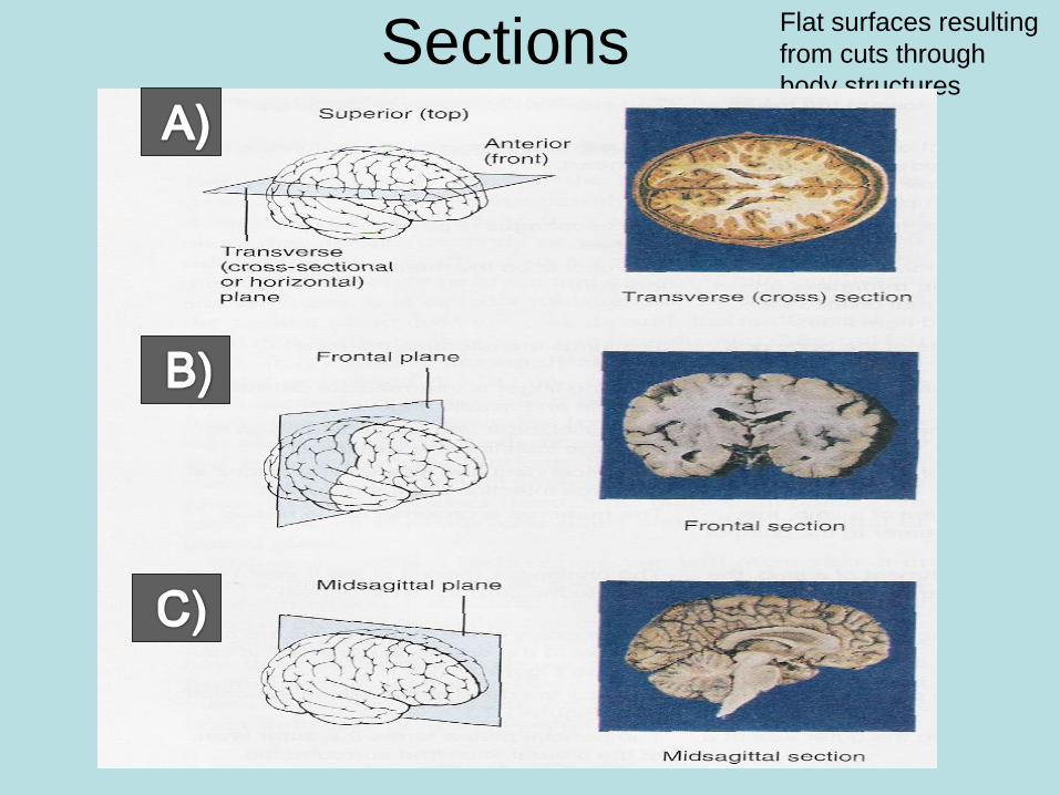
Sections
Flat surfaces resulting
from cuts through
body structures

Review:
• Complete: in Chapter One Packet:
– terminology questions 12-30
– labels and lists questions 1-3
• Assign HOMEWORK Terminology
coloring w.s.
• Introduce lab: turning a cucumber into a
frog!
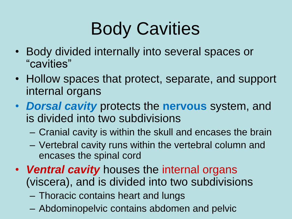
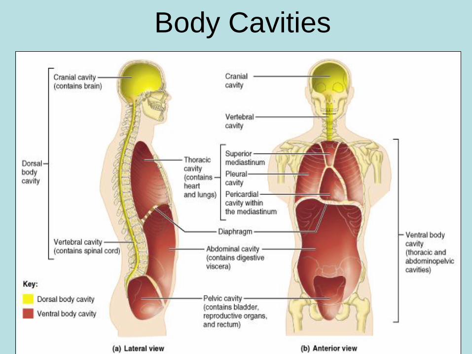
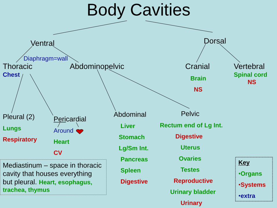
• Body divided internally into several spaces or “cavities”
• Hollow spaces that protect, separate, and support internal organs
• Dorsal cavity protects the nervous system, and is divided into two subdivisions – Cranial cavity is within the skull and encases the brain
– Vertebral cavity runs within the vertebral column and encases the spinal cord
• Ventral cavity houses the internal organs (viscera), and is divided into two subdivisions – Thoracic contains heart and lungs
– Abdominopelvic contains abdomen and pelvic
Body Cavities
Body Cavities
Body Cavities
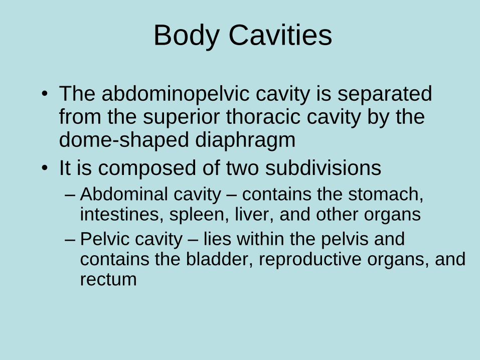
• The abdominopelvic cavity is separated from the superior thoracic cavity by the dome-shaped diaphragm
• It is composed of two subdivisions
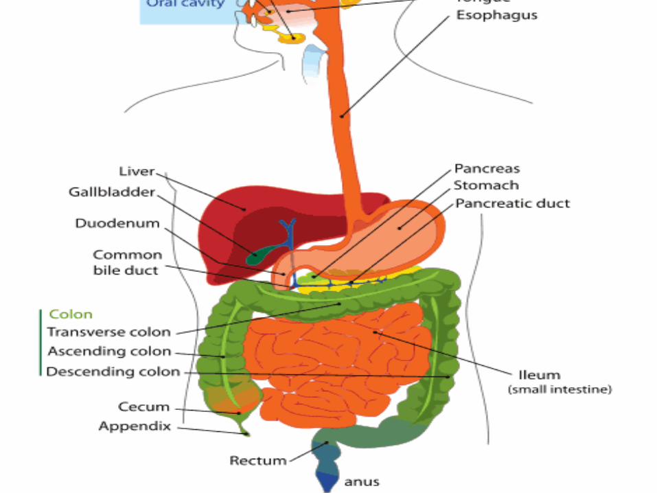
– Abdominal cavity – contains the stomach, intestines, spleen, liver, and other organs
– Pelvic cavity – lies within the pelvis and contains the bladder, reproductive organs, and rectum
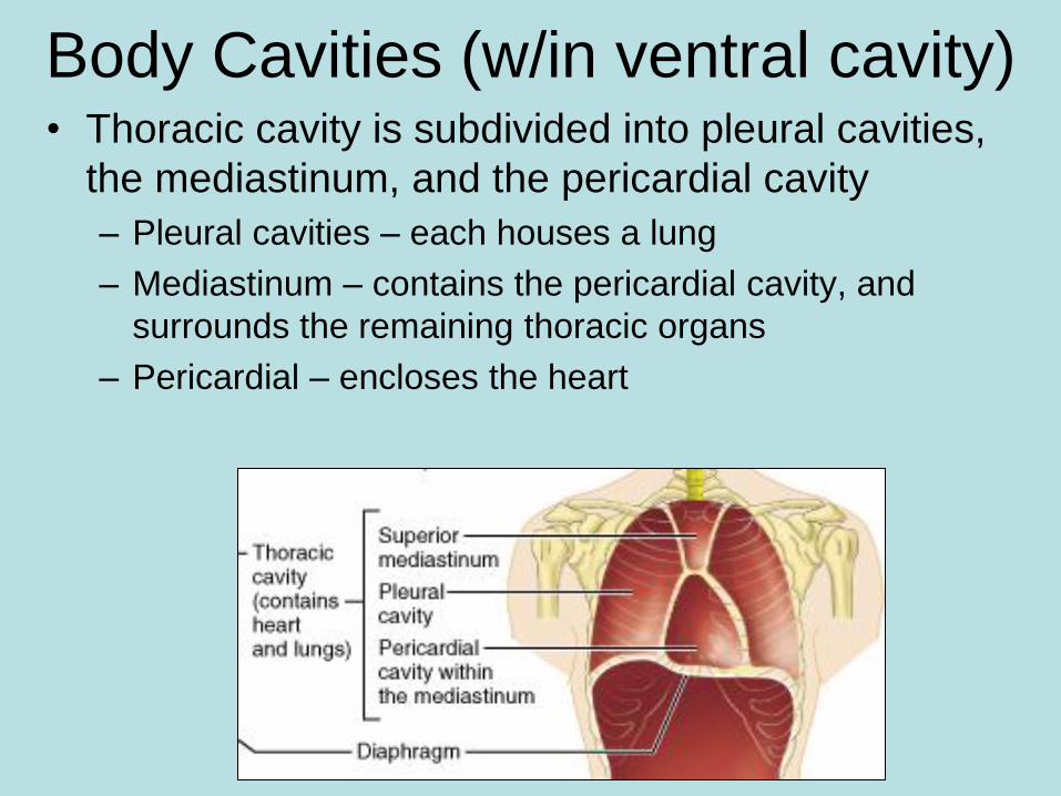
Body Cavities (w/in ventral cavity) • Thoracic cavity is subdivided into pleural cavities,
the mediastinum, and the pericardial cavity
– Pleural cavities – each houses a lung
– Mediastinum – contains the pericardial cavity, and
surrounds the remaining thoracic organs
– Pericardial – encloses the heart
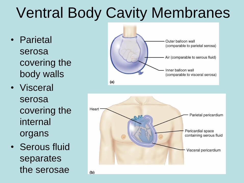
Ventral Body Cavity Membranes
• Parietal
serosa
covering the
body walls
• Visceral
serosa
covering the
internal
organs
• Serous fluid
separates
the serosae
Body Cavities
Ventral Dorsal
Thoracic Chest
Abdominopelvic
Pleural (2)
Lungs
Respiratory
Pericardial
Around
Heart
CV
Diaphragm=wall
Mediastinum – space in thoracic
cavity that houses everything
but pleural. Heart, esophagus,
trachea, thymus
Cranial
Brain
NS
Vertebral Spinal cord
NS
Abdominal
Liver
Stomach
Lg/Sm Int.
Pancreas
Spleen
Digestive
Pelvic
Rectum end of Lg Int.
Digestive
Uterus
Ovaries
Testes
Reproductive
Urinary bladder
Urinary
Key
•Organs
•Systems
•extra
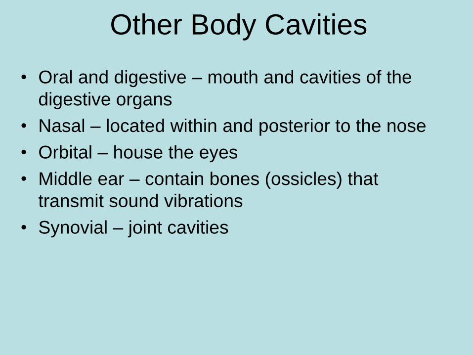
Other Body Cavities
• Oral and digestive – mouth and cavities of the
digestive organs
• Nasal – located within and posterior to the nose
• Orbital – house the eyes
• Middle ear – contain bones (ossicles) that
transmit sound vibrations
• Synovial – joint cavities
Nine Regions &
Four Quadrants
of the
Abdominopelvic
Cavity
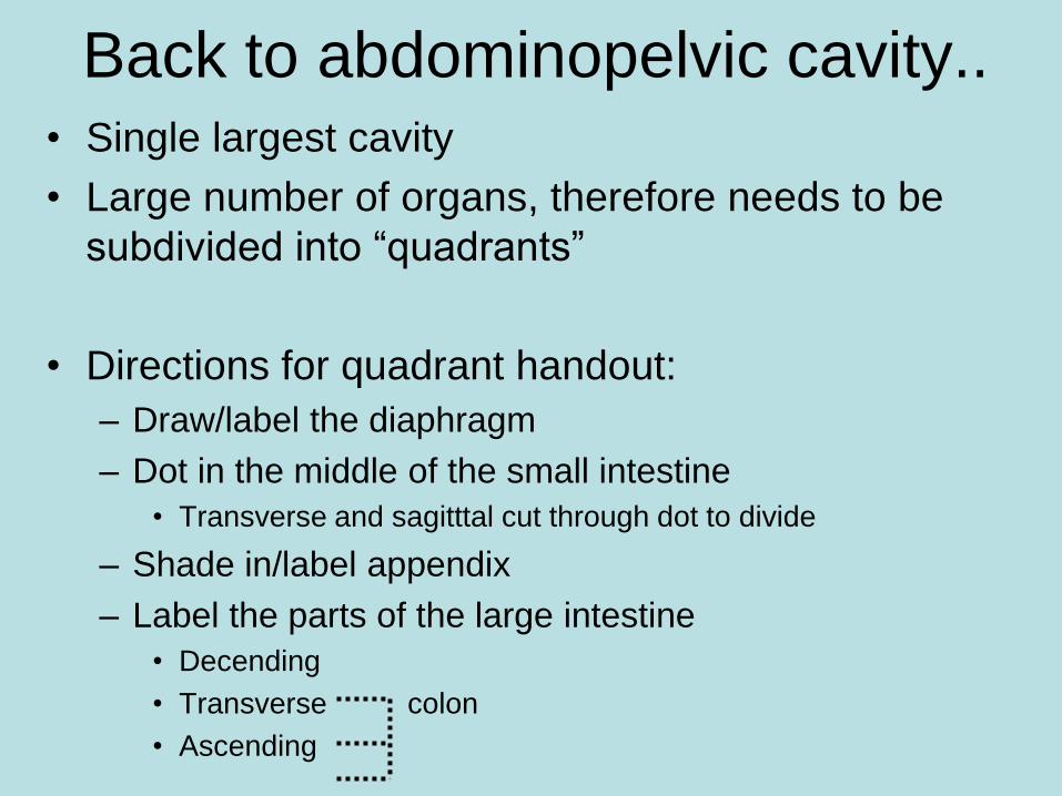
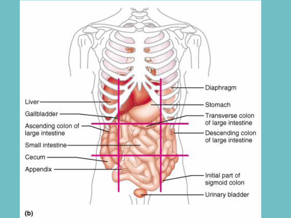
Back to abdominopelvic cavity.. • Single largest cavity
• Large number of organs, therefore needs to be
subdivided into “quadrants”
• Directions for quadrant handout:
– Draw/label the diaphragm
– Dot in the middle of the small intestine
• Transverse and sagitttal cut through dot to divide
– Shade in/label appendix
– Label the parts of the large intestine
• Decending
• Transverse colon
• Ascending
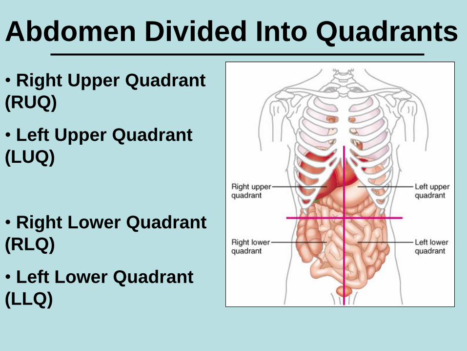
Abdomen Divided Into Quadrants
• Right Upper Quadrant
(RUQ)
• Left Upper Quadrant
(LUQ)
• Right Lower Quadrant
(RLQ)
• Left Lower Quadrant
(LLQ)
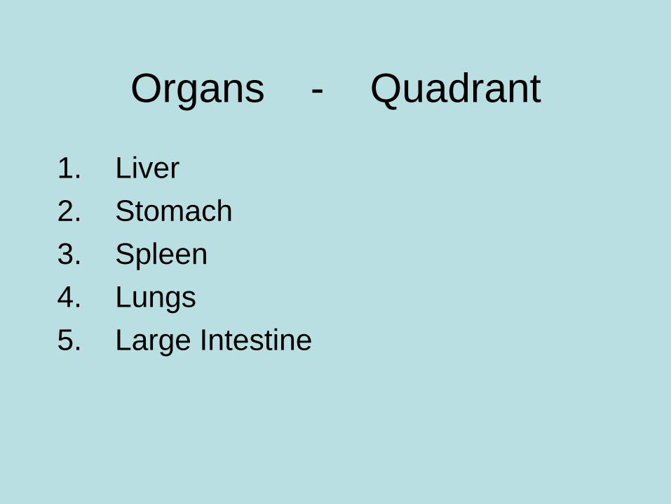
Organs - Quadrant
1. Liver
2. Stomach
3. Spleen
4. Lungs
5. Large Intestine
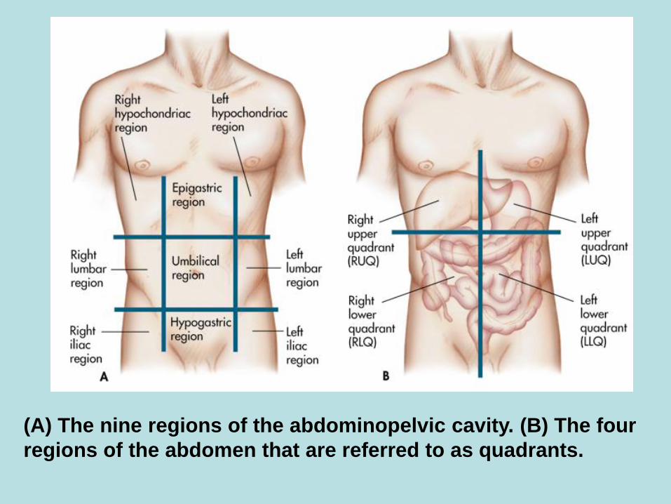
(A) The nine regions of the abdominopelvic cavity. (B) The four
regions of the abdomen that are referred to as quadrants.
Review
• http://www.wisc-
online.com/Objects/ViewObject.aspx?ID
=AP15605 (planes and sections and
body cavities)
Body Regions
Upper
appendages
Lower
appendages
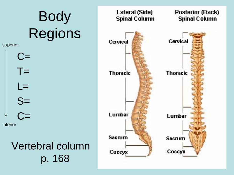
Body
Regions
C=
T=
L=
S=
C=
Vertebral column
p. 168
superior
inferior
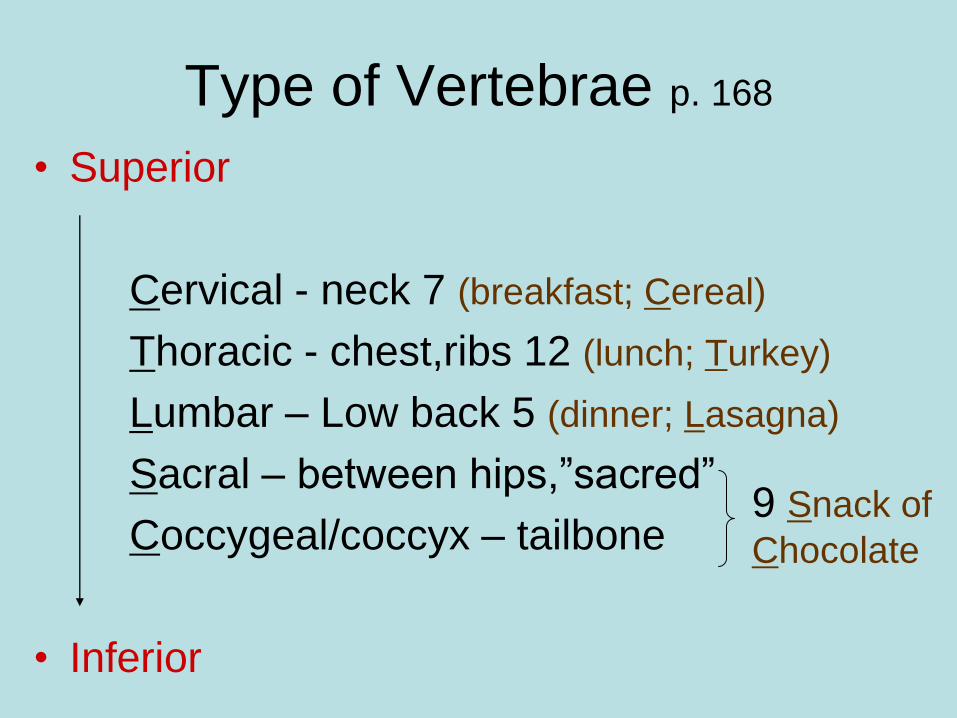
Type of Vertebrae p. 168
• Superior
Cervical - neck 7 (breakfast; Cereal)
Thoracic - chest,ribs 12 (lunch; Turkey)
Lumbar – Low back 5 (dinner; Lasagna)
Sacral – between hips,”sacred”
Coccygeal/coccyx – tailbone
• Inferior
9 Snack of
Chocolate