Embed Size (px)
Citation preview

1003
STEM CELLS AND DEVELOPMENTVolume 18, Number 7, 2009© Mary Ann Liebert, Inc.DOI: 10.1089/scd.2008.0300
Human Amniotic Fluid Stem Cells Do Not Differentiate Into Dopamine Neurons In Vitro or After Transplantation In Vivo
Angela E. Donaldson, Jingli Cai, Ming Yang, and Lorraine Iacovitti
Although embryonic stem (ES) cells can generate dopamine (DA) neurons that are potentially useful as a cell replacement therapy in Parkinson’s disease (PD), associated ethical and practical concerns remain major stum-bling blocks to their eventual use in humans. In this study, we examined human amniotic fl uid stem (hAFS) cells derived from routine amniocenteses for their potential to give rise to DA neurons in vitro and following transplantation into the 6-hydroxydopamine-lesioned rat brain. We show that undifferentiated hAFS cells con-stitutively expressed mRNAs and proteins typical of stem cells but also cell derivatives of all three germ layers, including neural progenitors/neurons (nestin, β-tubulin III, neurofi lament). Additionally, these cells expressed mRNAs of an immature DA phenotype (Lmx1a, Pitx-3, Nurr1, Aldh1a1) but not the corresponding proteins. Importantly, treatment with DA differentiation factors using a variety of protocols did not further promote the development of fully differentiated DA neurons from hAFS cells. Thus, Lmx1a, Aldh1a1, AADC, TH, and DAT proteins were not detected in hAFS cells in culture or after transplantation into the PD rat brain. Moreover, by 3 weeks after implantation, there were no surviving AFS cells in the graft, likely as a result of an acute immu-norejection response, as evidenced by the abundant presence of CD11+ macrophage/microglia and reactive GFAP+ astrocytes in the host brain. Taken together, these results suggest that further studies will be needed to improve differentiation procedures in culture and to prolong cell survival in vivo if hAFS cells are to be useful as replacement cells in PD.
Introduction
Over the last two decades, there has been intense focus on the potential use of stem cells to replace dead or dying
neurons in neurodegenerative diseases such as Parkinson’s disease (PD). Toward this end, stem cells from a variety of embryonic and adult sources have been studied in vitro and in vivo using a host of differentiation protocols (for review, refs. 1,2). To date, partial success has been achieved. While adult human bone marrow stromal stem cells (hMSCs) are capable of acquiring some dopaminergic traits, they do not fully trans-differentiate into dopamine (DA) neurons nor do they survive long term transplantation into the brain [3]. Greater success has instead been achieved using human embryonic stem (hES) cells that can be differentiated into neurons of a DA phenotype in vitro and in vivo, and which can ameliorate motor defi cits after transplantation into an animal model of PD [4–14].
However, ongoing legal, ethical, and practical issues associated with ES cells remain major stumbling blocks to
their eventual use in humans. Consequently, researchers continue to search for more practical sources of stem cells in humans. One such alternative source that has received much interest of late is human amniotic fl uid collected during routine amniocenteses. Amniotic fl uid contains a variety of cell types derived from both fetal and amnion tis-sues, ~1% of which are believed to be amniotic fl uid stem (AFS) cells [15,16]. Thus, it was of great interest when Atala and colleagues [17] reported recently that they had suc-cessfully established human stem cell lines from amniotic fl uid (hAFS). These hAFS cells were capable of unlimited self-renewal and differentiation into derivatives of all three germ layers in culture, sharing overlapping but not identi-cal properties with pluripotent hES cells [17]. Of particular signifi cance, was their ability to express traits of a neural lineage (i.e., nestin and β-tubulin III) in culture and after transplantation into the brain. Unlike their hES cell counter-parts, hAFS cells reportedly showed no sign of tumorigenic-ity in vivo. Thus, hAFS cell lines could provide an abundant,
Department of Neurology, Farber Institute for the Neurosciences, Thomas Jefferson University, Philadelphia, Pennsylvania.
ORIGINAL RESEARCH REPORT
06-SCD-2008_0300.indd 1003 8/27/2009 1:57:12 PM

DONALDSON ET AL.1004
~1 × 10⁶ of undifferentiated (N = 4) or semi-differentiated [cells treated with bFGF in Protocol A (N = 2) or neuro-spheres generated in Protocol B (N = 8)] hAFS cells, depos-ited at three stereotaxic levels (AP: +1.2mm, ML: −2.7 mm, DV: −5.4 mm, −4.9 mm and −4.2 mm) in the striatum on the side ipsilateral to the 6-OHDA lesion as described previ-ously [13]. All transplant recipients received cyclosporine A (15 mg/kg, i.p.) daily, beginning 1–2 days before transplan-tation. Animals with hAFS cell transplants were sacrifi ced at 3 days, 1 and 3 weeks and brains sectioned for immunocy-tochemical analysis.
Immunocytochemistry
Cultures were rinsed twice in PBS, fi xed with 4% para-formaldehyde and immunocytochemically stained (details described in 14). Antibodies used were: mouse monoclonal anti-Nestin (Chemicon 1:250), rabbit polyclonal anti-homeobox transcription factor 1 alpha (Lmx1; gift from Dr. M. German 1:1,000), mouse monoclonal anti-β-tubulin III (R&D Systems 1:400), goat polyclonal anti-retinaldehyde dehydrogenase (Aldh1a1) (Novus Biologicals 1:500), rabbit polyclonal anti-tyrosine hydroxylase (TH) (Pel-Freez 1:150), sheep polyclonal anti-TH (Abcam 1:1000), rabbit polyclonal anti-glial fi brillary acidic protein (GFAP) (Chemicon 1:1000), rabbit polyclonal anti-aromatic l-amino acid decarboxylase (AADC) (Protos Biotech 1:50) mouse monoclonal anti-dopamine transporter (DAT) (Chemicon 1:50). All secondary antibodies were Alexa Fluor antibodies from Invitrogen; donkey anti-rabbit 594, 1:300; don-key anti-rabbit 488, 1:300; donkey anti-mouse 594, 1:300; don-key anti-mouse 488, 1:300.Cultures were mounted in ProLong Gold antifade reagent with DAPI (Invitrogen). Stained cells were counted in representative fi elds (N = 5) on duplicate slides and expressed as a percentage of the total cells.
For immunocytochemical staining in transplantation experiments, rats were perfused with 500 mL of cold (4°C) periodate-lysine-paraformaldehyde (4%). Brains sections were cut at 30 μm on a freezing microtome and processed for immunocytochemistry as described previously [6,14]. Antibodies were used as above except for the following differences: (β-tubulin III; 1:200), HNA (1:40); GFAP from Chemicon (1:200) and mouse monoclonal anti-CD11 from BD Biosciences (1:25). Brain sections were analyzed along the length of the graft using a Nikon-Scanalytics Image System or a Zeiss LSM510 Confocal Image System.
RNA isolation and cDNA synthesis
Total RNA was isolated directly from freshly col-lected hAFS neurospheres and H9 StgIV cells with TRIzol (Invitrogen), a modifi cation of the guanidine isothiocyanate-phenol-chloroform extraction method. cDNA was synthe-sized by using 100 ng total RNA in a 20 μL reaction with Superscript III (Invitrogen) and oligo (dT)12–18 (Invitrogen). One microliter of RNase H (Invitrogen) was added to each reaction tube, and the tubes were incubated for 20 min at 37°C before proceeding to polymerase chain reaction (PCR).
PCR amplifi cation
A 0.5 μL cDNA template was used in a 20 μL reaction volume with the PCR master mix (Promega).The cycling parameters were: 94°C, 1 min; 55°C, 1 min; 72°C, 1min, for
nontumor-forming source of human stem cells free of ethi-cal dilemma that could be used in cell replacement therapies for diseases like PD. Therefore, in this study, we investigated whether cells derived from hAFS cell lines could indeed be differentiated into neurons, particularly those that express traits of a DA phenotype either in vitro or after transplanta-tion in vivo into PD rats.
Materials and Methods
Cell propagation/differentiation in culture
Cells of the AFS-A1 (Passage 15) and AFS-H1 (Passage 18) cells lines were generously supplied by A. Atala (Wake Forest University, NC). Human AFS cells were cultured on polystyrene (Petri) dishes in α-MEM medium (Invitrogen) containing 18% Chang’s Medium B, 2% Chang’s Medium C (Irvine Scientifi c), ES-qualifi ed 15% FBS (Invitrogen), 1% L-glutamine and 1% Penicillin/Streptomycin (Invitrogen) at 37°C with 5% CO2 atmosphere. Cells were passaged at 70% confl uence at a dilution of 1:4 to 1:6 and used for experiments at Passage 15–21 for A1 cells and Passage 18–27 for H1 cells. For differentiation protocols, cells were grown either accord-ing to Protocol A in defi ned serum-free (DM) media plus 20 ng/mL of basic fi broblast growth factor (bFGF) for 1 week followed by 1 week in 10 ng/mL aFGF, 200 nM TPA, 20 μM DA, 0.25 mM IBMX plus 50 μM forskolin; or Protocol B as described in Results. Essentially, neurosphere-like clusters were generated from 5 × 10⁵ cells grown in suspension in DM + Noggin (200 ng/mL) and maintained for 5–7 days (as described previously in ref. 6). Cells were gently lifted with a P1000 pipette and seeded in Ultra-low attachment dishes (Corning). In addition, cells were plated at a high density (100,000–150,000 cells in 25 μL seeded droplet) on chamber slide wells coated with polyornithine, 100ng/cm2 human collagen IV, 10 ng/mL laminin or 5 μg/mL human plasma fi bronectin and incubated with a variety of other differenti-ation protocols (Table 2).
6-OHDA lesions
Animals were maintained in accord with the Offi ce of Animal Resources at Thomas Jefferson University and IACUC policies. As described previously [14,18], 12 Fischer 344 rats (Taconic) were made Parkinsonian for these stud-ies. Briefl y, rats were anesthetized with sodium pentobarbi-tal [30 mg/kg, intraperitoneally (i.p.)], placed in a stereotaxic apparatus (Kopf Instruments) and a 26-gauge Hamilton syringe containing 6-OHDA (Sigma; 20 μg/mL in 4 μL phos-phate-buffered saline (PBS) containing 0.2 mg/mL ascor-bate) was lowered into the right median forebrain bundle (AP: −4.4mm, ML: −1.2mm, DV: −7.8 mm from bregma). The 6-OHDA solution was gradually injected at a rate of 1 μL/min. All lesions were verifi ed 3 and 6 weeks later by assessment of rotational behavior in an automated rotome-ter system (Columbus Instruments) following amphetamine challenge (5 mg/kg, i.p.). Only rats with consistent and sta-ble lesions (>10 ipsilateral turns/min on multiple tests) were used for transplantation studies.
Transplantation procedures
Animals with verifi able lesions (>10 ipsilateral turns/min) were implanted with a total of 10 μL of PBS containing
06-SCD-2008_0300.indd 1004 8/27/2009 1:57:12 PM
HUMAN AMNIOTIC FLUID STEM CELLS DO NOT GENERATE DOP 1005
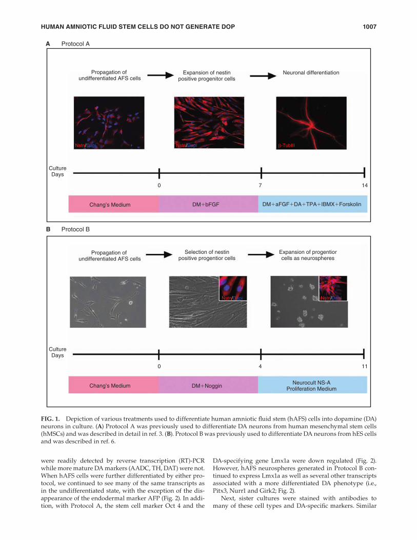
due to the presence of potentially toxic substances like for-skolin. Using the hES differentiation protocol developed in this laboratory (Protocol B) (Fig.1B), hAFS cells were fi rst incubated with 200 ng/mL noggin for 4 days to promote the formation of clusters of neural progenitors. This stage was next followed by a 1 week expansion phase during which cells were propagated as neurospheres in suspension before plating as dissociated cells. Unlike hES cells, hAFS cells did not readily form compact spheres nor did they require sub-cloning, suggesting that the nestin+ cells in these spheres were not dividing as robustly as in hES cell cultures. Finally, when hAFS neurospheres were dissociated into single cells for further differentiation in adherent cultures, we found poor attachment to substrates (polyornithine, laminin, fi bro-nectin, collagen type IV) previously used to adhere other types of stem cells [3,6,14,17]. Adherence of a small num-ber of dissociated neurosphere (Protocol B) hAFS cells only when polyornithine-coated dishes were also treated with fi bronectin.
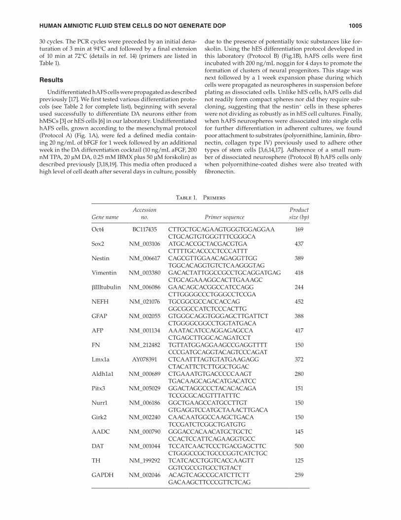
30 cycles. The PCR cycles were preceded by an initial dena-turation of 3 min at 94°C and followed by a fi nal extension of 10 min at 72°C (details in ref. 14) (primers are listed in Table 1).
Results
Undifferentiated hAFS cells were propagated as described previously [17]. We fi rst tested various differentiation proto-cols (see Table 2 for complete list), beginning with several used successfully to differentiate DA neurons either from hMSCs [3] or hES cells [6] in our laboratory. Undifferentiated hAFS cells, grown according to the mesenchymal protocol (Protocol A) (Fig. 1A), were fed a defi ned media contain-ing 20 ng/mL of bFGF for 1 week followed by an additional week in the DA differentiation cocktail (10 ng/mL aFGF, 200 nM TPA, 20 μM DA, 0.25 mM IBMX plus 50 μM forskolin) as described previously [3,18,19]. This media often produced a high level of cell death after several days in culture, possibly
Table 1. Primers
Gene nameAccession
no. Primer sequenceProduct size (bp)
Oct4 BC117435 CTTGCTGCAGAAGTGGGTGGAGGAA 169CTGCAGTGTGGGTTTCGGGCA
Sox2 NM_003106 ATGCACCGCTACGACGTGA 437CTTTTGCACCCCTCCCATTT
Nestin NM_006617 CAGCGTTGGAACAGAGGTTGG 389TGGCACAGGTGTCTCAAGGGTAG
Vimentin NM_003380 GACACTATTGGCCGCCTGCAGGATGAG 418CTGCAGAAAGGCACTTGAAAGC
βIIItubulin NM_006086 GAACAGCACGGCCATCCAGG 244CTTGGGGCCCTGGGCCTCCGA
NEFH NM_021076 TGCGGCGCCACCACCAG 452GGCGGCCATCTCCCACTTG
GFAP NM_002055 GTGGGCAGGTGGGAGCTTGATTCT 388CTGGGGCGGCCTGGTATGACA
AFP NM_001134 AAATACATCCAGGAGAGCCA 417CTGAGCTTGGCACAGATCCT
FN NM_212482 TGTTATGGAGGAAGCCGAGGTTTT 150CCCGATGCAGGTACAGTCCCAGAT
Lmx1a AY078391 CTCAATTTAGTGTATGAAGAGG 372CTACATTCTCTTGGCTGGAC
Aldh1a1 NM_000689 CTGAAATGTGACCCCCAAGT 280TGACAAGCAGACATGACATCC
Pitx3 NM_005029 GGACTAGGCCCTACACACAGA 151TCCGCGCACGTTTATTTC
Nurr1 NM_006186 GGCTGAAGCCATGCCTTGT 150GTGAGGTCCATGCTAAACTTGACA
Girk2 NM_002240 CAACAATGGCCAAGCTGACA 150TCCGATCTCGGCTGATGTG
AADC NM_000790 GGGACCACAACATGCTGCTC 145CCACTCCATTCAGAAGGTGCC
DAT NM_001044 TCCATCAACTCCCTGACGAGCTTC 500CTGGGCCGCTGCCCGGTCATCTGC
TH NM_199292 TCATCACCTGGTCACCAAGTT 125GGTCGCCGTGCCTGTACT
GAPDH NM_002046 ACAGTCAGCCGCATCTTCTT 259 GACAAGCTTCCCGTTCTCAG
06-SCD-2008_0300.indd 1005 8/27/2009 1:57:12 PM

DONALDSON ET AL.1006
regulated inwardly rectifying potassium channel (Girk2)] by PCR. We found that undifferentiated hAFS cells expressed not only stem cell transcripts, but also markers of the three germ layers, including those associated with an endodermal (AFP), mesodermal (FN) and neurectodermal lineage (nes-tin, β-tubulin III, human NEFH). Of particular interest was expression of mRNAs normally associated with more differ-entiated neuronal phenotypes (Fig. 2). Consequently, early markers of a DA phenotype, such as Lmx1a and Aldh1a1,
To assess the degree of differentiation achieved by these cells at various stages in these treatment protocols, we next examined the expression of stem cell (Oct4, Sox2), neural progenitor (nestin, vimentin), neural [β-tubulin III, neuro-fi lament (NEFP)] glial (GFAP), endodermal [α-fetoprotein (AFP)] and mesodermal (fi bronectin:FN) markers as well as specifi c markers of the DA system, including the DA tran-scription factors (Lmx1a, Nurr1, Pitx3), enzymes (Aldh1a1, AADC, TH), transporters (DAT) and channels [G-protein
Table 2. Differentiation of Amniotic Fluid Stem (AFS) Cells Using Various Treatment Protocols
Treatment Nestin Lmx1a Sox2 Aldh1a1 βtubIII TH GFAP
Undifferentiated AFS Cells 2+ – – 1+ 1+ – –
Defi ned Media – 7 div 2–3+ – – 1+ 1+ – –
Protocol A DM + bFGF (20 ng/mL) – 7 div 4+ – – 2+ 1+ – –DM + aFGF + DA + TPA + IBMX + Forskalin – 7 div 2+ – – 1+ 3+ – – *High cell death
Protocol B DM + Noggin (200 ng/mL) – 5–7 div Lift as neurospheres and seed in ultra-low attachment dish Neurocult proliferation media – 7 div 3–4+ Plate on P-Orn/fi bronectin and diff in: DM + cAMP (1μM) + Ascorbate – 2 div DM + cAMP (500μM) + Ascorbate – 2 div 2+ – – – 1+ – –
DM + bFGF (20 ng/mL) + Noggin (200 ng/mL) – 7 div 4+ – – 2+ 1+ – –
DM + bFGF (20 ng/mL) – 7 div DM + SHH (200 ng/mL) + FGF8 (100 ng/mL) + bFGF
(20 ng/mL) – 7 div
*Cell death at 5 div
NB media + B27 + SHH (250 ng/mL) + FGF8 (100 ng/mL) + bFGF (50 ng/mL) – 7 div
3–4+ – – – 1+ – –
DM + B27 + BDNF (25 ng/mL) + forskolin (15 μM) + DA(10 μM) – 7 div
2+
*No change in morphology
DM + B27 + Wnt1 (10 ng/mL) + bFGF (20 ng/mL) – 7 div 4+ – – – 1+ –
DM + B27 + ascorbate (200 μM) + cAMP (500 μM) 2+ – – – – – *High cell death
DM + aFGF + DA + TPA + IBMX + Forskalin – 7 div *Cells died within 5 div
DM + bFGF (20 ng/mL) + Wnt5A (100 ng/mL) – 7 div 4+ – – – 1+ – *Nestin + FoxA2 double-labeled cells (1–2+)
DM + substantia nigra conditioned media (1:1) – 7 div 2+ – – – – – *No change in cells
DM + glial conditioned media (1:1) – 7 div 2+ – – – – – *No change in cells
DM + H9 stg 5 conditioned media (1:1) *Cells died within 4–5 days
DM + MEF conditioned media (1:1) – 7 div 2+ – – – – – *No change in cells
AFS cells were stained for the presence of cell type specifi c markers after differentiation using a particular treatment protocol. For
cultures treated with conditioned media, defi ned media was incubated for 3 days on cultures of P1 rat glia, E14 rat substantia nigra,
mouse embryonic fi broblasts (MEF from specialty media), and stage V hES cells as described in ref. 6. Cultures of viable cells were
stained with the markers listed and evaluated for the intensity of staining as follows: none (–), lightly (1+), moderately (2–3+), or darkly
(4+) stained cells.
06-SCD-2008_0300.indd 1006 8/27/2009 1:57:13 PM
HUMAN AMNIOTIC FLUID STEM CELLS DO NOT GENERATE DOP 1007
DA-specifying gene Lmx1a were down regulated (Fig. 2). However, hAFS neurospheres generated in Protocol B con-tinued to express Lmx1a as well as several other transcripts associated with a more differentiated DA phenotype (i.e., Pitx3, Nurr1 and Girk2; Fig. 2).
Next, sister cultures were stained with antibodies to many of these cell types and DA-specifi c markers. Similar
were readily detected by reverse transcription (RT)-PCR while more mature DA markers (AADC, TH, DAT) were not. When hAFS cells were further differentiated by either pro-tocol, we continued to see many of the same transcripts as in the undifferentiated state, with the exception of the dis-appearance of the endodermal marker AFP (Fig. 2). In addi-tion, with Protocol A, the stem cell marker Oct 4 and the
Propagation of undifferentiated AFS cells
Propagation of undifferentiated AFS cells
CultureDays
0
Chang’s Medium DM�bFGF DM�aFGF�DA�TPA�IBMX�Forskolin
Chang’s Medium DM�NogginNeurocult NS-A
Proliferation Medium
7 14
CultureDays
0 4 11
A Protocol A
B Protocol B
Nstn/Dapi Nstn/Dapi
Nstn/Dapi Nstn/Dapi
β-TubIII
Expansion of nestinpositive progenitor cells
Selection of nestinpositive progentior cells
Expansion of progentiorcells as neurospheres
Neuronal differentiation
FIG. 1. Depiction of various treatments used to differentiate human amniotic fl uid stem (hAFS) cells into dopamine (DA) neurons in culture. (A) Protocol A was previously used to differentiate DA neurons from human mesenchymal stem cells (hMSCs) and was described in detail in ref. 3. (B). Protocol B was previously used to differentiate DA neurons from hES cells and was described in ref. 6.
06-SCD-2008_0300.indd 1007 8/27/2009 1:57:13 PM

DONALDSON ET AL.1008
denervated side. Fully differentiated cells were not used in transplantation experiments (i.e., Protocol A) because pro-cess-bearing neurons do not generally survive harvest and implantation. Of the 12 animals that were transplanted, only one graft of neurospheres (Protocol B) contained viable hAFS cells, as evidenced by the presence of the human nuclear antigen marker (HNA) (Fig. 4A), and only at their earliest time point examined (3 days after implantation). As shown in Fig. 4A, surviving cells exhibited an immature morphol-ogy, expressing markers of neural progenitors (nestin; Fig. 4A) and neurons (β-tubulin III) but not DA neuron traits (Lmx1a or TH; data not shown). Importantly, when grafts of these same cells were examined at 3 weeks after implan-tation, no hAFS cells remained at the implantation site (note the absence of HNA staining in Fig. 4B) nor were HNA+ cells found in surrounding brain regions. Residual nestin stain-ing observed in the vicinity of the graft was instead likely due to the presence of host brain progenitors. When these brains were further examined for the presence of reactive glia and macrophage/microglia, we found copious staining for their respective type-specifi c markers, GFAP and CD11 in host cells adjacent to the graft site at 3 days (data not shown) and 3 weeks (Fig. 4C and D) after implantation.
Discussion
This study has examined hAFS cells as an abundant, practical, and ethical source of stem cells to generate neurons for cell replacement therapies. In particular, our laboratory is interested in the differentiation of hAFS cells into func-tional DA neurons to replace cells lost in PD. We show here that undifferentiated hAFS cells from established cells lines [17] constitutively expressed mRNAs and proteins typical of stem cells but also cell derivatives of all three germ layers, including neural progenitors/neurons (nestin, β-tubulin III, NEFH). Additionally, these undifferentiated cells expressed specifi c transcripts appropriate to an immature DA pheno-type (Lmx1a, Pitx-3, Nurr1, Aldh1a1). However, treatment of hAFS cells with various DA differentiation factors using a number of published protocols did little to advance their development into fully differentiated DA neurons. Only cells grown as neurospheres and differentiated as described in Protocol B exhibited an increase in the expression of some DA transcripts. When AFS neurospheres were transplanted in vivo, cells did not survive more than several days fol-lowing their transplantation into the PD rat brain, likely due to an intense immunorejection response in host tissue. Transplanted cells that did survive in the short term, how-ever, did not express detectable levels of any of DA proteins (Lmx1a, Aldh1a1, AADC, TH, DAT).
The fact that hAFS cells, even after incubation with known neuron-promoting differentiation factors [3–14,20], continued to express transcripts of non-neuronal cell types indicates that hAFS cells behave quite differently from other pluripotent stem cell sources. Thus, hES cells that initially express transcripts from all three germ layers, if grown under the same conditions used here (Protocol B), will proceed down a neural progenitor pathway to selectively become neurons [4–14]. Likewise, adult stem cells (hMSCs) grown under similar culture conditions (Protocol A) down-regulate mesenchymal (FN) transcripts with the expression of neuronal markers (nestin, β-tubulin III) [3,20]. In con-trast, in this study, undifferentiated hAFS cells expressed
to our RT-PCR results, using immunocytochemistry, we found that progenitor protein nestin was robustly expressed in 20–40% of individual undifferentiated hAFS cells (Fig. 1A, 1B insets; Table 2). With further differentiation, cells changed in morphological shape but continued to express nestin. Moreover, the proportion of nestin+ cells increased, with the greatest number (>80%) found in semi-differenti-ated cultures (Protocol A) and in neurospheres (Protocol B) (Table 2). In contrast, relatively few (<20%) undifferentiated or semi-differentiated hAFS cells expressed the early neuron marker β-tubulin III (Fig. 3). The number of β-tubulin III+ cells did however increase (40–60%) after the fi nal differen-tiation step in Protocol A. Because in Protocol B dissociated neurosphere cells did not attach well to adherent substrates, we could not assess the degree of neuronal differentiation in these cultures (Table 2).
When semi-differentiated or fully differentiated cultures were further probed for the expression of DA marker pro-teins, importantly, no positively stained cells were observed regardless of their state of differentiation or the protocol used (Table 2). Thus, Lmx1a-staining cells were not detected by immunocytochemistry in these cultures despite detec-tion of Lmx1a mRNA in pooled cell cultures. Likewise, nei-ther Aldh1a1, AADC, TH nor DAT positive staining was observed in undifferentiated or differentiated AFS cells (Table 2), unlike other stem cell derivatives studied in our laboratory [3,6,14,18].
We next sought to determine whether transplantation of hAFS cells into the brain would further foster their develop-ment into DA neurons. Thus, undifferentiated (N = 4) and semi-differentiated hAFS cells (cells semi-differentiated with bFGF in Protocol A; N = 2 or neurospheres generated in Protocol B; N = 8) were transplanted into the striata of hemi-lesioned (6-OHDA) immunosuppressed rats on the
Oct4
Sox2
Nestin
Vimentin
�III tubulin
NEFH
GFAP
AFP
FN
Lmx1a
AFS-u
ndiff
AFS-s
emid
iff (bF
GF)
(Pro
toco
l A)
AFS-n
euro
sphe
re
(Pro
toco
l B)
AFS-u
ndiff
AFS-s
emid
iff (bF
GF)
(Pro
toco
l A)
AFS-n
euro
sphe
re
(Pro
toco
l B)
Pos
itive
con
t
Aldh1a1
Pitx3
Nurr1
Girk2
AADC
DAT
TH
G3PDH
FIG. 2. RT-PCR analysis of various stages in human amni-otic fl uid stem (hAFS) cell differentiation. Markers of stem cells (Oct4, Sox2), neural progenitors (nestin, vimentin), neurons (βIIItubulin, NEFH), glia (GFAP), ectoderm (AFP), mesoderm (FN), as well as DA-related markers (Lmx1a, Aldh1a1, Pitx3, Nurr1, Girk2, AADC, DAT, TH) were com-pared in undifferentiated (AFS-undiff) or partially differen-tiated cells (AFS-semidiff with bFGF according to Protocol A) or following neurosphere formation (in Protocol B). Positive controls taken from human embryonic stem (hES) cells dif-ferentiated into DA neurons (as described in ref. 14) were also included for AADC and TH as these were not expressed in AFS cells.
06-SCD-2008_0300.indd 1008 8/27/2009 1:57:15 PM

HUMAN AMNIOTIC FLUID STEM CELLS DO NOT GENERATE DOP 1009
there was no immunocytochemical evidence for Lmx1a, Aldh1a1, AADC, TH, or DAT in these cells, regardless of their differentiation status or their expression of DA mRNAs. The reasons for this disparity remain unknown but suggest that cells may be trapped in a primitive state, unable to produce critical proteins of a differentiated neuronal subtype using these treatment protocols. Consistent with this possibility is the fact that mature DA phenotypic traits, such as the expres-sion of the key DA rate-limiting enzyme TH or the second enzyme in DA biosynthesis AADC, were never observed at either the transcriptional or protein levels in these cells. Nonetheless, the DA-associated Girk2 channel [21–23] was detected by RT-PCR both in this study and by Atala and col-leagues who also demonstrated its functionality (i.e., barium sensitivity) in culture [17]. As Girk2 expression has also been reported in a variety of non-DA neurons [24–26), proof that its expression here represents the acquisition of a bona fi de DA neuronal trait will require experimental corroboration by other methods. Additionally, using different AFS lines and a different DA differentiation protocol, Tsai et al. [27]
transcripts for nestin, β-tubulin III and FN, with no shift in the mRNA expression profi le toward a neuronal fate after further differentiation in culture. Although differentiated hAFS cells acquired a neuronal like appearance and stained for neuronal marker proteins, it remains uncertain whether these cells can in fact function as neurons.
Further distinguishing the behavior of hAFS cells from other stem cell types was their acquisition of DA traits. Earlier studies on hMSCs and hES cells demonstrated that DA-related markers that were absent in undifferentiated cells could be partially or fully induced in vitro and in vivo using Protocols A and B, respectively [3,6,14,20]. In contrast, in this study, even in the undifferentiated state, hAFS cells expressed a number of mRNAs normally associated with DA-specifi ed progenitors/neurons but could not be further induced to complete their DA differentiation by incubation using published protocols (Table 2). Although in Protocol B, neurosphere cells did not survive plating and growth on dif-ferentiation medium in order to assess DA protein expres-sion, using Protocol A (and other differentiation schemes)
A B Protocol A
β-Tub III/Dapi Undiff β-Tub III/Dapi Semidiff β-Tub III/Dapi Diff
Protocol AC
FIG. 3. Immunocytochemical localization of β-tubulin III in amniotic fl uid stem (AFS) cells at various stages of differenti-ation. AFS cells were stained with β-tubulin III antibodies in the undifferentiated (Undiff) state (A) or after semi-differen-tiation (Semidiff) (B) and full differentiation (Diff) (C) using procedures described in Protocol A (as shown in Fig. 1). While many cells express β-tubulin III in all stages, unseen in imaged fi elds are other cells which do not express the neuronal marker. Note the change in cell shape as cells become more differentiated.
NSTN&HNA
GFAP&CD11
3 Days 3 Weeks
3 Weeks
LV
3 Weeks
NSTN&HNAA
C GFAP&CD11D
B
FIG. 4. Immunocytochemical localization of phe-notypic markers at various times after transplan-tation of amniotic fl uid stem (AFS) neurospheres into the striata of 6-OHDA treated rats. Cells were double labeled for: nestin and HNA at 3 days (A) and nestin and HNA (B) or GFAP and CD11 (C and D) at 3 weeks after transplantation. Panel D is a higher power view of the graft site shown in Panel C. Sections were mounted in ProLong Gold antifade reagent with DAPI (Invitrogen) and ana-lyzed along the length of the graft using a Nikon-Scanalytics Image System or a Zeiss LSM510 Confocal Image System. Bars = 100 μM.
06-SCD-2008_0300.indd 1009 8/27/2009 1:57:15 PM

DONALDSON ET AL.1010
2. Snyder BJ and CW Olanow. (2005). Stem cell treatment for
Parkinson’s disease: an update for 2005. Curr Opin Neurol
18:376–385.
3. Suon S, M Yang and L Iacovitti. (2006). Adult human bone mar-
row stromal spheres express neuronal traits in vitro and in a rat
model of Parkinson’s disease. Brain Res 1106:46–51.
4. Ben-Hur T, M Idelson, H Khaner, M Pera, E Reinhartz, A Itzik
and BE Reubinoff. (2004). Transplantation of human embryonic
stem cell-derived neural progenitors improves behavioral defi -
cit in Parkinsonian rats. Stem Cells 22:1246–1255.
5. Buytaert-Hoefen KA, E Alvarez and CR Freed. (2004). Generation
of tyrosine hydroxylase positive neurons from human embry-
onic stem cells after coculture with cellular substrates and
exposure to GDNF. Stem Cells 22:669–674.
6. Iacovitti L, AE Donaldson, CE Marshall, S Suon and M Yang.
(2007). A protocol for the differentiation of human embryonic
stem cells into dopaminergic neurons using only chemically
defi ned human additives: studies in vitro and in vivo. Brain Res
1127:19–25.
7. Itsykson P, N Ilouz, T Turetsky, RS Goldstein, MF Pera, I
Fishbein, M Segal and BE Reubinoff. (2005). Derivation of neural
precursors from human embryonic stem cells in the presence of
noggin. Mol Cell Neurosci 30:24–36.
8. Li Y, S Powell, E Brunette, J Lebkowski and R Mandalam (2005).
Expansion of human embryonic stem cells in defi ned serum-
free medium devoid of animal-derived products. Biotechnol
Bioeng 91:688–698.
9. Park S, KS Lee, YJ Lee, HA Shin, HY Cho, KC Wang, YS Kim, HT
Lee, KS Chung, EY Kim and J Lim. (2004). Generation of dopa-
minergic neurons in vitro from human embryonic stem cells
treated with neurotrophic factors. Neurosci Lett 359:99–103.
10. Sonntag KC, J Pruszak, T Yoshizaki, J van Arensbergen, R
Sanchez-Pernaute and O Isacson. (2007). Enhanced yield of
neuroepithelial precursors and midbrain-like dopaminergic
neurons from human embryonic stem cells using the bone mor-
phogenic protein antagonist noggin. Stem Cells 25:411–418.
11. Park CH, YK Minn, JY Lee, DH Choi, MY Chang, JW Shim,
JY Ko, HC Koh, MJ Kang, JS Kang, DJ Rhie, YS Lee, H Son, SY
Moon, KS Kim and SH Lee. (2005). In vitro and in vivo analy-
ses of human embryonic stem cell-derived dopamine neurons. J
Neurochem 92:1265–1276.
12. Perrier AL, V Tabar, T Barberi, ME Rubio, J Bruses, N Topf, NL
Harrison and L Studer. (2004). Derivation of midbrain dopa-
mine neurons from human embryonic stem cells. Proc Natl
Acad Sci USA 101:12543–12548.
13. Zeng X, J Cai, J Chen, Y Luo, ZB You, E Fotter, Y Wang, B Harvey,
T Miura, C Backman, GJ Chen, MS Rao and WJ Freed. (2004).
Dopaminergic differentiation of human embryonic stem cells.
Stem Cells 22:925–940.
14. Cai J, AE Donaldson, M Yang, M German, G Enikolopov and
L Iacovitti. (2008) The Role of Lmx1a in the differentiation of
human embryonic stem cells into midbrain dopamine neurons
in culture and after transplantation into a Parkinson’s disease
model. Stem Cells (Epub ahead of print).
15. Miki T, T Lehmann, H Cai, DB Stolz and SC Strom. (2005).
Stem cell characteristics of amniotic epithelial cell. Stem Cells
23:1549–1559.
16. Mimeault M, R Hauke, and SK Batra. (2007). Stem Cells: a revo-
lution in therapeutics-recent advances in stem cell biology and
their therapeutic applications in regenerative medicine and
cancer therapies. Nature 82:252–264.
17. Di Coppi P, G Bartsch, MM Siddiqui, T Xu, CC Santos, L Perin,
G Mostoslavsky, AC Serre, EY Snyder, JJ Yoo, ME Furth, S Soker
and A Atala. (2007) Isolation of amniotic stem cell lines with
potential for therapy. Nature Biotech 25:100–106.
18. Yang M, AE Donaldson, CE Marshall, J Shen and L Iacovitti.
(2004). Studies on the differentiation of dopaminergic traits
in human neural progenitor cells in vitro and in vivo. Cell
Transplant 13:535–547.
showed that small quantities of DA could be detected in the media following KCl-stimulated release. However, they did not provide evidence that AFS cells possessed the DA synthetic machinery (TH, AADC) to manufacture DA as opposed to simply taking up and re-releasing DA found in their (serum-containing) media.
Although the initial report describing these hAFS lines reported long-term survival of cells after transplanta-tion into the lateral ventricles of newborn mice [17], in our transplantation studies, hAFS cells did not persist in the adult PD rat brain. This was likely due to the acute immu-norejection of cells as evidenced by the copious expression of the microglial/macrophage marker CD11 and the reactive glial marker GFAP in host cells near the site of transplan-tation. This rejection response occurred despite the immu-nosuppression of rats with Cyclosporin A using a regimen that has successfully supported the long-term survival of other xenotransplanted human stem cells [3,6,14]. Similar to our observations, others using the same cell lines, also found an intense immunorejection response after hAFS transplantation into the immunosuppressed and immuno-defi cient rat myocardium [28]. Although hAFS cells were originally thought to be non-immunogenic because of their lack in major histocompatibility complex class II molecules, there is now evidence for expression of T-cell co-stimulatory molecules (normally found on antigen presenting cells) that may contribute to their immunorejection after transplanta-tion [28]. Possibly, as a result of the relative immaturity of the newborn immune system, these molecules are less effective thereby allowing the survival of transplanted hAFS cells into the perinatal brain [17]). Another possible explanation for the rejection of these cells may stem from their hetero-geneity, with mesodermal derivatives causing an intense immune/infl ammatory response in the brain [3]. Of the few cells that did survive in the short term after transplantation into the brain, there was little evidence of maturation into fully differentiated neurons or incorporation into surround-ing host tissues. A similar lack of neuronal differentiation and integration has recently been observed following the transplantation of human cord blood into the retina [29].
In sum, our studies have demonstrated that although hAFS cells constituitively express a number of traits nor-mally associated with neural progenitors/immature neu-rons, they cannot be induced to acquire a fully differentiated DA neuronal phenotype in vitro or in vivo. In addition, these cells are highly immunogenic, inducing an acute rejec-tion response after transplantation into the adult immuno-suppressed rat brain. Taken together, these results suggest that further studies will be needed to improve differentia-tion procedures in culture and to prolong cell survival in vivo if hAFS cells are to be useful as replacement cells in PD therapy.
Acknowledgments
We are grateful to Emily Foran for her dedicated assistance in the tissue culture studies. This work was generously sup-ported by a Michael J. Fox Foundation Rapid Response grant.
References
1. Lindvall O and A Bjorklund. (2004). Cell therapy in Parkinson’s
disease. NeuroRx 1:382–393.
06-SCD-2008_0300.indd 1010 8/27/2009 1:57:18 PM

HUMAN AMNIOTIC FLUID STEM CELLS DO NOT GENERATE DOP 1011
rectifying potassium current and dampens excitability in the
lateral amygdala. Mol Cell Neurosci 39:491–498.
27. Tsai M-S, S-M Hwang, Y-L Tsai, F-C Cheng, J-L Lee and Y-J
Chang. (2006) Clonal amniotic fl uid-derived stem cells express
characteristics of both mesenchymal and neural stem cells. Biol
Reprod 74: 545–551.
28. Chiavegato A, S Bollini, M Pozzobon, A Callegari, L Gasparotto,
J Taiani, M Piccoli, E Lenzini, G Gerosa, I Vendramin, E Cozzi,
A Angelini, L Iop, GF Zanon, A Atala, P De Coppi and S Sartore.
(2007). Human amniotic fl uid-derived stem cells are rejected
after transplantation in the myocardium of normal, ische-
mic, immuno-suppressed or immuno-defi cient rat. J Mol Cell
Cardiol 42:746–759.
29. Hill AJ, I Zwart, H Tam, J Chan, C Navarrete, LS Jen and R
Navarette. (2008) Human umbilical cord blood-derived mes-
enchymal stem cells do not differnetiate into neural cell types
or integrate into the retina after intrvitreal grafting in neonatal
rats. Stem Cell and Develop (Epub ahead of print).
Address correspondence to:Dr. Lorraine Iacovitti
Farber Institute for the NeurosciencesThomas Jefferson University
Philadelphia, PA 19107
E-mail: [email protected]
Received for publication October 6, 2008Accepted after revision December 1, 2008
Prepublished on Liebert Instant Online December 2, 2008
19. Guo Z, ND Stull and L Iacovitti. (1998). Molecular mechanisms
underlying the synergistic induction of tyrosine hydroxylase
gene expression by acidic fi broblast growth factor and co-acti-
vators. J Neuroscience 18:8163–8174.
20. Suon S, H Jin, AE Donaldson, EJ Caterson, RS Tuan, G
Deschennes, CE Marshall and L Iacovitti. (2004). Adult human
bone marrow stem cells transiently differentiate in culture to
express CNS proteins. Stem Cells Dev 13: 625–635.
21. Blum M, C Weickert and E Carrasco. (1999). The weaver GIRK2
mutation leads to decreased levels of serum thyroid hormone:
characterization of the effect on midbrain dopaminergic neuron
survival. Exp Neurol 160:413–424.
22. Chung CY, H Seo, KC Sonntag, A Brooks, L Lin and O Isacson.
(2005). Cell type-specifi c gene expression of midbrain dopami-
nergic neurons reveals molecules involved in their vulnerabil-
ity and protection. Hum Mol Genet 14:1709–1725.
23. Thompson L, P Barraud, E Andersson, D Kirik and A Björklund.
(2005). Identifi cation of dopaminergic neurons of nigral and
ventral tegmental area subtypes in grafts of fetal ventral mes-
encephalon based on cell morphology, protein expression, and
efferent projections. J Neurosci 25:6467–6477.
24. Aguado C, J Colón, F Ciruela, F Schlaudraff, MJ Cabañero, C
Perry, M Watanabe, B Liss, K Wickman and R Luján. (2008). Cell
type-specifi c subunit composition of G protein-gated potas-
sium channels in the cerebellum. J Neurochem 105:497–511.
25. Best TK, M Cho-Clark, RJ Siarey and Z Galdzicki. (2008)
Speeding of miniature excitatory post-synaptic currents in
Ts65Dn cultured hippocampal neurons. Neurosci Lett 438:
356–361.
26. Sosulina L, G Schwesig, G Seifert and HC Pape. (2008).
Neuropeptide Y activates a G-protein-coupled inwardly
06-SCD-2008_0300.indd 1011 8/27/2009 1:57:18 PM

06-SCD-2008_0300.indd 1012 8/27/2009 1:57:18 PM
This article has been cited by:
1. Roberto Soler, Claudius Füllhase, Ariel Hanson, Lysanne Campeau, Cesar Santos, Karl-Erik Andersson. 2012. Stem CellTherapy Ameliorates Bladder Dysfunction in an Animal Model of Parkinson Disease. The Journal of Urology 187:4,1491-1497. [CrossRef]
2. Weerapong Prasongchean , Marinella Bagni , Cinzia Calzarossa , Paolo De Coppi , Patrizia Ferretti . 2012. Amniotic FluidStem Cells Increase Embryo Survival Following Injury. Stem Cells and Development 21:5, 675-688. [Abstract] [Full TextHTML] [Full Text PDF] [Full Text PDF with Links]
3. Anna Jezierski, Kerry Rennie, Roger Tremblay, Bogdan Zurakowski, Andreé Gruslin, Marianna Sikorska, Mahmud Bani-Yaghoub. 2012. Human Amniotic Fluid Cells Form Functional Gap Junctions with Cortical Cells. Stem Cells International2012, 1-16. [CrossRef]
4. Petra AB Klemmt, Vida Vafaizadeh, Bernd Groner. 2011. The potential of amniotic fluid stem cells for cellular therapy andtissue engineering. Expert Opinion on Biological Therapy 1-18. [CrossRef]
5. M. Rosner, M. Mikula, A. Preitschopf, M. Feichtinger, K. Schipany, M. Hengstschläger. 2011. Neurogenic differentiation ofamniotic fluid stem cells. Amino Acids . [CrossRef]
6. Sarah Decembrini , Mara Cananzi , Sara Gualdoni , Alysia Battersby , Nick Allen , Rachael A. Pearson , Robin R. Ali , PaoloDe Coppi , Jane C. Sowden . 2011. Comparative Analysis of the Retinal Potential of Embryonic Stem Cells and AmnioticFluid-Derived Stem Cells. Stem Cells and Development 20:5, 851-863. [Abstract] [Full Text HTML] [Full Text PDF] [FullText PDF with Links] [Supplemental material]
7. Stefano Da Sacco, Roger E De Filippo, Laura Perin. 2011. Amniotic fluid as a source of pluripotent and multipotent stemcells for organ regeneration. Current Opinion in Organ Transplantation 16:1, 101-105. [CrossRef]
8. Mara Cananzi, Anthony Atala, Paolo de CoppiStem Cells from Amniotic Fluid 223-239. [CrossRef]
9. D. A. Davydova. 2010. Stem cells in human amniotic fluid. Biology Bulletin 37:5, 437-445. [CrossRef]
10. Shona Pfeiffer, David McLaughlin. 2010. In vitro differentiation of human amniotic fluid-derived cells: augmentation towardsa neuronal dopaminergic phenotype. Cell Biology International 34:9, 959-967. [CrossRef]
11. Jingli Cai , Ming Yang , Elizabeth Poremsky , Sarah Kidd , Jay S. Schneider , Lorraine Iacovitti . 2010. Dopaminergic NeuronsDerived from Human Induced Pluripotent Stem Cells Survive and Integrate into 6-OHDA-Lesioned Rats. Stem Cells andDevelopment 19:7, 1017-1023. [Abstract] [Full Text HTML] [Full Text PDF] [Full Text PDF with Links]