Embed Size (px)
Citation preview
Cell Tissue Res (2005) 319: 191–200DOI 10.1007/s00441-004-1002-7
REGULAR ARTICLE
Huai Zhen Ruan . Geoffrey Burnstock
The distribution of P2X5 purinergic receptors in the enteric
nervous system of mouse
Received: 8 December 2003 / Accepted: 10 September 2004 / Published online: 18 November 2004# Springer-Verlag 2004
Abstract The distribution of the P2X5 purinoceptor in theenteric nervous system of the mouse was studied byimmunohistochemistry. P2X5 receptor immunoreactivitywas widely distributed in myenteric and submucosalplexuses throughout the gastrointestinal tract. In myentericplexuses, immunoreactivity for the P2X5 receptor wasobserved in nerve fibres that enveloped ganglion cellbodies, and possibly on glial cell processes. P2X5 receptorimmunoreactivity was colocalised with vasoactive intes-tinal peptide and surrounded ganglion cells that containedcalretinin, calbindin or nitric oxide synthase. In thesubmucous plexus, P2X5 receptor immunoreactivityoccurred throughout the cytoplasm and on the surfacemembranes of the nerve cells. Double-labelling studiesshowed that 22%, 9%, 6% and 68% of P2X5 receptor-immunoreactive neurones were also immunoreactive forcalretinin, calbindin, nitric oxide synthase and vasoactiveintestinal peptide, respectively. Thus, the P2X5 receptorsubunit is expressed in specific functional groups ofneurones. P2X2 and P2X3 receptors were also present inthe mouse enteric plexuses but no immunoreactivity forP2X1, P2X4 or P2X6 receptors was found.
Keywords Purinergic receptors . ATP . Enteric nervoussystem . Neurotransmitter . Mouse
Introduction
ATP evokes responses in neuronal and non-neuronal cellsby activating various P2 receptor subtypes (Ralevic andBurnstock 1998). ATP and related purines have beenrecognised as neurotransmitters in the central, peripheral,and enteric nervous systems (Burnstock 2003). Twofamilies of purinoceptors have been identified: a P2Xionotropic ligand-gated ion channel family and a P2Ymetabotropic G-protein-coupled family (Abbracchio andBurnstock 1994). To date, seven mammalian P2X receptorsubunits (P2X1–P2X7) have been identified by molecularcloning; these can assemble to form homomeric andheteromeric receptors with different pharmacologicalproperties, including different responses to agonists andantagonists and differences in desensitisation properties(Khakh et al. 2001).
The P2X5 receptor was first cloned from rat coeliacganglia (Collo et al. 1996). It has been cloned andexpressed in heart, brain, spinal cord, peripheral ganglia,retina, endocrine and epithelial cells, adrenal gland, skin,gut and bladder (Collo et al. 1996; Brandle et al. 1998;Gröschel-Stewart et al. 1999a, 1999b; Lee et al. 2000;Dunn et al. 2001; Glass and Burnstock 2001; Greig et al.2003). Recently, the receptor has been implicated in thedifferentiation of satellite cells into mature multinucleatedmuscle fibres (Ryten et al. 2001, 2002). Less is knownabout the neurobiology of P2X5 receptors in the entericnervous system (ENS). The homomeric P2X5 receptor isrecognised as a slowly desensitising receptor sensitive toATP and 2-methylthio ATP but insensitive to α,β-methylene ATP.
ATP has been shown to elicit responses of neurones inboth plexuses of the ENS, indicating the presence of P2Xreceptors (for reviews, see Burnstock 2001; Dunn et al.2001). Application of ATP to enteric neurones evokes afast-activating depolarisation that is reminiscent of thedepolarising action of acetylcholine at nicotinic receptors(Galligan and Bertrand 1994; Barajas-López et al. 1996;Zhou and Galligan 1996). Furthermore, comparisons ofagonist potencies and susceptibility to antagonists confirm
H. Z. RuanDepartment of Neurobiology, Third Military MedicalUniversity,Chongqing, 400038, China
H. Z. Ruan . G. Burnstock (*)Autonomic Neuroscience Institute, Royal Free & UniversityCollege Medical School,Rowland Hill Street,London, NW3 2PF, UKe-mail: [email protected].: +44-207-8302948Fax: +44-207-8302949

that effects on both myenteric and submucosal neuronesare mediated through P2X receptors (Barajas-López et al.1996, 2000). This finding has led to the hypothesis thatP2X receptors are one of the mediators of fast excitatorypost-synaptic potentials (EPSPs) in the ENS (Galligan andBertrand 1994; LePard and Galligan 1999; Galligan et al.2000). However, at present, few selective agonists andantagonists discriminate clearly between subtypes of P2Xreceptors family. In both myenteric and submucosalneurones, the electrophysiology and pharmacology isambiguous as to which type of the seven known subtypesof P2X receptor (Ralevic and Burnstock 1998) is activated(Glushakov et al. 1998; Barajas-López et al. 2000).
The presence of P2X immunoreactivity in the ENS hasrecently been reported but most data has been concernedwith the presence of immunoreactivity to P2X2 and P2X3
receptors in guinea-pig, mouse and human ENS (Vulcha-nova et al. 1996; Facer et al. 2001; Hu et al. 2001;Yiangou et al. 2001; Castelucci et al. 2002; Giaroni et al.2002; Poole et al. 2002; Van Nassauw et al. 2002; Bian etal. 2003; De Man et al. 2003; Ren et al. 2003). The P2X2
receptor is expressed by specific subtypes of entericneurones, including inhibitory motor neurones, non-cho-linergic secretomotor neurones and intrinsic primaryafferent neurones (Castelucci et al. 2002). The P2X3
receptor subunit is expressed in excitatory and inhibitorymotor neurones to muscle, ascending interneurones andcholinergic secretomotor neurones (Poole et al. 2002; VanNassauw et al. 2002; Bian et al. 2003). P2X7 immuno-reactivity has been found in nerve fibres and somata inboth the myenteric plexuses (MPs) and the submucousplexuses (SMPs) of the guinea-pig small intestine (Hu etal. 2001). However, no studies of the involvement of theP2X5 receptor subtype have been reported for the ENS,apart from one report showing P2X5 receptor localisationon interstitial cells of Cajal (Burnstock and Lavin 2002).In the present work, we have used an antiserum specific tothe P2X5 receptor subtype in order to study the distribu-tion of this receptor in the ENS of the mouse.
Materials and methods
Animals
The breeding, maintenance and killing of the animals usedin this study followed principles of good laboratory animalcare and experimentation in compliance with Home Office(UK) regulations covering Schedule One Procedures andin accordance with the Animals (Scientific Procedures)Act, 1986, governing the use of animals. All protocolswere approved by the local animal ethics committee. Atotal of 16 T.O adult mice of both sexes, 19–25 g inweight, were used in this study. They were killed byasphyxiation with a rising concentration of CO2 (between0% and 100%) and death was confirmed by cervicaldislocation. Thereafter, animals were perfused immedi-ately through the heart (left ventricle) with 50 ml 0.9%NaCl solution followed by fixative containing 4% para-
formaldehyde, 0.2% saturated picric acid in 0.1 Mphosphate-buffered saline (PBS, pH 7.4).
Whole-mount preparations
Segments of stomach, jejunum, ileum and colon werequickly removed and washed with PBS. An incision wasmade along the midline of the gut and the tissue waspinned as a flat sheet onto Sylgard (Dow Corning,Wiesbaden, Germany) with the mucosa face-down. Tissuesamples were immersed in the same fixative (4% parafor-maldehyde, 0.2% saturated picric acid in 0.1 M PBS)overnight at 4 °C, unpinned, and washed in PBS (3×10min). After being cleared, the tissues were dissected intotwo layers: the outer musculature with adhering serosa andthe submucosa/mucosa. The circular muscle was removedto yield whole-mounts of longitudinal muscle with the MPattached. In a few whole-mounts, the circular muscle wasonly partially removed. In the whole-mounts containingthe SMP, the mucosa was scraped off by using a bluntscalpel. The whole-mounts were stored in PBS containing0.1% sodium azide.
Primary antibody directed against P2X5 receptor
The antibody directed against the P2X5 receptor, raised byRoche Bioscience (Palo Alto, Calif., USA), was asdetailed previously (Oglesby et al. 1999). The specificityof the P2X5 antibody was verified by immunoblotting withmembrane preparations from cloned P2X1–7 receptor-expressing CHO-K1 cells. The antibody recognised onlyone protein of the expected size in the heterologousexpression system and was shown to be receptor-subtype-specific (Oglesby et al. 1999). Preabsorption of theantibody with excess of the synthetic peptide used forgeneration of the antibody eliminated immunoreactivity(Xiang et al. 1998).
Immunocytochemistry
The antisera used in this study and their respectivedilutions are listed in Table 1. The preparations werewashed 3×5 min in PBS and then preincubated in 10%normal horse serum (NHS), 0.2% Triton X-100 in PBS for30 min. Preparations were subsequently incubated in theprimary antibodies, diluted with 10% NHS in PBScontaining 0.05% merthiolate and 0.2% Triton X-100,overnight and then in Cy3-conjugated donkey anti-rabbitIgG, diluted 1:300 in 1% NHS in PBS containing 0.05%merthiolate, for 1 h. All incubations were carried out atroom temperature and separated by 3×5 min washes inPBS. The preparations were mounted with Citifluor(Citifluor, London, UK) and examined by fluorescencemicroscopy.
To demonstrate the colocalisation of the P2X5 receptorwith calretinin, calbindin, neuronal nitric oxide synthase
192
(NOS) and vasoactive intestinal peptide (VIP), prepara-tions were immunostained for the P2X5 receptor, as above,and then incubated with these antibodies overnight.Subsequently, the preparations were incubated with fluo-rescein isothiocyanate (FITC)-conjugated goat anti-mouseIgG or FITC-conjugated donkey anti-sheep IgG. All theincubations and reactions were carried out at roomtemperature and separated by 3×10 min washes in PBS.The preparations were mounted with Citifluor andexamined by fluorescence microscopy.
The following control experiments were performed toestablish the specificity of the immunoreactivity: omissionof the primary antibodies and preabsorptions of theprimary antibodies with their homologous peptide anti-gens.
Photomicroscopy
Images of immunofluorescence labelling were taken witha Leica DC 200 digital camera (Leica, Switzerland)attached to a Zeiss Axioplan microscope (Zeiss, Ger-many). Filter sets included the following: for Cy3, 510–550 nm excitation, 590 nm emission; for FITC, 470 nmexcitation, 525 nm emission. Images were imported into agraphics package (Adobe Photoshop 5.0, USA). The two-channel readings for green and red fluorescence weremerged by using Adobe-Photoshop 5.0.
Analysis
All analyses were performed at ×20 objective magnifica-tion. The whole-mount preparations were also used toperform a quantitative analysis as described previously(Van Nassauw et al. 2001). Briefly, the immunoreactive-
positive neurone bodies in the submucosal and myentericganglia were counted per visual field (0.3 mm2) in thewhole-mount preparations. Ten randomly chosen fields ineach whole-mount preparation were analysed and thenumber of immunoreactive neurones was calculated.
Results
P2X5 receptor immunoreactivity in MPs and SMPs
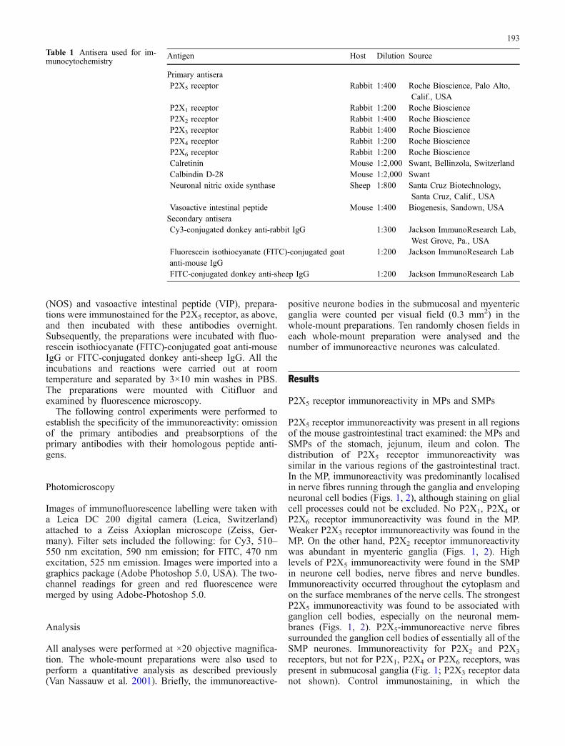
P2X5 receptor immunoreactivity was present in all regionsof the mouse gastrointestinal tract examined: the MPs andSMPs of the stomach, jejunum, ileum and colon. Thedistribution of P2X5 receptor immunoreactivity wassimilar in the various regions of the gastrointestinal tract.In the MP, immunoreactivity was predominantly localisedin nerve fibres running through the ganglia and envelopingneuronal cell bodies (Figs. 1, 2), although staining on glialcell processes could not be excluded. No P2X1, P2X4 orP2X6 receptor immunoreactivity was found in the MP.Weaker P2X3 receptor immunoreactivity was found in theMP. On the other hand, P2X2 receptor immunoreactivitywas abundant in myenteric ganglia (Figs. 1, 2). Highlevels of P2X5 immunoreactivity were found in the SMPin neurone cell bodies, nerve fibres and nerve bundles.Immunoreactivity occurred throughout the cytoplasm andon the surface membranes of the nerve cells. The strongestP2X5 immunoreactivity was found to be associated withganglion cell bodies, especially on the neuronal mem-branes (Figs. 1, 2). P2X5-immunoreactive nerve fibressurrounded the ganglion cell bodies of essentially all of theSMP neurones. Immunoreactivity for P2X2 and P2X3
receptors, but not for P2X1, P2X4 or P2X6 receptors, waspresent in submucosal ganglia (Fig. 1; P2X3 receptor datanot shown). Control immunostaining, in which the
Table 1 Antisera used for im-munocytochemistry
Antigen Host Dilution Source
Primary antiseraP2X5 receptor Rabbit 1:400 Roche Bioscience, Palo Alto,
Calif., USAP2X1 receptor Rabbit 1:200 Roche BioscienceP2X2 receptor Rabbit 1:400 Roche BioscienceP2X3 receptor Rabbit 1:400 Roche BioscienceP2X4 receptor Rabbit 1:200 Roche BioscienceP2X6 receptor Rabbit 1:200 Roche BioscienceCalretinin Mouse 1:2,000 Swant, Bellinzola, SwitzerlandCalbindin D-28 Mouse 1:2,000 SwantNeuronal nitric oxide synthase Sheep 1:800 Santa Cruz Biotechnology,
Santa Cruz, Calif., USAVasoactive intestinal peptide Mouse 1:400 Biogenesis, Sandown, USASecondary antiseraCy3-conjugated donkey anti-rabbit IgG 1:300 Jackson ImmunoResearch Lab,
West Grove, Pa., USAFluorescein isothiocyanate (FITC)-conjugated goatanti-mouse IgG
1:200 Jackson ImmunoResearch Lab
FITC-conjugated donkey anti-sheep IgG 1:200 Jackson ImmunoResearch Lab
193

194
primary antibodies were omitted or pre-absorbed with therelevant peptide, did not yield immunolabelling.
Double-labelling studies
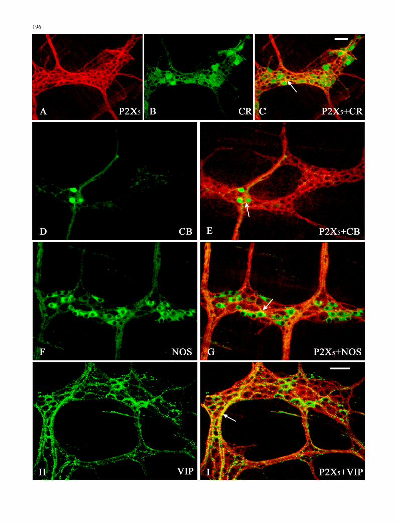
Double-labelling studies were conducted to identify majorclasses of neurones that had strong P2X5 receptorimmunoreactivity in the myenteric and submucosal gan-glia of the ileum (Figs. 3, 4). In the MP of ileum, double-labelling for the P2X5 receptor and calretinin, calbindin orNOS revealed that P2X5 receptor immunoreactivity wascolocalised with and surrounded neurones that expressedthese neurochemical markers (Fig. 3). Many of the nervefibres in the MP of the ileum were immunoreactive forP2X5 and VIP (Fig. 3).
Both the number and percentage of neurones colocalis-ing with various markers are summarised in Table 2. In theSMP of ileum, about 1/5 of all P2X5-positive neuroneswere found to display calretinin immunoreactivity but,conversely, most of the calretinin-expressing neuronesappeared to bear P2X5 receptor immunoreactivity (Table2; Fig. 3). Colocalisation of P2X5 receptors with calbindin(Table 2; Fig. 3) was observed. Of the P2X5-immunore-active cells, a few were also calbindin-immunoreactive.Some NOS-immunoreactive cells were present in theSMP; however, almost all NOS-immunoreactive cellswere also positive for the P2X5 subunit. Conversely, fewP2X5 -positive neurones expressed NOS (Table 2; Fig. 3).Double-immunofluorescent histochemistry showed thatabout 90% of VIP-immunoreactive nerve cells wereP2X5 receptor-immunoreactive in the submucosal ganglia.On the other hand, about 70% P2X5-immunoreactivesubmucosal nerve cells were immunoreactive for VIP(Table 2; Fig. 3).
Discussion
P2X receptors contribute to fast synaptic excitation in theENS (Galligan and Bertrand 1994; Johnson et al. 1999).High concentrations of ATP in subpopulations of myen-teric neurones in various regions of the gut of the guinea-pig, rabbit and rat have been reported (Crowe andBurnstock 1981; Belai and Burnstock 1994). In addition,after application of an appropriate stimulus, ATP isreleased from enteric nerves (Burnstock et al. 1978;McConalogue et al. 1996) and the subsequent activation ofspecific receptors on enteric neurones and muscle cellsmay evoke either the excitation or the inhibition of smoothmuscle function (Burnstock 2001). Furthermore, a com-parison of agonist potencies and susceptibility to antago-nists supports the contention that synaptic transmission inboth myenteric and submucosal neurones is mediated
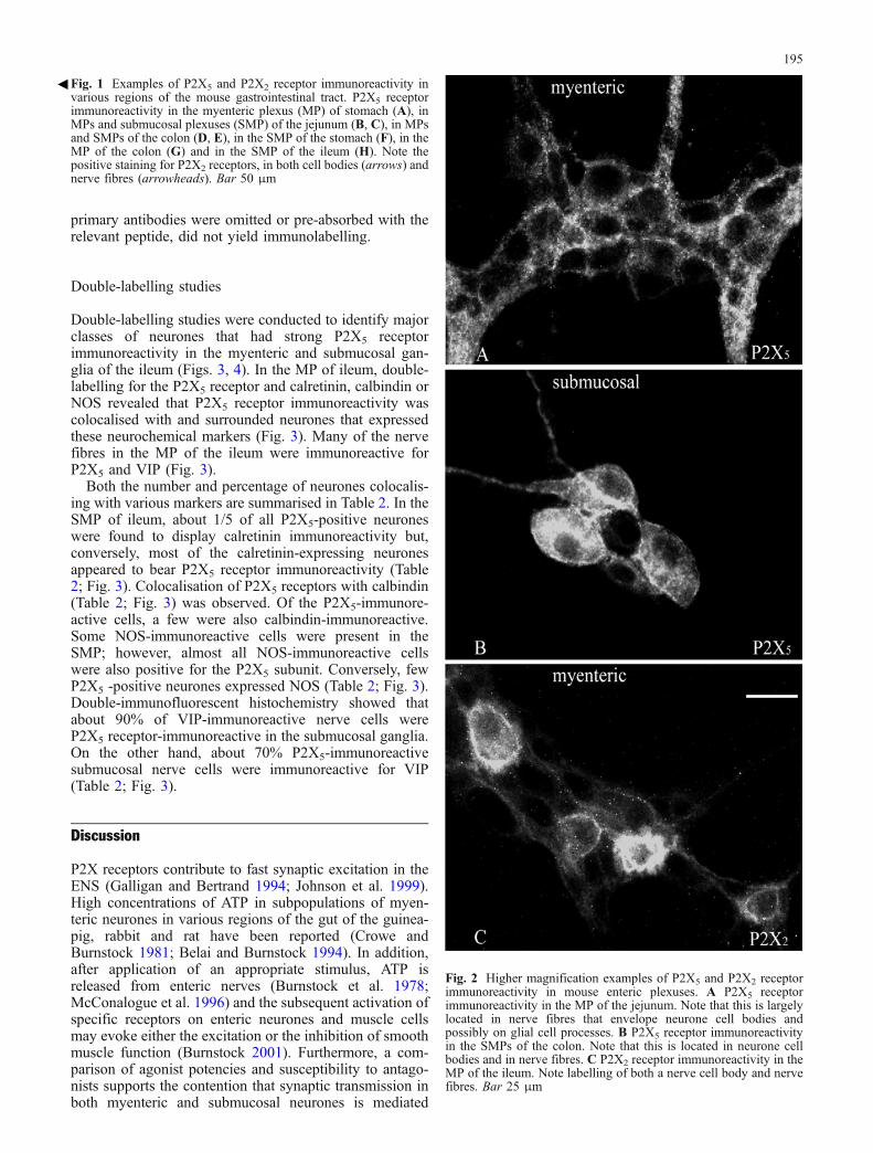
Fig. 2 Higher magnification examples of P2X5 and P2X2 receptorimmunoreactivity in mouse enteric plexuses. A P2X5 receptorimmunoreactivity in the MP of the jejunum. Note that this is largelylocated in nerve fibres that envelope neurone cell bodies andpossibly on glial cell processes. B P2X5 receptor immunoreactivityin the SMPs of the colon. Note that this is located in neurone cellbodies and in nerve fibres. C P2X2 receptor immunoreactivity in theMP of the ileum. Note labelling of both a nerve cell body and nervefibres. Bar 25 μm
3Fig. 1 Examples of P2X5 and P2X2 receptor immunoreactivity invarious regions of the mouse gastrointestinal tract. P2X5 receptorimmunoreactivity in the myenteric plexus (MP) of stomach (A), inMPs and submucosal plexuses (SMP) of the jejunum (B, C), in MPsand SMPs of the colon (D, E), in the SMP of the stomach (F), in theMP of the colon (G) and in the SMP of the ileum (H). Note thepositive staining for P2X2 receptors, in both cell bodies (arrows) andnerve fibres (arrowheads). Bar 50 μm
195
196
through P2X receptors (Barajas-López et al. 1996, 2000).Enteric neurones have been suggested predominantly toexpress P2X2 receptors (Zhou and Galligan 1996; LePardet al. 1997), whereas a minority heterologously expressesP2X1 and P2X3 receptors (Zhou and Galligan 1996).Barajas-López et al. (1996) have observed that myentericneurones express a P2X receptor showing pharmacologi-cal resemblances to P2X4 and P2X6 receptors. Moreover,they have noted that the electrophysiological and pharma-cological properties of P2X receptors are virtuallyidentical in both myenteric and submucous neurones,suggesting that similar P2X receptors are present in bothplexuses. The present study has shown that P2X2 receptorimmunoreactivity is widely distributed in both the MPsand SMPs throughout the entire length of the mousedigestive tract from the stomach to the colon. In the MPs,two kinds of P2X2-positive ganglion neurones (stronglystaining and weakly staining cells) have been seen. Theseresults are consistent with previous studies of P2X2
receptor localisation in guinea-pigs (Castelucci et al.2002). The P2X2 receptor is dominant but this does notpreclude the possibility that other P2X receptor subtypeson enteric neurones also play important roles. Our studyhas also shown that P2X5 receptor immunoreactivity iswidely distributed along the whole length of the mousegut. These observations of the distribution of P2X2 andP2X5 immunoreactivity in the small intestine are con-sistent with previous electrophysiological results (Zhouand Galligan 1996). Approximately 80% of culturedmyenteric ganglion cells in guinea-pig intestine arereported to respond to the P2X receptor antagonist,PPADS, which inhibits 97% of the hexamethonum-resistant fast EPSPs. Zhou and Galligan (1996) claimthat the electrophysiological properties of myentericneurones in the small intestine indicate that 10% of them
express P2X3 or P2X1 receptors, whereas most of themexpress P2X2 or P2X5 receptors.
To date, the evidence for the expression of four P2Xreceptor subtypes has been obtained in the ENS byelectrophysiological or immunohistochemical methods.Immunoreactivity for P2X2 (Castelucci et al. 2002;Giaroni et al. 2002), P2X3 (Poole et al. 2002; VanNassauw et al. 2002) and P2X7 (Hu et al. 2001) receptors,in addition to our results for the P2X5 receptor, has beenreported. Neither P2X1 nor P2X4 receptor immunoreac-tivity has been found in cultured myenteric neurones(Zhou and Galligan 2000) or whole-mount preparations ofenteric ganglia (unpublished observations).
In this study, we have demonstrated, for the first time,the presence of P2X5 receptor immunoreactivity in entericneurones of mouse. We have found abundant P2X5
receptor immunoreactivity at the surfaces of ganglioncell bodies in MPs and in association with nerve fibres,although the possibility that P2X5 receptors are alsolocalised on glial cell processes cannot be excluded. Thedistribution of the receptor on the surfaces of cell bodiessuggests that they could be activated by ATP released as aneurotransmitter or as a paracrine mediator released fromother cellular sources. Double-labelling of the P2X5
receptor in combination with immunoreactivity for calret-inin, calbindin and NOS has revealed P2X5 receptorimmunoreactivity variably colocalised with neuronesexpressing these neurochemical markers. Many of thenerve fibres in the MP of ileum are immunoreactive forP2X5 and for VIP. According to the chemical coding of theenteric neurone classes in the mouse small intestine (Sangand Young 1996), there are two major classes of circularmuscle motor neurones: one class is characterised by thepresence of NOS, VIP and neuropeptide Y (NPY),whereas the second class contains calretinin plus substanceP. In the guinea-pig small intestine (Costa et al. 1996;Brookes 2001), the neurones exhibiting calretinin immu-noreactivity can be identified as excitatory motor neuronesand ascending interneurones, the neurones expressingNOS immunoreactivity are thought to be inhibitory motorneurones and descending interneurones and the neuronesexpressing calbindin immunoreactivity are thought to beprimary afferent neurones. These findings suggest thatspecific roles are mediated via receptors containing P2X5
subunits and that P2X5 receptors could be heteromeric forother subunits, particularly P2X2.
3Fig. 3 Colocalisation of P2X5 receptor immunoreactivity in nervefibres enveloping enteric nerves, with various neurochemicalmarkers in the MP of mouse ileum. P2X5 receptor immunoreactivityis labelled with Cy3 (red) and the neurochemical markers with FITC(green). P2X5 receptor immunoreactivity was seen in nerve fibressurrounding ganglion cell soma with immunoreactivity for A–Ccalretinin (CR), D, E calbindin (CB) and F, G nitric oxide synthase(NOS). The nerve fibres with P2X5 receptor immunoreactivityexhibited colocalisation with H, vasoactive intestinal peptide (VIP).Sites of rare colocalisation (arrows) appear yellow. Bars 50 μm
Table 2 Quantitative analysis of double-labelling studies for P2X5with calretinin, calbindin, neuronal nitric oxide synthase (NOS) andvasoactive intestinal peptide (VIP) in mouse ganglia of the ileum
SMPs (n number of double-labelled cell profiles and the totalnumber of cell profiles counted, respectively, for each combinationof receptor)
Markers P2X5-immunoreactive neuronescontaining calretinin, calbindin,NOS or VIP
Calretinin-, calbindin-, NOS- orVIP-immunoreactive neuronescontaining P2X5
Calretinin n 22±4% (437/1986) 91±3% (437/481)Calbindin n 9±2% (169/1877) 86±5% (169/196)NOS n 6±2% (117/1923) 96±1% (117/122)VIP n 68±4% (1259/1851) 92±3% (1259/1368)
197
198
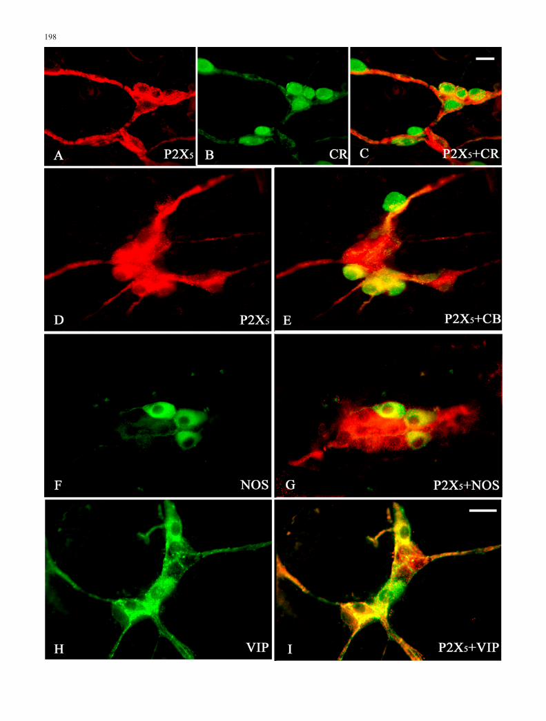
In the SMP, P2X5 receptor immunoreactivity occursthroughout the cytoplasm and on the surface membranesof the nerve cell bodies, nerve fibres and nerve bundles.Colocalisation of P2X5 receptors with calretinin, calbin-din, NOS and VIP in the SMP of mouse ileum has shownthat 22%, 9%, 6% and 68% of neurones with P2X5
receptor immunoreactivity are also immunoreactive forcalretinin, calbindin, NOS and VIP, respectively. Earlierimmunocytochemical studies have demonstrated that fourclasses of nerve cell bodies account for almost all neuronesin the submucosal ganglia of the guinea-pig ileum(Furness et al. 1984; Quinson et al. 2001). These are (1)Dogiel type II neurones immunoreactive for cholineacetyltransferase (ChAT), tachykinins and calbindin(about 10% of nerve cells), (2) neurones immunoreactivefor calretinin and ChAT (about 15%), (3) neurones withVIP immunoreactivity (about 45%) and (4) neurones withboth NPY and ChAT immunoreactivity (about 30%). Ourresults suggest that P2X5 receptor immunoreactivityoccurs at least on three classes of neurones in thesubmucosal ganglia of the mouse ileum.
In conclusion, abundant P2X5 receptors are expressed inthe MP and SMP of the mouse gastrointestinal tract. Thissubunit of P2X receptor may be specific to functionalsubtypes of enteric neurone. The P2X5 receptor and otherreceptors may form heteromultimers that would have aunique pharmacology.
Acknowledgements The authors thank Dr. Chrystalla Orphanidesfor editorial assistance.
References
Abbracchio MP, Burnstock G (1994) Purinoceptors: are therefamilies of P2X and P2Y purinoceptors? Pharmacol Therap64:445–475
Barajas-López C, Huizinga JD, Collins SM, Gerzanich V, Espinosa-Luna R, Peres AL (1996) P2X-purinoceptors of myentericneurones from the guinea-pig ileum and their unusualpharmacological properties. Br J Pharmacol 119:1541–1548
Barajas-López C, Espinosa-Luna R, Christofi FL (2000) Changes inintracellular Ca2+ by activation of P2 receptors in submucosalneurons in short-term cultures. Eur J Pharmacol 409:243–257
Belai A, Burnstock G (1994) Evidence for the coexistence of ATPand nitric oxide in non-adrenergic, non-cholinergic (NANC)inhibitory neurones in the rat ileum, colon and annococcygeusmuscle. Cell Tissue Res 278:197–200
Bian X, Ren J, DeVries M, Schnegelsberg B, Cockayne DA, FordAP, Galligan JJ (2003) Peristalsis is impaired in the smallintestine of mice lacking the P2X3 subunit. J Physiol (Lond)551:309–322
Brandle U, Guenther E, Irrle C, Wheeler-Schilling TH (1998) Geneexpression of the P2X receptors in the rat retina. Brain Res MolBrain Res 59:269–272
Brookes SJH (2001) Classes of enteric nerve cells in the guinea-pigsmall intestine. Anat Rec 262:58–70
Burnstock G (1978) A basis for distinguishing two types ofpurinergic receptor. In: Straub RW, Bolis L (eds) Cellmembrane receptors for drugs and hormones: a multidisciplin-ary approach. Raven Press, New York, pp 107–118
Burnstock G (2001) Purinergic signalling in gut. In: Abbracchio MP,Williams M (eds) Handbook of experimental pharmacology,vol 152/II. Purinergic and pyrimidinergic signalling. II.Cardiovascular, respiratory, immune, metabolic and gastroin-testinal tract function. Springer, Berlin Heidelberg New York,pp 141–238
Burnstock G (2003) Purinergic receptors in the nervous system. In:Schwiebert EM (ed) Current topics in membranes. Purinergicreceptors and signalling, vol 54. Academic Press, San Diego,pp 307–368
Burnstock G, Lavin S (2002) Interstitial cells of Cajal and purinergicsignalling. Autonom Neurosci 97:68–72
Castelucci P, Robbins HL, Poole DP, Furness JB (2002) Thedistribution of purine P2X2 receptors in the guinea-pig entericnervous system. Histochem Cell Biol 117:415–422
Collo G, North RA, Kawashima E, Merlo-Pich E, Neidhart S,Surprenant A, Buell G (1996) Cloning of P2X5 and P2X6receptors and the distribution and properties of an extendedfamily of ATP-gated ion channels. J Neurosci 16:2495–2507
Costa M, Brookes SJH, Steele PA, Gibbins I, Burcher E, KandiahCJ (1996) Neurochemical classification of myenteric neurons inthe guinea-pig ileum. Neuroscience 75:949–967
Crowe R, Burnstock G (1981) Comparative studies of quinacrine-positive neurons in the myenteric plexus of stomach andintestine of guinea-pig, rabbit and rat. Cell Tissue Res 221:93–107
De Man JG, De Winter BY, Seerden TC, De Schepper HU, HermanAG, Pelckmans PA (2003) Functional evidence that ATP or arelated purine is an inhibitory NANC neurotransmitter in themouse jejunum: study on the identity of P2X and P2Ypurinoceptors involved. Br J Pharmacol 140:1108–1116
Dunn PM, Zhong Y, Burnstock G (2001) P2X receptors inperipheral neurons. Prog Neurobiol 65:107–134
Facer P, Knowles CH, Tam PKH, Ford AP, Dyer N, Baecker PA,Anand P (2001) Novel capsaicin (VR1) and purinergic (P2X3)receptors in Hirschsprung’s intestine. J Pediatr Surg 36:1679–1684
Furness JB, Costa M, Keast JR (1984) Choline acetyltransferase andpeptide immunoreactivity of submucous neurons in the smallintestine of the guinea-pig. Cell Tissue Res 237:329–336
Galligan JJ, Bertrand PP (1994) ATP mediates fast synapticpotentials in enteric neurons. J Neurosci 14:7563–7571
Galligan JJ, LePard KJ, Schneider DA, Zhou X (2000) Multiplemechanisms of fast excitatory synaptic transmission in theenteric nervous system. J Auton Nerv Syst 81:97–103
Giaroni C, Knight GE, Ruan HZ, Glass R, Bardini M, Lecchini S,Frigo G, Burnstock G (2002) P2 receptors in the murinegastrointestinal tract. Neuropharmacology 43:1313–1323
Glass R, Burnstock G (2001) Immunohistochemical identification ofcells expressing ATP-gated cation channels (P2X receptors) inthe adult rat thyroid. J Anat 198:569–579
Glushakov AV, Glushakova Y, Skok VI (1998) Two types of P2X-purinoceptors in neurons of the guinea pig ileum submucousplexus. Neurophysiology 30:301–304
Greig AVH, Linge C, Terenghi G, McGrouther DA, Burnstock G(2003) Purinergic receptors are part of a functional signallingsystem for proliferation and differentiation of human epidermalkeratinocytes. J Invest Dermatol 120:1007–1015
Gröschel-Stewart U, Bardini M, Robson T, Burnstock G (1999a)P2X receptors in the rat duodenal villus. Cell Tissue Res297:111–117
3Fig. 4 Higher magnification of the colocalisation (yellow/orange)of P2X5 receptor immunoreactivity with various neurochemicalmarkers in the SMP of mouse ileum. P2X5 receptor immunoreac-tivity is labelled with Cy3 (red) and the neurochemical markers withFITC (green). P2X5 receptor immunoreactivity was seen withimmunoreactivity for A–C calretinin (CR), D, E calbindin (CB), F,G nitric oxide synthase (NOS), H, I vasoactive intestinal peptide(VIP). Bars 25 μm
199
Gröschel-Stewart U, Bardini M, Robson T, Burnstock G (1999b)Localisation of P2X5 and P2X7 receptors by immunohisto-chemistry in rat stratified squamous epithelia. Cell Tissue Res296:599–605
Hu H-Z, Gao N, Lin Z, Gao C, Liu S, Ren J, Xia Y, Wood JD (2001)P2X7 receptors in the enteric nervous system of guinea-pigsmall intestine. J Comp Neurol 440:299–310
Johnson PJ, Shum OR, Thornton PD, Bornstein JC (1999) Evidencethat inhibitory motor neurons of the guinea-pig small intestineexhibit fast excitatory synaptic potentials mediated via P2Xreceptors. Neurosci Lett 266:169–172
Khakh BS, Burnstock G, Kennedy C, King BF, North RA, SeguelaP, Voigt M, Humphrey PPA (2001) International union ofpharmacology. XXIV. Current status of the nomenclature andproperties of P2X receptors and their subunits. Pharmacol Rev53:107–118
Lee H-Y, Bardini M, Burnstock G (2000) Distribution of P2Xreceptors in the urinary bladder and the ureter of the rat. J Urol163:2002–2007
LePard KJ, Galligan JJ (1999) Analysis of fast synaptic pathways inmyenteric plexus of guinea pig ileum. Am J Physiol 276:G529–G538
LePard KJ, Messori E, Galligan JJ (1997) Purinergic fast excitatorypostsynaptic potentials in myenteric neurons of guinea pig:distribution and pharmacology. Gastroenterology 113:1522–1534
McConalogue K, Todorov L, Furness JB, Westfall DP (1996) Directmeasurement of the release of ATP and its major metabolitesfrom the nerve fibres of the guinea-pig taenia coli. Clin ExpPharm Physiol 23:807–812
Oglesby IB, Lachnit WG, Burnstock G, Ford APDW (1999) Subunitspecificity of polyclonal antisera to the carboxy terminalregions of P2X receptors, P2X1 through P2X7. Drug Dev Res47:189–195
Poole DP, Castelucci P, Robbins HL, Chiocchetti R, Furness JB(2002) The distribution of P2X3 purine receptor subunits in theguinea pig enteric nervous system. Auton Neurosci 101:39–47
Quinson N, Robbins HL, Clark MJ, Furness JB (2001) Calbindinimmunoreactivity of enteric neurons in the guinea-pig smallintestine. Cell Tissue Res 305:3–9
Ralevic V, Burnstock G (1998) Receptors for purines andpyrimidines. Pharmacol Rev 50:413–492
Ren J, Bian X, DeVries M, Schnegelsberg B, Cockayne DA, FordAP, Galligan JJ (2003) P2X2 subunits contribute to fastsynaptic excitation in myenteric neurons of the mouse smallintestine. J Physiol (Lond) 552:809–821
Ryten M, Hoebertz A, Burnstock G (2001) Sequential expression ofthree receptor subtypes for extracellular ATP in developing ratskeletal muscle. Dev Dyn 221:331–341
Ryten M, Dunn PM, Neary JT, Burnstock G (2002) ATP regulatesthe differentiation of mammalian skeletal muscle by activationof a P2X5 receptor on satellite cells. J Cell Biol 158:345–355
Sang Q, Young HM (1996) Chemical coding of neurons in themyenteric plexus and external muscle of the small and largeintestine of the mouse. Cell Tissue Res 284:39–53
Van Nassauw L, Bogers J, Van Marck E, Timmermans J-P (2001)Role of reactive nitrogen species in neuronal cell damageduring intestinal schistosomiasis. Cell Tissue Res 303:329–336
Van Nassauw L, Brouns I, Adriaensen D, Burnstock G, Timmer-mans JP (2002) Neurochemical identification of enteric neuronsexpressing P2X3 receptors in the guinea-pig ileum. HistochemCell Biol 118:193–203
Vulchanova L, Arvidsson U, Riedl M, Wang J, Buell G, SurprenantA, North RA, Elde R (1996) Differential distribution of twoATP-gated ion channels (P2X receptors) determined by immu-nocytochemistry. Proc Natl Acad Sci USA 93:8063–8067
Xiang Z, Bo X, Burnstock G (1998) Localization of ATP-gated P2Xreceptor immunoreactivity in rat sensory and sympatheticganglia. Neurosci Lett 256:105–108
Yiangou Y, Facer P, Baecker PA, Ford AP, Knowles CH, Chan CLH,Williams NS, Anand P (2001) ATP-gated ion channel P2X3 isincreased in human inflammatory bowel disease. Neurogas-troenterol Motil 13:365–369
Zhou X, Galligan JJ (1996) P2X purinoceptors in cultured myentericneurons of guinea-pig small intestine. J Physiol (Lond)496:719–729
Zhou X, Galligan JJ (2000) Properties of the nicotinic cholinergicreceptors and P2X receptors in myenteric neurons from guineapig small intestine. Soc Neurosci Abstr 719–729:1633
200