Embed Size (px)
Citation preview
HTA REPORT
Wireless Capsule Endoscopy
in the diagnosis of small bowel disease

This report should be cited as "Age.na.s. HTA Report - Wireless Capsule Endoscopy in the diagno-sis of small bowel disease, Rome, September 2008"

Contributions
Authors:
Fabio Bernardini, Marina Cerbo, Tom Jefferson, Alessandra Lo Scalzo, Marco Ratti
Age.na.s. Agenzia nazionale per i servizi sanitari regionali, Sezione ISS (Innovazione, Sperimentazione e Sviluppo), Rome (Italy)
Corresponding author:
Alessandra Lo Scalzo ([email protected])
Experts:
Lucio Capurso
Senior Consultant (Gastroenterology) - Azienda Ospedaliera "San Filippo Neri", Rome (Italy)
Antonio Migliore
Consultant (Biomedical Engineering) - age.na.s. Agenzia nazionale per i servizi sanitari regionali,Sezione ISS (Innovazione, Sperimentazione e Sviluppo), Rome (Italy)
External Reviewers:
Roberto De Franchis
Department of Medical Sciences, University of Milan (Italy)
Brendan C. Delaney
Primary Care Clinical Sciences, University of Birmingham, Birmingham (UK)
Francesco Martelli
Dipartimento Tecnologie e Salute, ISS – Istituto Superiore di Sanità, Rome (Italy)
In this report Given Imaging GmbH and MG Lorenzatto S.p.A. are not cited as “external revie-wers” since, although they had been involved in its production process, they communicated thatthey do not agree with the report’s conclusions. Given Imaging GmbH and MG Lorenzatto S.p.A.stated that the report does not include/consider some of the important comments and pointsthey made.
Il presente report non contiene il riferimento delle aziende Given Imaging GmbH e MGLorenzatto S.p.A. come “external reviewers”. Tali aziende, pur essendo state contattate durantel’elaborazione del report, hanno comunicato di non riconoscersi nelle conclusioni dello stesso,dal momento che non ritengono essere stati presi in considerazione i commenti e le modifichedalle stesse proposti.

Editing and layout
Dario FellaAge.na.s. Agenzia Nazionale per i Servizi Sanitari Regionali
Acknowledgements:The Agenzia Nazionale per i Servizi Sanitari Regionali (age.na.s.) thanks all the external revie-
wers who kindly provided comments on an earlier draft of this report.
The Age.Na.S. also thanks those who contributed to the realisation of this report providingmarket details, Paolo Gazzaniga and Fernanda Gellona from Association of producers and distri-butors of medical devices (ASSOBIOMEDICA) and the responding medical centres of endoscopyand gastroenterology (see Appendix 7).
Age.Na.S. also thanks Carlo Di Pietrantonj and Alessandro Rivetti from SEREMI (Servizio diriferimento Regionale di Epidemiologia, ASL 20, Alessandria, Italy) for their help in designing andcarrying out the search strategy.
HTA REPORT
Wireless Capsule Endoscopy
in the diagnosis of small bowel disease
Executive summary . . . . . . . . . . . . . . . . . . . . . . . . . . . . . . . . . . . . . . . . . . . . . . . . . . . . .11
Sintesi . . . . . . . . . . . . . . . . . . . . . . . . . . . . . . . . . . . . . . . . . . . . . . . . . . . . . . . . . . . . . . . . . . .13
1. Background 1.1 Clinical problem and indications . . . . . . . . . . . . . . . . . . . . . . . . . . . . . . .191.1.2. Obscure gastrointestinal bleeding (OGIB) . . . . . . . . . . . . . . . . . .191.1.3 Crohn Disease (CD) . . . . . . . . . . . . . . . . . . . . . . . . . . . . . . . . . . . . . . . . . . .201.1.4 Coeliac disease (COD) . . . . . . . . . . . . . . . . . . . . . . . . . . . . . . . . . . . . . . . .201.1.5 Familial Adenomatous Polyposis (FAP) . . . . . . . . . . . . . . . . . . . . . .20
2. Technology, procedure and alternatives2.1. Technology . . . . . . . . . . . . . . . . . . . . . . . . . . . . . . . . . . . . . . . . . . . . . . . . . . . . . .232.2 Procedure . . . . . . . . . . . . . . . . . . . . . . . . . . . . . . . . . . . . . . . . . . . . . . . . . . . . . . . .232.3 The alternatives . . . . . . . . . . . . . . . . . . . . . . . . . . . . . . . . . . . . . . . . . . . . . . . . .24
3. The marketing status of WCE and current reimbursement arrangements . . . . . . . . . . . . . . . . . . . . . . . .27
4. Report’s objectives: policy question and research questions . . . . .29
5. Assessing the available evidence5.1 Methods . . . . . . . . . . . . . . . . . . . . . . . . . . . . . . . . . . . . . . . . . . . . . . . . . . . . . . . . . .315.1.1 Health Technology Assessments reports
and systematic reviews . . . . . . . . . . . . . . . . . . . . . . . . . . . . . . . . . . . . . . . . . .315.1.2 Primary studies . . . . . . . . . . . . . . . . . . . . . . . . . . . . . . . . . . . . . . . . . . . . . . . .31


5.1.3 Inclusion criteria . . . . . . . . . . . . . . . . . . . . . . . . . . . . . . . . . . . . . . . . . . . . . .315.2 Assessing the available evidence: diagnostic accuracy . . . . . . .325.3 Assessing the available evidence: safety of WCE . . . . . . . . . . . . .385.4 Assessing the available evidence:
Systematic reviews and meta analyses . . . . . . . . . . . . . . . . . . . . . . . . .405.5 Assessing the available evidence:
WCE acceptability to patients . . . . . . . . . . . . . . . . . . . . . . . . . . . . . . . . . . .415.6 Results . . . . . . . . . . . . . . . . . . . . . . . . . . . . . . . . . . . . . . . . . . . . . . . . . . . . . . . . . . .41
6. Collecting context specific data 6.1 Appropriateness of WCE use
and related costs in the Italian context . . . . . . . . . . . . . . . . . . . . . . .456.1.1 Objective, materials and methods . . . . . . . . . . . . . . . . . . . . . . . . . . .456.1.2 Results . . . . . . . . . . . . . . . . . . . . . . . . . . . . . . . . . . . . . . . . . . . . . . . . . . . . . . . . .466.2 An assessment of patient's acceptability
of the WCE procedure . . . . . . . . . . . . . . . . . . . . . . . . . . . . . . . . . . . . . . . . . .626.2.1 Objective, materials and methods . . . . . . . . . . . . . . . . . . . . . . . . . . .626.2.2 Results . . . . . . . . . . . . . . . . . . . . . . . . . . . . . . . . . . . . . . . . . . . . . . . . . . . . . . . . .636.2.2.1 Socio economical characteristics of patients . . . . . . . . . . . . . .636.2.2.2 Acceptability dimensions
for the WCE and other endoscopies . . . . . . . . . . . . . . . . . . . . . . .65
7. Discussion . . . . . . . . . . . . . . . . . . . . . . . . . . . . . . . . . . . . . . . . . . . . . . . . . . . . . . . . . . . .69
8. Recommandations . . . . . . . . . . . . . . . . . . . . . . . . . . . . . . . . . . . . . . . . . . . . . . . . . . .71
9. Funding . . . . . . . . . . . . . . . . . . . . . . . . . . . . . . . . . . . . . . . . . . . . . . . . . . . . . . . . . . . . . . . .73
10. Conflict of interest . . . . . . . . . . . . . . . . . . . . . . . . . . . . . . . . . . . . . . . . . . . . . . . . . . . .75
Glossary . . . . . . . . . . . . . . . . . . . . . . . . . . . . . . . . . . . . . . . . . . . . . . . . . . . . . . . . . . . . . . . . .77
Abbreviations . . . . . . . . . . . . . . . . . . . . . . . . . . . . . . . . . . . . . . . . . . . . . . . . . . . . . . . . . . . .83
Bibliography . . . . . . . . . . . . . . . . . . . . . . . . . . . . . . . . . . . . . . . . . . . . . . . . . . . . . . . . . . . . .85
Appendix
1. Expert Opinion Questionnaire Wireless Capsule Endoscopy (WCE) . . . . . . . . . . . . . . . . . . . . . . . . . . . .89
2a. Wireless capsule endoscopy (WCE) . . . . . . . . . . . . . . . . . . . . . . . . . . . . . . .91


2b. Standard technology for diagnosis of bowel diseases . . . . . . . . . . .97
3. Search Strategy . . . . . . . . . . . . . . . . . . . . . . . . . . . . . . . . . . . . . . . . . . . . . . . . . . . .105
4. Tables of comparative studies . . . . . . . . . . . . . . . . . . . . . . . . . . . . . . . . . . . . .107
5. Tables on Safety . . . . . . . . . . . . . . . . . . . . . . . . . . . . . . . . . . . . . . . . . . . . . . . . . . . .129
6. List of citations and reasons for esclusion . . . . . . . . . . . . . . . . . . . . . . . .133
7. Author questions . . . . . . . . . . . . . . . . . . . . . . . . . . . . . . . . . . . . . . . . . . . . . . . . . . .159
8. Responding Centres . . . . . . . . . . . . . . . . . . . . . . . . . . . . . . . . . . . . . . . . . . . . . . . .161
9. Questionnaire Wireless Capsule Endoscopy . . . . . . . . . . . . . . . . . . . . . .163
10. Wireless capsule and traditional Endoscopy:patient acceptability . . . . . . . . . . . . . . . . . . . . . . . . . . . . . . . . . . . . . . . . . . . . . .173

11
Executive summary
One-linerA quick diagnosis for occult bleeding in the gut is necessary. We summarised the evidence of
performance and impact of the video capsule developed for this purpose.
BackgroundThe Wireless Capsule Endoscopy (WCE) is a recent technology that allows imaging of the small
intestine, an anatomic site that has proved peculiarly difficult to visualise. The patient swallows asmall capsule, which whilst moving through the gastrointestinal tract, captures images. The mainindication for WCE use is diagnosis of Obscure Gastrointestinal Bleeding in adults, which is defi-ned as bleeding of unknown origin that persists, or recurs, or is visible after a negative colonsco-py and/or upper endoscopy result. Obscure Gastrointestinal Bleeding is a syndrome or group ofsymptoms rather than a single pathology, and as such makes it difficult to make reliable estima-tes of prevalence. In recent years indications for WCE use are increasing, but this may not be sup-ported by sufficient evidence. Anecdotal evidence and expert opinion about the diffusion of WCEsimply that Italy is the country where its use is most widespread. A systematic assessment of thisdevice for the Italian context has become urgent since new models of the WCE at higher pricesare currently becoming available on the International market.
ObjectivesTo identify and summarise available evidence about the diagnostic accuracy and safety of WCE
for Obscure Gastrointestinal Bleeding, Crohn’s disease, familiar polyposis, and Celiac disease in thesmall bowel, its related costs and acceptability.
MethodsWe ran searches on Medline, Embase and Cochrane Library, looking for evidence of safety, dia-
gnostic accuracy, economic impact and patient’s acceptability. We identified recent evidence syn-thesis studies on which to base our assessment, updated and transferred results to the Italian con-text and collected primary data on safety, use and acceptability of WCE in Italy.
ResultsWe included the latest HTA report (with 10 studies fulfilling our inclusion criteria) and 17 pri-
mary studies to update the HTA report. We identified only one randomised controlled trial compa-ring WCE with Push Enteroscopy. The remainder of the studies were affected by poor study design

12
and their results could not be interpreted. We sent 116 questionnaires to centres performing theWCE requesting information on indications, diagnosis, safety and costs. We received 56 answers(48%) indicating very high variability of WCE use across the country. The main indication was apositive fecal occult blood test (45%), followed by Inflammatory Bowel Disease (19%).Angiodysplasia was the most frequent diagnosis (39.5%) followed by Inflammatory Bowel Disease(18.3%) and polyposis (7.8%). In 2006, 2457 WCE procedures were carried out, with 17 seriousharm cases (1%) and 375 failed procedures (15.3%) reported. The average cost per procedure in2006 was €1.108 in a medium throughput centre (44 WCEs per annum). We surveyed 126 patientsin the 5 centres with the highest annual WCE throughput. Sixty eight percent of patients (84)found the WCE procedure tolerable compared with 33% (39 patients) for colonoscopy or oeso-phago-gastro-duodenoscopy patients. The equivalent data for the induction of anxiety were 25(20%) and 60 (52%).
ConclusionsBased on evidence from one randomised controlled trial the WCE appears dominant for the
diagnosis of Obscure Gastrointestinal Bleeding in the small bowel compared to Push Enteroscopy.However its dominance is based on tolerability, rather than proof of a superior diagnostic accura-cy. The WCE procedure has a high failure rate, results in serious harms in 1% of cases, but is moreacceptable to patients than its alternatives. Given its tiny evidence base, high cost, and potential-ly high failure rate, the WCE procedure should be only be reimbursed if used in a valid evidence-generating framework.

13
Sintesi
Problema clinico e indicazioni per l’utilizzo della VCE Le particolari caratteristiche dell’intestino tenue, una configurazione complessa e una lunghez-
za che si aggira in media intorno ai 6 metri, rendono difficile la sua visualizzazione che è possibi-le solo in parte attraverso endoscopia tradizionale e/o esami radiologici. Quando queste procedu-re danno esito negativo, è necessario poter visualizzare anche le parti di intestino non raggiuntericorrendo a tecniche alternative, come la Video Capsula Endoscopica (VCE) che permette la visua-lizzazione dell’intero tratto gastrointestinale. Le indicazioni per la VCE sono oggi molteplici: san-gue occulto nelle feci (OGIB), ma anche diagnosi e valutazione della severità della malattia diCrohn (CD), diagnosi per la celiachia (COD) e poliposi familiare (FAP).
Per OGIB, acronimo di Obscure Gastrointestinal Bleeding, si intende la presenza, permanentee/o ricorrente, di sangue di origine sconosciuta nelle feci, sospetto e/o identificato a seguito di testIDA (Iron Deficiency Anemia), test FOBT (Faecal Occult Blood Test) o sanguinamento visibileanche dopo colonoscopia e gastroscopia, che abbiano dato esito negativo (Zuckerman, 1999). Sistima che l’OGIB sia dovuto nel 5% dei casi a lesioni nell’intestino tenue (AmericanGastroenterological Association, 2007). La causa più comune delle lesioni nel tenue è l’angiodi-splasia, all’origine del 70%-80% di tutti i casi di OGIB, seguita dal tumore (Lewis, 1994).
La malattia di Crohn (CD) è un’enterite subcronica idiopatica, che interessa soprattutto l’ileoterminale ed è caratterizzata da ulcerazioni che possono causare fistole e stenosi dell’intestino.L’incidenza in Europa meridionale è di 3.6/100,000, mentre in Italia una recente ricerca ha stima-to che 1 persona ogni 100.000 è affetta da CD (Schivanada et al, 1996). La Celiachia (COD) è unamalattia dovuta all’intolleranza al glutine, che provoca infiammazione cronica e atrofia della muco-sa dell’intestino. Secondo la National Digestive Diseases Information Clearinghouse, in Italia circa1 persona su 250 è celiaca. La Poliposi familiare ereditaria comporta la tendenza a sviluppare poli-pi intestinali precancerosi che, se non trattati in tempo, possono trasformarsi in tumori. In caso dipoliposi adenomatosa la crescita dei polipi si presenta come molto lenta. L’incidenza della FAP variada 1 ogni 7,000 a 1 ogni 22,000 indivdui (Genetics Home Reference).
Descrizione della tecnologia e sue alternative L’endoscopia mediante Video Capsula Endoscopica (VCE) può essere eseguita in ambulatorio,
in ricovero ordinario o in day hospital. Il paziente ingoia la capsula dopo avere digiunato durante lanotte (8-12 ore), e questa riprende con una micro video camera tutto il tratto intestinale mentre lopercorre. Dopo circa 8 ore la batteria della videocamera si esaurisce e le immagini ed i dati regi-strati dalla capsula sono scaricati sul PC, dal registratore posto su una cintura applicata sul corpodel paziente. La procedura non è raccomandata in pazienti che hanno una storia di restringimentiintestinali e/o ostruzioni, portatori di pacemaker cardiaci o di altre apparecchiature elettronicheimpiantate. Rispetto ai possibili comparatori della VCE, alcune tecniche endoscopiche che vengonoannoverate tra le alternative in realtà non permettono la visualizzazione di tutto l’intestino tenue.In letteratura, gli studi individuati si basano sul confronto tra VCE e diversi altri comparatori (vediAppendici n.2a e 2b nel testo del report). Tra esse, come confermato da una nostra indagine cheha coinvolto 56 gastroenterologi, la DBE rappresenterebbe l’unico comparatore valido.
14
ObiettiviIn questo quadro, a fronte di una diffusione della VCE in Italia che gli esperti giudicano ampia,
il lavoro svolto ha avuto due obiettivi:
1) valutare le evidenze disponibili in letteratura per le diverse indicazioni di utilizzo dellacapsula;
2) quantificare l’effettiva diffusione della tecnologia in Italia, le sue modalità di rimbor-so ed utilizzo, i costi ad essa connessi, l’accettabilità da parte del paziente.
MetodiNel caso del primo obiettivo, è stata svolta una revisione sistematica della letteratura. Per il
secondo obiettivo sono state effettuate un’analisi di contesto, per identificare la situazione di mer-cato della VCE in Italia, ed un’indagine nazionale per la raccolta di dati primari (survey), data lanatura, fortemente legata al contesto d’uso, di questo tipo di informazioni e l’assenza, in lettera-tura, di informazioni relative a costi e diffusione della VCE in Italia.
Per la revisione sistematica, le fonti consultate sono state Medline, Embase e la CochraneLibrary e, per il reperimento di report di HTA sulla VCE, è stato consultato il database dello “YorkCentre for Review and Dissemination”. Il report più recente sulla VCE, “Endoscopie par Capsule”del Health Care Knowledge Centre (KCE) belga, pubblicato nel 2005, è stato il nostro punto di par-tenza: la ricerca bibliografica compiuta è stata, infatti, finalizzata all’aggiornamento, per gli annidal 2005 al 2007, della revisione sistematica presente nel report del KCE. L’applicazione dei nostricriteri di inclusione ha portato alla identificazione di un totale di 27 studi (per i dettagli completirelativi a strategia di ricerca e criteri di inclusione vedi cap. 5.1 e Appendici 1 e 3 del report). Glioutcome considerati sono stati, oltre all’accuratezza diagnostica, anche l’accettabilità da parte delpaziente, l’impatto economico e la sicurezza della VCE.
Per l’analisi di contesto e l’indagine nazionale abbiamo dovuto prima individuare tutti i centridi endoscopia e/o gastroenterologia italiani che, nell’anno 2006, hanno erogato prestazioni dia-gnostiche con VCE. A questo scopo è stato utilizzato il database clienti fornitoci dal distributoreitaliano, M.G. Lorenzatto S.p.a., di cui abbiamo controllato la completezza attraverso il confrontocon le fonti ministeriali (database di tutti i centri di gastroenterologia e endoscopia in Italia) e conaltri database non istituzionali (database dei gastroenterologi iscritti al CICE – Club Italiano dellaVideo Capsula). Ai centri identificati è quindi stato inviato un primo questionario finalizzato alla rile-vazione di dati e informazioni sulle modalità d’utilizzo della VCE e sui suoi costi, in termini di mate-riali, ma anche di tempo speso da parte dei professionisti impegnati nella procedura (periodo rile-vazione: dicembre 2007 - marzo 2008). Un secondo questionario rivolto ai pazienti è stato invia-to a 5 centri, selezionati ex post sulla base dei volumi annui di utilizzo della capsula, allo scopo divalutare l’effettiva soddisfazione del paziente relativamente all’utilizzo della VCE, nonché alcunicosti indiretti, come il tempo speso per essere sottoposto alla procedura, in confronto ad altriesami endoscopici (periodo rilevazione: febbraio-maggio 2008).
15
Risultati
Revisione sistematicaÈ stato individuato un unico trial randomizzato (De Leusse et al.) pubblicato, tra l’altro, di
recente, nel 2007, dopo 6 anni dall’entrata della VCE sul mercato italiano. Questo studio su pazien-ti OGIB è di buona qualità, ma oltre ad avere alcuni limiti di potenza campionaria, non confrontala VCE con il suo effettivo comparatore (la DBE), ma con la PE che non visualizza, come accenna-to, l’intero tratto intestinale. Gli autori, infatti, concludono consigliando l’utilizzo della capsula comemetodo complementare, e non alternativo, alla DBE.
Rispetto all’accuratezza diagnostica, tutti gli studi sull’utilizzo della VCE in pazienti OGIB, indi-cano per la VCE una maggiore accuratezza diagnostica se confrontata con quella di PE, DBE e altretecniche radiologiche. D’altra parte, eccetto il caso del trial di De Leusse, vi sono limiti nel disegnoadottato da tutti gli altri studi, che rendono le conclusioni non attendibili. Essi presentano, infatti,un disegno di tipo sequenziale, in cui lo stesso paziente a intervalli di tempo variabile da studio astudio, non sempre riportati, viene sottoposto alla VCE e quindi al comparatore, o viceversa, fun-gendo sostanzialmente da “controllo” di sé stesso. Il tempo che intercorre tra una procedura e l’al-tra appare però molto rilevante in termini diagnostici nel caso dei pazienti OGIB, dato che il san-guinamento occulto è spesso dovuto a lesioni angiodisplasiche. Queste lesioni, possono, infatti,variare molto velocemente, con il risultato che uno stesso paziente dopo una sola settimana, puònon presentarsi più con le stesse caratteristiche. Il disegno dunque, se punta a minimizzare levariabili confondenti, con il confronto dello stesso paziente in tempi diversi, rischia di ottenere ilrisultato opposto. Si confrontano infatti i dati clinici ottenuti su un individuo con una procedura epoi con l’altra, ma dal punto di vista delle lesioni da identificare quello stesso soggetto potrebbeessere molto diverso da sé stesso, anche dopo una sola settimana.
Gli studi individuati, inoltre, oltre a non avere un disegno appropriato, sono anche in numeronon alto, una volta distinti in base sia al tipo di indicazioni, sia al tipo di comparatore. Per i pazien-ti OGIB abbiamo, infatti, identificato 7 studi che comparano PE e VCE, 5 DBE vs VCE, mentre 5riguardano varie procedure di tipo radiologico vs VCE. Per il morbo di Crohn sono stati identifica-ti 6 studi, che comparano la VCE a varie tecniche radiologiche e/o alla PE, mentre per la FAP sonostati identificati solo 3 studi. Per la celiachia nessuno tra gli studi individuati, ha soddisfatto i cri-teri di selezione per la revisione sistematica.
Rispetto alla sicurezza, aggregando i dati degli studi che riportano informazioni su questadimensione, risulta che su un totale di 1236 pazienti il numero di eventi avversi è molto alto ed èpari a circa il 3%, mentre si incorre in problemi tecnici nel 7% circa dei casi. Per la parte econo-mica nessuno degli studi selezionati contiene informazioni, mentre per la accettabilità da parte deipazienti solo 3 studi su 27, trattano l’argomento sebbene, per lo più, in modo non metodologica-mente fondato, ed evidenziando una generica preferenza del paziente per la tecnica diagnosticain analisi.
16
Analisi di contesto e raccolta di dati primari (survey nazionale)L’analisi di contesto ci ha permesso di identificare l’esistenza, dal 2001 al 2006, di un solo
distributore sul mercato italiano la M.G. Lorenzatto S.p.a, e di un solo produttore, la GIVENImaging Ltd (nel 2007 la Olympus Corporation è entrata come produttore alternativo). I dati divendita per l’Italia, forniti da M.G. Lorenzatto S.p.a, mostrano che nel periodo dal 2001 al 2006,sono state comprate 12.451 capsule, con un aumento negli acquisti pari al 20% annuo a partiredal 2005. Dal 2001 al 2007, per la sola strumentazione d’esame (VCE e macchinari di lettura risul-tati esami), sono stati spesi un totale di €16.608.000. La VCE risulta rimborsata come proceduraambulatoriale, in Piemonte, Basilicata, Sardegna e Valle d’Aosta (Orecchia, 2007), ma dai dati dellanostra survey risulta eseguita anche in regime di ricovero ordinario e in Day Hospital. Rispetto alnumero di VCE vendute i dati aggregati a livello regionale, mostrano molta variabilità: per esem-pio, il numero maggiore di VCE vendute ogni 100.000 abitanti (13/100.000) è in Liguria e nelleMarche, mentre in Calabria il dato scende molto (1/100.000).
La survey per la raccolta di dati relativi all’utilizzo e ai costi della VCE ha coinvolto tutti i 116centri individuati (Cap 6 e Appendice 7) con una percentuale di ritorno del questionario su utiliz-zo e costi, del 48% (56 centri su 116). I centri rispondenti sono nel 91% dei casi pubblici e, nel2006, risultano avere effettuato un totale 2457 endoscopie con VCE. Il regime ambulatoriale vieneutilizzato nel 31% dei casi, il regime di ricovero nel 43% e il day hospital nel 26%. Dal punto divista clinico, in media, ogni paziente prima di essere sottoposto alla VCE risulta avere già esegui-to almeno 2 altri esami endoscopici con esito negativo e, nel 40% dei casi la diagnosi principaledopo la VCE è di angiodisplasia (vedi Figura 6.10 nel testo del report). Dal punto di vista della sicu-rezza, nell’1% dei casi si sono verificati eventi quali ritenzione della capsula, occlusioni intestinalie/o subintestinali. Più alte sono le percentuali di fallimento dell’esame (15%), dovuto nel 6% deicasi a scaricamento della batteria pre termine, e nel 9% dei casi alle caratteritiche fisiche deipazienti. Il 3% dei pazienti ha invece ripetuto l’esame per inadeguata preparazione (vedi Tabella6.12 nel testo del report).
Dal punto di vista dei costi, una forte variabilità è stata rilevata per i prezzi di hardware e soft-ware (€ 43.108 in media), mentre il prezzo di vendita della capsula appare invece costante (inmedia € 642,00 IVA compresa). I dati relativi al tempo dedicato da medici, infermieri e altro per-sonale nello svolgimento della procedura, hanno permesso una stima del budget impact in trediversi scenari, distinti in base al numero alto (190/anno), medio (44/anno) o basso (10/anno) diesami VCE effettuati in un anno. Il numero di esami annuali risulta influire, in ragione della varia-bile tempo/operatore, sui costi unitari: l’impatto economico di un singolo esame nel caso di uncentro con volumi bassi di utilizzo è di € 1.624,30, mentre nel caso di volumi medi scende a €1.108,41, per ridursi ulteriormente in centri con almeno 190 VCE anno, il cui costo unitario è sti-mato pari a € 998,28 (Tavola 6.14 nel testo del report ).
L’indagine sui pazienti ha interessato 116 individui evidenziando che non c’è risparmio ditempo da parte del paziente nel sottoporsi alla VCE piuttosto che ad altre procedure più invasive:il 70% di coloro che si sottopongono alla VCE perdono più di 7 ore della propria giornata (Figura6.15 nel testo del report). Dal punto di vista della facilità della procedura e dei livelli di stress con-nessi la VCE, questa appare invece meglio tollerata di altre procedure, e ciò anche dal punto divista del dolore, che non è assolutamente percepito dall’83% dei rispondenti nel caso della VCE(Figura 6.18 nel testo del report).
17
DiscussioneLa valutazione relativa alla qualità degli studi è negativa e questo determina una qualche incer-
tezza rispetto alla affidabilità delle loro conclusioni su accuratezza diagnostica e sicurezza. Esisteal momento un solo trial randomizzato, pubblicato nel 2007 (De Leusse et al), e riguarda i pazien-ti con OGIB e la comparazione tra performance della VCE e della PE, che però non raggiunge tuttoil tratto dell’intestino tenue. Gli altri studi sono distinguibili in studi che coinvolgono pazienti OGIB,FAP o Crohn, ed ogni gruppo di studi è ulteriormente diviso in base al “comparatore”. In ogni grup-po e sottogruppo è presente lo stesso disegno di studio “sequenziale” fonte di forte bias nei risul-tati finali, dato che le lesioni che causano sanguinamento sono, in altissima percentuale, di tipoangiodisplasico e, dunque, con una alta variabilità morfologica in tempi molto brevi (dato presen-te in letteratura e confermato dalla nostra indagine nazionale). Questo rende il disegno utilizzatoinaffidabile, ed i risultati ottenuti su questa base di evidenza, potenzialmente non affidabili.L’analisi di contesto e la survey hanno evidenziato un’alta diffusione della procedura, ed un usoche non pare essere basato su criteri di efficienza ed economie di scala, se si considerano i risul-tati della budget analysis effettuata sui tre scenari. Alla raccolta di dati di costo non è stato pos-sibile fare seguire una valutazione economica di costo efficacia per la mancanza di un chiaro com-paratore, di evidenze solide e di stime di prevalenza attendibili per le varie indicazioni.
Raccomandazione Sarebbe importante che nel futuro il rimborso della VCE fosse legato alla produzione di evi-
denze fondate su trial randomizzati ben costruiti, il cui obiettivo sia testare le performance dellatecnologia per le sue diverse indicazioni, sotto la supervisione di una commissione scientifica edetica. Questo tipo di approccio, che a livello internazionale è già in uso (ed è chiamato Coveragewith Evidence Development), dovrebbe essere adottato per tutte le tecnologie emergenti primache queste si diffondano, in modo non governato e spesso senza sufficienti evidenze, in Italia. Inparticolare, rispetto al contesto italiano alcune nuove indicazioni proposte, come per la diagnosi diceliachia, appaiano inutili in quanto per tale diagnosi è comunque necessaria una biopsia che affer-mi l’effettiva presenza della patologia.

18
Bibliografia
American Gastroenterological Association (AGA) Institute Technical Review on ObscureGastrointestinal Bleeding. Gastoenterology 2007;133:1697 1717.
De Leusse A, Vahedi K, Edery J, et al. Capsule endoscopy or push enteroscopy for first-lineexploration of obscure gastrointestinal bleeding? eng. Gastroenterology. 2007 Mar;132(3):855-62; quiz 1164-5; ISSN: 0016-5085
Genetics Home Reference (http://ghr.nlm.nih.gov)
Lewis BS, Small Intestinal Bleed. Gastroenterology Clincs of North America,1994;23(1):67 91.
Orecchia G, Oral communication at 2° CONGRESSO NAZIONALE sulla Videoendoscopia -Second Italian Congress on WCE - 28-30 June 2007, Feltre (Italy).
Schivanada S, Lennard Jones J, Logan R et al. Incidence of inflammatory bowel diseaseacross Europe: is there a difference between north and south? Results of the EuropeanCollaborative Study on Inflamma tory Bowel Disease (EC IBD) Gut 1996;39:690 697.
Zuckerman GR, Prakash C, Askin MP, Lewis BS. AGA technical review on the evaluation andmanagement of occult and obscure gastrointestinal bleeding. Gastroenterology1999;118:201 221.

19
1. Background
1.1 Clinical problem and indications
The characteristics of the human small bowel, a complex looped configuration/structure anda huge length (around 6 metres), make its examination difficult. For the diagnosis of small boweldiseases endoscopy can be used to determine the causes of obscure gastro intestinal bleeding(OGIB), Crohns disease (CD) and coeliac disease (COD) (see relative sections for definitions).
Visualisation of the small bowel is possible using different radiological and traditional endosco-pic procedures, the latter such as Push Enteroscopy (PE) does not always allow the examinationof the entire organ (see section 2). When a source of bleeding cannot be detected with the aboveprocedures, and this occurs in 5-10% of OGIB cases (Myers1, and results of the Italian survey heldat Torgiano’s Gastroenterologist meeting in 2007, see Appendix 1), the lesion causing the bleedingcould be located in the part of the small bowel not yet visualised.
Wireless Capsule Endoscopy (WCE), approved in 2001 by the Food and Drug Administration inthe United States, allows visualisation of the entire small bowel. The capsule is being used forOGIB; for the diagnosis of CD, and for the assessment of its extent and severity; in the diagnosisof small bowel tumours; in the detection of small bowel injury associated with the use of non ste-roidal anti-inflammatory drugs; in the delineation of type of abdominal pain and in the assessmentof COD.
1.1.2. Obscure gastrointestinal bleeding (OGIB)
OGIB is defined as bleeding of unknown origin that persist or recurs, as in recurrent or persi-stent Iron Deficiency Anemia (IDA), positive Faecal Occult Blood Test (FOBT), or visible bleedingafter a negative colonoscopy and/or upper endoscopy result. Obscure bleeding can thus have twoclinical forms: obscure-occult, as manifested by recurrent IDA and/or recurrent positive FOBTresults, and obscure-overt, with recurrent passage of visible blood (Zuckerman2).
OGIB could be due to lesions that are overlooked in the esophagus, stomach, and colon duringinitial workup or lesions in the small intestine that are difficult to visualise with conventional endo-scopy and radiologic imaging. Overall lesions in the small intestine account for approximately 5%of causes of OGIB (American Gastroenterological Association, 20073). Medical imaging of the smallintestine has been a very difficult due to limited visualisation of the lumen.
The principal causes of small bowel bleeding are:
• Angiodysplasia;
• Vascular lesions;
• Adenocarcinoma;
• Lymphoma;
• Carcinoid Tumour;

20
• Ulcers;
• Crohn’s disease;
• Polyps.
The most common cause of small intestine bleeding is Angiodysplasias: 70%-80% of all OGIBare due to these kind of lesions, while tumours are in fact the second most common cause(Lewis4).
1.1.3 Crohn Disease (CD)
CD is a subacute chronic enteritis, of unknown cause, involving the terminal ileum and lessfrequently other parts of the gastrointestinal tract; characterised by patchy deep ulcers that maycause fistulas, and narrowing and thickening of the bowel by fibrosis and lymphocytic infiltration,with noncaseating tuberculoid granulomas that may also be found in regional lymph nodes.Symptoms include fever, diarrhea, cramping abdominal pain, and weight loss (Stedman’s MedicalDictionary5).
Incidence rates for CD were generally lower and were similar for men and women, with ratesfor both sexes declining with increasing age. The European Collaborative Study on InflammatoryBowel Disease (EC-IBD) indicated that in Northern Europe theincidence rates of CD patients for100,000 population aged 15 years or over 6.3/100,000, while in Southern Europe the number ofCD patients was 3.6/100,000. Data from the 4 Italian centres involved in the reaserch showed that1/100.000 had CD (Schivanada et al.6).
1.1.4 Coeliac disease (COD)
COD occurs in both children and adults and is characterised by sensitivity to gluten, with chro-nic inflammation and atrophy of the mucose of the upper small intestine; symptoms include diar-rhoea, malabsorption, steatorrhea, nutritional and vitamin deficiencies, and failure to thrive, orshort stature (Stedman’s Medical Dictionary5). This digestive disease damages the small intestineand interferes with the absorption of nutrients.
Data on the prevalence of COD is spotty and more research is needed to learn the true pre-valence, as in Italy, a National register on inflammatory bowel diseases (to which COD belongs) isnot yet available. According to the National Digestive Diseases Information Clearinghouse7, in Italyabout 1 in 250 people have COD. However, it could be underdiagnosed due to symptoms whichare common to other pathologies, unfamiliarity with the disease and lack of specific expertise.
1.1.5 Familial Adenomatous Polyposis (FAP)
FAP is an inherited disorder characterised by cancer of the large intestine (colon) and rectum.People with the classic type of familial adenomatous polyposis begin to develop multiple noncan-cerous (benign) growths (polyps) in the colon, which may become malignant (cancerous). Some

people have a variant of the disorder, called attenuated familial adenomatous polyposis, in whichpolyp growth is delayed. In both classic familial adenomatous polyposis and its attenuated variant,benign and malignant tumours are sometimes found in other places in the body, including the duo-denum (a section of the small intestine), stomach, bones, skin and other tissues. A milder type offamilial adenomatous polyposis, called autosomal recessive familial adenomatous polyposis, hasalso been identified. People with the autosomal recessive type of this disorder have fewer polypsthan those with the classic type. The colonoscopy consents disease diagnosis. The reported inci-dence of familial adenomatous polyposis varies from 1 in 7,000 to 1 in 22,000 individuals (GeneticsHome Reference8).
21

22

23
2. Technology, procedure and alternatives
2.1. Technology
Wireless Capsule Endoscopy (WCE) (figure 2.1) is a recent technology primarily designed toprovide imaging of the small intestine, an anatomic site that has proved paticularly difficult tovisualise. Devised by an Israeli engineer, Gavriel Iddan in 1981: 15 years later, the capsule wastested on animals, whilst the first trials on adult humans began in 2001 (see Appendix 2a).
Figure 2.1 Video Capsule Endoscopy
1) optical cupola;
2) lock for the lens;
3) lens;
4) lighting system to LED (Light Emitted Diode);
5) CMOS imager (Complementary Metal Oxide Semiconductor);
6) Batteries;
7) Transmitter ASIC (Application Specific Integrated Circuit);
8) Antenna.
2.2 Procedure
The procedure can be performed in an ambulatory or hospital setting on an outpatient basis.The patient swallows a small capsule after fasting overnight (8-12 hours). The capsule contains amicro-imaging video technology, a light source and a wireless circuit for the acquisition and tran-smission of images. The system also includes a software that provides localisation of the deviceduring its passage through the intestine. While moving through the gastrointestinal tract, imagesare captured at the rate of two per second. These images are transmitted to a data recorder wornon a belt outside the patient’s body and approximately eight hours after ingestion, the patientreturns to the clinic where images and data are downloaded. The capsule is passed in the patien-t’s stools within 24-48 hours. It is not reusable.
The WCE procedure involves a high degree of expertise and providers of the service should
24
be specialists who have undertaken a specific training program. The procedure is not recommen-ded in patients suspected of, or with a history of, intestinal stricture or obstruction, or who carrya cardiac pacemaker or other implanted electronic devices. The main indication for WCE use is thediagnosis of the site of Obscure Gastrointestinal Bleeding (OGIB) in adults. OGIB is defined asbleeding of an unknown origin that persists or recurs after a negative initial endoscopy (colono-scopy and/or upper gastrointestinal endoscopy). The capsule is being used for the diagnosis,assessment of its extent and severity of CD, in the diagnosis of small bowel tumours, in the detec-tion of small bowel injury associated with the use of non steroidal anti-inflammatory drugs, deli-neation of the type of abdominal pain and in the assessment of COD.
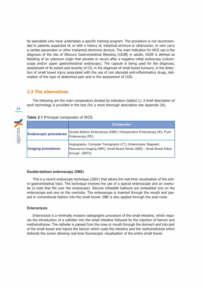
2.3 The alternatives
The following are the main comparators divided by indication (table2.1). A brief description ofeach technology is provided in the text (for a more thorough description see Appendix 2b).
Table 2.1 Principal comparator of WCE
Double-balloon enteroscopy (DBE)
This is a recent endoscopic technique (2001) that allows the real-time visualisation of the enti-re gastrointestinal tract. The technique involves the use of a special enteroscope and an overtu-be (a tube that fits over the endoscope). Silicone inflatable balloons are embedded one on theenteroscope and one on the overtube. The enteroscope is inserted through the mouth and pas-sed in conventional fashion into the small bowel. DBE is also applied through the anal route.
Enteroclysis
Enteroclysis is a minimally invasive radiographic procedure of the small intestine, which requi-res the introduction of a catheter into the small intestine followed by the injection of barium andmethylcellulose. The catheter is passed from the nose or mouth through the stomach and into partof the small bowel and injects the barium which coats the intestine and the methylcellulose whichdistends the lumen allowing real-time fluoroscopic visualisation of the entire small bowel.
Comparator
Endoscopic proceduresDouble Balloon Enteroscopy (DBE), Intraoperative Enteroscopy (IE), Push
Enteroscopy (PE).
Imaging procedures
Angiography, Computer Tomography (CT), Enteroclysis, Magnetic
Resonance Imaging (MRI), Small Bowel Series (SBS) - Small Bowel follow
through (SBTS)

25
Intraoperative Enteroscopy (IE)
IE (per-orally, trans-nasally, per-rectum, or through single or multiple intestinal incisions) isusually applied in cases of bleeding that has not been localised in spite of extensive diagnostic eva-luation and in which the risks of continued bleeding are judged to outweigh the risks of laparotomy.
Standard push enteroscopy (PE)
This standard procedure allows visualisation of the bowel. It requires the oral insertion of along endoscope and allows examination of the distal duodenum and proximal jejunum. Due to theanatomical features of gastrointestinal tract, this kind of device cannot reach all the parts of thebowel and therefore some “dark areas” cannot be observed.
Angiography
Angiography is the x-ray study of the blood vessels. An angiogram uses a radio opaquemedium to highlight the blood vessels in a fluoroscopy suite. Angiography requires the injectionof a contrast medium that makes the blood vessels visible to x-rays. The patient’s vascular systemis displayed on a monitor in real-time. For examination of the small bowel, the procedure is cal-led mesenteric angiography and involves x-ray exploration of the celiac and mesenteric arteries,the arterial branches that supply blood to the abdomen and digestive system.
Computer Tomography (CT)
Computer Tomography scanning (CT or CAT, Computer Assisted Tomography) is a non-invasi-ve, painless imaging technique. CT uses special x-ray equipment to produce multiple images ofthe inside of the body and a computer joins them together in cross-sectional views of the areabeing studied. CT scans of internal organs, bone, soft tissue and blood vessels provide greater cla-rity than conventional x-ray examinations.
Magnetic Resonance Imaging (MRI)
Unlike conventional x-ray examinations and CT scans, MRI does not depend on radiation.Instead, radio waves are directed at hydrogen atoms, in a strong magnetic field.
Small Bowel Series (SBS) or Small Bowel follow-Through (SBTS)
STS or SBTS is an x-ray examination of the small intestine. This procedure requires that thepatient swallows a radio-opaque contrast medium, usually barium sulphate, and then is placed invarious positions on the x-ray table while the radiologist uses a fluoroscope connected to a moni-tor to acquire x-ray images usually every 20 to 30 minutes (this exam often takes 2 hours or moreto complete).

26

27
3. The marketing status of WCE and current reimbur-sement arrangements
Two main companies are competing in the worldwide market:
• Given Imaging Ltd, with its PillCam;
• Olympus Corporation, with its EndoCapsule
The latter in 2007 developed a wireless capsule endoscope (EndoCapsule EC type 1) using adifferent image sensor with electronic enhancement of image quality. In Italy Olympus started-commercialisation of the Endo Capsule in 2007.
The capsule by Given Imaging Ltd has been on the world market since 2001 (M2A capsule),receiving approval from the U.S. Food and Drug Administration (FDA) in August 2001 for use inthe United States. As regards the European market, Given Imaging Ltd received approval (CEMark) by the Regulatory Authorities of the European Union in May 2001. The European directiveson biomedical devices have been incorporated into the Italian legislative system. Its main requi-rements are related mainly to safety and technical performance. In Italy, market introduction ofbiomedical technologies, is not subordinated to further regulation by any national or regionalbodies. However the 2003 financial law (L. 266/2002) forsaw the establishment of a national data-base of all medical devices available in the Italian market (“Repertorio generale dei dispositivimedici commercializzati”). Therefore the use of biomedical devices such as the WCE is usuallystarted and developed in a bottom up fashion. The WCE capsule by Given Imaging Ltd has beensold in Italy since 2001, and its use is widespread. Data form Given Imaging Ltd, shows that over3,500 centres from more than 65 countries use WCE (Orecchia G9). Data provided byM.G.Lorenzatto S.p.a., exclusive distributor of the Given Imaging Ltd WCE in Italy, show that inthe period 2001 - 2006, 12.451 capsules were purchased, with a yearly increase of 20% from 2005onwards. WCE is reimbursed as an outpatient procedure in 4 Regions: Piemonte, Basilicata,Sardegna and Valle d’Aosta (Orecchia9).

28

29
4. Report’s objectives: policy question and researchquestions
According to expert opinion, the WCE has been widely distributed and used in Italy since itsintroduction in 2001. However no data on its use has ever been collected.
Our policy question was: what is the evidence of diagnostic accuracy underpinning the use ofWCE and what are its uses and costs in Italy?
Our research question was: what is the scientific evidence of WCE’s diagnostic accuracy com-pared to other commonly used techniques and what is the evidence of its safety, patients' accep-tability and economic impact in Italy?
The report’s objectives are:
• To retrieve, assess and appraise the available evidence about diagnostic accuracy,safety, cost effectiveness, and patient’s acceptability of WCE for OGIB, Crohn’sDisease, Familiar Polyposis and Celiac Disease.
• To produce context-specific data and information by collecting primary data and infor-mation from all the Italian providers of WCE, from patients, physicians and from theItalian Regional health services.

30

31
5. Assessing the available evidence
5.1 Methods
We performed a systematic review of the evidence from primary and secondary studies toidentify:
a) Health Technology Assessments reports and systematic reviews;
b) Primary studies to update the reports identified in step a);
c) Primary economic evaluations and patients acceptability studies.
5.1.1 Health Technology Assessments reports and systematic reviews
We conducted searches on the database of the “York Centre for Review and Dissemination”(CRD). We selected reports published from January 2001 to July 2007, we chose the online ver-sions in English, and assessed them on the base of quality criteria (see INAHTA and EUnetHTA’schecklist for transferability of HTA reports: http://www.inahta.org/HTA/). We identified threereports (from Australia10, Britain11 and Belgium12) for an in-depth analysis of data transferability.No report contained an economic evaluation or a survey of patient acceptability.
5.1.2 Primary studies
We selected the most recent reports to update the Belgian WCE diagnostic efficacy systema-tic review12. As this had been published in early 2006, we overlapped the searches to June 2005to minimise the risk of missing studies. On the basis of our own inclusion criteria we selected stu-dies dated before 2005 included in the Belgian report. Searches were conducted on the followingdatabases:
• Medline
• Embase
• Cochrane Library (CL)
Complete details on the strategy criteria and search terms are presented in Appendix 3.
5.1.3 Inclusion criteria
We included comparative studies (excluding editorials, letters, news articles, clinical guideli-nes, conference papers, interviews, surveys, opinion pieces, anonymous articles and non syste-matic reviews) on patients with OGIB, Crohn’s disease, COD and Familiar Polyposis reporting anappropriate outcomes (diagnostic performance, effect on clinical management and/or health out-come, tolerability, efficiency and direct and indirect costs) comparing WCE to different diagnostic

32
techniques (DBE, Enteroclysis, IE, PE, Angiography, CT, MRI, SBTS). Studies with fewer than 10participants and those not carried out on humans were excluded.
5.2 Assessing the available evidence: diagnostic accuracy
WCE versus PE diagnostic accuracy in patients with OGIB
Six studies compared the performance of WCE with that of PE in OGIB: Mylonaki et al.13, Mataet al.14, Adler DG. et al.15, Saurin et al.16, Neu et al.17, De Leusse et al.18. PE does not allow thevisualisation of the entire small bowel, therefore it does not seem to be a suitable or fair compa-rator, nevertheless most of the included studies compared the diagnostic accuracy of PE versusWCE. All patients included in the study had OGIB, and had already undergone upper and lowerendoscopic procedures. Among selected studies there was only one randomised trial (De Leusseet al.18) published in 2007 (see below), while the other studies show varied and numerous designand reporting limitations, as described below.
Randomised controlled trials
De Leusse et al.18 is the only randomised trial in our evidence base. Minor shortcomings arerelated to the generalisabilty of results due to the low number of patients enrolled (78) and to theclinical characteristics of patients, which seem to be very specific to the French context (for exam-ple very high haemoglobin levels at baseline). Reasons for patients lost to follow up are not repor-ted. This trial shows that WCE has a higher diagnostic yield than PE in patients with OGIB. The twostrategies tested (WCE or PE first, followed if negative, by the alternative) were not (DY, 95% CI)statistically different in terms of diagnostic yields, clinical remission rate, therapeutic impact, needof alternative exploration during first year. However, the authors conclude that using the WCE firstwould be the best option, since it is simpler and better tolerated. The diagnostic yield of WCE vsPE for definite sources of bleeding were 17 of 40 patients (43%; 95% CI: 29-59) and 4 of 38patients (11%; 95% CI: 4-25), respectively for small bowel lesions (P=.02), 3 of 40 patients (8%:95% CI: 3-20) and 4 of 38 patients (11%; 95% CI: 4-25) for gastric lesions and 0 of 40 patients(0%; 95% CI: 0-7) and 1 of 38 patients (3%; 95% CI: 1-14) for colonic lesions. Performance ofWCE for all lesions were: sensitivity 79% (60-86); specificity 87% (67-90); PPV 88% (75-90) andNPV 77% (50-85). For small bowel lesions: sensitivity 100% (61-100); specificity 90% (77-92); PPV85%(69-88) and NPV 100%(71-100). Performance of PE for all lesions was: sensitivity 41% (30-53); specificity 100% (91-100); PPV 100% (89-100) and NPV 56% (35-72). For small bowel lesions:sensitivity 33% (21-43); specificity 100 (93-100); PPV 100% (83-100) and NPV 62% (41-75).
Non randomised studies
In Mylonaki et al.13, Mata et al.14, Adler et al.15, Neu et al.17 all patients enrolled served astheir own controls, with PE performed within 3-14 days after WCE (in two studies, Adler et al.15,Neu et al.17, the time range is not reported).
Saurin et al.16 is a “follow up study”, involving 58 patients already enrolled in a previous pro-spective study (comparing WCE with PE). Patients were contacted after a year. The authors con-cluded that WCE is a sensitive examination for the detection of small-bowel lesions in patients with
33
OGIB, with a specificity lower than that of PE and a high negative predictive value, making it auseful first line technique before PE (sensitivity 95% CI) of WCE 0,92 (0.82-1.00) and PE 0.69(0.53-0.87) - specificity (95% CI) of WCE 0.48 (0.32-0.68) and PE 0.80 (0.54-0.94). PPV of WCE0.62, and of PE 0.75 - NPV of WCE 0.87 and NPV 0.74).
According to the studies by Mylonaki et. al.13, Mata et al.14, Neu et al.17 Adler et al.15, WCEdiagnostic accuracy would seem superior to that of PE. Mylonaki et al.13 is based on 52 consecu-tive patients and report that WCE can provide small intestinal imaging comparable with PE, andcan diagnose intestinal bleeding sources at sites beyond the reach of PE: WCEs diagnostic yield inthe small bowel was 68% and in total was 76% compared with PE. PE identified a bleeding sour-ce in the same location in 32% of patients, while its total diagnostic yield was 38%.
The Mata et al.14 study is based on 42 consecutive patients undergoing WCE first and PE after1 week. The authors report that WCE increases the diagnostic yield in patients with OGIB andallows modification of the therapy strategy in a remarkable proportion of patients: WCE diagno-stic yield: 31/42 (74%) and PE diagnostic yield: 8/42 (19%). Most of the findings detected by WCEwere located in the distal jejunum and ileum, probably out of reach of the enteroscope. WCE ledto a change in treatment strategy in 7/31 patients (22%). The authors suggest that WCE shouldbe used before PE and after a negative upper and lower conventional endoscopy.
Neu et al.17 is a multicenter prospective study carried out in five German centres and compa-re the diagnostic yield of WCE to a group of other technologies (OT). Time range and sequenceof technologies is not reported, since it varies from centre to centre. The authors underline thatthe diagnostic yield of WCE vs PE is higher above all in the parts of small bowel not reachable byPE and that WCE tends to visualise and identify many small lesions which were too small to bethe cause of bleeding. The report is confusing, with aggregate findings, although tables showingresults for each single technology are presented, but not discussed. WCE detected 42% of lesionswith low probability of being a bleeding source, and 58% of lesions with a high probability. PEdetected 27% of minor lesions and 73% of major lesions.
Adler et al.15 is a study based on 20 consecutive patients undergoing WCE first and then PE(time range not reported). According to the authors WCE affects long term management ofpatients with clearly seen lesions, but does not affect the management of patients with lesionswhich are not necessary the source of bleeding). Definitive sources of bleeding in the small bowelwere identified by WCE in 6 out of 20 patients (30%) and only two of them were found to havesmall bowel angioectasias at PE. Five of them underwent targeted endoscopic or surgical therapybased on WCE and PE findings.
WCE and DBE diagnostic accuracy in patients with OGIB
Randomised controlled trials
None were identified

34
Non randomised studies
Five studies involving patients with OGIB compared WCE versus DBE: Matsumoto et al.19,Hadithi et al.20, Nakamura et al.21, Gay et al.22, Xiao-bo et al.23. DBE is probably a fairer compa-rator than PE, as the use of two balloons should allow the exploration of the entire small bowel,although this can not be taken for granted due to each patient’s individual variability. Three stu-dies are based on very small numbers. In addition the same patient undergoes both proceduresat different times, introducing a strong time bias affecting results. Time between the two inter-ventions is always reported and ranges from 2 to 14 days. In only one study patients undergo DBEfirst (Matsumoto et al.19) this may be due to the fact that authors aim was to evaluate how muchfarther WCE travels behind the last part of intestine reached by DBE. All the studies, exceptMatsumoto’s, seem to reach similar conclusions, indicating WCE as the first option in OGIB casesand DBE as last option given its therapeutic ability and histopathologic capacity.
Matsumoto et al.19 enrolled 22 patients to compare WCE and DBE diagnostic accuracy. Thevalue for diagnosing OGIB is similar in the two procedures for the area reached by DBE. Accordingto the authors DBE appears superior to WCE in the diagnosis of small-intestinal polyps. DBE iden-tified positive findings in 12 patients (54.5%), while WCE identified positive findings in the areaexplored by DBE in 8 patients (36.4%), and in the unexplored area in 11 patients (50%). Five ofthem underwent a new DBE. In 2 cases it did not discern the small intestinal pathology suggestedby the WCE.
The Hadithi et al.20 study is based on 35 consecutive patients and the authors report that thediagnostic detection rate of WCE is 80% (28/35) and that of DBE 60% (21/35). The authors reportthat WCE has a higher detection rates than DBE, but also has limits lacking any related therapeu-tic potential (unlike DBE) and tends to overestimate the density of lesions by also visualising tri-vial ones (high number of false positive). According to the authors WCE and DBE are complemen-tary: WCE is a good candidate to be the first diagnostic step in approaching selected patients withOGIB, DBE may verify WCE findings and provide therapeutic options. The study by Nakamura etal.21 includes 32 consecutive patients. The authors evaluated the access rate to the entire smallintestine of the two procedures. WCE succeeded in accessing the entire intestine in 90.6% ofpatients (29/32), while DBE in just 62% (10/16 p<0.05). The diagnostic rate of WCE is 59.4%(19/32) and for DBE is 42.9% (12/28 p=0.30). According to the authors in many suspected smallbowel bleeding cases WCE should be selected for the initial diagnosis and DBE for treatment orhistopathologic diagnosis after detection of the bleeding site by WCE. However if it is suspectedthat the bleeding site is located in the distal ileum, DBE may be chosen initially since WCE is una-ble to reach this due to food residues or battery failure.
The study by Gay et al.22 involves a group of 160 consecutive patients. Half of them under-went just WCE, while 42 had DBE, 87 cases were confirmed as OGIB. The objective of this studywas not to compare the diagnostic accuracy of WCE vs that of DBE. The authors assume the dia-gnostic dominance of WCE on the basis of the evidence provided by previous studies (Saurin etal.16, Ell et al.24) and they conclude that WCE should be used as a filter for DBE.
The study by Xiao-bo et al.23 published in 2007 is large: 218 participants with OGIB, diarrho-ea or abdominal pain who had undergone other diagnostic procedures. They were “categorised”into 2 groups undergoing first either WCE or DBE . Patients with negative or equivocal findings onWCE underwent DBE and vice versa. The time range between DBE and WCE was 12.9 days (2-50
35
days.) As a whole the “categorisation” is not well explained and it is not possible to say if any ran-domisation occurred. The authors report that the detection rate of small bowel diseases with DBEis relatively lower than that with WCE. The two procedures are complementary but the authorsconclude that WCE is a better initial diagnostic approach for suspected small bowel diseases espe-cially for OGIB. With regard to the relatively high non-diagnostic (i.e. unknown pathology causingbleeding) rate (28%) of small bowel diseases and inability to provide diagnostic sampling, DBE stillappears to be a viable instrument to complete and/or confirm the negative and non-diagnosismade by WCE
WCE versus other techniques in patients with OGIB
Randomised controlled trials
None were identified
Non randomised studies
Five studies compared WCE with different diagnostic techniques. One of the studies compa-red WCE with Intraoperative Enteroscopy (IE), while the other comparisons are all imagingtechnologies, such as Small bowel barium follow through (SBFT), Computed Tomography (CT),Magnetic Resonance Enteroclysis (MRE), Standard Angiography (ANGIO).
The study by Costamagna et al.25, compares the clinical outcomes of small bowel radiographs(SBFT) with WCE in a prospective study, where 22 consecutive patients with suspected smallbowel disease were enrolled and underwent both barium follow-through and the WCE at differenttimes was carried out 4 days before WCE. The endoscopist was blinded to the SBFT results. Theauthors report that WCE was superior to small bowel radiograph for the evaluation of small boweldisease. For OGIB, the diagnostic potential of barium follow-through was much worse comparedwith that of WCE (5% vs 31%, P<0.05). Findings were classified as diagnostic, suspicious or fai-led (no source of bleeding identified). Barium follow-through was normal in 17 patients and sho-wed ileal nodularity in 3 patients. WCE was normal in 3 patients and showed positive findings inthe remaining 17 patients. The barium study was considered diagnostic in 4 (20%) patients, suspi-cious in 0 and failed in 15 (73%). The capsule endoscopy was considered diagnostic in 9 (45%)patients, suspicious in 8 (40%) patients and failed in 3 (15%) patients.
The study by Hara et al.26 compared WCE findings with barium studies or computed tomogra-phy (CT) in 22 patients. This is a retrospective study where 36 patients underwent SBFT, 4 ente-roclysis, and 19 CT of abdomen and pelvis. Imaging results were retrospectively reviewed andcompared to WCE, standard endoscopy, and surgical results. Findings of any examinations betwe-en WCE and imaging that were discrepant were retrospectively reviewed by a radiologist not blin-ded to WCE results. The proportion of positive WCE findings was compared with the proportion ofpositive findings from barium studies and CT in the same patients. In patients without a small-bowel stricture in the barium study, more small-bowel diseases were found with WCE when fin-dings were retrospectively compared with barium examination and CT findings. Barium examina-
36
tion findings were positive in one (3%) of 40 patients; WCE findings were positive in 22 (55%)(P<0.001). CT demonstrated small-bowel findings in four (21%) of 19 patients, but WCE demon-strated findings in 12 (63%) of 19 patients (P=0.02). The most common WCE findings (11 casesof angioectasia), were not detected at any imaging study. More ulcers (n=8) were detected withWCE than with barium study (one of eight) and CT (three of six). At WCE, three of five surgical-ly confirmed masses (carcinoid, intussusceptions, lymphangioma) were identified, but two jejunaltumours were not detected in a patient with poor bowel preparation. Barium studies detected nomasses (zero of five), CT detected one of four masses.
The study by Golder et al.27 assessed the diagnostic yield of WCE compared with that of MRenteroclysis in the detection of small bowel pathologies. This is a prospective study involving 36consecutive patients, of whom 14 had OGIB and it is focused mainly on CD patients. Although ingeneral, the diagnostic yield of WCE was different on each part of the small bowel the authorsconclude that MRE had no diagnostic benefit in patients with OGIB versus WCE, which is clearlysuperior to MRE.
The study by Saperas et al.28 compared the diagnostic yield of WCE with that of ComputedTomography (CT) or Standard Angiography (ANGIO) in patients with OGIB. Twenty eight patientsunderwent WCE within 7 days of imaging. A source of bleeding was detected by WCE in 72%patients (18/25), by CTA in 24% of patients (6/25), by ANGIO in 56% patients (14/25). In thisstudy WCE detected more lesions than CTA or ANGIO in patients with OGIB.
Hartman et al.29, compared the diagnostic yield of WCE with that of IntraoperativeEnteroscopy (IE), which could be considered the only proper comparator. The study involved 46patients that underwent IE 6 days after having WCE. The authors report that the diagnostic yieldof WCE is different according to the kind of OGIB. In overt ongoing bleeding a diagnosis was madein 100% of patients (11/11), in overt previous bleeding 67% (16/24) of patients had a lesiondetected by WCE and in cases of occult bleeding, WCE found a lesion in the same percentage ofpatients 67% (8/12). IE performed similarly in overt ongoing bleeding (100%, 11/11), better thanWCE in overt previous bleeding 70.8% (17/24) and in occult bleeding 50.0% (6/12). The authorsreport that WCE performs better in occult bleeding cases.
WCE versus other techniques in patients with CD
Randomised controlled trials
None were identified
Non randomised studies
Seven studies were carried out in patients with CD: Albert et al.30, Buchman et al.31, Eliakimet al.32, Ho Chong et al.33, Golder et al.27, Gay et al.22, Voderholzer et al.34. In those studies WCEis compared to different imaging diagnostic procedures: SBFT (2 studies), CT (1), MRI (2). Gay etal.22 (14 out of 160 enrolled patients had CD) compared WCE to both endoscopic and imaging pro-cedures (PE and Enteroclysis). The first group of studies assessing WCE compared with visual

37
techniques, appear to show a similar sensitivity for WCE (except Eliakim et al.32), but there is astrong attrition bias in the studies with almost 10-15% of patients lost to follow up or for whomWCE is contraindicated due to strictures in the small bowel which were detected previously withthe comparator (Appendix 4).
Albert et al.30 report a prospective blinded study comparing WCE with MRI on 52 consecutivepatients with suspected recurrence of CD. They were enrolled on the basis of abdominal pain, diar-rhoea, anaemia, and/or arthralgias. Results showed that WCE and MRI are complementary techni-ques having very similar sensitivity in both suspected and diagnosed CD. Buchman et al.31 invol-ved 30 patients with clinical suspected CD recurrence and compared WCE to SBFT. The authorsreport that that WCE and SBFT have similar sensitivity and accuracy for the diagnosis of CD. Thestudy by Eliakim et al.32 involved 35 consecutive patients with suspected CD and the authorsreport that WCE is a more sensitive diagnostic tool than SBFT and entero-CT in the target popu-lation. The study by Ho Chong33 compares WCE with PE and Enteroclysis. The authors report thatWCE has a higher yield than the other two procedures in patient with suspected CD. The studyby Gay et al.22 involves a group of 160 consecutive patients. Fourteen of them had CD. The objec-tive of this study was not to compare the WCE diagnostic accuracy versus that of the DBE. Thesuperiority of WCE is assumed by the authors also for CD on the basis of the evidence providedby previous studies. The authors report that WCE should be used as a filter for DBE. Voderholzeret al’s34 prospective study involved 56 patients (of whom 14 were excluded due to strictures detec-ted with CT). The study compared WCE to CT enteroclysis and the authors report that WCE impro-ves the diagnosis of small bowel CD and changes patient’s therapeutic plan.
WCE versus other technniques in patients with FAP
Randomised controlled trials
None were identified
Non randomised studies
Three prospective studies assess the performance of WCE in FAP: Caspari et al.35, Schulmanet al.36 and Wong et al.37. Comparators were PE (2 studies) and MRI (1). The total numbers ofpatients involved in the three studies were 92. The authors report that WCE tends to have pro-blems in detecting large polyps, while it can overestimate the number of polyps, having a highsensitivity (Appendix 4).
Systematic reviews and meta analyses
In Marmo et al.38 the studies were examined with respect to the following criteria: studydesign; inclusion and exclusion criteria; patient characteristics; technical detail of CE and otherdiagnostic procedures; definition of study outcomes and their monitoring methods. Information onobjective quality-related characteristics was also collected and the quality of the studies includedwas assessed using 17 of the 22 items of the CONSORT statement (Altmant et al.39). Results fromthe trial reports were reproduced where possible. The systematic review was performed accordingto the QUOROM statement (Mother et al.40).

38
Marmo et al.38 report that WCE proved significantly superior to PE and small bowel radiologyin the diagnosis of ileal disease. Patients evaluated were 526 (289 assigned to WCE for OGIB, 237assigned for known or suspected CD). The rate difference between WCE and alternative techni-ques for small bowel disease was: 41% (95% CI 35.6-45.9); 37% (95% CI 29.6-44.1) for OGIB;and 45% (95% CI 30.9-58) for CD. Failure to visualise the caecum occurred in 13%, significantlymore often in occult bleeders (17%) than in patients with CD (8%) (P<0.006).
In Triester et al.41 the studies were examined for prospective trials comparing WCE to one ormore alternate modalities for evaluation of the small intestine in patients with suspected or esta-blished CD using the MEDLINE, EMBASE and Cochrane Central Trials databases.
The review by Triester et al.41, included only studies assessing WCE for the diagnosis of CD.The authors report that WCE is better than all other considered techniques for the diagnosis ofnon stricturing small bowel CD, with a number needed to test NNT of 3 to yield one additional dia-gnosis of CD over small bowel barium radiography and a NNT of 7 over colonoscopy with ileosco-py. Nine studies (N=250) reported the yield for WCE versus BR equal to 40%. Four studies(N=114) report that the yield for WCE versus colonoscopy with ileoscopy was 61% e 46%. Threestudies (n=93) assessing WCE vs CT enterography showed 69% and 30% diagnosed patientsrespectively.
5.3 Assessing the available evidence: safety of WCE
In the identified studies the issue of safety is not always reported and when present, is notdealt with in a systematic manner: 21 out of 27 studies describe events related to safety events.
We analysed the information reported in the studies by categorising it into two major groups:Adverse Events (AE) and Technical Problems (TP). The events within the first category are direc-tly related to the patient’s safety, while TPs are indirectly related. Those events can cause repeti-tion of a procedure which has an impact on the patient’s safety as they undergo another proce-dure, increasing their overall chance of having an AE. The AE category can be further divided intofour types of events on the basis of their severity: None, Self Resolving Symptoms (SR), e.g. pro-blems in swallowing capsule, problems in passing the stomach, Therapy (T), e.g. retention of cap-sule for more than 1 day but not requiring invasive interventions, Surgery/Intervention (SI), e.g.retention of capsule requiring intervention with PE or surgical, and Mortality (M). The TP catego-ry can be divided into two types: the Battery Failure (BF) and Battery expiry (BE). In the first caseit is a technical problem due to the technology itself, while in the second case the WCE batterycan end up by running out before reaching the small bowel, this being related to the specific cha-racteristics of the patients.
In OGIB the five studies comparing WCE with DBE (Matsumoto et al.19, Hadithi et al.20,Nakamura et al.21, Gay et al.22, Xiao-bo et al.23) report safety information, although in Gay et al.22
was limited to a one-line sentence stating that both procedures had no “major complications”.WCE had no complications in 3 out of 5 studies, and DBE in 2 out of 5. All events reported aboutDBE are SR: Haditi et al.20 describe abdominal pain in 29% patients, while in 2 patients the pro-cedure had to be interupted (3%); Nakamura et al.21 report that 1 patient refused DBE (3%),while Xiao-bo et al.23 state that 18% of patients reported discomfort after DBE. Complicationsreported for WCE were almost all self resolving, but some did require surgery, and were relatedto the expiry of the battery. In the study by Xiao-bo et al.23 involving 218 patients, 0.5% had thecapsule detained in the lower oesophagus and one of them interrupted the examination. In 2%

39
of patients the capsule was retained, and 2 patients underwent surgical removal. 19% patient’scapsule battery expired before reaching the caecum. Three of the 4 studies comparing WCE andPE (Mylonaki et al.13, Mata et al.14, Adler et al.15) provide information about safety. PE did not haveany complication, while WCE had both different types of adverse events, such as self resolvingand surgical interventions, and technical problems. Mylonaki et al.13 describe that in 1 out of 50patients the WCE remained in the oesophagus for 7 hours and was pushed in the stomach by anendoscope. In 7 patients the capsule passed into the pylorus and returned to the stomach (in onepatient this occurred 7 times), while Mata et al.14 report that 1 patient retained asymptomaticallyWCE for 48 days (natural expulsion) and in 1 patients it was removed by laparoscopy. Mylonaki etal.13 describe the battery running out in 16 patients (28%), while in 3 patients there was a lossof images due to temporary electrical disconnection. In 1 patient the battery expired after 2 hours.Mata et al.14 in 3 patients (7%) illustrate that WCE did not reach the ileocecal valve by the end ofthe recording time.
Costamagna et al.25 Golder et al.27 and Hartman et al.29 report just 1 case (5%) of battery fai-lure (Costamagna et al.25) and 1 case of malfunctioning (3%) of the capsule (Golder27), whileHartmann29, comparing WCE versus Intraoperative endoscopy reports 1 mortality case for IE.Percentages and data for the two main categories of complications in all the studies about OGIBpatients are summarised in Tab. 5.1. (see Appendix 5, for a detailed analysis of each category ofcomplications and type of problems in each study).
Table. 5.1 Complications with WCE in OGIB patients N° of patients %
Techinical problems 80 9%
Adverse events 23 3%
Total OGIB patients 863
In CD studies adverse events occurred on average in 2% of patients (Appendix 4). In an ave-rage of 2.5% of cases the capsule battery expired (Golder et al.27; Voderholzer et al.34), while HoChong33 reports that the capsule failed in 29% of patients. For CD studies we had to introduce afurther variable while synthetising data on safety, otherwise the incidence of capsule retentionscould have been understimated. In most of the studies testing the diagnostic accuracy of WCE indetecting CD lesions, patients often undergo RX first, which is also a means of selecting patientswith no intestinal strictures (a widespread characteristic of CD disease) and enrolling them in thestudy as eligible to WCE. As a result 10% of the CD patients cannot undergo the WCE procedureas retention problems might occur (see tab. 5.2).
Table 5.2 Complications with WCE in CD patients N° of patients %
Techinical problems 2 1%
Adverse events 6 2%
N° patients not eligible
for strictures detected
before WCE 29 10%
Total eligible CD patients 281
For FAP, just two of the three studies reported data on safety. In Wong et al.37, 8 patientsretained the capsule which was later removed, and in Schulmann al.36, the battery expired in 1
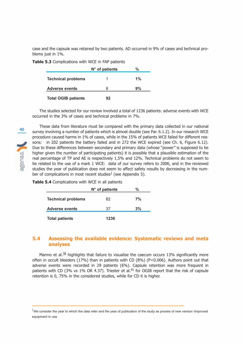
40
case and the capsule was retained by two patients. AD occurred in 9% of cases and technical pro-blems just in 1%.
Table 5.3 Complications with WCE in FAP patients
N° of patients %
Technical problems 1 1%
Adverse events 8 9%
Total OGIB patients 92
The studies selected for our review involved a total of 1236 patients: adverse events with WCEoccurred in the 3% of cases and technical problems in 7%.
These data from literature must be compared with the primary data collected in our nationalsurvey involving a number of patients which is almost double (see Par. 6.1.2). In our research WCEprocedure caused harms in 1% of cases, while in the 15% of patients WCE failed for different rea-sons: in 202 patients the battery failed and in 272 the WCE expired (see Ch. 6, Figure 6.12).Due to these differences between secondary and primary data (whose “power” is supposed to behigher given the number of participating patients) it is possible that a plausible estimation of thereal percentage of TP and AE is respectively 1,5% and 12%. Technical problems do not seem tobe related to the use of a mark 1 WCE: data of our survey refers to 2006, and in the reviewedstudies the year of publication does not seem to affect safety results by decreasing in the num-ber of complications in most recent studies1 (see Appendix 5).
Table 5.4 Complications with WCE in all patients
N° of patients %
Technical problems 82 7%
Adverse events 37 3%
Total patients 1236
5.4 Assessing the available evidence: Systematic reviews and metaanalyses
Marmo et al.38 highlights that failure to visualise the caecum occurs 13% significantly moreoften in occult bleeders (17%) than in patients with CD (8%) (P<0.006). Authors point out thatadverse events were recorded in 29 patients (6%). Capsule retention was more frequent inpatients with CD (3% vs 1% OR 4.37). Triester et al.41 for OGIB report that the risk of capsuleretention is 0, 75% in the considered studies, while for CD it is higher.
1We consider the year to which the data refer and the year of publication of the study as proxies of new version /improved
equipment in use.

41
5.5 Assessing the available evidence: WCE acceptability to patients
Some of the selected studies dealt with the acceptability and tolerability aspects, although fewof them did it in a systematic and structured way, e.g. by using questionnaires and explaining themethodology used to collect data from patients. For instance, short passages are dedicated to thisissue in many of the assessed articles, but authors tend to take it for granted that patients preferWCE over any other endoscopic procedures. This means that usually no good evidence is collec-ted in support of this statement, by either qualitative or quantitative methods. We did not consi-der citations and statements of this kind in our analysis unless supported by evidence. Out of 27studies, only 3 reported information and data in a more or less systematic manner: Mylonaki etal.13, Hadithi et al.20 and Buchaman et al.31.
The first three studies compared WCE to procedures considered “more invasive”, while the lastone compared it to SBFT, which is an imaging technology supposed to be less invasive. Themethods used, although described, are not always well explained: they range from multiple choi-ce questionnaire filled by patients, or views collected by physician’s interviews with patients. Notable summing up data and number of respondents is provided and data are reported in a discur-sive manner in most of cases. The studies by Hadithi et al.20 and Buchaman et al.31 do not con-sider a comprehensive range of dimensions that may define acceptability, focusing above all onpain or swallowing difficulties. All of them report that WCE was preferred to the other procedures.
In the study by Mylonaki et al.13, patients were interviewed at follow up and asked to compa-re WCE and PE painfulness and state their preference: 49/50 preferred WCE over PE, while just 2patients found it difficult to swallow. PE was painful for 34/50 (p<0.05). Hadithi et al.20 used que-stionnaires to gather information on tolerability, administrating them during follow up. Questionswere about discomfort and other problems related to procedure: DBE resulted to be less tolera-ted than WCE (40% vs 94%, p<0.001).
5.6 Results
The systematic review of this report is based on 27 studies. Seventeen of them were publi-shed after the Belgian report12 and date from 2005 to 2007. A total of 349 primary studies wereidentified by the search, of which 17 met our criteria. The chart in Figure 5.1 summarises the flowof studies in the review. A complete list of the studies identified in the literature search and exclu-ded is at Appendix 6. In addition we selected 10 studies published before 2005 from the KCEreport12 by applying our inclusion criteria, and we re-assessed them.
Among the selected literature we could not find any study dealing with costs or economic eva-luations. Two studies were supposedly meta analyses. One is from Marmo et al.38, and is aboutWCE and different comparators used for OGIB and Crohn’s Disease (CD), while the other is fromTriester et al.31. The authors assessed the evidence comparing WCE with various technologies andfor the diagnosis of non stricturing small bowel CD (Appendix 4).
We could identify only one randomised trial out of the 27 assessed studies. Almost all prima-ry studies, no matter the indication and/or comparator, were found to have an unusual studydesign. This kind of design is simply called by authors “prospective” or “blinded and prospective”when physicians performing the alternative are blinded to the WCE’s results. A rather small num-

42
ber of consecutive patients are enrolled and serve as their own controls. This means that theyundergo both WCE and then comparator (or vice versa) after a certain range of time: in 7 studiesthe range was not reported, in 6 it went form 1-6 days, and in 9 from 7 to 14 days, while in twostudies the time range between one procedure and the other was more than 15 days (Figure 5.2).
This design has two major linked biases. One due to the absence of randomisation and theother related to the time range in the various studies. The lesions causing OGIB, in many cases,may vary and change in a few days. The most frequent reason for OGIB is angiodyplasia (Lewis4)which, by its very nature, undergoes very rapid morphological changes (Norman43). This impliesthat the results of the final studies are not fully reliable due to the too long or even not declaredtime range between one procedure and the other. In conclusion the studies compared the incom-parable.
Figure 5.1 Flow Chart A total of 349 primary studies were identified by the search, of which 17met the criteria for the comparative assessment of the effectiveness/efficacy and safety.
Key: EVC=Video Capsule Endoscopy

43
Figure 5.2 Time range (days) in the eligible twenty-four primary studies.
Given the absence of randomised trials and the peculiarities of the chosen design, we tried tounderstand reasons for design and comparator asking the authors directly, by sending them a for-mal e-mail with some questions. We received 7 answers (out of 18 authors contacted) from whichwe understood that choosing the same group of patient was seen as the best option guaranteeingcomparability of results. The confounding variable related to the morphological change of manypotential sources of bleeding was usually underestimated or ignored. In addition no motivationwas provided about not choosing to randomise patients into two groups undergoing different dia-gnostic procedures (see Appendix 7).
In the preceding sections we described shortcomings, characteristics and results of each studyby dividing and grouping them according to the disease/s they referred to and to the comparator.For each study we focus on three main dimensions and related outcomes: diagnostic accuracy,safety and patient’s acceptability of WCE and comparator. The two systematic reviews are descri-bed and assessed in a separate paragraph.
However findings and subsequent conclusions from the selected literature about WCE and itsdiagnostic accuracy, safety etc. have to be regarded as uncertain, as these results are based onlow quality evidence. The only reliable results are those from the RCT by De Leusse et al.18.

44

45
6. Collecting context specific data
The collection of context specific data was important to gain a complete picture of the diffu-sion and use of the WCE, its costs, appropriateness of use and patient’s tolerability in Italy. In ourcountry WCE was introduced in the market with no governance at regional or national level in2001 and according to experts it is being used in many centres.
We carried out two surveys. The first was aimed at collecting data on the actual diffusion ofWCE in the year 2006, its direct costs and appropriateness of its use from Italian centres delive-ring WCE; the second was about patient satisfaction with WCE compared to other endoscopicinterventions, and involved 5 centres, chosen amongst those which completed the first survey’squestionnaires, on the basis of the highest use of WCEs per year. The following sections providea detailed description of materials, methods and main results for each of the two surveys.
6.1 Appropriateness of WCE use and related costs in the Italian context
6.1.1 Objective, materials and methods
The aim of the survey was to collect primary information and data on the diffusion of WCE(2006), appropriateness of WCE use and on costs to generate an economic impact scenario forthe use of the technology.
We identified all Italian centres that could potentially provide WCE, to obtain a comprehensi-ve population denominator for the survey. Due to Italian informative system shortcomings this wasnot an easy task and the merging of three different databases and sources of information wasnecessary. Our final denominator was 116 Italian centres providing WCE diagnostics in 2006(Lorenzatto s.p.a. data). This inclusive list was drawn from three sources:
a) Ministry of Health Database (gastroenterology and endoscopy centres)
b) Italian Club of Wireless Capsule Endoscopy(CICE)
c) Data from the Italian distributor M.G. Lorenzatto S.p.a..
A structured questionnaire was sent by post to all the 116 physicians identified as responsibleof the various endoscopic and gastroenterology centres providing WCE. The collection of datalasted from December 2007 to April 2008.
Responding centres (Appendix 8) were 48% of the total (56/116 filled questionnaires retur-ned). The questionnaire aimed at gathering information on three areas: characteristics of the cen-tre, clinical information about patients undergoing WCE, costs of WCE procedure in terms ofhuman resources, device and equipment and consumption material (Appendix 9).

6.1.2 Results
Characteristics of responding centres
Among the 56 responding centres delivering WCE, the majority were public: 51 were publicstructures (91%, 46 public hospitals, 2 university hospitals, 2 research hospitals-IRCSS), whileonly 4 were private accredited providers (2 hospital trusts, 1 university hospital, 1 residentialhospital) with a contractual agreement (7%) and 1 was a completely private centre (Figure 6.1).Accredited centres with contractual agreement can provide services on behalf of the public healthservice, and are reimbursed. Completely private centres provide services which are paid directlyby patients or private insurance. The majority of responding centres (32, 57%) were located inNorthern Italy, 8 were from Central Italy (14%), while 16 (29%) were from Southern Italy andIslands.
Figure 6.1 Type of centres delivering WCE (year 2006)
Source: data from responding centres analysed by age.na.s
Comparing the data provided by the only Italian distributor of WCE in 2006, with the dataobtained from the survey, we can see that the highest number of centres providing WCE is inLombardy (22 centres) of which 9 (41%) answered to the questionnaire (Figure 6.2), while noneof the centres of Basilicata (3 centres), Umbria (2 centres) and Trentino Alto Adige (2 centres)replied to the questionnaire. At regional level responsiveness was 58%.
46

Figure 6.2 regional distributions of centres
Source: age.na.s and M.G. Lorenzatto S.p.a., 2006
The data provided by M.G. Lorenzatto S.p.a./Given Imaging Ltd show that the highest num-ber of WCE sold per 100.000 population is in the Liguria and Marche Regions, where 13 capsulesevery 100.000 population were used. More than 10 capsules per 100.000 population were used inEmilia Romagna and Piemonte (10 and 11 WCE/100.000 respectively). The Region with the smal-lest number of WCE per 100.000 population is the Calabria region with 1/100.000 population(Figure 6.3). Comparing these data with data obtained from the survey highlights the fact that insome cases the total number of WCE purchased in 2006 was higher than the total of WCE exami-nations performed in same year.
Figure 6.3 Regional distribution of WCE per 100.000 population and WCE/100.000 inhabitantfor volume of sales - year 2006
Source: age.na.s and M.G. Lorenzatto S.p.a., 2006
47
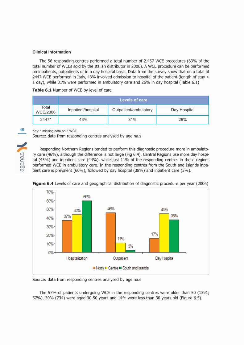
48
Clinical information
The 56 responding centres performed a total number of 2.457 WCE procedures (63% of thetotal number of WCEs sold by the Italian distributor in 2006). A WCE procedure can be performedon inpatients, outpatients or in a day hospital basis. Data from the survey show that on a total of2447 WCE performed in Italy, 43% involved admission to hospital of the patient (length of stay >1 day), while 31% were performed in ambulatory care and 26% in day hospital (Table 6.1)
Table 6.1 Number of WCE by level of care
Key: * missing data on 8 WCE
Source: data from responding centres analysed by age.na.s
Responding Northern Regions tended to perform this diagnostic procedure more in ambulato-ry care (46%), although the difference is not large (Fig 6.4). Central Regions use more day hospi-tal (45%) and inpatient care (44%), while just 11% of the responding centres in those regionsperformed WCE in ambulatory care. In the responding centres from the South and Islands inpa-tient care is prevalent (60%), followed by day hospital (38%) and inpatient care (3%).
Figure 6.4 Levels of care and geographical distribution of diagnostic procedure per year (2006)
Source: data from responding centres analysed by age.na.s
The 57% of patients undergoing WCE in the responding centres were older than 50 (1391;57%), 30% (734) were aged 30-50 years and 14% were less than 30 years old (Figure 6.5).
Levels of care
Total
WCE/2006Inpatient/hospital Outpatient/ambulatory Day Hospital
2447* 43% 31% 26%
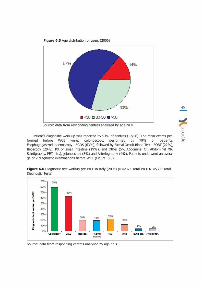
49
Figure 6.5 Age distribution of users (2006)
Source: data from responding centres analysed by age.na.s
Patient’s diagnostic work up was reported by 93% of centres (52/56). The main exams per-formed before WCE were: colonoscopy, performed by 79% of patients,Esophagogastroduodenoscopy - EGDS (63%), followed by Faecal Occult Blood Test - FOBT (22%),Ileoscopy (20%), RX of small intestine (19%), and Other (5%:Abdominal CT, Abdominal MR,Scintigraphy, PET, etc.), jejunoscopy (5%) and Arteriography (4%). Patients underwent an avera-ge of 2 diagnostic examinations before WCE (Figure. 6.6).
Figure 6.6 Diagnostic test workup pre WCE in Italy (2006) (N=2374 Total WCE N =5300 TotalDiagnostic Tests)
Source: data from responding centres analysed by age.na.s

In the three geographical areas, the diagnostic tests performed prior to WCE are similar,however in the central Regions’ centres FOBT is less common (0.7%), compared to its averageuse (10%) (Figure 6.7).
Figure 6.7 Diagnostic Test workup prior to WCE by geographical location (n=2374 WCE; n=5300 DT)
Source: data from responding centres analysed by age.na.s
Figure 6.8 Indications for WCE use (n=2504)
Source: data from responding centres analysed by age.na.s
50
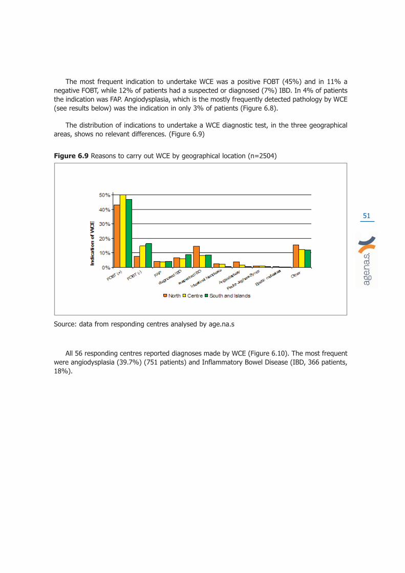
The most frequent indication to undertake WCE was a positive FOBT (45%) and in 11% anegative FOBT, while 12% of patients had a suspected or diagnosed (7%) IBD. In 4% of patientsthe indication was FAP. Angiodysplasia, which is the mostly frequently detected pathology by WCE(see results below) was the indication in only 3% of patients (Figure 6.8).
The distribution of indications to undertake a WCE diagnostic test, in the three geographicalareas, shows no relevant differences. (Figure 6.9)
Figure 6.9 Reasons to carry out WCE by geographical location (n=2504)
Source: data from responding centres analysed by age.na.s
All 56 responding centres reported diagnoses made by WCE (Figure 6.10). The most frequentwere angiodysplasia (39.7%) (751 patients) and Inflammatory Bowel Disease (IBD, 366 patients,18%).
51
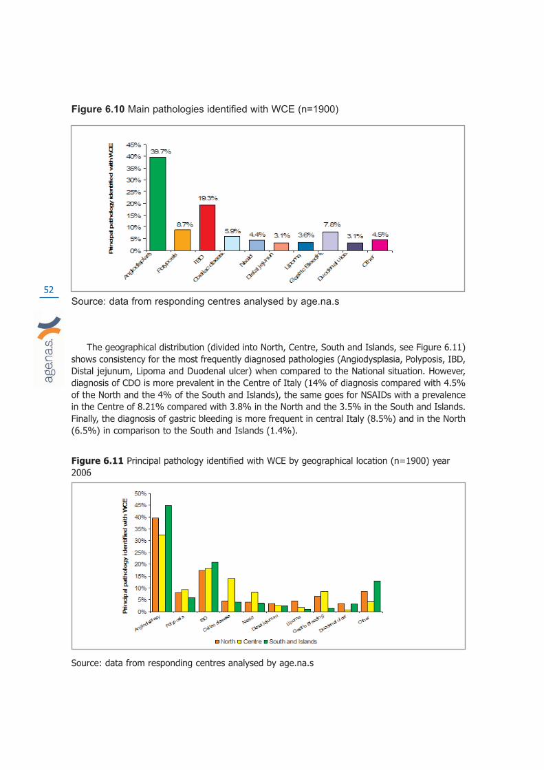
52
Figure 6.10 Main pathologies identified with WCE (n=1900)
Source: data from responding centres analysed by age.na.s
The geographical distribution (divided into North, Centre, South and Islands, see Figure 6.11)shows consistency for the most frequently diagnosed pathologies (Angiodysplasia, Polyposis, IBD,Distal jejunum, Lipoma and Duodenal ulcer) when compared to the National situation. However,diagnosis of CDO is more prevalent in the Centre of Italy (14% of diagnosis compared with 4.5%of the North and the 4% of the South and Islands), the same goes for NSAIDs with a prevalencein the Centre of 8.21% compared with 3.8% in the North and the 3.5% in the South and Islands.Finally, the diagnosis of gastric bleeding is more frequent in central Italy (8.5%) and in the North(6.5%) in comparison to the South and Islands (1.4%).
Figure 6.11 Principal pathology identified with WCE by geographical location (n=1900) year2006
Source: data from responding centres analysed by age.na.s
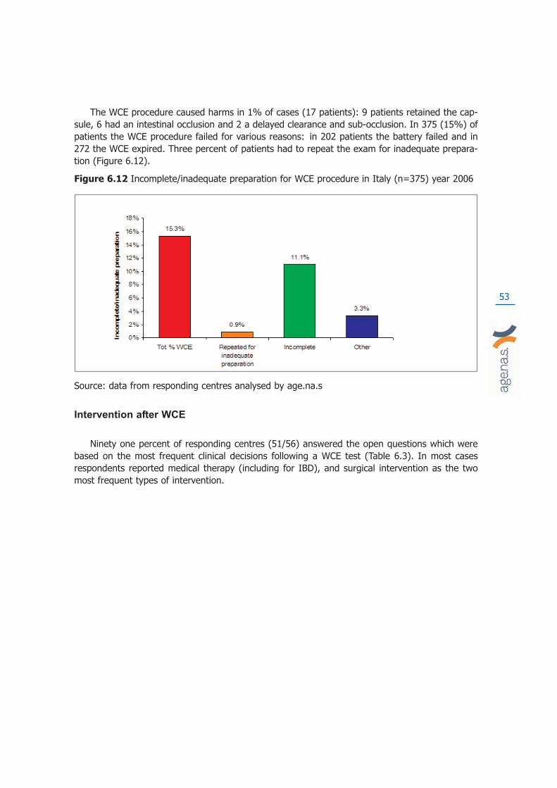
53
The WCE procedure caused harms in 1% of cases (17 patients): 9 patients retained the cap-sule, 6 had an intestinal occlusion and 2 a delayed clearance and sub-occlusion. In 375 (15%) ofpatients the WCE procedure failed for various reasons: in 202 patients the battery failed and in272 the WCE expired. Three percent of patients had to repeat the exam for inadequate prepara-tion (Figure 6.12).
Figure 6.12 Incomplete/inadequate preparation for WCE procedure in Italy (n=375) year 2006
Source: data from responding centres analysed by age.na.s
Intervention after WCE
Ninety one percent of responding centres (51/56) answered the open questions which werebased on the most frequent clinical decisions following a WCE test (Table 6.3). In most casesrespondents reported medical therapy (including for IBD), and surgical intervention as the twomost frequent types of intervention.

54
Costs related to the use of the WCE
Health personnel and average time per WCE procedure
Responding centres were asked to indicate the average time (in minutes) spent by four typesof hospital personnel: physicians, nurses, support operators, administrative officers. Procedureswere divided into three phases, and we asked how much time each figure spent in each phase.
• Preparation: the time for the preparation of the patient (wearing of the recordingbelt, WCE swallowing, etc.);
• Medical reporting: scanning and analysis of the acquired images and diagnosis;
• Consultation: time spent in meeting the patient and discussing procedure anddiagnosis.
Almost all the responding centres (53/56, 94.5%) returned data on average time spent by thedifferent actors on a standard WCE procedure (Table 6.4). The distribution of reported time valuesare reported as the mean time spent, its 95% confidence interval (CI), the minimum, the maxi-mum and the mode as calculated from data returned from responding centres.
Table 6.3 Interventions after the WCE test
Key: IBD=inflammatory bowel disease
Surgical/clinical decisions after performing WCE
Medical Therapy
Medical management of IBD
Surgery
Push Enteroscopy
Gluten-free diet
Laparoscopy
Endoscopic therapy
Enteroscopic treatment with argon plasma
Chemotherapy
Clinical remission within three years
Enterography to remove polyps
Chemotherapy against lymphoma
Therapy with argonplasma coagulation
Thermic coagulation therapy for bleeding from Angiodyplasia
Endoscopic examination for Angiodyplasia
Follow up
Laser therapy
Embolisation
Iliac-release mesalazine for patients with iliac CD
Angio-Tac Scan
Gastroscopy with General Anaesthetic
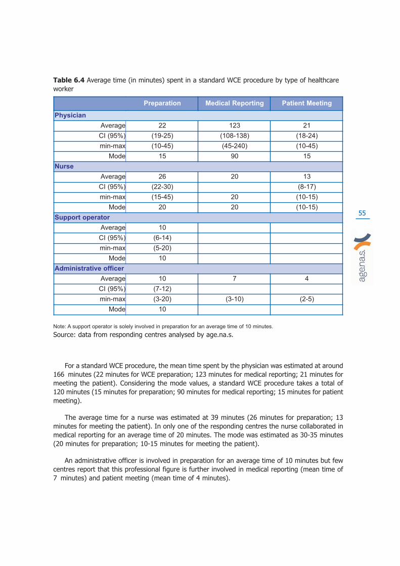
55
For a standard WCE procedure, the mean time spent by the physician was estimated at around166 minutes (22 minutes for WCE preparation; 123 minutes for medical reporting; 21 minutes formeeting the patient). Considering the mode values, a standard WCE procedure takes a total of120 minutes (15 minutes for preparation; 90 minutes for medical reporting; 15 minutes for patientmeeting).
The average time for a nurse was estimated at 39 minutes (26 minutes for preparation; 13minutes for meeting the patient). In only one of the responding centres the nurse collaborated inmedical reporting for an average time of 20 minutes. The mode was estimated as 30-35 minutes(20 minutes for preparation; 10-15 minutes for meeting the patient).
An administrative officer is involved in preparation for an average time of 10 minutes but fewcentres report that this professional figure is further involved in medical reporting (mean time of7 minutes) and patient meeting (mean time of 4 minutes).
Preparation Medical Reporting Patient Meeting
Physician
Average 22 123 21
CI (95%) (19-25) (108-138) (18-24)
min-max (10-45) (45-240) (10-45)
Mode 15 90 15
Nurse
Average 26 20 13
CI (95%) (22-30) (8-17)
min-max (15-45) 20 (10-15)
Mode 20 20 (10-15)
Support operator
Average 10
CI (95%) (6-14)
min-max (5-20)
Mode 10
Administrative officer
Average 10 7 4
CI (95%) (7-12)
min-max (3-20) (3-10) (2-5)
Mode 10
Table 6.4 Average time (in minutes) spent in a standard WCE procedure by type of healthcareworker
Note: A support operator is solely involved in preparation for an average time of 10 minutes.
Source: data from responding centres analysed by age.na.s.
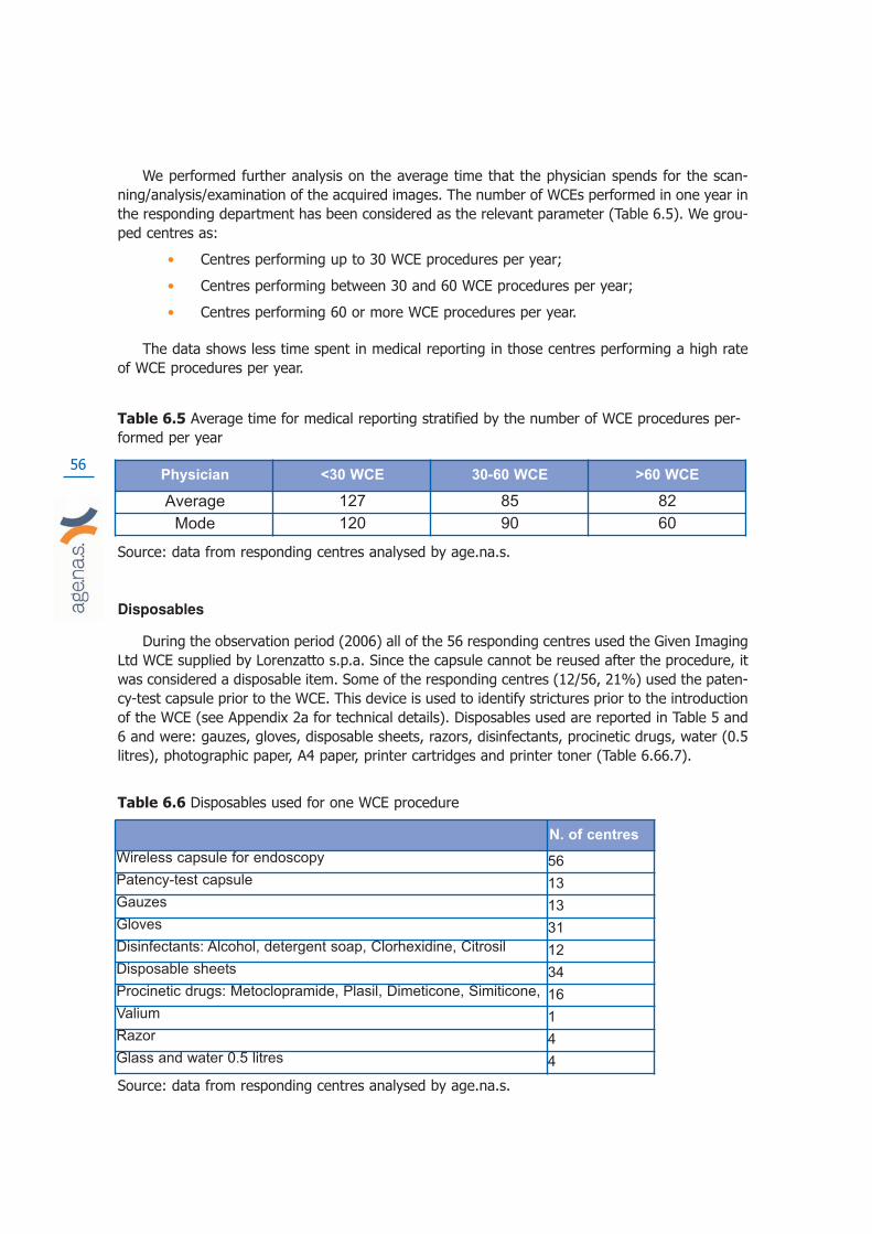
56
We performed further analysis on the average time that the physician spends for the scan-ning/analysis/examination of the acquired images. The number of WCEs performed in one year inthe responding department has been considered as the relevant parameter (Table 6.5). We grou-ped centres as:
• Centres performing up to 30 WCE procedures per year;
• Centres performing between 30 and 60 WCE procedures per year;
• Centres performing 60 or more WCE procedures per year.
The data shows less time spent in medical reporting in those centres performing a high rateof WCE procedures per year.
Table 6.5 Average time for medical reporting stratified by the number of WCE procedures per-formed per year
Source: data from responding centres analysed by age.na.s.
Disposables
During the observation period (2006) all of the 56 responding centres used the Given ImagingLtd WCE supplied by Lorenzatto s.p.a. Since the capsule cannot be reused after the procedure, itwas considered a disposable item. Some of the responding centres (12/56, 21%) used the paten-cy-test capsule prior to the WCE. This device is used to identify strictures prior to the introductionof the WCE (see Appendix 2a for technical details). Disposables used are reported in Table 5 and6 and were: gauzes, gloves, disposable sheets, razors, disinfectants, procinetic drugs, water (0.5litres), photographic paper, A4 paper, printer cartridges and printer toner (Table 6.66.7).
Table 6.6 Disposables used for one WCE procedure
Source: data from responding centres analysed by age.na.s.
N. of centres
Wireless capsule for endoscopy 56
Patency-test capsule 13
Gauzes 13
Gloves 31
Disinfectants: Alcohol, detergent soap, Clorhexidine, Citrosil 12
Disposable sheets 34
Procinetic drugs: Metoclopramide, Plasil, Dimeticone, Simiticone, 16
Valium 1
Razor 4
Glass and water 0.5 litres 4
Physician <30 WCE 30-60 WCE >60 WCE
Average 127 85 82
Mode 120 90 60

57
Table 6.7 Other items used for one WCE procedure
Source: data from responding centres analysed by age.na.s.
Equipment
Thirty four of the 56 responding centres (61%) returned data about the equipment requiredfor WCE procedures purchased in the period between 2000 and 2006. All the equipment was sup-plied by Lorenzatto s.p.a. Different methods of acquisition have been identified: purchasing (25centres), service and commodatum (6 centres), donation (3 centres) (Table 6.13) .
The questionnaire (Appendix 9) allows data reporting of equipment used in aggregate (har-dware and software) or as a single element form (hardware and software separately). Data areshown in Table 7.
The majority of centres (34/56, 61%) returned equipment data in an aggregate form. Everyacquisition procedure differed in relation to the configuration selected, i.e., basic configuration(Table 6.8) or customised configuration (Table 6.9) with optional equipment.
Figure 6.13 Method of acquisition of WCE equipment
Source: data from responding centres analysed by age.na.s.
N. of centres
Toner 14
Printer cartridge 36
Photographic paper 20
Paper (A4 format) 6

58
Table 6.8 Equipment used in a single WCE
Source: data from responding centres analysed by age.na.s.
Table 6.9 Standard equipment WCE Given Imaging Ltd (2008)
Data from M.G. Lorenzatto S.p.a.
Quantity Standard Equipment WCE
1
Data recording system for WCE
(recording data DR2C with battery, recording belt, 1
battery charger, 1 recording sensor SB, 1 recording
sensor ESO, briefcase, 2 tables of instructions)
1
Processing unit Pillcam capsule SB-ESO-COLON
(Computer, monitor lcd 20, software rapid 5.2, colour
printing, accessory).
N. of centres
Hardware+software 34
Software 3
Monitor 4
Computer 5
Equipmant trolley 4
Other equipment:
Software update 3
Battery 1
Imaging Equipment 4

59
Table 6.10 Optional Equipment WCE Given Imaging Ltd (2008)
Data from M.G. Lorenzatto s.p.a.
Budget impact of WCE for the Italian NHS
For the assessment of the elements of cost the method of the standard costing has been used.The costs attributed to the various elements used in the WCE was derived as an average of thedeclared values by centres or when incomplete were derived from market prices. All costs areinclusive of VAT (20%). The average personnel costs (table 6.11) were estimated using theNational Collective Contract of Category 2004 inclusive of employer’s contributions.
Quantity Optional equipment WCE
1
Real time micro read (support for verification of
the functionality capsule before the beginning of
the examination and for the verification that the
WCE transits over the stomach)
1
Software rapid reader (used for viewing and to
see the videotaped video rapid 5.1 again on
personal computer and to manage an WCE
from peripheral unit Given Imaging Ltd)
1
Agile patency capsule Given Imaging Ltd (for
the follow-up of the intestinal patency through
dissolute capsule)
1Scanner for the follow-up of the agile patency
capsule in the intestine given imaging Ltd
1 Portable hard disk 320 GB Given Imaging Ltd
1 Package of clinical technical training
1Kit of training to the videoendoscopy capsulare
pillcam given imaging

60
Table 6.11 Average cost of personnel involved in the WCE procedure in Euros
Source: data from responding centres analysed by age.na.s.
The costs of consumed materials were valued as indicated by the responding centres andwhen incomplete were based on market prices (Table 6.12). For some items (standard WCE andpatency test WCE) price data were provided by the only manufacturer of WCEs in 2006(Lorenzatto s.p.a. authorised distributor of Given Imaging Ltd).
Table 6.12 Cost of consumables for a single WCE procedures
*note that the “Patency test capsule” is only used in some centres.
Source: data from responding centres analysed by age.na.s.
Quantity Unit Costs in Euros
WCE 1 € 642,00
Patency test WCE 1 € 117,60
Surgical Gauze 4 € 0,08
Surgical gloves 2 € 0,02
Disinfectant € 0,05
Disposable sheets 1 € 0,18
Procinetics drugs: Metoclopramide,
Plasil, Dimeticone, Simiticone,10 mg € 0,24
Valium 2 mg € 0,34
Razor 1 € 0,10
Paper cup + 1/2 lt. Water 1 € 0,40
Paper and computer consumables € 0,60
WCE Medical Reporting Consultation
Physician
average € 24,06 € 135,36 € 23,06
range €20,90-€27,50 €118,80-€151,80 €19,80-€26,40
Nurse
average € 11,09 € 8,53 € 5,33
range €9,39-€12,80 €3,41-€7,25
Support Operator
average € 3,38
range €2,03-€4,74
Administrative costs
average € 3,38 € 2,37 €1,35
range €2,37-€4,06
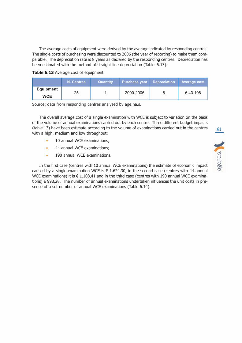
61
The average costs of equipment were derived by the average indicated by responding centres.The single costs of purchasing were discounted to 2006 (the year of reporting) to make them com-parable. The depreciation rate is 8 years as declared by the responding centres. Depreciation hasbeen estimated with the method of straight-line depreciation (Table 6.13).
Table 6.13 Average cost of equipment
Source: data from responding centres analysed by age.na.s.
The overall average cost of a single examination with WCE is subject to variation on the basisof the volume of annual examinations carried out by each centre. Three different budget impacts(table 13) have been estimate according to the volume of examinations carried out in the centreswith a high, medium and low throughput:
• 10 annual WCE examinations;
• 44 annual WCE examinations;
• 190 annual WCE examinations.
In the first case (centres with 10 annual WCE examinations) the estimate of economic impactcaused by a single examination WCE is € 1.624,30, in the second case (centres with 44 annualWCE examinations) it is € 1.108,41 and in the third case (centres with 190 annual WCE examina-tions) € 998,28. The number of annual examinations undertaken influences the unit costs in pre-sence of a set number of annual WCE examinations (Table 6.14).
N. Centres Quantity Purchase year Depreciation Average cost
Equipment
WCE
25 1 2000-2006 8 € 43.108
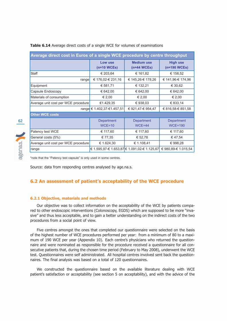
62
Table 6.14 Average direct costs of a single WCE for volumes of examinations
*note that the “Patency test capsule” is only used in some centres.
Source: data from responding centres analysed by age.na.s.
6.2 An assessment of patient’s acceptability of the WCE procedure
6.2.1 Objective, materials and methods
Our objective was to collect information on the acceptability of the WCE by patients compa-red to other endoscopic interventions (Colonoscopy, EGDS) which are supposed to be more “inva-sive” and thus less acceptable, and to gain a better understanding on the indirect costs of the twoprocedures from a social point of view.
Five centres amongst the ones that completed our questionnaire were selected on the basisof the highest number of WCE procedures performed per year: from a minimum of 80 to a maxi-mum of 190 WCE per year (Appendix 10). Each centre’s physicians who returned the question-naire and were nominated as responsible for the procedure received a questionnaire for all con-secutive patients that, during the chosen time period (February to May 2008), underwent the WCEtest. Questionnaires were self administrated. All hospital centres involved sent back the question-naires. The final analysis was based on a total of 120 questionnaires.
We constructed the questionnaire based on the available literature dealing with WCE patient’s satisfaction or acceptability (see section 5 on acceptability), and with the advice of the
Average direct cost in Euros of a single WCE procedure by centre throughput
Low use
(n=10 WCEs)
Medium use
(n=44 WCEs)
High use
(n=190 WCEs)
Staff € 203,64 € 161,82 € 158,52
range € 176,02-€ 231,16 € 145,26-€ 178,26 € 141,96-€ 174,96
Equipment € 581,71 € 132,21 € 30,62
Capsule Endoscopy € 642,00 € 642,00 € 642,00
Materials of consumption € 2,00 € 2,00 € 2,00
Average unit cost per WCE procedure €1.429,35 € 938,03 € 833,14
range € 1.402,37-€1.457,51 € 921,47-€ 954,47 € 816,58-€ 851,58
Other WCE costs
Department
WCE=10
Department
WCE=44
Department
WCE=190
Patency test WCE € 117,60 € 117,60 € 117,60
General costs (5%) € 77,35 € 52,78 € 47,54
Average unit cost per WCE procedure € 1.624,30 € 1.108,41 € 998,28
range € 1.595,97-€ 1.653,87 € 1.091,02-€ 1.125,67 € 980,89-€ 1.015,54
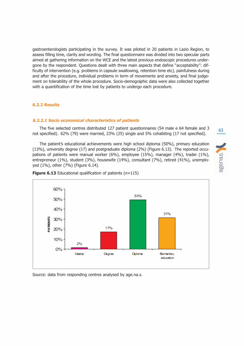
63
gastroenterologists participating in the survey. It was piloted in 20 patients in Lazio Region, toassess filling time, clarity and wording. The final questionnaire was divided into two specular partsaimed at gathering information on the WCE and the latest previous endoscopic procedures under-gone by the respondent. Questions dealt with three main aspects that define “acceptability”: dif-ficulty of intervention (e.g. problems in capsule swallowing, retention time etc), painfulness duringand after the procedure, individual problems in term of movements and anxiety, and final judge-ment on tolerability of the whole procedure. Socio-demographic data were also collected togetherwith a quantification of the time lost by patients to undergo each procedure.
6.2.2 Results
6.2.2.1 Socio economical characteristics of patients
The five selected centres distributed 127 patient questionnaires (54 male e 64 female and 3not specified). 62% (79) were married, 23% (25) single and 5% cohabiting (17 not specified).
The patient’s educational achievements were high school diploma (50%), primary education(13%), university degree (17) and postgraduate diploma (2%) (Figure 6.13). The reported occu-pations of patients were manual worker (6%), employee (15%), manager (4%), trader (1%),entrepreneur (1%), student (3%), housewife (14%), consultant (7%), retired (41%), unemplo-yed (1%), other (7%) (Figure 6.14).
Figure 6.13 Educational qualification of patients (n=115)
Source: data from responding centres analysed by age.na.s.
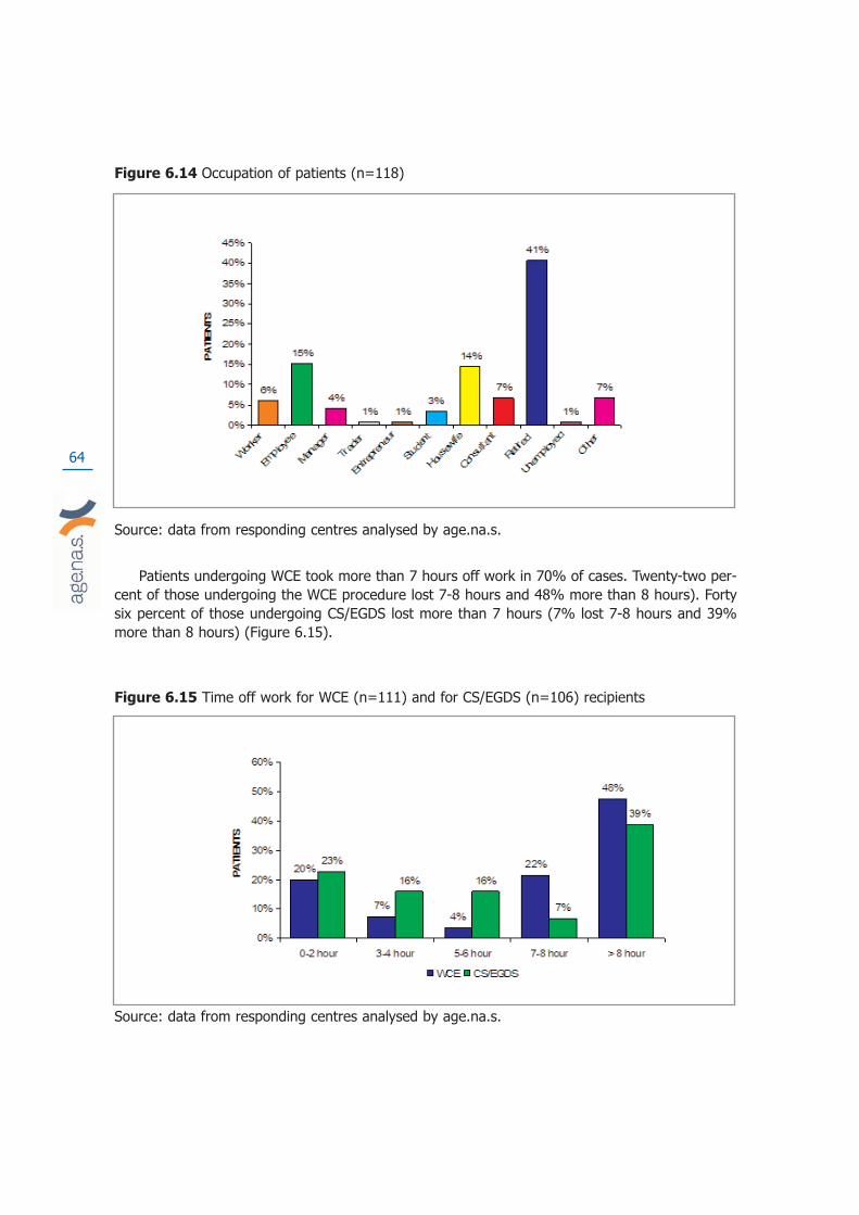
64
Figure 6.14 Occupation of patients (n=118)
Source: data from responding centres analysed by age.na.s.
Patients undergoing WCE took more than 7 hours off work in 70% of cases. Twenty-two per-cent of those undergoing the WCE procedure lost 7-8 hours and 48% more than 8 hours). Fortysix percent of those undergoing CS/EGDS lost more than 7 hours (7% lost 7-8 hours and 39%more than 8 hours) (Figure 6.15).
Figure 6.15 Time off work for WCE (n=111) and for CS/EGDS (n=106) recipients
Source: data from responding centres analysed by age.na.s.

65
6.2.2.2 Acceptability dimensions for the WCE and other endoscopies
Patients indicated a greater ease of performing the WCE in comparison to CS/EGDS (Figure6.16). Sixteen percent of patients undergoing CS/EGDS indicated a degree of difficulty in the pro-cedure (11% difficult, 5% very difficult).
Figure 6.16 Ease of execution of the procedure WCE (n=125) and CS/EGDS (n=125)
Source: data from responding centres analysed by age.na.s.
During the procedures 84% of patients undergoing WCE had no perceivable pain, comparedto 33% with CS/EGDS (Figure 6.17). Patients undergoing CS/EGDS felt severe pain in 10% ofcases and extreme in 3% of cases.
Figure 6.17 Pain during the procedure with WCE (n=123) and CS/EGDS (n=126)
Source: data from responding centres analysed by age.na.s.

66
After the WCE procedure, 83% of patients experienced no perceivable pain compared to 48%with CS/EGDS (Figure 6.18). Patients who underwent CS/EGDS, felt severe (3%) or extreme (2%)pain after the procedure.
Figure 6.18 Pain after the procedure with WCE (n=110) and CS/EGDS (n=119)
Source: data from responding centres analysed by age.na.s.
Twenty nine percent of patients undergoing the WCE procedure recorded difficulty of move-ment during the recording and procedure compared with of 22% undergoing CS / EGDS (Figure6.19).
Figure 6.19 Difficulty of movement during the recording/procedure with WCE (n=126) andCS/EGDS (n=117)
Source: data from responding centres analysed by age.na.s.
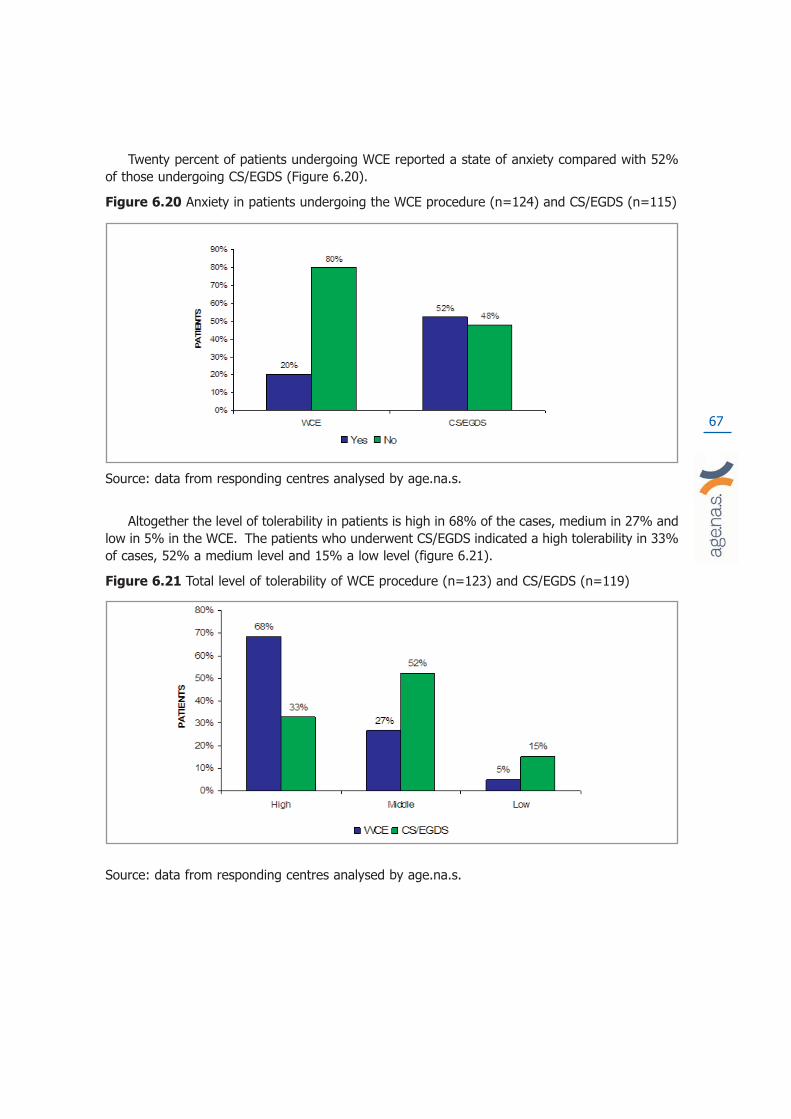
67
Twenty percent of patients undergoing WCE reported a state of anxiety compared with 52%of those undergoing CS/EGDS (Figure 6.20).
Figure 6.20 Anxiety in patients undergoing the WCE procedure (n=124) and CS/EGDS (n=115)
Source: data from responding centres analysed by age.na.s.
Altogether the level of tolerability in patients is high in 68% of the cases, medium in 27% andlow in 5% in the WCE. The patients who underwent CS/EGDS indicated a high tolerability in 33%of cases, 52% a medium level and 15% a low level (figure 6.21).
Figure 6.21 Total level of tolerability of WCE procedure (n=123) and CS/EGDS (n=119)
Source: data from responding centres analysed by age.na.s.

68

69
7. Discussion
We found a sizeable amount of low quality literature which dramatically reduced our capabili-ty to draw conclusions on the diagnostic accuracy of the wireless capsule for endoscopy. Most stu-dies had faulty design and faulty reporting. The typical sequential study design used by the majo-rity of researchers in which the same patient underwent the WCE procedure and the referencestandard, introduced a notable element of time bias into the observation period. As angiodyplasiaand other variable morphology lesions were the most diagnosed and often the most frequent indi-cations for use of the WCE, the capability of comparing like with like (i.e. of having the same pro-bability that similar patients had lesions of roughly the same appearance and evolution stage)would have been the basic pre-requisite for a meaningful comparison. Such comparability can onlybe achieved by a randomised design in which a set of people are given an equal chance of beingassigned to the WCE procedure or its comparator so that all differences between participants arebalanced and results are comparable. This is the reason why the trial by De Leusse et al18 is theonly credible piece of evidence at our disposal, however small its denominator. The majority ofresearchers justified the choice of study design with the idea that each patient provided his or herown control, thus minimising the effect of confounders. Far from this being the case, in rapidlyevolving lesions the time bias present in the sequential design used maximised the effects of con-founding making images from the same patient non-comparable in the majority of cases.Respondents to our questionnaire appeared not to have understood the basic requirement to com-pare like with like. In addition some experts expressed the view that conducting a clinical trial ofsuch a dominant and safe device as the WCE would be unethical. This view is particularly dange-rous because as we have shown the WCE has not been tested is sufficient numbers against acomparator allowing visualisation of the entire small bowel mucosa (i.e. double balloon enterosco-py with double access or intraoperative enteroscopy. The De Lusse et al18 study was correctly desi-gned (i.e. the methods used could answer the study objectives). However its results should beinterpreted with caution. The trial is small (78 participants) highly contextualised and the authorsdraw conclusions not based on the finding of superior diagnostic accuracy but on the better tole-rability of the WCE compared to PE.
Overall other problems with the rationale, design and reporting of the studies further weake-ned the validity of our evidence base. Few studies clearly stated the rationale for the choice ofcomparator and some even reported a “cross over” design, mistaking it for the sequential designactually used. Twenty nine percent of included studies with a sequential design failed to report thetime interval between one procedure and the other and few reported funding source and/or con-tained an authors’ conflicts of interest statement. The WCE seems to be better tolerated than arange of other procedures and our patient questionnaire results confirm this view. The WCE is nothowever free from harms and in 1% of cases its use and retention led to a major surgical proce-dure such as laparotomy for removal being carried out or in an anxious wait for the expulsion ofa retained capsule. Few studies reported harms due to their small size, so our survey may provi-de the first reliable estimates of their incidence. In addition a thorough preparation of the patientand correct use of the WCE are necessary as shown by the high proportion of failures or of incom-plete procedures (nearly 15% of those carried out in the 56 responding centres in 2006).
Our safety dataset of over two thousand observations is to our knowledge the largest in exi-stence from a single nation although we are aware of large international case series (such as theone comprising 733 cases reported by Rondonotti et al44).

70
Our data do not support the view that technical failures were more common in the earlier sta-ges of WCE use, although we used date of publication as a proxy for date of execution of the studyand of device type.
Our enquiries on the use of the WCE in the Italian context paint a varied picture with peaksand troughs of use probably unrelated to clinical need and some frankly disturbing indications forits use. For example a biopsy is necessary in all cases to make a definitive diagnosis in coeliacdisease and its ethical use in situations of high likelihood of intestinal stenosis such as Crohn‘sdisease is debatable. The enthusiasm of a few centres far outstrips the evidence base on whichthe appropriate use of the WCE procedure should take place. Variability of use is also reflected inthe variability of costs which persist even after adjustment for the optional configuration of theWCE set. The WCE in Italy is an expensive procedure, but we cannot say whether it is cost-effec-tive if compared to the alternatives available in 2006 or likely to be cost-effective compared to thecurrent alternatives, as we lack clear evidence to guide indications and unbiased evidence of itscomparative performance. Our evidence shows that centres in which the highest numbers of pro-cedures are carried out have the lowest costs. This however should not be interpreted as a rea-son to increase WCE use in the absence of credible evidence of diagnostic accuracy.

71
8. Recommendations
Given the present and future development of the technology a reasonable way forward maybe to link reimbursement of the WCE to its use in adequately designed and powered randomisedcontrolled trials with a potential crossover design similar to that of the De Leusse et al trial18.These should test the performance of the WCE for present and future indications under the super-vision of scientific and ethical committees. We further recommend that this process (called cove-rage with evidence generation), widely adopted abroad should be adopted in Italy for all promi-sing new technologies.

72
9. Funding
Production of this report was made possible by financial contributions from the Italian Ministryof Health (CUD, Commissione Unica Dispositivi) and the Age.Na.S.
The Age.Na.S. takes sole responsibility for the final form and content of this report. The viewsexpressed herein do not necessarily represent the views of the Italian Ministry of Health or anyregional government.
73

74

75
10. Competing interests declaration
The authors declare that they will not receive either benefits or harms from the publication ofthis report. None of the authors have or have held shares, consultancies or personal relationshipswith any of the producers of the devices assessed in this document.

76

77
Glossary
Coeliac Disease (COD)COD is a disease occurring in children and adults characterized by sensitivity to gluten, with
chronic inflammation and atrophy of the mucosa of the upper small intestine; manifestations inclu-de diarrhoea, malabsorption, steatorrhea, nutritional and vitamin deficiencies, and failure to thri-ve, or short stature.
Cochrane Library (CLIB)A collection of databases, published on disk, CD-ROM and the Internet and updated quarter-
ly, containing the Cochrane Database of Systematic Reviews, the Cochrane Controlled TrialsRegister, the Database of Abstracts of Reviews of Effectiveness, the Cochrane Review MethodologyDatabase, and information about the Cochrane Collaboration and other information.
Cochrane ReviewA Cochrane Review is a systematic, up-to-date summary of reliable evidence of the benefits
and risks of healthcare. Cochrane Reviews are intended to help people make practical decisions.For a review to be called a “Cochrane Review” it must be in the Parent Database maintained bythe Cochrane Collaboration. The Parent Database is composed of modules of reviews submittedby Collaborative Review Groups (CRGs) registered with the Cochrane Collaboration. The reviewscontributed to one of the modules making up the Parent Database are refereed by the editorialteam of the CRG, as described in the CRG module. Reviewers adhere to guidelines published inthe Cochrane Reviewers’ Handbook.
CostA negative variation in utility.
Crohn’s DiseaseCrohn’s Disease (CD) is a subacute chronic enteritis, of unknown cause, involving the termi-
nal ileum and less frequently other parts of the gastrointestinal tract; characterised by patchy deepulcers that may cause fistulas, and narrowing and thickening of the bowel by fibrosis and lympho-cytic infiltration, with non caseating tuberculoid granulomas that also may be found in regionallymph nodes.
Decision TreeDecision tree is a basic decision making tool used when in need of analysing a clinical problem
with an underlining time and logic structure. The structure of the tree illustrates the three struc-

78
tural components: alternative actions that the decision maker can take; events that result from aspecific decision and the influences on such actions (i.e. clinical information obtained or resultingclinical outcomes); patient outcomes connected to each possible action scenario.
Direct costA cost entirely related to the product, activity or service provided by the cost evaluation pro-
gram. These costs can be allocated to a specific cost centre only if they can be directly attribu-ted to the specified cost centre.
Discount rateDiscounting describes a method that calculates how much one euro is worth today if paid after
one or more years.
Economic EvaluationA comparative analysis of costs and outcomes of several alternative actions. Therefore the
main function of such evaluation is to identify, measure, assign values and compare costs and out-comes of examined alternatives.
Fixed costFixed cost is a cost that does not very with the activity level. It is relatively stable and it is
usually expressed in the units of time (usually as annual cost) rather than units of volume.
Incremental costIncremental cost is the cost necessary to increase the total production. It indicates the diffe-
rence in terms of cost or effect, between two or more evaluated programs or treatments.
Indirect costIs the value that cannot be directly traced back to a specific activity, service or product used
in the program.
Likelihood Ratios1. Positive Diagnostic Likelihood Ratios
Diagnostic likelihood ratios (DLR), can be a valuable tool for comparing the accuracy of seve-ral tests to the gold standard, and they are not dependent upon the prevalence of disease.

79
The positive DLR represents the odds ratio that a positive test result will be observed in aninfected population compared to the odds that the same result will be observed among a noninfected population.
2. Negative Diagnostic Likelihood Ratios
The negative DLR represents the odds ratio that a negative test result will be observed in aninfected population compared to the odds that the same result will be observed among a noninfected population.
Mean costThe mean cost for one unit of product is the total cost of N units of product divided by N.
Meta AnalysisMeta analysis uses statistical methods to combine and integrate results from a number of pre-
vious experiments or studies examining the same question, in an attempt to summarise the tota-lity of evidence relating to a particular issue. This combination will produce a more robust conclu-sion than the one obtained in a single study.
Obscure bleedingRecurrent or persistent IDA, positive FOBT, or visible bleeding with no bleeding source found
at original endoscopy
Obscure occult bleedingSubcategory of obscure bleeding characterised by recurrent or persistent IDA and/or positive
FOBT with no source found at original endoscopy; no visible blood in faeces.
Obscure overt bleedingSubcategory of obscure bleeding characterised by recurrent or persistent overt/visible blee-
ding with no source found at original endoscopy; bleeding manifest as visible blood in emesis orfaeces.
Occult bleedingInitial presentation of IDA and/or positive FOBT; no visible blood in faeces
80
Odds ratio (OR)Both the odds ratio and the relative risk compare the likelihood of an event between two
groups.
Overt or visible bleedingGastrointestinal bleeding manifest as visible bright red or altered blood in emesis or faeces
OutcomeOutcome is used to indicate results and value of a health intervention. Mortality and similar units
are often used as outcome measurements to evaluate the effect on a population or a health caresystem. In other cases outcome is the result of a specific diagnostic or therapeutic procedure.
Relative risk (RR)In statistics and mathematical epidemiology, relative risk (RR) is the risk of an event (or of
developing a disease) relative to exposure. Relative risk is a ratio of the probability of the eventoccurring in the exposed group versus the control (non-exposed) group.
Sensitivity analysisAn analysis used to determine how sensitive the results of a study or systematic review are to
changes in how it was done. Sensitivity analyses are used to assess how robust the results are touncertain decisions or assumptions about the data and the methods that were used.
SensitivityThe sensitivity of a test is the probability that the test is positive when given to a group of
patients with the disease. Sensitivity is sometimes abbreviated Sn.
A large sensitivity means that a negative test can rule out the disease. David Sackett coinedthe acronym “SnNOut” to help us remember this.
SpecificityThe specificity of a test is the probability that the test will be negative among patients who do
not have the disease. Specificity is sometimes abbreviated Sp.
A large specificity means that a positive test can rule in the disease. David Sackett coined theacronym “SpPIn” to help us remember this.
81
Positive predictive valueThe positive predictive value of a test is the probability that the patient has the disease when
restricted to those patients who test positive. This term is sometimes abbreviated as PPV.
If the prevalence of the disease in your situation is different from the prevalence of the disea-se in the research study you are examining, then you can use likelihood ratios to estimate the PPV.
Negative predictive valueThe negative predictive value of a test is the probability that the patient will not have the
disease when restricted to all patients who test negative.
If the prevalence of the disease in your situation is different from the prevalence of the disea-se in the research study you are examining, then you can use likelihood ratios to estimate the NPV.
Time Preference for Health test:This unit indicates the social discount rate at which decision makers exchange the present with
future consumptions. This parameter is often approximated with the real interest rate (net infla-tion rate) of state bonds.

82
83
Abbreviations
AGA American Gastroenterology Association
CCOHTA Canadian Coordinating Office of Health Technology
CD Crohn’s Disease
COD Coeliac Disease
CS Colonoscopy
CT Computer Tomography
CRD Centre for Reviews and Dissemination
DBE Double Balloon Enteroscopy
EGD Esophago Gastro Duodenoscopy
FAP Familial Adenomatous Polyposis
FDA Food and Drug Administration
FOBT Faecal Occult Blood Test
HTA Health Technology Assessment
KCE Centre Fédéral d’Expertise de soins de Santé
IE Intraoperative Enteroscopy
MRI Magnetic Resonance Imaging
NICE National Institute for Clinical Excellence (UK)
NDDIC National Digestive Diseases Information Clearinghouse
OGIB Obscure Gastrointestinal Bleeding
PE Push Enteroscopy

84
SBS Small Bowel Series
SBTS Small bowel follow through
SI Small Intestine
SR Systematic Review
WCE Video Capsule Endoscopy

85
Bibliography1. Mayers RT, Diagnosis and Management of occult gastrointestinal bleeding:
visualization of the small bowel lumen by fiberoptic colonscope, American
Surgeon, 1976;42(2):92-95.
2. Zuckerman GR, Prakash C, Askin MP, Lewis BS. AGA technical review on the evalua-tion and management of occult and obscure gastrointestinal bleeding.Gastroenterology 1999;118:201221.
3. American Gastroenterological Association (AGA) Institute Technical Review onObscure Gastrointestinal Bleeding. Gastoenterology 2007;133:1697-1717.
4. Lewis BS, Small Intestinal Bleed. Gastroenterology Clincs of North America,1994;23(1):67-91.
5. Stedman medical dictionary, 2008. www.stedman.com accessed on the 7th
April 2008
6. Schivanada S, Lennard-Jones J, Logan R et al. Incidence of inflammatorybowel disease across Europe: is there a difference between north and south?Results of the European Collaborative Study on Inflammatory Bowel Disease(EC-IBD) Gut 1996;39:690-697.
7. National Digestive Diseases Information Clearinghouse
(http://digestive.niddk.nih.gov/) accessed on the 7th April 2008
8. Genetics Home Reference (http://ghr.nlm.nih.gov) accessed on the 7th April
2008
9. Orecchia G, Oral communication at 2° CONGRESSO NAZIONALE sullaVideoendoscopia - Second Italian Congress on WCE - 28-30 June 2007, Feltre (Italy).
10. MSAC. M2A(R) capsule endoscopy for the evaluation of obscure gastrointestinal blee-ding in adult patients. Medical Services Advisory Committee 2003 (MSAC Application1507), 2003;159.
11. NICE Wireless capsule endoscopy. For investigation of the small bowel. NationalInstitute for Clinical Excellence (NICE) Interventional Procedure Guidance 101.2004.Availablefrom www.nice.org.uk
12. KCE Endoscopie par capsule, Centre Federal d’Expertise des Soins de Santè,2006;25B
13. Mylonaki M, Fritscher A, Swain P, Wireless capsule endoscopy: a comparison withpush enteroscopy in patients with gastroscopy and colonscopy negative gastrointe-stinal bleeding. Gut. 2003; 52:1122-1126
14. Mata A, Bordas M, Feu F, et al, Wireless capsule endoscopy in patients with obscuregastrointestinal bleeding: a comparative study with pusch enteroscopy, AlimentPharmacol Ther, 2004;20:189-194.
15. Adler DG, Knipschield M, Cristopher G, A prospective comparison of capsule endosco-py and push enteroscopy in patients with GI bleeding of obscure origin.Gastrointestinal Endoscopy, 2004;59(4):492-498.
86
16. Saurin JC, Delvaux M, et al, Clinical impact of Capsule Endoscopy Comparedto Push Enteroscopy: 1 year follow up study. Endoscopy 2005;37:318-323.
17. Neu B, Ell C, May A, Schmid E, et al. Capsule endoscopy versus standardtests in influencing management of obscure digestive bleeding: Results froma German multicentre trial. Am. J. Gastroenterol. 2005; 100(8):1736-1742;
ISSN: 0002-9270.
18. De Leusse A, Vahedi K, Edery J, et al. Capsule endoscopy or pushenteroscopy for first-line exploration of obscure gastrointestinal bleeding?eng. Gastroenterology. 2007; 132(3):855-62; quiz 1164-5; ISSN: 0016-5085
19. Matsumoto T, Esaki M, Moriyama T, et al. Comparison of capsule endoscopyand enteroscopy with the double-balloon method in patients with obscurebleeding and polyposis. eng. Endoscopy. 2005; 37(9):827-32; ISSN: 0013-
726X.
20. Hadithi M, Heine GD, Jacobs MA, et al. A prospective study comparingWireless Capsule Endoscopywith double-balloon enteroscopy in patients withobscure gastrointestinal bleeding, eng. Am J Gastroenterol. 2006; 101(1):52-
7; ISSN: 0002
21. Nakamura M, Niwa Y, Miyahara R, et al. Preliminary comparison of capsuleendoscopy and double-balloon enteroscopy in patients with suspected small-bowel bleeding. Endoscopy. 2006; 38(1):59-66; ISSN: 0013-726X.
22. Gay G, Delvaux M and Fassler I, Outcome of capsule endoscopy in determin-ing indication and route for push-and-pull enteroscopy, eng. Endoscopy.
2006; 38(1):49-58; ISSN: 0013-726X
23. Xiao-Bo L, ZZ G, Dai J, et al., The role of capsule endoscopy combined withdouble-balloon enteroscopy in diagnosis of small bowel diseases, eng. Chin
Med J (Engl). 2007; 120(1):30-5; ISSN: 0366-6999.
24. Ell C, Remke S, May A, et al. The first prospective controlled trial comparingwireless capsule endoscopy with push enteroscopy in chronic gastrointestinalbleeding. Endoscopy 2002;34:685689.
25. Costamagna G, Shah S, Riccioni ME, et al, A prospective trial comparingsmall bowel radiographs and Wireless Capsule Endoscopy for suspectedsmall bowel disease. Gastroenterology 2002;123:999-1005.
26. Hara AK, Leighton AJ, Sharma VK, Fleischer DE, Small Bowel: preliminarycomparison of capsule endoscopy with barium study and CT, Radiology,
2003;230(1):260-265.
27. Golder SK, Schreyer AG, Endlicher E, et al. Comparison of capsuleendoscopy and magnetic resonance (MR) enteroclysis in suspected smallbowel disease. eng. Int J Colorectal Dis. 2006; 21(2):97-104; ISSN: 0179-
1958.
28. Saperas E, Dot J, Videla S, et al. Capsule endoscopy versus computed tomo-graphic or standard angiography for the diagnosis of obscure gastrointestinalbleeding, eng. Am J Gastroenterol. 2007 Apr.
29. Altman DG, Schulz KF, Moher D, et al, The revised CONSORT statement forreporting randomized trials: explanation and elaboration. Ann Intern Med
2001;134:663694.

87
30. Albert JG, Martiny F, et al, Diagnosis of small bowel Crohn’s disease: aprospective comparison of capsule endoscopy with magnetic resonanceimaging and fluoroscopic enteroclysis. Gut 2005;54:1721-1727.
31. Buchman AL, Miller FH, Wallin A, et al. Videocapsule Endoscopy versusBarium Contrast Studies for Diagnosis of CDRecurrence Involving the SmallIntestine. American Journal of Gatroenterology, 2004;99:2171-2177.
32. Eliakim R, Suissa A, Yassin K, et al. Wireless capsule video endoscopy com-pared to barium follow-through and computerised tomography in patients withsuspected Crohn’s Disease final report. Digestive and liver disease,
2004;36:519-522
33. Ho Chong AK, Taylor A, Miller A, et al. Capsule endoscopy vs. push entero-scopy and enteroclysis in suspected small-bowel Crohn’s disease.Gastrointest. Endosc. 2005; 61(2):255-261; ISSN: 0016-5107.
34. Voderholzer WA, Beinhoelzl J, Rogalla P, et al. Small bowel involvement inCrohn’s disease: a prospective comparison of wireless capsule endoscopyand computed tomography enteroclysis. eng. Gut. 2005; 54(3):369-73; ISSN:
0017-5749.
35. Caspari R, Falkenhausen M, Krautmacher C, et al. Comparison of CapsuleEndoscopy and Magetic Resonance Imaging for the detection of polyps of thesmall intestine in patients with familial adenomatous polyposis or with peutz-jeghers’ syndrome. Endoscopy 2004;36:1054-1059.
36. Schulmann K, Hollerbach S, Kraus K, Feasibility and Diagnostic Utility ofWireless Capsule Endoscopyfor the Detection of Small Bowel Polyps inPatients with Hereditary Polyposis Syndromes, American Journal
Gastroenterol, 2005;100:27-37.
37. Wong RF, Tuteja AK, Haslem DS, et al. Wireless CapsuleEndoscopycompared with standard endoscopy for the evaluation of small-bowel polyps in persons with familial adenomatous polyposis (with video.eng. Gastrointest Endosc. 2006; 64(4):530-7; ISSN: 0016-5107.
38. Marmo R, Rotondano G, Piscopo R, et al. Meta-analysis: Capsule enterosco-py vs. conventional modalities in diagnosis of small bowel diseases. Aliment.
Pharmacol. Ther. 2005; 22(7):595-604; ISSN: 0269-2813.
39. Hartmann D, Schmidt H, Bolz G, et al. A prospective two-centre study compa-ring wireless capsule endoscopy with intraoperative enteroscopy in patientswith obscure GI bleeding, eng. Gastrointest Endosc. 2005; 61(7):826-32;
ISSN: 0016-5107.
40. Mother D, Cook DJ, Eastwood S, et al. Improving the quality of reports ofmeta-analyses of randomised controlled trial: the QUORUM statement.Quality of reporting of metaanalyses. Lancet 1999; 354:18961900.
41. Triester SL, Leighton JA, Leontiadis GI, et al. A meta-analysis of the yield ofcapsule endoscopy compared to other diagnostic modalities in patients withobscure gastrointestinal bleeding. Am. J. Gastroenterol. 2005; 100(11):2407-
2418; ISSN: 0002-9270.
42. Van Gossum A, Hittelet A, Schmit A, et al. A prospective comparative study ofpush and wireless-capsule enteroscopy in patients with obscure digestivebleeding. Acta Gastro-enterologica Belgica, 2003;66:199-205.

88
43. Norman M. Dy, Gostout CJ, Balm RK. Bleeding from the Endoscopically-iden-tified Dieulafoy Lesion of the proximal small intestine and colon. Am. J.
Gastroenterol. 1995; 90(1):108-111; ISSN: 0002-9270.
44. Rondonotti E, Herrerias JM, Pennazio M et al. Complications, limitations,andfailures of capsule endoscopy: a review of 733 cases. Gastrointest. Endosc.
2005; 62:712-716.

89
Appendix 1
Expert Opinion Questionnaire Wireless Capsule Endoscopy (WCE)
1) How many OGIB cases require the use of WCE?
� 0%
� 1%
� 2%
� 3%
� 4%
� 5%
� 6%
� 7%
� 8%
� 9%
� 10%
Other (define):
2) Which is the best diagnostic alternative to the WCE?
� Double Baloon Enteroscopy
� Push Enteroscopy
� Small Bowel RX
� Computer Tomography (CT)
� Magnetic Resonance Imaging (MRI)
Other (define):

90
Questionnaire results
Figure 1. Expert’s opinion percentage of OGIB cases requiring a WCE after negative endoscopies
Figure 2. Diagnostic alternatives* to the WCE (n=82)
* SBRX= Small Bowel RX; PE= Push Enteroscopy; DBE=Double Ballon Enteroscopy; MRI=Magnetic Risonance Imaging;
CT= Computer Tomography; Other= Scintigraphy, Arteriography, Angiography

91
Appendix 2a
Wireless capsule endoscopy (WCE)
Miniaturisation represents a continuing trend in technology toward ever-smaller scales formechanical, optical and electronic devices. In the industry of medical devices this trend has car-ried out a variety of diagnostic and therapeutic procedures that are less invasive than only fewyears ago. In the field of endoscopy, miniaturisation means that, theoretically, even the deepestcavity of the body is reachable without difficulty.
A key-point is surely represented by the image sensors which have to be embedded on the tipof the endoscope for acquire the images that will be transmitted to the monitor outside the body.An image sensor is a device that converts a visual image (light) to an electric signal. The smallerthe sensor the smaller the tip of the endoscope and thus less invasive will be the procedure.
In this field the CCD images sensor and the CMOS sensor are the most used technologies.Both types of sensor accomplish the same task of capturing light and converting it into electricalsignals.
• CCD (Charge-Coupled Device): every pixel’s charge is transferred through a very lim-ited number of output nodes (often just one) to be converted to voltage, buffered,and sent off-chip as an analogical signal.
• CMOS (Complementary Metal-Oxide-Semiconductor): each pixel has its own charge-to-voltage conversion, and the sensor often also includes amplifiers, noise-correction,and digitisation circuits, so that the chip outputs digital bits. These other functionsincrease the design complexity and reduce the area available for light capture.
Both CCDs and CMOS imagers can offer excellent imaging performance.
CCDs offer good image quality and flexibility at the expense of system size. They remain themost suitable technology for high-end imaging applications, such as digital photography, broad-cast television, high-performance industrial imaging, and most scientific and medical applications(Litwiller 2001).
CMOS imagers offer small system size at the expense of image quality (particularly in low light)and flexibility. They are the technology of choice for high-volume, space constrained applicationswhere image quality requirements are low. This makes them a natural fit for security cameras, PCvideoconferencing, wireless handheld device videoconferencing, bar-code scanners, fax machines,consumer scanners, toys, biometrics and some automotive in-vehicle uses (Litwiller 2001).

92
Wireless Capsules for Endoscopy (WCE)WCE is also known as Video-Capsule for Endoscopy (VCE), capsule camera, video pill, or cap-
sule endoscopy. It consists in a camera with the size and shape of a large pill used to acquire ima-ges of the gastrointestinal tract.
At the present time, the capsule camera is primarily used to visualise the small bowel (fromthe jejunum to ileum). Unlike the upper gastrointestinal tract (esophagus, stomach, and duode-num) and the colon (large intestine), that can be very adequately visualised with endoscopes, thesmall bowel is very long (more than 5 meters), very convoluted and thus hardly observable withendoscopes.
WCE represent a monumental advance in the diagnosis of small bowel diseases but at themoment biopsies and endoscopic interventions are not possible.
Operating principle of video-capsules for endoscopyAlthough there are 2 different devices available on the market, the principle of operation of
the endoscopic capsule is quite similar (at a first sight, the only difference seem to be the imagesensor). Due to its mini-invasivity, this technology seems to be very promising and for this reason,the two manufacturers were involved in a long and very expensive patent litigation settled in April2008. Under the terms, Olympus will pay Given Imaging Ltd. $ 2.3 million. The companies willcross licence existing patents to each other without royalty payments and have agreed to workout royalty payments for future patents (marketwatch website).
The two devices are the size of a large pill, and are composed by:
• one or more batteries;
• a strong light source (light-emitting diodes, LED);
• an image sensor;
• a small transmitter.
Once swallowed, the capsule begins transmitting images (for about 8 hours) of the inside of thegastrointestinal tube. Sensors are attached to the patient’s abdomen and wired to a portable recor-der. The recorder is battery-operated and is composed of a receiver, a processor, and a hard disk tostore the images. These video images are transmitted by means of ultra-high-frequency radio tele-metry to the sensor array, which subsequently sends the data to the recorder (Figure A2.1).
After some hours the patient returns the belt to the physician who loads the information intoa computer and then can review in detail the pictures of the intestine, looking for abnormalities.Image interpretation takes from 30 minutes to 2 hours, depending on experience, but is tipicallyone hour (Hara 2005). The capsule is disposable, propelled by peristalsis and excreted after 24-48 hours.

93
Figure A2.1: Sensors attached to the abdomen along with the battery pack and recorder
State of the ArtTwo companies share the worldwide market for this technology:
• Given Imaging Ltd. with its PillCam SB;
• Olympus Corporation with its EndoCapsule;
PillCam SB (Given Imaging Ltd.)The first capsule endoscope in the worldwide market was the M2A capsule by Given Imaging
Ltd. M2A received approval from the U.S. Food and Drug Administration (FDA) and permission toaffix the CE mark in 2001. In 2005, Given Imaging Ltd. brought the second generation capsuleendoscope to the market (PillCam SB) with some new features like the real-time view system.PillCam SB consists of: a capsule “endoscope”, an external receiving antenna with attached por-table hard-drive, and a customised PC workstation with a dedicated software for reviewing andinterpretation of images. The capsule weighs 3.7 grams and measures 11 mm in diameter x 26mm in length (Figure A2.2). It includes a CMOS chip camera of 256x256 pixels, a short focal lengthlens, 4 to 6 white LED, two silver oxide batteries, and a UHF band radio telemetry transmitter.Image features include a 140° field of view, 1:8 magnification, 1 to 30 mm depth of view, and aminimum size of detection of about 0.1 mm. The activated capsule provides images at a frequen-cy of 2 frames per second until the battery expires after 7±1 hours, which enables the device totake up to 55,000 images in jpg format (Figure A2.3) (Shelat 2006).
From the end of 2007 a newer version of PillCam SB is on the market: PillCam SB2. The mainimprovements consist of a better lens system, and a wider field of view (156° instead of 140°).(Given website). One of the concerns in the WCE procedure is the capsule retention that mayoccur when strictures of the lumen are present. To assess the patency of the gastro-intestinaltract, Given Imaging Ltd. has developed the Agile Patency System. This system consists of a radio-
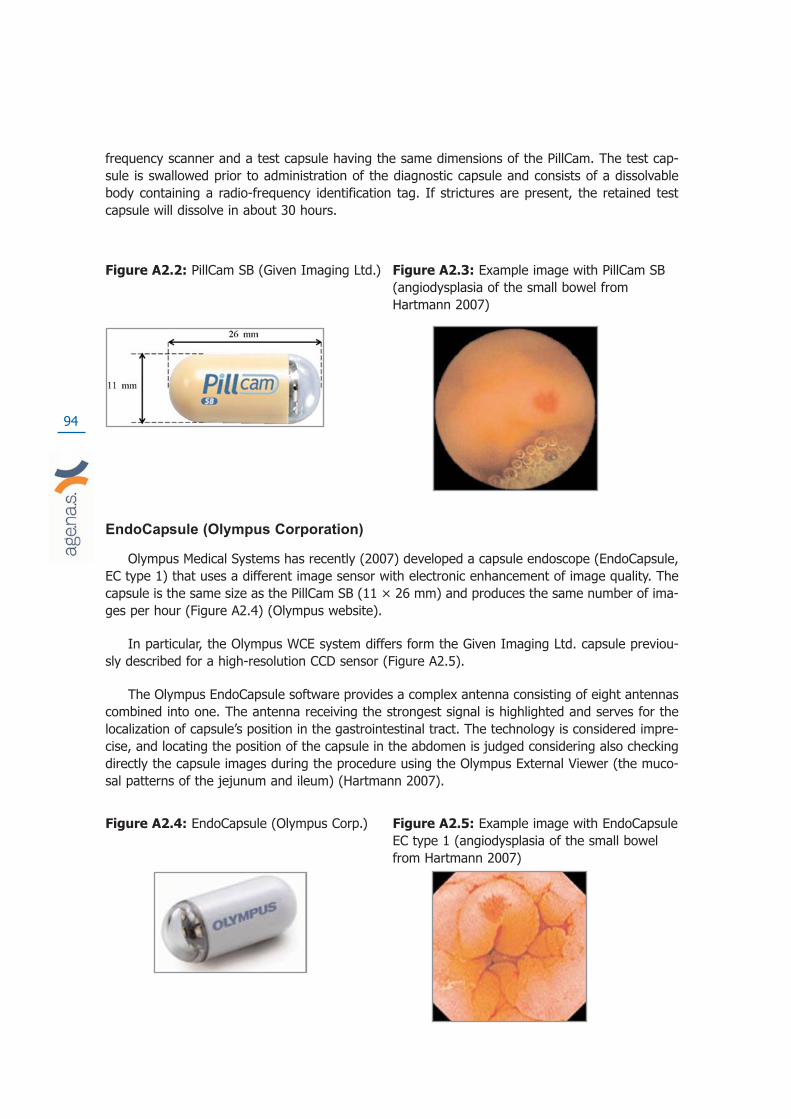
94
frequency scanner and a test capsule having the same dimensions of the PillCam. The test cap-sule is swallowed prior to administration of the diagnostic capsule and consists of a dissolvablebody containing a radio-frequency identification tag. If strictures are present, the retained testcapsule will dissolve in about 30 hours.
Figure A2.2: PillCam SB (Given Imaging Ltd.) Figure A2.3: Example image with PillCam SB (angiodysplasia of the small bowel from Hartmann 2007)
EndoCapsule (Olympus Corporation)
Olympus Medical Systems has recently (2007) developed a capsule endoscope (EndoCapsule,EC type 1) that uses a different image sensor with electronic enhancement of image quality. Thecapsule is the same size as the PillCam SB (11 × 26 mm) and produces the same number of ima-ges per hour (Figure A2.4) (Olympus website).
In particular, the Olympus WCE system differs form the Given Imaging Ltd. capsule previou-sly described for a high-resolution CCD sensor (Figure A2.5).
The Olympus EndoCapsule software provides a complex antenna consisting of eight antennascombined into one. The antenna receiving the strongest signal is highlighted and serves for thelocalization of capsule’s position in the gastrointestinal tract. The technology is considered impre-cise, and locating the position of the capsule in the abdomen is judged considering also checkingdirectly the capsule images during the procedure using the Olympus External Viewer (the muco-sal patterns of the jejunum and ileum) (Hartmann 2007).
Figure A2.4: EndoCapsule (Olympus Corp.) Figure A2.5: Example image with EndoCapsuleEC type 1 (angiodysplasia of the small bowelfrom Hartmann 2007)

95
The FutureGiven have recently presented two new video-capsules, equipped with two imaging device and
light source (one at both ends): PillCam ESO and PillCam COLON (Figure A2.6).
The PillCam ESO takes up to 14 images per second, as it passes down the esophagus duringthe 20-minute procedure. PillCam ESO was developed for patients suffering from esophagealdisorders, such as esophageal varices, which can result in fatal bleeding, and Barrett’s esophagus,which is an early indication for esophageal cancer. The improved version, PillCam ESO2, was clea-red for marketing by U.S. FDA in May 2007. It is the same size as the PillCam ESO video capsulebut has a wider angle of view and captures 18 frames per second. The indications for this videocapsule are similar to PillCam ESO however, the Company believes that screening for gastroeso-phageal reflux disease could be possible in the near future.
The PillCam COLON 11 mm × 31 mm (slightly longer than other PillCams) captures 4 imagesper second during the 10-hour procedure. PillCam COLON was cleared for marketing in theEuropean Union in October 2006 and Israel in February 2008. The video capsule was initially deve-loped to help physician’s visualise the colon and is currently being used for patient’s that have hadan incomplete colonoscopy, for those contraindicated for a colonoscopy or for those who prefernot to have a colonoscopy. Multi-centre clinical trials are underway in Europe (Given website).
Figure A2.6: Given imaging Ltd. products (PillCam ESO, PillCam SB2 and PillCam COLON)
A new-concept device could be available in the next few years. RF System Lab. has declaredthat their capsule for endoscopy (Sayaka Capsule Camera) will be on the market in 2010. Theinnovations carried by this device consisting in the ability to change the camera position and enabethe camera to rotate. While conventional capsules typically have cameras at one end of the devi-ce, Sayaka’s camera has been positioned on the side, where it has a lateral view of the intestinalwalls. In addition, a tiny stepper motor rotates the camera as the capsule passes through the dige-stive tract.
Sayaka’s power is supplied wirelessly from an external source (and it contains no battery).Photos acquired are sent to a receiver located near the body. Image “mosaicking” technology isthen used to combine multiple images taken from various angles into a flat, high-resolution rec-tangular map of the intestines, which can be magnified up to 75 times. Sayaka is characterised bya double-structured capsule made up of an outer and an inner shell. Whereas the outer capsuletraverses through the gastrointestinal tract, the inner capsule alone spins. This spinning is derivedby an electromagnet and a small permanent magnet and which causes the “stepping rotation”.
Over an 8-hour period procedure, Sayaka will generate approximately 870.000 images of thewhole digestive tract at a rate of 30 frames per second. (RFSystem website ).
96
Bibliography
Litwiller D. CCD vs. CMOS: Facts and Fiction; PHOTONICS SPECTRA, January 2001.
http://www.marketwatch.com (accessed in July 2008)
Hara AK, Capsule endoscopy: the end of the barium small bowel examination? Abdom Imaging2005;30:179–83.
Shelat V, Shelat G. “Wireless Capsule Endoscopy”; Journal of Postgraduate Medical Education,Training & Research Vol. I, No. I & II (2006).
http://www.givenimaging.com
http://www.olympusamerica.com/msg_section/endocapsule/index.asp
Hartmann D, Eickhoff A, Damian U, Riemann JF. “Diagnosis of small?bowel pathology using pai-red capsule endoscopy with two different devices: a randomized study”; Endoscopy 2007; 39:1041-1045.
http://www.rfamerica.com/sayaka/index.html

97
Appendix 2b
Standard technology for diagnosis of bowel diseases
One of the most common symptom imputable to a bowel disease is the obscure gastro-inte-stinal bleeding (OGIB) defined as “bleeding from the GI tract that persists or recurs without anobvious aetiology after esophagogastroduodenoscopy, colonoscopy, and radiologic evaluation ofthe small bowel such as small bowel follow-through or enteroclysis” (AGA).
As showed in Table 1, the diagnosis for such conditions is very difficult since a lot of diseasespresent OGIB.
Table 1: Differential diagnosis of occult gastrointestinal bleeding (OGIB)
Notes: †These abnormalities are the most common; ‡These are linear erosions within a hiatus hernia; §This is a large
superficial artery underlying a small mucosal defect (Zuckerman 2000).
Mass lesions Infectious diseases
Carcinoma (any site)† Hookworm
Large (>1.5 cm) adenoma (any site) Whipworm
Inflammation Strongyloidiasis
Erosive esophagitis† Ascariasis
Ulcer (any site)† Tuberculous enterocolitis
Cameron lesions‡ Amebiasis
Erosive gastritis Surreptitious bleeding
Celiac disease Hemoptysis
Ulcerative colitis Oropharyngeal bleeding (including epistaxis)
Crohn’s disease Other causes
Colitis (nonspecific) Hemosuccus pancreaticus
Idiopathic cecal ulcer Hemobilia
Vascular disorders Long-distance running
Vascular ectasia (any site)† Factitious cause
Portal hypertensive gastropathy or colopathy
Watermelon stomach
Varices (any site)
Hemangioma
Dieulafoy’s vascular malformation§

98
Figure A2.7: A typical endoscope
Endoscopy is the examination and inspection of the interior of body organs, joints or cavitiesthrough flexible tubes called endoscopes. An endoscope uses optical fibres and powerful lenssystems to provide lighting and visualisation of the interior of the body. The latest developed devi-ces have an image sensor at the tip instead of the lens system.
An endoscope can consist of
• a rigid or flexible tube;
• a light delivery system to illuminate the organ or object under inspection (the lightsource is normally outside the body and the light is typically directed via an opticalfibre system);
• a lens system transmitting the image to the viewer from the tip;
• an additional channel to allow entry of medical instruments or manipulators.
Endoscopy is classified as mini-invasive procedure but is not generally well accepted by thepatients since it causes pain and discomfort and for this reason different ways to look inside thebody have been developed.
Unlike imaging technologies, endoscopy allows the real-time intervention on the tissues beingexamined by means of distending the tissues, taking samples for biopsy, cleaning, cauterising, etc.
Figure A2.8: Gastro-intestinal apparatus

99
Push Enteroscopy (PE)One of the standard procedures for looking inside the bowel is Push Enteroscopy (PE). This
practice requires per oral insertion of a long endoscope (usually a dedicated enteroscope or pae-diatric colonoscope), and allows examination of the distal duodenum and proximal jejunum.
PE has been considered a useful diagnostic procedure for identifying small bowel lesions.However, this procedure is poorly tolerated, technically difficult (often requires deep sedation oranaesthesia, skilled operators and is time-consuming) and it is not free of complications.
Despite substantial improvements to PE in the last 10 years with manufacturers producing lon-ger instruments (240 cm) with high quality video systems and overtubes, these instruments canonly examine the upper gastrointestinal tract up to the proximal jejunum.
The procedure is usually done under anaesthesia (general anaesthesia or conscious sedation).The enteroscope is inserted through the mouth, with or without an overtube embedding an infla-table silicone balloon, and thanks to extraction-insertion manoeuvres or using the friction at theinterface of the balloon and intestinal wall, it passed into the small bowel.
Due to the anatomical features of gastrointestinal tract, this kind of devices cannot reach all thepoints of the small bowel and then some “dark areas” cannot be observed (Gómez Rodríguez 2006).
Figure A2.9: Single-balloon enteroscope. a b c
a) endoscope; b) disposable sliding tube; c) assembled deviceFrom Olympus website
Double-balloon enteroscopy (DBE)Endoscopic examination of the entire small bowel is technically very difficult. Push enterosco-
py is often possible only to the proximal jejunum. In 2001, Yamamoto and Kito developed the dou-ble balloon method as an insertion technique for the diagnosis and treatment of small bowel disor-ders (Yamamoto 2005).
The technique, called Double-Balloon Enterescopy (DBE) or Push-and-Pull Enteroscopy, invol-ves the use of a special enteroscope and a overtube (a tube that fits over the endoscope). Siliconeinflatable balloons are embedded one on the enteroscope and one on the overtube.

100
The procedure is usually done under anaesthesia (general anaesthesia or conscious sedation).The enteroscope is inserted through the mouth (oral approach) and passed in conventional fashioninto the small bowel. Subsequently, the enteroscope is inserted through the rectum (rectal appro-ach) and arrives at the portion of small bowel that is unreachable through the oral route. The exa-mination is performed by advancing the endoscope for small distances in front of the overtube andinflating the balloon at the end. Using the assistance of friction at the interface of the enterosco-pe and intestinal wall, the small bowel is accorded back to the overtube. The overtube balloon isthen deployed, and the enteroscope balloon is deflated. The process is continued until the entiresmall bowel is visualised.
The main advantage of DBE is the possibility of visualise the entire small bowel to the termi-nal ileum. The disadvantage of this procedure is patient discomfort and the laborious and timeconsuming nature of the modality. Further, the procedure should be approached with caution inpatients with prior small bowel resections (MacKalski 2006).
Figure A2.10:a b
a) Double-balloon enteroscope; b) Phases of DBE. Oral approach: endoscope and overtube are inserted through the
mouth; the balloon on overtube is inflated; the scope is inserted further and the balloon on the scope is inflated; the over-
tube is advanced along the endoscope; the balloon on overtube is inflated; the balloon on the endoscope is deflated; the
scope is inserted further the procedures are repeated and the scope can reach deeper and deeper locations.
Rectal approach: the same procedure is performed inserting the endoscope through the rectum.
Adapted from www.doubleballoonenteroscopy.com/procedure.html and Fujinon website.
Computer Tomography (CT)Computer Tomography scanning (CT or CAT, Computer Assisted Tomography) is a non-invasi-
ve, painless imaging technique. CT uses special x-ray equipment to produce multiple images ofthe inside of the body and a computer to join them together in cross-sectional views of the areabeing studied. CT scans of internal organs, bone, soft tissue and blood vessels provide greater cla-rity than conventional x-ray exams.
This technology offer better quality images than standard radiography: completely eliminatesthe superimposition of images of structures outside the area of interest; discriminates between

101
tissues differing slightly in physical density and processing data from a single CT procedure, axial,coronal, or sagittal planes images can be obtained.
In small bowel examination CT can depict segmental thickening, extraluminal lesions, andcomplications such as sinus tracts, fistulas, and abscesses. Like magnetic resonance imaging(MRI), CT provides cross sectional imaging of extraintestinal organs, but its radiation (greater thanthat of small bowel x ray) present a significant disadvantage.
Conventional CT is limited in its assessment of the small bowel because artefacts are produ-ced by collapsed bowel loops and there is no distension of the small bowel.
A relatively new diagnostic tool is CT enteroclysis which combines conventional small bowelenteroclysis with helical CT. The advantage offered by enteroclysis is distension of the small boweland therefore intraluminal visualisation. The advantage of conventional CT is speed, resolution ofvolumetric data set, and optimal utilisation of the contrast media.
The disadvantage of CT scan compared with conventional barium studies is the lack of dyna-mic information (differentiation between peristalsis and skip lesions may be difficult). Comparedwith MRI, CT has greater availability, is less expensive, and is less time consuming. MRI, however,offers static and dynamic imaging capabilities (MacKalski).
CT is regarded as a moderate to high radiation diagnostic technique. While technical advan-ces have improved radiation efficiency, there has been simultaneous pressure to obtain higher-resolution imaging and use more complex scan techniques, both of which require higher doses ofradiation. The radiation dose for a particular study depends on multiple factors: volume scanned,patient build, number and type of scan sequences, and desired resolution and image quality. CTscans of children have been estimated to produce non-negligible increases in the probability oflifetime cancer mortality leading to calls for the use of reduced current settings for CT scans ofchildren.
Magnetic Resonance Imaging (MRI)Unlike conventional x-ray examinations and CT scans, MRI does not depend on radiation.
Instead, radio waves are directed at hydrogen atoms, in a strong magnetic field.
As the patient lies inside the MRI unit, radio waves are directed at the hydrogen atoms in thearea of his body being studied. In the magnetic field, these atoms, produce signals that are detec-ted by the coils. A computer processes the signals and generates a series of images each of whichshows a thin slice of the body. The computer provides three-dimensional representation of thebody, which can be studied from many different angles on a computer monitor.
Since radio waves have effects on the hydrogen atoms, MR images can show differences inwater content between various body tissues. As a result, MRI is especially suited to detectingdisorders that increase fluid in diseased areas of the body, for example, areas affected by tumours,infection and inflammation. Overall, the differentiation of abnormal (diseased) tissue from normaltissues is significantly easier with MRI than with other imaging modalities such as x-ray, CT andultrasound.

102
Although the strong magnetic field is not harmful in itself, medical devices that contain metalmay malfunction or cause problems during an MRI exam.
Historically, intestinal assessment with MRI was suboptimal as slow sequences caused respira-tory and peristaltic artefacts. Faster pulse sequences are now routinely used and allow both bre-ath-held and breathing independent images with no physiological artefacts. Luminal distension withoral contrast material provides assessment of the bowel wall and its thickness and regularity.
MRI is a potentially ideal imaging modality for patients with Crohn’s disease who requirerepeated cross sectional imaging. Lack of ionising radiation, particularly in patients of reproducti-ve age, is appealing. It is safe in pregnancy and in patients with renal failure, it is helpful inpatients with equivocal findings by other imaging modalities. The greatest advantage is the abili-ty to identify active inflammation and distinguish whether obstructions are from fibrotic strictures,adhesions, or active inflammation. Patients with small bowel obstruction can be examined with aminimally invasive approach (no intravenous contrast required) (MacKalski 2006).
Small Bowel Series (SBS) or Small Bowel follow-Through (SBTS)STS or SBTS is an x-ray exam of the small intestine. This procedure requires that the patient
swallows a radio-opaque contrast medium, usually barium sulphate, in various positions on the x-ray table while the radiologist uses a fluoroscope connected to a monitor for acquire x-ray imagesusually every 20 to 30 minutes (this exam often takes 2 hours or more to complete). Because fluo-roscopy involves the use of x-rays all fluoroscopic procedures pose a potential health risk to thepatient. Radiation doses to the patient depend greatly on the size of the patient as well as lengthof the procedure. More modern improvements in technology have allowed for increased imagequality while minimizing the radiation dose to the patient (Hara 2005).
EnteroclysisEnteroclysis is a minimally invasive radiographic procedure of the small intestine, which requi-
res the introduction of a catheter into the small intestine followed by the injection of barium andmethylcellulose. The catheter is passed from the nose or mouth through the stomach and into partof the small bowel and injects the barium which coats the intestine and the methylcellulose whichdistends the lumen allowing real-time fluoroscopic visualization of the entire small bowel. Thereare several different types of enteroclysis catheters available, and the choice is usually determi-ned by radiologists’ preference, but cost can also be a factor in the decision. There are severaladvantages to having an enteroclysis in comparison to the traditional SBS: this examination ismuch quicker than a routine SBS and there is an increase in the distension of the lumen, which isvery important for tissue characterisation (tissue properties are related to pathological condition).
Nevertheless this procedure presents two main drawbacks: the first and greatest disadvanta-ge is the placement of the catheter, it can be uncomfortable for the patient, even with the use ofanaesthetic spray and jelly; The second is that the patient will receive higher doses of radiation incomparison to the traditional SBS exam.
As stated for CT, for what concerns diagnosis of bowel diseases, suboptimal bowel distensionand overlapping bowel loops could negatively affect all the radiographic imaging techniques.

103
Intraoperative Enteroscopy (IE)IE is usually applied in cases of bleeding that is not localised in spite of extensive diagnostic
evaluation and in which the risks of continued bleeding are judged to outweigh the risks of lapa-rotomy. IE can be performed per-orally, trans-nasally, per-rectum, or through single or multipleintestinal incisions. The choice of enteroscope type and entry site will necessarily depend oninstrument availability, familiarity with the diagnostic approaches, and the experience and techni-cal expertise of the surgeon and endoscopist.
Esophago-Gastro Duodenoscopy (EGD)EGD (or upper endoscopy) enables the physician to take inside the oesophagus, stomach, and
duodenum (first part of the small intestine).
After an anaesthetic has suppressed the gag reflex, an endoscope is advanced through theoesophagus to the stomach and duodenum. Air is introduced slowly through the endoscope toexpand the folds of tissue enhancing view. The internal mucosal surface (lining of the intestines)can be examined and, if the need arises, biopsy samples can be obtained through the endoscopeitself. The procedure takes about 30 minutes.
AngiographyAngiography is the x-ray study of the blood vessels. An angiogram uses a radio opaque
medium to highlight the blood vessels in a fluoroscopy suite. This examination is usually perfor-med at a hospital, in an x-ray or fluoroscopy suite, by a radiologist and assisting technician ornurse. Angiography requires the injection of a contrast medium that makes the blood vessels visi-ble to x-rays. The patient’s vascular system is displayed on a monitor in real-time.
Angiograms can vary depending on the vascular system being studied. For examination of thesmall bowel, the procedure is called mesenteric angiography and involves x-ray exploration of theceliac and mesenteric arteries, the arterial branches that supply blood to the abdomen and dige-stive system.
The test is commonly used to detect aneurysm, thrombosis, and signs of ischemia in the celiacand mesenteric arteries, and to locate the source of gastrointestinal bleeding. The procedure cantake up to 3 hours, depending on the number of blood vessels studied (Gale Encyclopaedia).
Mesenteric angiography can detect active bleeding but is often considered unsatisfactory andinfeasible for the investigation of OGIB because of its highly variable diagnostic yield and risk ofcomplications (Saperas 2007).
Patients with kidney disease or injury may suffer further kidney damage from the contrastmediums used for angiography. Pregnant women are advised to avoid this procedure because xrays carry risks of ionizing radiation exposure to the foetus.

104
Bibliography
AGA - American Gastroenterological Association, Institute Technical Review on ObscureGastrointestinal Bleeding. Gastroenterology 2007;133:1697–1717
Zuckerman GR, Prakash C, Askin MP, Lewis BS. AGA technical review on the evaluation and mana-gement of occult and obscure gastrointestinal bleeding. Gastroenterology 2000;118:201-21.
Gómez Rodríguez BJ, Ortiz Moyano C, Romero Castro R, Caunedo Alvarez A, Hernández Durán MD,Hergueta Delgado P, Pellicer Bautista F, Herrerías Gutiérrez J. Diagnostic yield of 335 push video-enteroscopiesRev Esp Enferm Dig. 2006;98(2):82-92.
http://www.olympusamerica.com
Yamamoto H, Kito H. Double-balloon endoscopy. Curr Opin Gastroenterol 2005;21:573–7.
MacKalski BA, Bernstein CN.New diagnostic imaging tools for inflammatory bowel disease.Gut.2006;55(5):733-41.
www.doubleballoonenteroscopy.com/procedure.html
http://www.fujinonendoscopy.com/default.aspx?pageid=1
Hara AK, Leighton JA, Sharma VK, et al. Imaging of Small Bowel Disease: Comparison of CapsuleEndoscopy, Standard Endoscopy, Barium Examination, and CTRadioGraphics 2005; 25: 697-711.
Gale Encyclopedia of Medicine, Published December, 2002 by the Gale Group.
Saperas E, Dot J, Videla S et al. Capsule endoscopy versus computed tomographic or standardangiography for the diagnosis of obscure gastrointestinal bleeding. Am J Gastroenterol.2007;102(4):731-7.
105
APPENDIX 3
Search Strategy
EMBASE:
#1. ‘capsule endoscopy’/syn OR ‘video *1 capsule’ OR ‘wireless *1 capsule’ AND
([cochrane
review]/lim OR [controlled clinical trial]/lim OR [meta analysis]/lim OR [randomized con-
trolled
trial]/lim OR [systematic review]/lim) AND [humans]/lim AND [embase]/lim
PUB MED:
#9 Search “Capsule Endoscopy”[Mesh] OR Video capsule*[Title/Abstract] OR “wireless
capsule”[Title/Abstract]
CL:
video next capsule* in All Text; WCE in Title, Abstract or Keywords; “Capsule
Endoscopy” in Title,
Abstract or Keywords; wireless next capsule* in All Text

106

App
endi
x 4
Ta
ble
A:
OG
IB_
WC
E v
ers
us
PE
107
OU
TC
OM
ES
Stu
dy
Aim
De
sig
nT
ime
ra
nge a
nd/o
r
follow
up
Part
icip
ants
Dia
gnostic a
ccura
cy
Safe
ty*
Acce
pta
bility
Con
clu
sio
ns
Com
me
nts
Mylo
na
ki M
. e
t a
l.
20
03
,
UK
(Lo
nd
on
)
To
co
mpa
re
effic
acy a
nd
clin
ica
l
impa
ct
of
WC
E v
s P
E
in p
atie
nts
with
OG
IB
an
d n
eg
ative
ga
str
osco
py
an
d
co
lon
osco
py.
Th
e lim
ita
-
tio
ns,
tech
ni-
ca
l fa
ilu
res
an
d p
ractica
l
issu
es o
f
usin
g W
CE
we
re
exp
lore
d.
Ap
rosp
ective
blin
de
d
stu
dy.
Fifty
tw
o p
atie
nts
we
re e
nro
lle
d.
Tw
o w
ere
lost
(re
aso
ns r
ep
ort
ed
).
Th
ey a
ll h
ad
ha
d
co
lon
osco
py a
nd
ga
s-
tro
sco
py,
an
d m
ost
of
the
m
ha
d u
nd
erg
on
e m
ultip
le
pro
ce
du
res.
Th
ey h
ad
WC
E w
ith
in t
wo
we
eks
be
fore
un
de
rgo
ing
PE
. A
ll
ga
str
ic a
bn
orm
alitie
s
de
tecte
d b
y W
CE
ca
n b
e
co
nfirm
ed
by P
E.
Th
e
co
lon
ic a
bn
orm
alitie
s w
ere
tre
ate
d w
ith
su
bse
qu
en
t
co
lon
osco
py.
WC
E w
as
pe
rfo
rme
d a
nd
re
po
rte
d b
y
diffe
ren
t e
xa
min
ers
un
aw
are
of
the
fin
din
gs o
f
the
oth
er
exa
min
atio
n.
Tw
o
en
do
sco
pis
ts c
arr
ied
ou
t
PE
an
d a
ph
ysic
ian
ad
min
-
iste
red
WC
E a
nd
re
ad
ima
ge
s.
Tw
o r
evie
we
rs
exa
min
ed
all W
CE
im
ag
es,
an
d a
th
ird
en
do
sco
pis
t
arb
itra
ted
on
an
y d
iscre
p-
an
cie
s in
th
e in
terp
reta
tio
n
of
fin
din
gs. A
furt
he
r g
rou
p
of
40
vo
lun
tee
rs w
as a
lso
en
rolle
d b
efo
re t
he
be
gin
-
nin
g o
f th
e s
tud
y t
o a
cq
uire
info
rma
tio
n o
n t
he
no
rma
l
ap
pe
ara
nce
of
the
sm
all
bo
we
l w
ith
WC
E a
nd
PE
.
Tim
e r
ange:
WC
E
with
in 2
weeks
be
fore
PE
.
Fo
llo
w u
p:
two
we
eks o
r m
ore
(no
t sp
ecifie
d)
Fifty
patients
(21
male
, 29
fem
ale
), A
vera
ge
age 5
0.3
(ra
nge
17-8
0).
They a
ll
had h
ad
colo
noscopy a
nd
gastr
oscopy,
and
most
of
them
had u
nderg
one
multip
le p
roce-
dure
s.
Fort
y
seven w
ere
transfu
sed,
15 o
f
them
had m
ore
than 1
00 u
nits o
f
blo
od,
15 m
ore
than 4
00 u
nits.
WC
E d
iagnostic
yie
ld in s
mall inte
s-
tine w
as 6
8%
(34/5
0)
(angio
dypla
-
sia
(16),
fre
sh f
aecal
ble
edin
g (
8),
apt-
hous u
lcera
tion s
ug-
gestive o
f C
rohn’s
dis
ease (
3),
tum
our
(2).
The W
CE
tota
l
dia
gnostic y
ield
was
76%
(38/5
0).
The
yie
ld o
f P
E in d
is-
covering t
he s
ourc
e
of
obscure
ble
edin
g
was 3
2%
(16/5
0).
The t
ota
l dia
gnostic
yie
ld o
f P
E w
as 3
8%
(19/5
0).
WC
E
- T
P:
in 1
6
patients
the
ba
t-
tery
expired b
efo
re
caecum
(28%
); I
n
3 p
atie
nts
the
re
was a
loss o
f
images d
ue t
o
tem
pora
ry e
lectr
i-
cal dis
con
nection
;
In 1
patie
nt
the
batt
ery
expired
after
2 h
ours
;
- A
E:
in 1
patien
t
the W
CE
rem
ain
ed
in t
he
oesopha
gus f
or
7
hours
an
d w
as
pushed in
to t
he
sto
mach b
y a
n
endosco
pe.
In
seven t
he W
CE
passed into
the
pylo
rus a
nd
retu
rned t
o t
he
sto
mach b
efo
re
passin
g in
the
inte
stine a
t le
ast
once.
In o
ne
patient
this
occurr
ed 7
tim
es.
PE
- N
o c
om
plica
tio
ns
Me
thod:
not
cle
ar-
ly r
eport
ed. A
t fo
l-
low
up a
ll p
atie
nts
we
re in
terv
iew
ed
an
d a
sked t
o c
om
-
pare
WC
E a
nd
PE
pain
fuln
ess a
nd
wh
ich w
as t
hey
pre
ferr
ed
.
Re
su
lts:
WC
E w
as
pre
ferr
ed
to P
E:
49
/50
; W
CE
was
un
co
mfo
rta
ble
for
sw
allow
ing 2
/50
;
PE
is p
ain
ful
(34/5
0,
p<
0.0
5).
Th
e a
uth
ors
cla
im
that
this
stu
dy
sh
ow
s t
hat
the
WC
E c
an
pro
vid
e
sm
all in
testin
al
imag
ing
co
mpa
ra-
ble
with
PE
an
d
ca
n d
iag
no
se s
ites
of
inte
stin
al b
lee
d-
ing
beyo
nd
the
rea
ch
of
PE
. W
CE
was s
afe
, pain
less
and w
ell t
ole
rate
d.
Th
is is n
ot
a r
an
-
dom
ise
d t
ria
l, a
nd
pa
tien
ts s
erv
ed a
s
their o
wn
co
ntr
ol.
PE
is d
efin
ed a
s
an u
nfa
ir c
om
-
pa
rato
r sin
ce it
doe
s n
ot
rea
ch
th
e
low
er
sm
all inte
s-
tin
e.
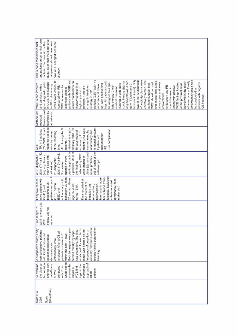
Ma
ta e
t a
l.
20
04
Spa
in
(Ba
rce
lon
a)
To
exa
min
e
the
dia
gn
os-
tic p
recis
ion
an
d t
he
clin
i-
ca
l e
ffic
acy
of
WC
E
co
mpa
red
with
PE
in
OG
IB a
nd
to
an
aly
se
th
e
impa
ct
of
WC
E f
ind
-
ing
s o
n t
he
the
rap
eu
tic
ap
pro
ach
of
OG
IB
pa
tie
nts
.
Ap
rosp
ective
stu
dy.
Fo
rty
two
co
nse
cu
tive
pa
tie
nts
with
OG
IB a
nd
no
rma
l
oe
so
ph
ag
og
astr
od
uo
-
de
no
sco
py a
nd
co
lon
osco
py w
ere
an
aly
se
d. A
fte
r W
CE
all
pa
tie
nts
un
de
rwe
nt
PE
with
in t
he
ne
xt
7 d
ays.
Bo
th t
ech
niq
ue
s w
ere
pe
r-
form
ed
“b
lin
dly
” b
y s
epa
-
rate
exa
min
ers
. T
he
dia
g-
no
stic y
ield
fo
r e
ach
te
ch
-
niq
ue
wa
s d
efin
ed
as t
he
fre
qu
en
cy o
f d
ete
ctio
n o
f
clin
ica
lly r
ele
va
nt
inte
stin
al
lesio
ns h
avin
g p
ote
ntia
l fo
r
ble
ed
ing
.
Tim
e r
an
ge
: P
E
with
in a
we
ek a
fter
WC
E.
Fo
llo
w u
p:
no
t
rep
ort
ed
Fort
y t
wo c
onsec-
utive p
atients
with
OG
IB (
overt
ble
edin
g in 2
6
patients
and o
ccult
in 1
6),
norm
al
EG
D a
nd
colo
noscopy w
ith
ileoscopy.
22 m
en,
20 w
om
en.
Mean
age 5
5 y
ears
,
range 1
6-8
4.
Data
record
ed a
t
the m
om
ent
of
inclu
sio
n a
re
report
ed (
e.g
.
haem
oglo
bin
,
haem
ato
crit,
num
-
ber
of
blo
od t
rans-
fusio
ns).
Exclu
sio
n
crite
ria r
eport
ed
(pre
gnancy,
pace-
maker
etc
.)
WC
E d
iagnostic
yie
ld:
(74%
) 31/4
2,
(angio
dypla
sia
>
fresh b
lood w
ith-
out
lesio
ns)
PE
dia
gnostic
yie
ld:
(19%
) 8/4
2.
Successfu
l
change in t
hera
-
peutic a
ppro
ach in
7 p
atients
. M
ost
of
the f
indin
gs
dete
cte
d b
y W
CE
were
locate
d a
t
dis
tal je
junum
and
ileum
, pro
bably
out
of
reach o
f th
e
ente
roscope.
WC
E
-TP
: In
3 p
atients
(7%
) W
CE
did
not
reach t
he ile
oceca
l
valv
e b
y t
he e
nd
of
the r
ecord
ing
tim
e;
-AE
: A
mong t
he 3
patients
, 1
reta
ined a
sym
pto
-
matically W
CE
fo
r
48 d
ays (
natu
ral
expuls
ion);
In 1
patient
(2%
) W
CE
was r
em
oved b
y
laparo
scopy d
ue
to jeju
nal str
ictu
re;
1 p
atient
not
accounte
d f
or.
PE
- N
o c
om
plication
Meth
od
: n
ot
rep
ort
ed
.
Results:
well
tole
rate
d b
y
all p
atie
nts
.
WC
E is n
on invasiv
e
and p
ain
less,
with
a
good
dia
gno
stic y
ield
in O
GIB
. It
is s
upe
rior
to P
E in d
iag
nosin
g
sm
all b
ow
el le
sio
ns.
If
co
mpa
red
with
PE
,
WC
E in
cre
ases t
he
dia
gn
osis
yie
ld in
pa
tients
with O
GIB
an
d
allo
ws m
od
ific
atio
n o
n
the
rap
y s
tra
tegy in
a
hig
h p
rop
ort
ion
of
pa
tients
. It led
to
a
ch
ang
e in t
rea
tme
nt
str
ate
gy in 7
/31
pa
tients
(22
%)
(with n
o
recu
rre
nce a
t fo
llo
w
up)
with
positiv
e f
ind-
ings. A
ll p
atie
nts
co
uld
be a
ssig
ned
to a
spe-
cific
th
era
py:
2 w
ith
med
ical tr
ea
tmen
t
(Cro
hn
), 2
with e
nd
o-
sco
pic
th
era
py (
jeju
nal
angio
dyspla
sia
), 3
sur-
gery
(1 C
rohn
and
2
tum
ou
rs in
ile
um
). O
nly
two
of
the 1
2 d
iag
no
sis
of
an
gio
dyspla
sia
we
re
effe
ctive
ly t
rea
ted.
Th
e
auth
ors
su
gge
st
that
WC
E s
hou
ld b
e u
sed
firs
t ch
oic
e a
fte
r a
neg
-
ative u
pp
er
an
d low
er
co
nven
tio
nal
endo
sco
py a
nd
PE
sh
ould
be u
sed
in
ca
ses w
ith
positiv
e
WC
E f
indin
gs lo
ca
ted
at
the
pro
xim
al sm
all
bow
el w
ithin
th
e r
ea
ch
of
en
tero
scop
y.
Fin
ally
ente
roscopy c
ou
ld a
lso
be c
onsid
ere
d in
pa
tients
with n
eg
ative
CE
fin
din
gs.
This
is n
ot
a r
an
do
mis
ed t
ria
l,
an
d p
atie
nts
se
rve a
s t
heir o
wn
con
tro
ls.
The m
ain
aim
of
this
stu
dy,
giv
en
th
at
PE
is n
ot
a f
air
com
pa
rato
r shou
ld h
ave b
een
ho
w W
CE
cha
ng
ed
tre
atm
en
t
str
ate
gy.

Ad
ler
DG
. e
t a
l.
20
04
US
A
(Ro
ch
este
r-
Min
ne
so
ta)
To
co
mpa
re W
CE
an
d P
E in
th
e
inve
stig
atio
n o
f th
e
sm
all b
ow
el in
pa
tie
nts
with
OG
IB.
Ap
rosp
ective
“blin
de
d”
stu
dy
en
rollin
g 2
0
pa
tie
nts
with
GI
ha
em
orr
ha
ge
. A
ll
pa
tie
nts
un
de
rwe
nt
WC
E f
ollo
we
d b
y
PE
. T
he
ph
ysic
ian
pe
rfo
rmin
g t
he
en
tero
sco
py (
se
nio
r
en
do
sco
pis
t) in
ter-
pre
ted
th
e W
CE
resu
lts in
an
un
blin
de
d m
an
ne
r,
wh
ile
a s
eco
nd
blin
de
d r
evie
we
r
(en
do
sco
py a
ssis
-
tan
t) in
terp
rete
d
the
m t
o e
sta
blish
the
re
lia
bility o
f th
e
inte
rpre
tatio
n.
Tim
e r
an
ge
: not
rep
ort
ed
.
Fo
llo
w u
p:
11-2
4
mo
nth
s,
bu
t only
for
pa
tie
nts
who
rece
ive
d c
are
at
the
ce
ntr
e s
ubse-
qu
en
t to
th
e p
roce-
du
re.
Tw
enty
patients
with O
GIB
, nega-
tive E
GD
and
colo
noscopy in last
2 m
onth
s. A
ll h
ad
docum
ente
d iro
n
deficie
ncy
anaem
ia.
8 m
en,
12 w
om
en.
Mean
age 6
5.5
years
,
range 3
8-8
0.
Exclu
sio
n c
rite
ria
report
ed (
age less
than 1
8 y
ears
,
pre
gnancy,
car-
dio
pulm
onary
dis
-
ease e
tc.,
GI
tract
obstr
uction).
Dia
gnostic y
ield
of
WC
E.
Definitiv
e s
ourc
es
of
ble
edin
g in t
he
sm
all b
ow
el w
ere
identified b
y W
CE
in 6
out
of
20
patients
(30%
) and
only
tw
o o
f th
em
were
found t
o h
ave
sm
all b
ow
el
angio
ecta
sia
s a
t
PE
. 5 o
f th
em
underw
ent
targ
ete
d
endoscopic
or
sur-
gic
al th
era
py b
ased
on W
CE
and P
E
findin
gs.
The W
CE
did
not
affect
the
managem
ent
of
patients
found t
o
have indete
rmin
ate
lesio
ns.
WC
E
- N
o c
om
plica
tio
n
PE
- N
o c
om
plica
tio
n
No
t re
po
rted
WC
E h
as a
n
impa
ct
on lon
g
term
man
ag
em
ent
of
patie
nts
with
cle
arly d
efin
ed
lesio
ns (
an
gio
eca
t-
sia
, fo
ca
l u
lce
r),
but
do
esn’t a
ffe
ct
the
mana
gem
ent
of
pa
tie
nts
with
less
de
fined
le
sio
ns.
WC
E a
llo
ws t
he
de
tectio
n o
f sm
all
or
triv
ial le
sio
ns
tha
t are
not
evi-
de
nced
du
rin
g a
sta
nd
ard
PE
an
d
do
not
ne
cessa
rily
ca
use b
lee
din
g.
The
stu
dy u
sed t
he
firs
t so
ftw
are
ve
r-
sio
n f
or
WC
E’s
wh
ich
did
no
t pro
-
vid
e lo
ca
lisa
tion. A
larg
e t
ria
l w
ith a
co
ntr
ol a
rm w
ith
no
rmal a
sym
pto
-
matic in
div
idu
als
sh
ould
be
und
er-
taken
. T
he
in
ter-
pre
ters
’re
liab
ility
wa
s h
igh
.
Th
is is n
ot
a r
an-
dom
ised
tria
l. A
nd
pre
se
nts
se
vera
l
facto
rs t
o b
e c
on-
sid
ere
d
a)
PE
ma
y n
ot
be
a
pro
per
co
mpara
tor,
sin
ce
it
do
esn
’t
rea
ch
th
e p
art
of
sm
all b
ow
el
rea
ch
ed b
y W
CE
.
B)
Th
at
a b
asic
req
uire
men
t of
all
the s
tud
ies s
ho
uld
be t
he
defin
itio
n o
f
the k
ind o
f le
sio
ns
(de
fin
itiv
e o
r no
t)
foun
d w
ith
WC
E v
s
those
fo
und b
y P
E
C)
Ea
ch s
tudy
sh
ould
hig
hlig
ht
the
ch
ang
es in t
he
man
agem
en
t of
pa
tients
bro
ug
ht
abou
t w
ith
or
with-
out
WC
E.
109

Sa
urin
et
al.,
20
05
FR
AN
CE
(Lyo
n a
nd
Pa
ris)
To
eva
lua
te t
he
lon
g t
erm
ou
tco
me
in W
CE
pa
tie
nts
an
d t
o u
nd
ers
tan
d
the
clin
ica
l re
le-
va
nce
of
lesio
ns
de
tecte
d w
ith
WC
E.
To
co
mpa
re
WC
E a
nd
PE
sp
ecific
ity a
nd
se
n-
sitiv
ity,
usin
g p
revi-
ou
s r
esu
lts in
term
s o
f clin
ica
l
ou
tco
me
, a
s a
go
ld
sta
nd
ard
.
A“f
ollo
w u
p s
tud
y”,
it in
vo
lve
s 5
8
pa
tie
nts
alre
ad
y
en
rolle
d in
a p
revi-
ou
s p
rosp
ective
stu
dy c
om
pa
rin
g
WC
E a
nd
PE
.
Pa
tie
nts
we
re c
on
-
tacte
d a
fte
r 1
ye
ar,
an
d e
ach
ca
se
wa
s
cla
ssifie
d in
to
tru
e/fa
lse
po
sitiv
e
or
tru
e/fa
lse
ne
ga
-
tive
fin
din
gs a
t P
E
an
d W
CE
. T
he
resu
lts w
ere
co
m-
pa
red
with
th
e in
i-
tia
l cla
ssific
atio
n o
f
lesio
ns o
bse
rve
d a
t
WC
E:
hig
hly
re
le-
va
nt
(P2
), a
nd
le
ss
rele
va
nt
(P0
, P
1)
lesio
ns.
Tim
e r
an
ge
: P
E
with
in 3
da
ys a
fter
WC
E
Fo
llo
w u
p:
no
t
rep
ort
ed
Six
ty p
atients
,
mean a
ge 5
8
(±
18 y
ears
, ra
nge
21-7
9);
27 m
en
and 3
3 w
om
en.
28
patients
(47%
) pre
-
sente
d r
ecurr
ent
overt
GI
ble
edin
g.
Fifty
eig
ht
patients
were
eligib
le.
Dia
gnostic y
ield
(sm
all b
ow
el
lesio
ns w
ith p
ote
n-
tial fo
r ble
edin
g)
CE
: 40/6
0 (
67%
),
19 p
atients
with
lesio
ns b
oth
on C
E
and P
E.
PE
:22/6
0
(37%
), inclu
din
g 3
patients
mis
sed
with C
E.
Sensitiv
ity (
95%
CI)
of
WC
E 0
.92
(0.8
2-1
.00)
and P
E
0.6
9 (
0.5
3-0
.87)
-
Specific
ity
(95%
CI)
of
WC
E 0
.48
(0.3
2-0
.68)
and P
E
0.8
0 (
0.5
4-0
.94).
PP
V o
f W
CE
0.6
2,a
nd o
f P
E
0.7
5 -
NP
V o
f
WC
E 0
.87 a
nd
NP
V 0
. 74.
WC
E
- N
ot
report
ed
PE
- N
ot
report
ed
No
t re
port
ed
WC
E is a
se
nsitiv
e
exa
min
ation
for
the
de
tection
of
lesio
ns
of
the
sm
all-b
ow
el
in p
atie
nts
with
OG
IB,
its s
pe
cific
i-
ty is low
er
than
that
of
PE
wh
en
the c
lin
ica
l o
ut-
com
e is u
se
d a
s
the g
old
sta
nd
ard
.
Ta
kin
g into
accou
nt
the r
ele
va
nce o
f all
lesio
ns id
en
tified
in
the s
mall b
ow
el by
WC
E initia
l, 1
5 o
f
18
we
re h
igh
ly r
el-
eva
nt
lesio
ns
(83.3
%)
were
ulti-
ma
tely
cla
ssifie
d
as t
rue p
ositiv
e
case
s c
om
pare
d
with s
eve
n o
f 2
2
less r
ele
vant
lesio
ns (
31%
p=
0.0
00
3).
T
he
no
n inva
siv
e
na
ture
of
WC
E a
nd
its h
igh n
ega
tive
pre
dic
tive
va
lue
sug
gest
tha
t it
sho
uld
be u
se
d
be
fore
an
y t
hera
-
pe
utic P
E,
wh
ich
mig
ht
subse
quen
tly
be
requ
ired
.
This
is n
ot
a r
an
-
do
mis
ed
stu
dy.
It
trie
s t
o ide
ntify
a
wa
y t
o d
ete
rmin
e
sen
sitiv
ity a
nd
spe
cific
ity o
f W
CE
vs P
E,
find
ing
a
pro
pe
r g
old
sta
n-
da
rd a
nd a
vo
idin
g
a t
oo s
tric
t d
efin
i-
tion
of
true
positiv
e
an
d f
als
e p
ositiv
e
case
s o
f oth
ers
(Pen
na
zio
, 20
03
).
It s
ee
ms t
ha
t m
ost
of
the lesio
ns ide
n-
tified
by W
CE
are
sm
all,
and
the
stu
dy u
nd
erlin
es
the p
roble
m o
f
lesio
ns n
ot
de
tect-
ed
beca
use
they
are
out
of
the a
rea
exa
min
ed b
y P
E.
It
is u
ncle
ar
whe
ther
they c
alc
ula
ted
spe
cific
ity a
nd s
en-
sitiv
ity r
efe
rrin
g
on
ly t
o t
he p
art
of
the s
ma
ll b
ow
el
that
bo
th p
roce
-
du
res c
an
re
ach
.

111
Ne
u e
t a
l.,
20
05
GE
RM
AN
Y
(Ha
mb
urg
, M
un
ich
,
Ma
inz, A
lto
na
,
Wie
sb
ad
en
)
To
eva
lua
te t
he
influ
en
ce
of
WC
E
fin
din
gs in
th
e f
ur-
the
r m
an
ag
em
en
t
of
pa
tie
nts
(m
ajo
r
ch
an
ge
s in
ma
n-
ag
em
en
t),
pa
rtic
u-
larly w
ith
OG
IB.
An
aly
sis
of
ou
t-
co
me
ch
an
ge
s
su
ch
as a
sig
nifi-
ca
nt
red
uctio
n o
f
furt
he
r b
lee
din
g.
Am
ultic
en
ter
pro
sp
ective
stu
dy
ca
rrie
d o
ut
in f
ive
diffe
ren
t te
rtia
ry
refe
rra
l ce
ntr
es.
Th
e d
iag
no
stic
yie
ld o
f W
CE
wa
s
co
mpa
red
to
th
ree
oth
er
tests
, P
E a
nd
Sm
all B
ow
el
Do
ub
le c
on
tra
st
rad
iog
rap
hy (
SB
E)
an
d S
ele
ctive
Ab
do
min
al
An
gio
gra
ph
y,
bu
t in
diffe
ren
t se
qu
en
ce
acco
rdin
g t
o t
he
ce
ntr
es f
acilitie
s.
An
en
do
sco
pis
t
with
sim
ila
r e
xp
eri-
en
ce
re
vie
we
d
WC
E v
ide
os.
WC
E
resu
lts o
nly
we
re
un
blin
de
d f
or
PE
.
Pa
tie
nts
we
re f
ol-
low
ed
up
fo
r a
t
lea
st
6 m
on
ths.
Pa
ram
ete
rs t
ha
t
led
to
ma
jor
ma
n-
ag
em
en
t ch
an
ge
s
we
re a
na
lyse
d
(e.g
. su
rgic
al o
r
en
do
sco
py in
ter-
ve
ntio
ns,
sp
ecific
me
dic
al th
era
pie
s)
as w
ell a
s t
he
ir c
or-
rela
tio
n t
o f
urt
he
r
ble
ed
ing
.
Tim
e r
an
ge:
not
rep
ort
ed
Fo
llo
w u
p:
6
mo
nth
s
Fifty
six
patients
17
wom
en a
nd 3
0
men,
mean a
ge 6
3
years
(ra
nge 1
8-
82).
OG
IB (
OB
D)
was a
n inclu
sio
n
crite
ria,
bein
g t
his
overt
in 3
7
patients
, and o
ccult
in 1
9 p
atients
.
Dia
gnostic y
ield
of
WC
E c
om
pare
d t
o
the d
iagnostic y
ield
of
thre
e o
ther
com
-
para
tor
tests
(O
T).
WC
E d
ete
cte
d
42%
of
lesio
ns w
ith
low
pro
bability o
f
ble
edin
g,
and 5
8%
of
lesio
ns w
ith a
hig
h p
robability.
PE
dete
cte
d 2
7%
of
min
or
lesio
ns a
nd a
73%
of
majo
r
lesio
ns (
table
s
show
results f
or
each s
ingle
tech-
nolo
gy a
nd a
lso
aggre
gate
d
results).
WC
E
- N
ot
rep
ort
ed
PE
- N
ot
rep
ort
ed
Not
rep
ort
ed
WC
E h
elp
s w
ith
ma
nag
em
en
t d
eci-
sio
ns a
nd
ca
n
repla
ce
oth
er
more
com
ple
x a
nd r
isky
sta
nda
rd t
ests
.
Neve
rthe
less,
clin
i-
cal para
mete
rs a
re
eq
ually im
po
rtant
for
pre
dic
ting f
ur-
ther
ble
ed
ing a
nd
sho
uld
als
o b
e
use
d t
o d
ecid
e o
n
furt
he
r m
an
age
-
me
nt.
It is n
ot
a r
an-
dom
ised
stu
dy.
Th
e
text
sta
tes t
hat
sin
ce
vis
ualisation
of
the s
ma
ll b
ow
el
by P
E is p
art
ial, its
DY
ma
y h
ave b
een
unde
restim
ate
d.
In
additio
n t
he
auth
ors
sta
te t
ha
t
WC
E o
fte
n ide
nti-
fie
s m
an
y le
sio
ns
whic
h a
re n
ot
the
ca
use o
f b
lee
din
g.
Th
e s
tudy’s
co
nclu
-
sio
ns a
re d
ifficu
lt t
o
inte
rpre
t as t
he
auth
ors
dis
cuss
aggre
ga
te d
ata
while p
resen
ting
data
in a
dis
ag
gre
-
gate
fo
rm.K
11

De
Le
usse
et
al.,
20
07
FR
AN
CE
(Pa
ris)
To
asse
ss d
iag
no
stic
yie
ld o
f W
CE
vs P
E
as f
irst
lin
e e
xp
lo-
ratio
n in
OG
IB;
Asse
ss c
lin
ica
l re
le-
va
nce
of
WC
E a
nd
PE
an
d a
ccu
racy o
f
the
exp
lora
tio
n.
Pro
sp
ective
ra
n-
do
mis
ed
tria
l. A
ll c
on
-
se
cu
tive
pa
tie
nts
refe
rre
d f
or
ob
scu
re
ga
str
oin
testin
al
ble
ed
ing
we
re r
an
-
do
mis
ed
to
eith
er
WC
E o
r P
E a
s t
he
firs
t-lin
e e
xp
lora
tio
n.
Th
e a
lte
rna
tive
me
tho
d w
as o
nly
use
d if
the
first
lin
e
me
tho
d r
eve
ale
d n
o
de
fin
ite
ble
ed
ing
so
urc
e,
or
if r
eq
uire
d
for
clin
ica
l re
aso
ns
du
rin
g f
ollo
w-u
p.
Fo
llo
w u
p:
12
mo
nth
s
Seventy
eig
ht
patients
, 30 w
om
en
and 4
8 m
en,
avera
ge
age o
f 54±16 y
ears
(range 2
2-8
5 y
ears
).
All p
atients
had
underg
one o
ther
pro
-
cedure
s b
efo
re.
The d
iagnostic y
ield
of
WC
E a
nd P
E f
or
definite s
ourc
es o
f
ble
edin
g w
ere
17/4
0
patients
(43%
; 95%
CI:
13-4
0)a
nd 4
/38
patients
(11%
; 95%
CI:
4-2
5),
respective-
ly f
or
sm
all b
ow
el
lesio
ns (
P=
.02),
3/4
0
patients
(8%
: 95%
CI:
3-2
0)
and 4
/38
patients
(11%
; 95%
CI:
4-2
5)
for
gastr
ic
lesio
ns a
nd 0
/40
patients
(0%
; 95%
CI:
0-7
) and 1
/38
patients
(3%
; 95%
CI;
: 1-1
4)
for
colo
nic
lesio
ns.
Perf
orm
ances o
f
WC
E f
or
all lesio
ns:
sensitiv
ity° 7
9 (
60-
86);
specific
ity°
87(6
7-9
0);
PP
V 8
8
(75-9
0)
and N
PV
77
(50-8
5).
For
sm
all
bow
el le
sio
ns:
sensi-
tivity° 1
00 (
61-1
00);
specific
ity° 9
0 (
77-
92);
PP
V 8
5 (
69-8
8)
and N
PV
100 (
71-
100).
Perf
orm
ances
of
PE
for
all lesio
ns:
sensitiv
ity 4
1(3
0-5
3)*
;
specific
ity 1
00 (
91-
100);
PP
V 1
00 (
89-
100)
and N
PV
56
(35-7
2).
For
sm
all
bow
el le
sio
ns:
sensi-
tivity 3
3 (
21-4
3)*
;
specific
ity 1
00 (
93-
100);
PP
V 1
00 (
83-
100)
and N
PV
62
(41-7
5).
*P
=.0
25 v
s
WC
E.
°C
orr
ecte
d f
or
non v
erified c
ases.
WC
E
- 1 p
atient
(1%
)
refu
sed t
o s
wa
llow
the c
apsu
le a
nd
had
it intr
oduce
d e
nd
o-
scopic
ally;
- In
6 p
atie
nts
(9%
)
the c
apsu
le d
id n
ot
reach t
he
ca
ecum
.
PE
- N
o s
ignific
ant
co
m-
plication
s
Not
rep
ort
ed
Th
is r
an
do
mis
ed
stu
dy c
on
firm
s t
hat
WC
E h
as a
hig
he
r
dia
gn
ostic y
ield
(D
Y)
tha
n P
E in
pa
tien
ts
with O
GIB
. T
he t
wo
str
ate
gie
s t
este
d
(WC
E o
r P
E f
irst
on
e
the
n t
he o
the
r, if
ne
g-
ative
) w
ere
no
t sta
tis-
tically d
iffe
ren
t in
term
s o
f D
Y,
clin
ical
rem
issio
n r
ate
, th
era
-
pe
utic im
pact, n
ee
d
alte
rna
tive e
xp
lo-
ratio
n d
uring f
irst
yea
r.
Ho
we
ver
CE
firs
t str
ate
gy is s
im-
ple
r a
nd b
ett
er
tole
r-
ate
d a
s f
irst
str
ate
gy.
The
on
ly p
rop
er
ran-
do
mis
ed
stu
dy o
f th
e
wh
ole
syste
ma
tic
revie
w.
It is a
ve
ry
go
od
stu
dy.
Sho
rtco
min
gs r
ela
ted
to t
he g
ene
ralisab
ilty
of
the r
esu
lts d
ue
to
the n
um
be
r o
f
patie
nts
enro
lled
(7
8)
an
d t
o t
he c
linic
al
cha
racte
ristics o
f
patie
nts
, w
hic
h s
ee
m
to b
e v
ery
spe
cific
to
Fre
nch c
onte
xt
(see
very
hig
h h
aem
oglo
-
bin
leve
ls).
Re
aso
ns
for
patie
nts
lo
st
to f
ol-
low
up a
re n
or
rep
ort
-
ed
.
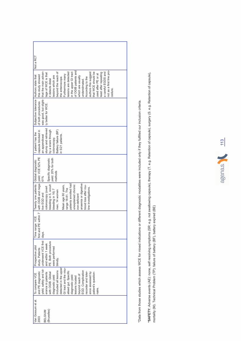
113
*D
ata
fro
m t
hose s
tudie
s w
hic
h a
ssess W
CE
for
mix
ed indic
ations o
r diffe
rent
dia
gnostic m
odalities w
ere
inclu
ded o
nly
if
they f
ulfille
d o
ur
inclu
sio
n c
rite
ria.
*S
AF
ET
Y:
Advers
e e
vents
(A
E):
none, self r
esolv
ing s
ym
pto
ms (
SR
: e.g
. not sw
allow
ing c
apsule
), thera
py (
T. e.g
. R
ete
ntion o
f capsule
), s
urg
ery
(S
. e.g
. R
ete
ntion o
f capsule
),
mort
ality
(M
). T
echnic
al P
roble
m (
TP
): f
ailure
of
battery
(B
F),
battery
expired (
BE
)
Va
n G
ossu
m e
t a
l.
20
03
BE
LG
IUM
(Bru
xe
lle
s)
To
co
mpa
re V
CE
an
d P
E d
iag
no
stic
yie
ld,
sa
fety
an
d t
ol-
era
nce
in
pa
tie
nts
with
OG
IB.
Glo
ba
l
dia
gn
ostic y
ield
inclu
de
d a
ll le
sio
ns
de
tecte
d in
th
e u
pp
er
GI
tra
ct
an
d t
he
vis
u-
alise
d g
ut. S
pe
cific
dia
gn
ostic y
ield
:
lesio
ns lo
ca
ted
be
yo
nd
re
ach
of
EG
D.
Co
mp
lica
tio
n
reco
rde
r a
nd
to
ler-
an
ce
asse
sse
d b
y
pa
tie
nt’s q
ue
stio
n-
na
ire
.
Pro
sp
ective
pilo
t
stu
dy.
Pa
tie
nts
un
de
rwe
nt
VC
E f
irst
an
d w
ith
in 1
we
ek
PE
. B
oth
pro
ce
du
re
we
re p
erf
orm
ed
blin
dly
.
Tim
e r
an
ge
: V
CE
firs
t a
nd
PE
with
in 7
da
ys.
Tw
enty
one p
atients
with O
GIB
and n
ega-
tive E
GD
and
colo
noscopy (
overt
ble
edin
g in 5
, occult
ble
edin
g in 1
6)
7
men,
14 w
om
en
Mean a
ge 6
0 y
ears
,
range 1
8-8
1.
The
patients
enro
lled h
ad
unexpla
ined c
hro
nic
iron-d
eficie
nt
anaem
ia o
r dig
estive
blo
od loss a
fter
rou-
tine investigations.
Glo
bal dia
gnostic
yie
ld:
VC
E 5
2%
PE
61%
Specific
dia
gnostic
yie
ld:
20%
for
both
meth
ods
1 p
atient
has t
he
capsule
blo
cked in
an a
ppendic
eal
stu
mp a
nd r
etr
ieved
by a
snare
thro
ug
h
colo
noscopy.
Batt
ery
failure
(B
F)
in 5
/21 p
atients
.
Su
bje
ctive
tole
ran
ce
of
both
pro
cedu
res
was g
oo
d,
but
sligh
t-
ly b
ett
er
for
WC
E.
Au
tho
rs s
tate
tha
t
this
stu
dy s
ho
wed
that
the m
ain
advan
-
tag
e o
f W
CE
is t
hat
it d
ete
cts
dis
tal
lesio
ns w
hic
h a
re
be
yo
nd t
he r
ea
ch o
f
the e
nte
roscop
e.
Fu
rth
erm
ore
ma
ny
lesio
ns a
re d
ete
cte
d
in t
he
up
per
GI
tra
ct
of
OG
IB p
atien
ts a
nd
whic
h a
re u
sua
lly
un
ders
tim
ate
d.
Acco
rdin
g t
o t
he
au
thors
th
is s
ug
ge
st
that
WC
E s
ho
uld
be
use
d a
fter
PE
or
at
lea
st
afte
r re
pea
tin
g
a c
are
ful E
GD
S a
nd
no
t as a
first
lin
e p
ro-
ced
ure
.
No
t a
RC
T
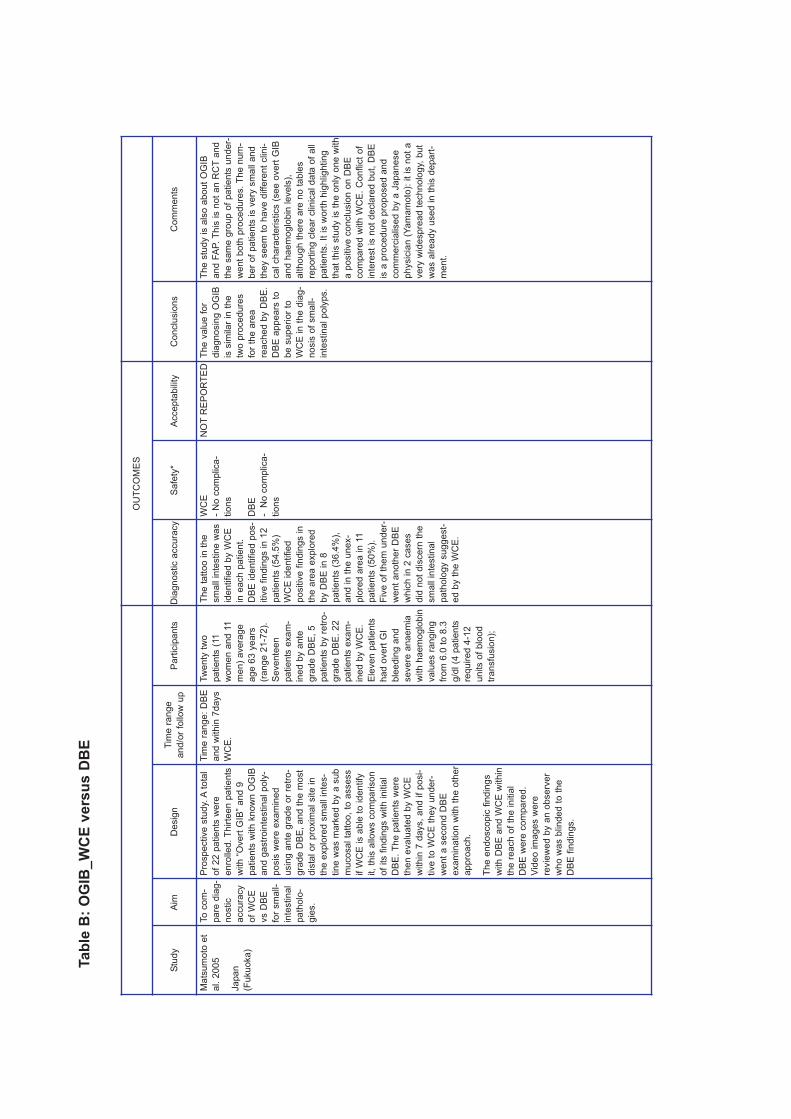
Ta
ble
B:
OG
IB_
WC
E v
ers
us
DB
E
OU
TC
OM
ES
Stu
dy
Aim
De
sig
nT
ime
ra
ng
e
an
d/o
r fo
llo
w u
pP
art
icip
ants
Dia
gnostic a
ccura
cy
Safe
ty*
Acce
pta
bility
Conclu
sio
ns
Com
men
ts
Ma
tsu
mo
to e
t
al. 2
00
5
Ja
pa
n
(Fu
ku
oka
)
To
co
m-
pa
re d
iag
-
no
stic
accu
racy
of
WC
E
vs D
BE
for
sm
all-
inte
stin
al
pa
tho
lo-
gie
s.
Pro
sp
ective
stu
dy. A
tota
l
of
22
pa
tie
nts
we
re
en
rolle
d.
Th
irte
en
pa
tie
nts
with
“O
ve
rt G
IB”
an
d 9
pa
tie
nts
with
kn
ow
n O
GIB
an
d g
astr
oin
testin
al p
oly
-
po
sis
we
re e
xa
min
ed
usin
g a
nte
gra
de
or
retr
o-
gra
de
DB
E,
an
d t
he
mo
st
dis
tal o
r p
roxim
al site
in
the
exp
lore
d s
ma
ll in
tes-
tin
e w
as m
ark
ed
by a
su
b
mu
co
sa
l ta
tto
o,
to a
sse
ss
if W
CE
is a
ble
to
id
en
tify
it,
this
allo
ws c
om
pa
riso
n
of
its f
ind
ing
s w
ith
in
itia
l
DB
E.
Th
e p
atie
nts
we
re
the
n e
va
lua
ted
by W
CE
with
in 7
da
ys,
an
d if
po
si-
tive
to
WC
E t
he
y u
nd
er-
we
nt
a s
eco
nd
DB
E
exa
min
atio
n w
ith
th
e o
the
r
ap
pro
ach
.
Th
e e
nd
osco
pic
fin
din
gs
with
DB
E a
nd
WC
E w
ith
in
the
re
ach
of
the
in
itia
l
DB
E w
ere
co
mpa
red
.
Vid
eo
im
ag
es w
ere
revie
we
d b
y a
n o
bse
rve
r
wh
o w
as b
lin
de
d t
o t
he
DB
E f
ind
ing
s.
Tim
e r
an
ge
: D
BE
an
d w
ith
in 7
da
ys
WC
E.
Tw
enty
tw
o
patients
(11
wom
en a
nd 1
1
men)
avera
ge
age 6
3 y
ears
(range 2
1-7
2).
Sevente
en
patients
exam
-
ined b
y a
nte
gra
de D
BE
, 5
patients
by r
etr
o-
gra
de D
BE
. 22
patients
exam
-
ined b
y W
CE
.
Ele
ven p
atients
had o
vert
GI
ble
edin
g a
nd
severe
anaem
ia
with h
aem
oglo
bin
valu
es r
angin
g
from
6.0
to 8
.3
g/d
l (4
patients
required 4
-12
units o
f blo
od
transfu
sio
n);
The t
att
oo in t
he
sm
all inte
stine w
as
identified b
y W
CE
in e
ach p
atient.
DB
E identified p
os-
itiv
e f
indin
gs in 1
2
patients
(54.5
%)
WC
E identified
positiv
e f
indin
gs in
the a
rea e
xplo
red
by D
BE
in 8
patients
(36.4
%),
and in t
he u
nex-
plo
red a
rea in 1
1
patients
(50%
).
Fiv
e o
f th
em
under-
went
anoth
er
DB
E
whic
h in 2
cases
did
not
dis
cern
the
sm
all inte
stinal
path
olo
gy s
uggest-
ed b
y t
he W
CE
.
WC
E
- N
o c
om
plica-
tions
DB
E
- N
o c
om
plica-
tions
NO
TR
EP
OR
TE
DT
he v
alu
e f
or
dia
gn
osin
g O
GIB
is s
imilar
in t
he
two
pro
ce
dure
s
for
the a
rea
rea
ch
ed b
y D
BE
.
DB
E a
ppe
ars
to
be s
upe
rior
to
WC
E in
the d
iag-
nosis
of
sm
all-
inte
stina
l p
oly
ps.
The
stu
dy is a
lso a
bou
t O
GIB
an
d F
AP.
This
is n
ot
an R
CT
an
d
the s
am
e g
rou
p o
f patie
nts
un
de
r-
we
nt
both
pro
ce
dure
s.
The
num
-
be
r o
f pa
tie
nts
is v
ery
sm
all a
nd
they s
eem
to
have
diffe
ren
t clin
i-
cal ch
ara
cte
ristics (
se
e o
vert
GIB
an
d h
aem
oglo
bin
levels
),
alth
ou
gh
there
are
no t
able
s
rep
ort
ing c
lea
r clinic
al data
of
all
patie
nts
. It is w
ort
h h
igh
lig
htin
g
that
this
stu
dy is t
he
on
ly o
ne w
ith
a p
ositiv
e c
onclu
sio
n o
n D
BE
com
pa
red
with W
CE
. C
on
flic
t o
f
inte
rest
is n
ot
decla
red b
ut, D
BE
is a
pro
ced
ure
pro
po
sed a
nd
com
me
rcia
lise
d b
y a
Ja
pane
se
ph
ysic
ian (
Yam
am
oto
): it
is n
ot
a
very
wid
esp
rea
d t
echno
log
y,
but
wa
s a
lrea
dy u
se
d in t
his
depa
rt-
men
t.
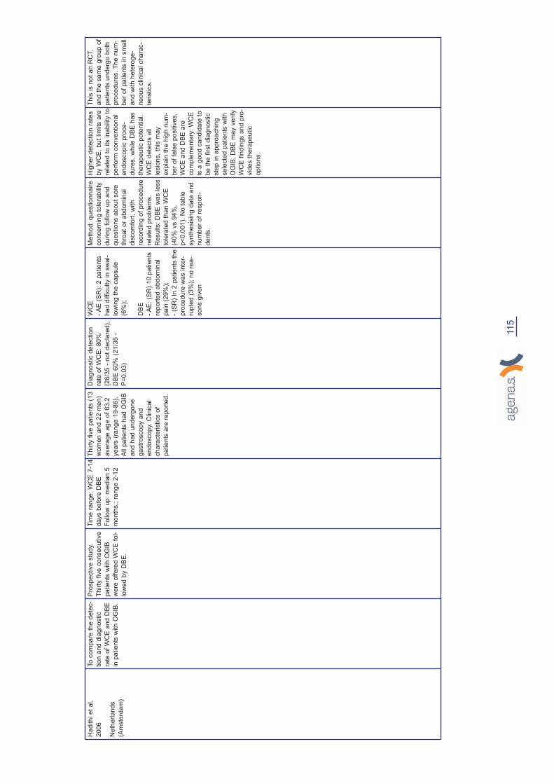
Ha
dith
i e
t a
l,
20
06
Ne
the
rla
nd
s
(Am
ste
rda
m)
To
co
mpa
re t
he
de
tec-
tio
n a
nd
dia
gn
ostic
rate
of
WC
E a
nd
DB
E
in p
atie
nts
with
OG
IB.
Pro
sp
ective
stu
dy.
Th
irty
fiv
e c
on
se
cu
tive
pa
tie
nts
with
OG
IB
we
re o
ffe
red
WC
E f
ol-
low
ed
by D
BE
.
Tim
e r
an
ge
: W
CE
7-1
4
da
ys b
efo
re D
BE
Fo
llo
w u
p:
me
dia
n 5
mo
nth
s,; r
an
ge
2-1
2
Thirty
fiv
e p
atients
(13
wom
en a
nd 2
2 m
en)
avera
ge a
ge o
f 63.2
years
(ra
nge 1
9-8
6).
All p
atients
had O
GIB
and h
ad u
nderg
one
gastr
oscopy a
nd
endoscopy.
Clinic
al
chara
cte
ristics o
f
patients
are
report
ed.
Dia
gnostic d
ete
ction
rate
of
WC
E:
80%
(28/3
5 -
not
decla
red),
DB
E 6
0%
(21/3
5 -
P=
0.0
3)
WC
E
- A
E (
SR
): 2
patien
ts
had
difficulty in s
wa
l-
low
ing
the
ca
psu
le
(6%
);
DB
E
- A
E:
(SR
) 10 p
atie
nts
report
ed a
bd
om
inal
pain
(29
%);
- (S
R)
In 2
pa
tients
th
e
pro
ced
ure
wa
s inte
r-
rupte
d (
3%
); n
o r
ea
-
sons g
iven
Meth
od:
qu
estionn
aire
co
ncern
ing
to
lera
bility
during
follow
up a
nd
questio
ns a
bou
t sore
thro
at
or
ab
do
min
al
dis
com
fort
, w
ith
reco
rdin
g o
f p
roce
dure
rela
ted
pro
ble
ms.
Results:
DB
E w
as le
ss
tole
rate
d t
ha
n W
CE
(40
% v
s 9
4%
,
p<
0.0
01
). N
o t
ab
le
syn
the
sis
ing d
ata
and
num
ber
of
resp
on
-
dents
.
Hig
her
de
tectio
n r
ate
s
by W
CE
, b
ut
lim
its a
re
rela
ted t
o its
ina
bility t
o
perf
orm
conve
ntiona
l
end
osco
pic
pro
ce-
dure
s,
wh
ile
DB
E h
as
thera
pe
utic p
ote
ntia
l.
WC
E d
ete
cts
all
lesio
ns,
this
ma
y
expla
in t
he
hig
h n
um
-
ber
of
fals
e p
ositiv
es.
WC
E a
nd D
BE
are
co
mple
me
nta
ry:
WC
E
is a
go
od c
an
did
ate
to
be t
he
first
dia
gn
ostic
ste
p in a
ppro
ach
ing
se
lecte
d p
atie
nts
with
OG
IB,
DB
E m
ay v
erify
WC
E f
indin
gs a
nd
pro
-
vid
es t
he
rap
eu
tic
optio
ns.
Th
is is n
ot
an
RC
T,
and t
he s
am
e g
roup
of
patie
nts
un
de
rgo
both
pro
ce
du
res.
The
num
-
ber
of
patie
nts
in
sm
all
and w
ith h
ete
rog
e-
neou
s c
lin
ica
l ch
ara
c-
teristics.
115
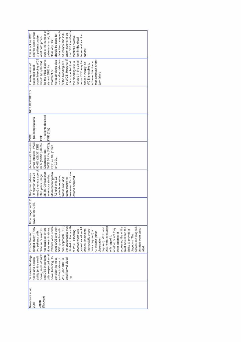
Na
ka
mu
ra e
t a
l,
20
06
Ja
pa
n
(Na
go
ya
)
To
asse
ss t
he
dia
g-
no
stic a
nd
acce
ss
ab
ility (
en
tire
sm
all
bo
we
l) r
ate
s o
f W
CE
an
d D
BE
in
pa
tie
nts
with
su
sp
ecte
d s
ma
ll
bo
we
l b
lee
din
g.
To
co
nsid
er
the
ro
les
an
d in
dic
atio
ns o
f
WC
E a
nd
DB
E in
sm
all b
ow
el b
lee
d-
ing
.
Pro
sp
ective
an
d
blin
de
d s
tud
y.
Th
irty
two
pa
tie
nts
with
ob
scu
re o
r o
ve
rt G
I
no
t lo
ca
lise
d b
y p
re-
vio
us e
xa
min
atio
ns.
Pa
tie
nts
we
re u
nd
er-
we
nt
WC
E a
nd
th
en
DB
E (
po
ssib
ly w
ith
du
al a
pp
roa
ch
. D
BE
)
the
en
do
sco
pis
t w
as
blin
de
d t
o t
he
re
su
lts
of
WC
E.
Ble
ed
ing
so
urc
es w
ere
ca
te-
go
rise
d a
s e
ith
er
A1
lesio
ns (
imm
ed
iate
ha
em
osta
tic p
roce
-
du
res r
eq
uire
d)
or
A2
le
sio
ns (
clo
se
ob
se
rva
tio
n
req
uire
d).
WC
E a
nd
DB
E w
ere
eva
lua
ted
with
re
ga
rd t
o
wh
eth
er
or
no
t th
ey
we
re c
apa
ble
of
acce
ssin
g t
he
en
tire
sm
all b
ow
el a
nd
th
e
ab
ility t
o p
rovid
e a
dia
gn
osis
. T
he
acce
ss a
nd
dia
gn
os-
tic r
ate
s w
ere
ca
lcu
-
late
d.
Tim
e r
an
ge
: W
CE
2
da
ys b
efo
re D
BE
.
Thirty
-tw
o p
atients
(11 w
om
en a
nd 2
1
men)
avera
ge a
ge o
f
58.5
years
, ra
nge
25-8
5.
Clinic
al char-
acte
ristics s
imilar.
Mean h
aem
oglo
bin
7.3
g/d
l w
ith 2
2
patients
requirin
g
transfu
sio
n a
nd
som
e r
eceiv
ing
treatm
ent.
Exclu
sio
n
crite
ria d
ecla
red.
Access r
ate
to e
ntire
sm
all inte
stine:
WC
E
90.6
% (
29/3
2)
DB
E
62%
(10/1
6 p
<0.0
5).
Dia
gnostic r
ate
:
WC
E 5
9.4
% (
19/3
2),
DB
E 4
2.9
% (
12/2
8
p=
0.3
0).
WC
E
- N
o c
om
plica
tions
DB
E
- 1 p
atien
ts d
eclined
DB
E (
3%
)
NO
TR
EP
OR
TE
DIn
ma
ny c
ase
s o
f
su
specte
d s
ma
ll
bow
el ble
edin
g W
CE
sh
ould
be s
ele
cte
d
for
the
initia
l dia
gno
-
sis
and
DB
E f
or
tre
atm
en
t o
r
his
topath
olo
gic
dia
g-
nosis
after
de
tectio
n
of
the b
leed
ing s
ite
by W
CE
. H
ow
ever
if
it is s
usp
ecte
d t
hat
the b
lee
din
g s
ite is
loca
ted
in
th
e d
ista
l
ile
um
, D
BE
ma
y b
e
ch
osen
in
itia
lly,
as
WC
E is u
na
ble
to
achie
ve
th
is d
ue
to
food
re
sid
ue
s o
r bat-
tery
fa
ilu
re
Th
is is n
ot
an R
CT
and t
he s
am
e g
roup
of
pa
tien
ts u
nd
er-
wen
t bo
th p
roce
-
dure
s;
the
num
ber
of
pa
tients
is s
ma
ll;
Not
cle
ar
wh
y D
BE
sh
ould
be u
se
d f
or
dis
tal ile
um
su
spe
ct-
ed le
sio
ns:
the
ind
i-
ca
tion s
eem
s t
o b
e
that
DB
E id
entifie
d a
Mecke
l’s d
ivert
icu
-
lum
in t
he
dis
tal
ile
um
, and
a c
olo
n
ca
ncer.

Xia
o-b
o e
t a
l,
20
07
CH
INA
(Sh
an
gh
ai)
To
eva
lua
te t
he
de
tectio
n r
ate
an
d
dia
gn
ostic a
ccu
racy
of
WC
E a
nd
DB
E
alo
ne
an
d c
om
bin
ed
,
for
pa
tie
nts
with
su
s-
pe
cte
d s
ma
ll b
ow
el
dis
ea
se
.
Pro
sp
ective
“ca
te-
go
rise
d”
stu
dy. A
tota
l
of
21
8 p
atie
nts
in
wh
om
it
ha
d n
ot
be
en
po
ssib
le t
o d
ete
rmin
e
ca
use
s o
f O
GIB
,
ab
do
min
al pa
in o
r
dia
rrh
oe
a u
sin
g c
on
-
ve
ntio
na
l d
iag
no
stic
pro
ce
du
res,
we
re c
at-
eg
orise
d in
to 2
gro
ups u
nd
erg
oin
g
bo
th W
CE
an
d D
BE
.
Pa
tie
nts
with
ne
ga
-
tive
or
eq
uiv
oca
l fin
d-
ing
s o
n W
CE
un
de
r-
go
DB
E a
nd
vic
e
ve
rsa
.
Tim
e r
an
ge
DB
E/W
CE
(if n
ega-
tive
, th
en
th
e a
ltern
a-
tive
): m
ea
n in
terv
al
tim
e b
etw
ee
n C
E a
nd
DB
E w
as 1
2.9
days
(2-5
0 d
ays).
Follow
up
: n
ot
rep
ort
ed (
just
3 p
atie
nts
ne
gative t
o
DB
E w
ere
fo
llow
ed
for
6 m
on
ths)
Tw
o h
undre
d a
nd
eig
hte
en p
atients
were
cate
gorised into
two g
roups u
nderg
o-
ing e
ither
CE
or
DB
.
Am
ong t
he 1
65
patients
(94 m
ale
, 71
fem
ale
; avera
ge a
ge
50.5
, ra
nge 1
6-8
7
years
) underg
oin
g
CE
first,
101 c
ases
had o
bscure
gastr
oin
-
testinal ble
edin
g a
nd
64 c
ases h
ad
obscure
abdom
inal
pain
or
dia
rrhoea.
Fifty
-thre
e p
atients
(32 m
ale
, 21 f
em
ale
;
avera
ge a
ge 4
6.8
years
, ra
nge 1
8-7
5
years
) underw
ent
DB
E f
irst;
inclu
din
g
15 c
ases w
ith
obscure
gastr
oin
testi-
nal ble
edin
g a
nd 3
8
with o
bscure
abdom
i-
nal pain
or
dia
rrhoea.
Dete
ction r
ate
- o
ver-
all:
WC
E 7
2%
(118/1
64),
DB
E
41.2
% (
21/5
1);
Dia
gnostic a
ccura
cy -
overa
ll:
WC
E 5
1.8
%
(85/1
64)
DB
E 3
9.2
%
(20/5
1).
Dete
ction
rate
- O
GIB
: W
CE
88%
(88/1
00)
DB
E
60%
(9/1
5;
P=
0.0
054).
Dia
gnostic a
ccura
cy -
OG
IB:
WC
E 6
5%
(65/1
00)
DB
E 3
9%
(20/5
1).
WC
E
- 2 p
atie
nts
re
tain
ed
the
ca
psule
at
low
er
oesop
hag
us a
nd o
ne
of
the
m r
enou
nced
contin
uin
g t
he
exa
mi-
nation
- 5 p
atie
nts
re
tain
ed
capsule
(T
)
- 2
of
the
m u
nd
er-
went
surg
ica
l re
moval
(S)
- C
apsu
le h
ad
not
reached
the
ca
ecum
by t
he e
nd
of
the
reco
rdin
g t
ime
in 4
4
patien
ts (
BE
)
DB
E
- 39 p
atie
nts
repo
rted
dis
co
mfo
rt a
fte
r D
BE
(SR
)
- 2 p
atie
nts
faile
d t
o
com
ple
te t
he D
BE
(due t
o p
art
ial g
as-
trecto
my);
(SR
)
NO
TR
EP
OR
TE
DT
he d
ete
ction
ra
te o
f
sm
all b
ow
el d
isease
s
with D
BE
is r
ela
tive
ly
low
er
than
tha
t w
ith
WC
E.
The
tw
o p
roce
-
dure
s a
re c
om
ple
-
me
nta
ry:
WC
E a
s ini-
tia
l dia
gn
ostic
app
roa
ch f
or
su
spect-
ed s
mall b
ow
el dis
-
ease
s e
spe
cia
lly f
or
OG
IB.
With
re
ga
rd t
o
the
rela
tively
hig
h
ind
efinite d
iagn
ostic
rate
(2
8%
) o
f sm
all
bow
el dis
ease a
nd
the
ina
bility t
o p
rovid
e
dia
gnostic s
am
pling,
DB
E s
till a
ppea
rs t
o
be a
via
ble
in
str
u-
me
nt
to c
om
ple
te
and
/or
co
nfirm
the
neg
ative
and
in
defi-
nite d
iagno
sis
ma
de
by W
CE
.
Th
is s
tud
y a
na
lyses
OG
IB a
nd
oth
er
ca
ses w
ith o
bscure
abd
om
ina
l pa
in a
nd
ch
ron
ic d
iarr
hoe
a.
It
app
ea
rs t
o b
e a
com
-
para
tive
stu
dy,
bu
t it
is n
ot
“ran
do
mis
ed
”.
Ran
do
mis
atio
n (
if
any)
is n
ot
cle
arly
expla
ined
. T
he
text
doe
s n
ot
expla
in w
hy,
of
the
in
itia
l g
rou
p o
f
216
pa
tien
ts,
165
und
erw
en
t W
CE
first,
while 5
3 u
nde
rwent
DB
E.
Clinic
al cha
rac-
teristics o
f th
e
patie
nts
are
not
repo
rte
d.
117
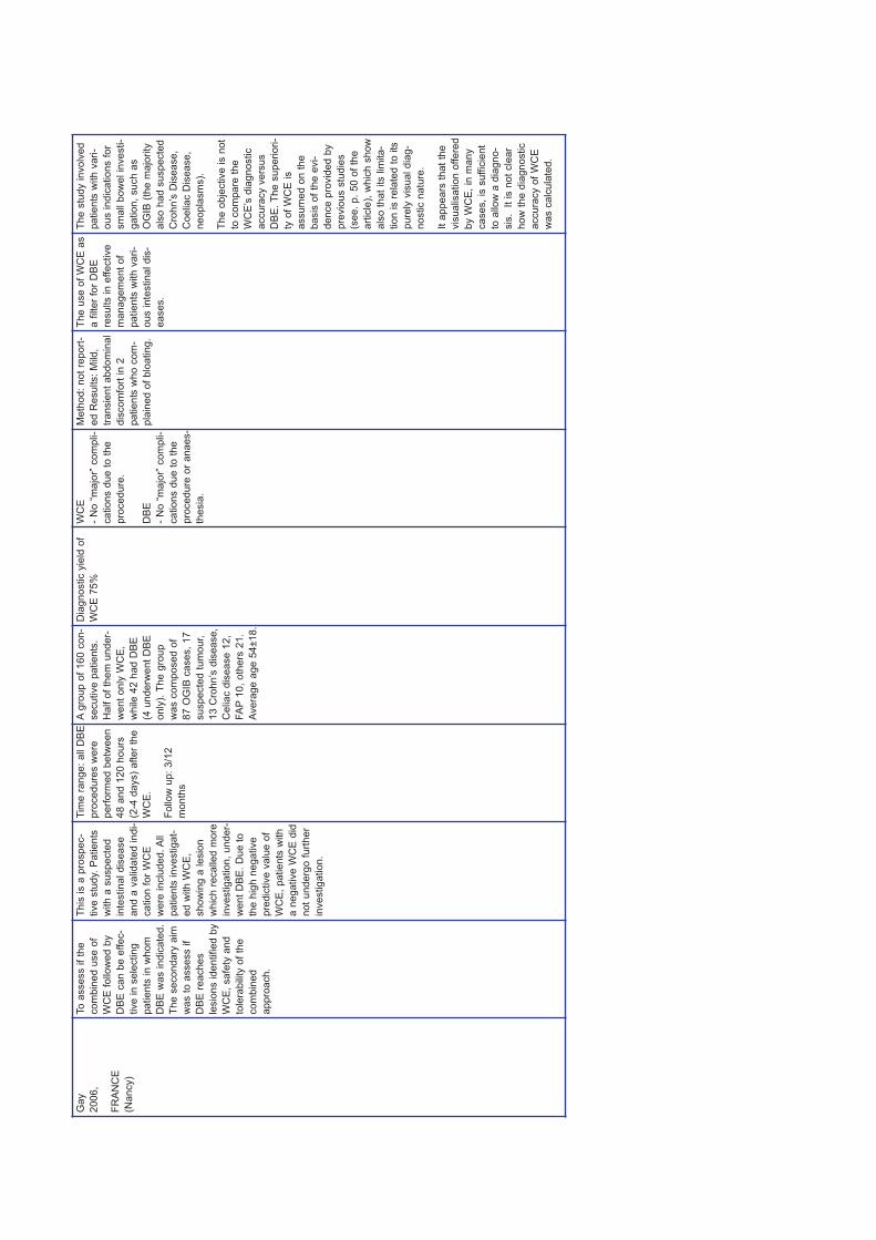
Ga
y
20
06
,
FR
AN
CE
(Na
ncy)
To
asse
ss if
the
co
mb
ine
d u
se
of
WC
E f
ollo
we
d b
y
DB
E c
an
be
effe
c-
tive
in
se
lectin
g
pa
tie
nts
in
wh
om
DB
E w
as in
dic
ate
d.
Th
e s
eco
nd
ary
aim
wa
s t
o a
sse
ss if
DB
E r
ea
ch
es
lesio
ns id
en
tifie
d b
y
WC
E,
sa
fety
an
d
tole
rab
ility o
f th
e
co
mb
ine
d
ap
pro
ach
.
Th
is is a
pro
sp
ec-
tive
stu
dy.
Pa
tie
nts
with
a s
usp
ecte
d
inte
stin
al d
ise
ase
an
d a
va
lid
ate
d in
di-
ca
tio
n f
or
WC
E
we
re in
clu
de
d. A
ll
pa
tie
nts
in
ve
stig
at-
ed
with
WC
E,
sh
ow
ing
a le
sio
n
wh
ich
re
ca
lle
d m
ore
inve
stig
atio
n,
un
de
r-
we
nt
DB
E.
Du
e t
o
the
hig
h n
eg
ative
pre
dic
tive
va
lue
of
WC
E,
pa
tie
nts
with
a n
eg
ative
WC
E d
id
no
t u
nd
erg
o f
urt
he
r
inve
stig
atio
n.
Tim
e r
an
ge
: a
ll D
BE
pro
ce
du
res w
ere
pe
rfo
rme
d b
etw
een
48
an
d 1
20
hours
(2-4
da
ys)
after
the
WC
E.
Fo
llo
w u
p:
3/1
2
mo
nth
s
Agro
up o
f 160 c
on-
secutive p
atients
.
Half o
f th
em
under-
went
only
WC
E,
while 4
2 h
ad D
BE
(4 u
nderw
ent
DB
E
only
). T
he g
roup
was c
om
posed o
f
87 O
GIB
cases,
17
suspecte
d t
um
our,
13 C
rohn’s
dis
ease,
Celiac d
isease 1
2,
FA
P10,
oth
ers
21.
Avera
ge a
ge 5
4±18.
Dia
gnostic y
ield
of
WC
E 7
5%
WC
E
- N
o “
majo
r” c
om
pli-
cations d
ue
to
the
pro
cedure
.
DB
E
- N
o “
majo
r” c
om
pli-
cations d
ue
to
the
pro
cedure
or
an
aes-
thesia
.
Meth
od
: n
ot
repo
rt-
ed R
esu
lts:
Mild,
transie
nt
abdo
min
al
dis
com
fort
in
2
pa
tients
who c
om
-
pla
ined o
f blo
ating
.
Th
e u
se o
f W
CE
as
a f
ilte
r fo
r D
BE
resu
lts in
effective
man
ag
em
ent
of
patie
nts
with
va
ri-
ou
s inte
stin
al dis
-
ea
ses.
Th
e s
tud
y in
volv
ed
patie
nts
with
va
ri-
ou
s ind
icatio
ns f
or
sm
all b
ow
el in
ve
sti-
ga
tion
, such a
s
OG
IB (
the m
ajo
rity
als
o h
ad
su
spe
cte
d
Cro
hn’s
Dis
ea
se
,
Coeliac D
isease
,
ne
op
lasm
s).
Th
e o
bje
ctive is n
ot
to c
om
pare
th
e
WC
E’s
dia
gno
stic
accu
racy v
ers
us
DB
E.
The
su
pe
riori-
ty o
f W
CE
is
assu
med
on t
he
ba
sis
of
the
evi-
de
nce
pro
vid
ed b
y
pre
vio
us s
tudie
s
(see
. p.
50
of
the
art
icle
), w
hic
h s
how
als
o t
hat
its lim
ita
-
tion
is r
ela
ted t
o its
pu
rely
vis
ual dia
g-
no
stic n
atu
re.
It a
ppe
ars
th
at
the
vis
ualisatio
n o
ffe
red
by W
CE
, in
man
y
case
s,
is s
uffic
ient
to a
llow
a d
iagn
o-
sis
. I
t is
no
t cle
ar
ho
w t
he d
iagno
stic
accu
racy o
f W
CE
was c
alc
ula
ted
.

Ta
ble
C:
OG
IB_
WC
E v
ers
us
Oth
ers
OU
TC
OM
ES
Stu
dy
Aim
Co
mpa
rato
rD
esig
nT
ime r
ange
an
d/o
r fo
llow
up
Part
icip
ants
Dia
gnostic a
ccura
cy
Safe
tyA
ccep
tab
ility
Co
nclu
sio
ns
Com
me
nts
Co
sta
ma
gn
a e
t a
l.
20
02
ITA
LY
(Ro
me
)
To
co
mpa
re t
he
clin
ica
l o
utc
om
es o
f
sm
all b
ow
el ra
di-
og
rap
hs -
SB
FT
-
with
WC
E
SB
ra
dio
gra
ph
s
(Ba
riu
m F
ollo
w
Th
rou
gh
)
Th
is is a
pro
sp
ec-
tive
stu
dy.
Co
nse
cu
tive
pa
tie
nts
with
su
s-
pe
cte
d s
ma
ll b
ow
el
dis
ea
se
we
re
en
rolle
d a
nd
un
de
r-
we
nt
ba
riu
m f
ollo
w-
thro
ug
h a
nd
WC
E,
to c
om
pa
re t
he
ir
fin
din
gs.
Th
e e
nd
o-
sco
pis
t w
as b
lin
de
d
to r
ad
iog
rap
h’s
resu
lts.
Tim
e r
ange:
Barium
follo
w-t
hro
ugh t
hen
afte
r 4 d
ays
en
do
scopy F
ollow
up
: n
ot
report
ed
Tw
enty
tw
o p
atients
with s
mall b
ow
el
dis
eases (
13 m
en,
8 w
om
en).
Tw
o
were
exclu
ded d
ue
to ile
al ste
nosis
.
Avera
ge a
ge 5
2.5
years
, ra
nge 2
9-7
8.
Thirte
en h
ad O
GIB
,
3 s
uspecte
d
Cro
hn’s
dis
ease/r
ecurr
ence,
1 s
uspecte
d s
arc
o-
ma r
ecurr
ence,
1
unexpla
ined c
hro
n-
ic d
iarr
hoea,
1 h
ad
FA
P,
1 h
ad s
mall
bow
el poly
posis
. A
ll
patients
had u
nder-
gone o
ther
dia
g-
nostic p
rocedure
s.
Contr
ain
dic
ations
report
ed
Fin
din
gs w
ere
cla
ssi-
fied a
s:
- dia
gnostic -
if
the f
ind-
ings c
ould
expla
in
sym
pto
ms o
r w
ere
late
r
confirm
ed b
y o
ther
meth
ods;
- suspic
ious -
if
furt
her
investigation is r
equire
d
to c
onfirm
clinic
al re
le-
vance;
- fa
iled -
if
no a
bnor-
mality
was d
ete
cte
d
despite a
definite indi-
cation o
f an e
xis
ting
lesio
n.
Barium
follow
-thro
ugh
was n
orm
al in
17
patients
and s
how
ed
ileal nodula
rity
in 3
patients
. W
CE
was n
or-
mal in
3 p
atients
and
show
ed p
ositiv
e f
ind-
ings in t
he r
em
ain
ing
17 p
atients
. T
he b
arium
stu
dy w
as c
onsid
ere
d:
- dia
gnostic in 4
(20%
)
patients
,
- suspic
ious in 0
and
- fa
iled in 1
5 (
73%
).
Capsule
endoscopy
was c
onsid
ere
d:
- dia
gnostic in 9
(45%
)
patients
,
- suspic
ious in 8
(40%
)
patients
,
- fa
iled in 3
(15%
)
patients
.
For
OG
IB,
the d
iagnos-
tic p
ote
ntial of
barium
follow
-tro
ugh w
as m
uch
wors
e c
om
pare
d w
ith
WC
E (
5%
vs 3
1%
,
P<
0.0
5).
WC
E:
batt
ery
fail-
ure
in 1
patien
t.
SB
FT:
no
pro
ble
ms
rep
ort
ed
NO
TR
EP
OR
TE
DW
CE
fou
nd t
o b
e
sup
erio
r to
sm
all
bo
wel ra
dio
gra
ph
for
evalu
atio
n o
f
sm
all b
ow
el d
is-
ea
se
.
The
stu
dy is a
bout
OG
IB,
susp
ecte
d
Cro
hn
’s d
ise
ase
and
Fam
ilia
r
Ad
en
om
ato
us
Po
lyp
osis
.
(Co
nsid
ering
Cro
hn
’s d
ise
ase
and
FA
Pth
e n
um
-
ber
of
pa
tien
ts w
as
less t
han
10
) It
is
not
cle
arly s
tate
d
whic
h o
ther
dia
g-
nostic p
roced
ure
was u
sed
fo
r O
GIB
pa
tien
ts.
Data
in
the
te
xt
did
not
co
r-
respo
nd
with
data
in t
ab
les.
119

Ha
ra e
t a
l.
20
03
Lo
ca
tio
n n
ot
rep
ort
ed
To
co
mpa
re W
CE
fin
din
gs w
ith
ba
ri-
um
stu
die
s o
r
co
mp
ute
d t
om
og
-
rap
hy (
CT
) in
pa
tie
nts
with
no
evid
en
ce
of
sm
all
bo
we
l str
ictu
re a
t
ba
riu
m e
xa
min
a-
tio
n.
CT
an
d b
ariu
m
stu
die
s
Th
is is a
re
tro
-
sp
ective
stu
dy.
Fifty
tw
o p
atie
nts
un
de
rwe
nt
WC
E,
su
bse
qu
en
tly 3
6
un
de
rwe
nt
SB
FT,
4 e
nte
rocly
sis
,
an
d 1
9 C
To
f
ab
do
me
n a
nd
pe
lvis
. Im
ag
ing
resu
lts w
ere
re
tro
-
sp
ective
ly
revie
we
d a
nd
co
mpa
red
with
WC
E,
sta
nd
ard
en
do
sco
py,
an
d
su
rgic
al re
su
lts. A
rad
iolo
gis
t, w
ho
wa
s n
ot
blin
de
d
to t
he
WC
E
resu
lts,
retr
osp
ec-
tive
ly r
evie
we
d
dis
cre
pa
ncie
s
be
twe
en
WC
E
an
d im
ag
ing
exa
min
atio
ns.
Pro
po
rtio
n o
f p
os-
itiv
e f
ind
ing
s a
t
WC
E w
as c
om
-
pa
red
with
pro
-
po
rtio
n o
f p
ositiv
e
fin
din
gs a
t b
ariu
m
stu
die
s a
nd
CT
in
the
sa
me
pa
tie
nts
.
Sta
tistica
l sig
nifi-
ca
nce
wa
s c
alc
u-
late
d w
ith
McN
em
ar
sta
tis-
tic.
Tim
e r
ange:
Barium
stu
die
s
and/o
r C
Tw
ithin
6 m
onth
s (
either
befo
re o
r after)
of
CE
.
Fifty
tw
o p
atients
,
31 m
en,
avera
ge
age 6
4 y
ears
,
range 2
1-9
2 a
nd
21 w
om
en,
aver-
age a
ge 6
3 y
ears
,
range 2
8-8
2
years
. F
ort
y t
hre
e
patients
under-
went
WC
E f
or
OG
IB
Barium
exam
ina-
tion f
indin
gs w
ere
positiv
e in o
ne
(3%
) of
40
patients
; W
CE
findin
gs w
ere
positiv
e in 2
2
(55%
) (P
<0.0
01).
CT
dem
onstr
ate
d
sm
all-b
ow
el find-
ings in f
our
(21%
)
of
19 p
atients
, but
WC
E d
em
onstr
at-
ed f
indin
gs in 1
2
(63%
) of
19
patients
(P
=0.0
2).
The m
ost
com
-
mon C
E f
indin
gs,
angio
ecta
sia
(11
cases),
was n
ot
dete
cte
d a
t any
imagin
g s
tudy.
More
ulc
ers
(8)
were
dete
cte
d
with W
CE
than
with b
arium
stu
dy
(one o
f eig
ht)
and
CT
(thre
e o
f six
).
At
WC
E,
thre
e o
f
five s
urg
ically
confirm
ed m
asses
(carc
inoid
, in
tus-
susceptions,
lym
-
phangio
ma)
were
identified,
but
two
jeju
nal tu
mours
were
not
dete
cte
d
in a
patient
with
poor
bow
el pre
pa-
ration. A
t barium
stu
dy,
no m
asses
(zero
of
five)
were
dete
cte
d;
at
CT,
one o
f fo
ur
mass-
es w
as d
ete
cte
d.
NO
TR
EP
OR
TE
DN
OT
RE
PO
RT
ED
In p
atien
ts w
itho
ut
a s
mall-b
ow
el
str
ictu
re a
t ba
rium
stu
dy,
more
sm
all-b
ow
el dis
-
eases w
ere
foun
d
with W
CE
wh
en
fin
din
gs w
ere
re
t-
rosp
ectively
co
m-
pa
red
with b
ariu
m
exam
ina
tion
and
CT
find
ing
s.
Neve
rth
ele
ss
lesio
n lo
calisatio
n
and e
va
lua
tion
ca
n b
e d
ifficult
with W
CE
tha
t
ca
nno
t m
ea
sure
abno
rma
lity
or
co
ntr
ol th
e c
am
-
era
wh
en
the
abno
rma
lity
wh
en
ide
ntifie
d.
It is d
if-
ficu
lt t
o lo
calise
pre
cis
ely
the
abno
rma
lity
dete
cte
d a
fter
WC
E.
Th
e t
ime r
ang
e
be
twe
en o
ne
dia
gn
ostic e
xa
mi-
na
tio
n a
nd a
noth
-
er
is n
ot
cle
ar.

*D
ata
fro
m t
hose s
tudie
s w
hic
h a
ssess W
CE
for
mix
ed indic
ations o
r diffe
rent
dia
gnostic m
odalities w
ere
inclu
ded o
nly
if
they f
ulfille
d o
ur
inclu
sio
n c
rite
ria.
*S
AF
ET
Y:
Advers
e e
vents
(A
E):
none, self r
esolv
ing s
ym
pto
ms (
SR
: e.g
. not sw
allow
ing c
apsule
), thera
py (
T. e.g
. R
ete
ntion o
f capsule
), s
urg
ery
(S
. e.g
. R
ete
ntion o
f capsule
),
mort
ality
(M
). T
echnic
al P
roble
m (
TP
): f
ailure
of
battery
(B
F),
battery
expired (
BE
)
Go
lde
r e
t a
l. 2
00
6
Ge
rma
ny (
Re
ge
nsb
urg
)
To
asse
ss t
he
dia
gn
os-
tic y
ield
of
WC
E w
ith
MR
en
tero
cly
sis
in
th
e
de
tectio
n o
f sm
all b
ow
el
pa
tho
log
ies.
MR
en
tero
-
cly
sis
Th
is is a
pro
sp
ective s
tudy. A
tota
l o
f 3
6 p
atie
nts
were
inclu
de
d in
th
e s
tud
y.
Ind
ica
tio
ns f
or
ima
gin
g o
f th
e
sm
all b
ow
el w
ere
: p
roven o
r
su
sp
ecte
d s
ma
ll b
ow
el
Cro
hn
’s d
ise
ase
, o
bscure
ga
str
oin
testin
al b
lee
din
g a
nd
tum
ou
r su
rve
illa
nce
. T
here
we
re 1
4pa
tie
nts
with
OG
IC,
16
with
CD
, a
nd
2 s
uspecte
d
CD
. T
he
re
ma
inin
g p
atients
ha
d F
AP
an
d t
um
ou
rs.
Diffe
ren
ce
s in
th
e d
ete
ction
of
pa
tho
log
ica
l fin
din
gs in t
he
sm
all b
ow
el w
ere
ca
lcula
ted
usin
g t
he
McN
em
ar
test.
Ap
va
lue
of
<0
.05
wa
s c
onsid
-
ere
d s
ign
ific
an
t.
Tim
e r
ange:
not
report
ed
Thirty
six
patients
(19 w
om
en
and 1
7 m
en)
avera
ge a
ge 4
7
years
, ra
nge 1
8-8
4.
Ä y
ield
: U
ppe
r sm
all b
ow
el
77.1
WC
E (
27/3
5)
MR
72
.2
(26/3
6);
Mid
dle
sm
all b
ow
el
WC
E 6
2.9
(22/3
5)
MR
77
.8
(28.3
6)
Lo
wer
sm
all b
ow
el
WC
E 3
7.1
(13/3
5)
MR
77
.8
(28/3
6).
Malfu
ncti
on
ing
of
WC
E.
in
1 p
atie
nt
NO
TR
EP
OR
T-
ED
Th
e r
esu
lts
de
mon
str
ate
tha
t W
CE
is
sup
erior
to M
R
for
Cro
hn’s
dis
ea
se
whe
n
con
sid
erin
g t
he
firs
t a
nd s
ec-
on
d s
ectio
n o
f
the
sm
all
bo
we
l, b
ut
not
for
third
se
c-
tion
The
stu
dy
is a
lso
ab
out
Cro
hn
’s d
is-
ea
se
Sa
pe
ras e
t a
l.,
20
07
Spa
in (
Ba
rce
lon
a)
Co
mpa
riso
n o
f th
e d
iag
-
no
stic y
ield
s W
CE
vs
Co
mp
ute
d T
om
og
rap
hy
(CT
) o
r S
tan
da
rd
An
gio
gra
ph
y (
AN
GIO
)
in p
atie
nts
with
OG
IB.
Co
mp
ute
d
To
mo
gra
ph
y
(CT
) o
r sta
n-
da
rd m
ese
n-
teric a
ng
iog
ra-
ph
y (
AN
GIO
)
Th
is is a
pro
sp
ective s
tudy.
Co
nse
cu
tive
pa
tie
nts
adm
it-
ted
fo
r O
GIB
un
de
rwent
both
CT
an
d A
NG
IO,
follow
ed b
y
WC
E p
erf
orm
ed
blin
dly
by
ind
ep
en
de
nt
exa
min
ers
with-
in t
he
fo
llo
win
g 7
da
ys.
Tim
e r
ange:
WC
E w
ithin
7 d
ays
Tw
enty
eig
ht
patients
(12
wom
en a
nd 1
6 m
en)
avera
ge
age 7
4 y
ears
, ra
nge 3
6-8
5.
Ä y
ield
: U
ppe
r sm
all b
ow
el
77.1
WC
E (
27/3
5)
MR
72
.2
(26/3
6);
Mid
dle
sm
all b
ow
el
WC
E 6
2.9
(22/3
5)
MR
77
.8
(28.3
6)
Lo
wer
sm
all b
ow
el
WC
E 3
7.1
(13/3
5)
MR
77
.8
(28/3
6).
NO
T
RE
PO
RT
-
ED
NO
TR
EP
OR
T-
ED
WC
E d
ete
cte
d
more
lesio
ns
tha
n C
TA
or
AN
GIO
in
patie
nts
with
OG
IB.
Ha
rtm
an
n e
t a
l. 2
00
5
Ge
rma
ny (
Be
rlin
)
To
co
mpa
re d
iag
no
stic
yie
ld o
f W
CE
vs
Intr
ao
pe
rative
En
tero
sco
py (
IE)
Intr
ao
pe
rative
En
tero
sco
py
(IE
)
Th
is is a
pro
sp
ective s
tudy.
Fo
rty s
eve
n c
on
se
cutive
pa
tie
nts
with
ob
scu
re G
I
ble
ed
ing
fro
m t
wo
gastr
oen-
tero
log
y c
en
tre
s w
ere
inclu
d-
ed
.
Th
e s
tan
da
rd e
va
luation o
f
the
se
pa
tie
nts
in
clu
ded u
pper
en
do
sco
py,
co
lon
oscopy w
ith
a r
etr
og
rad
e e
xa
min
ation o
f
the
dis
tal ile
um
, a
nd
push
en
tero
sco
py.
Tim
e r
ange:
WC
E a
nd
after
6 d
ays
IE.
Fort
y s
even p
atients
(17
wom
en a
nd 3
0 m
en)
avera
ge
age o
f 61±17 y
ears
, ra
nge
18-8
8.
Ä y
ield
: W
CE
overt
on
goin
g
ble
edin
g 1
00%
(11/1
1)
overt
pre
vio
us b
leed
ing 6
7.0
%
(16/2
4)
occu
lt b
lee
din
g
67.0
% (
8/1
2);
IE
overt
on
go-
ing b
leed
ing 1
00
% (
11
/11)
overt
pre
vio
us b
lee
din
g
70.8
% (
17
/24)
occu
lt b
lee
d-
ing 5
0.0
% (
6/1
2);
WC
E:
IE:
AE
(M
) 1
patie
nt
die
d o
f
pe
rito
nitis
afte
r
laparo
to-
my.
NO
TR
EP
OR
T-
ED
WC
E
Dia
gn
ostic
yie
ld v
arie
s
acco
rdin
g t
o
the
type
of
ble
ed
ing.
Auth
ors
su
g-
ge
st
the
use o
f
WC
E f
or
patie
nts
with
OG
IB,
iron
de
ficie
ncie
s,
an
d f
or
those
ne
ga
tive t
o
bid
ire
ction
al
en
do
scop
y.
No
t an
RC
T.
NP
P
an
d P
PV
we
re c
alc
u-
late
d.
But
the
IE
was
use
d a
s t
he
refe
rence
sta
nd
ard
.
121

Ta
ble
D:
Cro
hn
’s d
ise
as
e_
WC
E v
ers
us
Oth
ers
OU
TC
OM
ES
Stu
dy
Aim
Co
mpa
rato
rD
esig
nT
ime r
ange
and/o
r fo
llow
up
Part
icip
ants
Dia
gnostic a
ccura
cy
Safe
tyA
ccep
tab
ility
Co
nclu
sio
ns
Co
mm
en
ts
Bu
ch
ma
n e
t a
l.
20
04
,
US
A(C
hic
ag
o)
To
co
mpa
re t
he
dia
gn
ostic y
ield
of
the
WC
E w
ith
SB
FT
an
d
En
tero
cly
sis
in
CD
pa
tie
nts
.
SB
FT
Th
is is a
pro
sp
ec-
tive
blin
de
d s
tudy.
Th
e g
astr
oe
n-
tero
log
ist
wa
s n
ot
aw
are
of
the
SB
FT
resu
lts.
Co
nse
cu
tive
pa
tie
nts
with
su
s-
pe
cte
d r
ecu
r-
ren
ce
of
CD
on
the
ba
sis
of
ab
do
min
al pa
in,
dia
rrh
oe
a,
an
ae
mia
, a
nd
/or
art
hra
lgia
s.
SB
FT
wa
s p
erf
orm
ed
an
d t
ho
se
pa
tie
nts
with
str
ic-
ture
s a
nd
pro
xi-
ma
l b
ow
el d
ila
ta-
tio
n w
ere
exclu
d-
ed
. F
or
SB
FT,
resu
lts w
ere
gra
d-
ed
no
rma
l (0
), 1
(<f
5 c
m),
2 (
<5
-
10
), 3
(>
10
cm
).
Fo
r W
CE
we
re
gra
de
d n
orm
al
(0),
1 (
erith
em
a,
iso
late
d v
illi lo
ss)
2 (
ero
sio
n,
no
ulc
er)
3 (
ulc
ers
,
sp
on
tan
eo
us
ble
ed
ing
, a
nd
/or
str
ictu
re).
Tim
e r
ange:
SB
FT
and W
CE
after
1 w
eek.
Follow
up:
NO
T
RE
PO
RT
ED
Thirty
consecu-
tive p
atients
with
clinic
ally s
us-
pecte
d C
D
recurr
ence 2
2
fem
ale
, 8 m
ale
.
Mean a
ge 3
6.9
years
, ra
nge
21-8
0.
Active C
D w
as v
isu-
alised in 2
1 o
f 30
patients
with W
CE
and in 2
0 o
f 30 w
ith
SB
FT.
Com
ple
te
agre
em
ent
occurr
ed
in 1
3 o
f 30 p
atients
.
In 1
3 o
f 17 s
core
s
diffe
red b
y 1
gra
de.
SR
: In
2 (
7%
)
patie
nts
WC
E
wa
s r
eta
ine
d f
or
respective
ly,
9
mo
nth
s a
nd
3
we
eks.
Meth
od:
a m
ulti-
ple
cho
ice
pa
tient’s s
atisfa
c-
tio
n q
ue
stio
nna
ire
adm
inis
tra
ted t
o
all o
nce
WC
E
was c
om
ple
ted
.
All q
uestio
ns a
nd
answ
ers
are
rep
ort
ed
in
te
xt.
Results:
WC
E is
the
pre
ferr
ed
pro
-
ce
dure
com
pa
red
to S
BF
T.
Resu
lts s
ug
ge
st
that
WC
E a
nd
SB
FT
have
sim
i-
lar
sen
sitiv
ity a
nd
accura
cy f
or
the
dia
gn
osis
of
CD
.
Th
e t
ime r
an
ge
betw
ee
n t
he t
wo
pro
ce
dure
s is
quite s
ho
rt a
nd
pa
tho
log
y is n
ot
su
ppo
se
d t
o
ch
ang
e. A
uth
ors
sta
te t
ha
t W
CE
has b
ee
n s
how
n
to b
e a
safe
an
d
effe
ctive t
oo
l fo
r
the d
iag
nosis
of
CD
invo
lvin
g t
he
sm
all in
testin
e.
Capsu
le r
ete
ntio
n
is g
rate
r in
pa
tients
with C
D.
Accord
ing
to
the
auth
ors
this
, in
so
me
ca
se
s,
rath
er
tha
n a
co
mp
lica
tio
n,
sh
ould
be v
iew
ed
as d
iagn
ostic
tool.
Th
e s
tud
y is n
ot
a
ran
do
mis
ed
tria
l,
it is a
stu
dy w
ith
man
y s
hort
com
-
ings.
Pa
tien
t
acce
pta
bility:
am
ong
the
re
po
rt-
ed
que
stion
s
the
re is o
ne
whic
h a
ddre
sses
this
issue.
The
au
thors
rep
ort
tha
t W
CE
is t
he
pre
ferr
ed
me
thod
,
an
d t
hat
is less
invasiv
e a
nd
tim
e
con
sum
ing.
Th
ose t
wo
la
st
sen
tences a
re n
ot
sup
po
rte
d b
y e
vi-
de
nce
fro
m t
he
qu
estio
nn
aire
su
r-
vey a
nd s
ee
m t
o
be
auth
or’s
assu
mptio
ns.
No
resu
lts t
able
s a
re
rep
ort
ed
for
an
y
dim
ensio
n.
Th
e
ab
str
act
sta
tes
tha
t “t
welv
e
patie
nts
we
re
exclu
de
d f
or
sm
all b
ow
el
ob
str
uction
”, b
ut
text
and
data
are
no
t co
nsis
tent
with t
his
. S
om
e o
f
the
au
tho
r’s
co
n-
clu
sio
ns a
re n
ot
sup
po
rte
d b
y
da
ta.
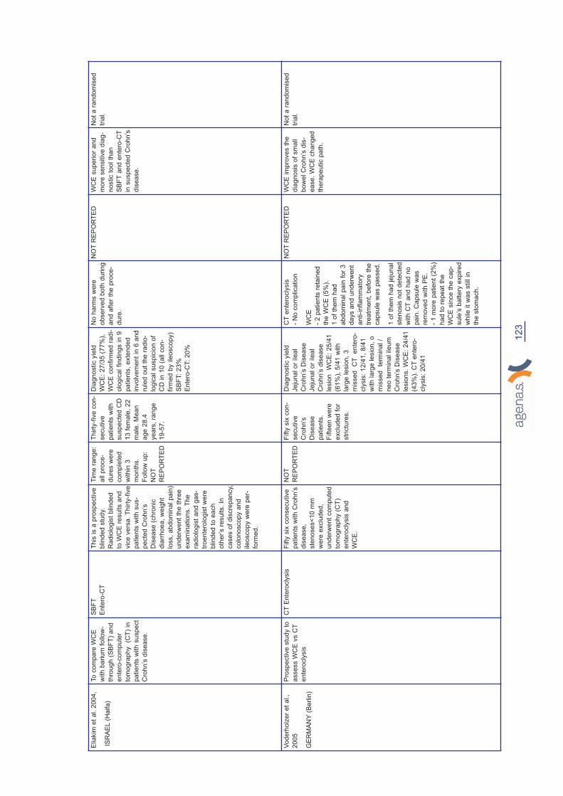
Elia
kim
et
al. 2
00
4,
ISR
AE
L(H
aifa
)
To
co
mpa
re W
CE
with
ba
riu
m f
ollo
w-
thro
ug
h (
SB
FT
) a
nd
en
tero
-co
mp
ute
r
tom
og
rap
hy
(CT
) in
pa
tie
nts
with
su
sp
ect
Cro
hn
’s d
ise
ase
.
SB
FT
En
tero
-CT
Th
is is a
pro
sp
ective
blin
de
d s
tud
y.
Ra
dio
log
ist
blin
de
d
to W
CE
re
su
lts a
nd
vic
e v
ers
a.
Th
irty
-fiv
e
pa
tie
nts
with
su
s-
pe
cte
d C
roh
n’s
Dis
ea
se
(ch
ron
ic
dia
rrh
oe
a,
we
igh
t
loss,
ab
do
min
al pa
in)
un
de
rwe
nt
the
th
ree
exa
min
atio
ns.
Th
e
rad
iolo
gis
t a
nd
ga
s-
tro
en
tero
log
ist
we
re
blin
de
d t
o e
ach
oth
er’s
re
su
lts.
In
ca
se
s o
f d
iscre
pa
ncy,
co
lon
osco
py a
nd
ile
osco
py w
ere
pe
r-
form
ed
.
Tim
e r
ange:
all p
roce-
dure
s w
ere
com
ple
ted
within
3
month
s.
Follow
up:
NO
T
RE
PO
RT
ED
Thirty
-fiv
e c
on-
secutive
patients
with
suspecte
d C
D
13 f
em
ale
, 22
male
. M
ean
age 2
8.4
years
, ra
nge
19-5
7.
Dia
gnostic y
ield
WC
E:
27/3
5 (
77%
),
WC
E c
onfirm
ed r
adi-
olo
gic
al findin
gs in 9
patients
, exte
nded
involv
em
ent
in 6
and
rule
d o
ut
the r
adio
-
logic
al suspic
ion o
f
CD
in 1
0 (
all c
on-
firm
ed b
y ile
oscopy)
SB
FT:
23%
Ente
ro-C
T:
20%
No h
arm
s w
ere
observ
ed
bo
th d
uring
and a
fter
the
pro
ce
-
dure
.
NO
TR
EP
OR
TE
DW
CE
sup
erior
and
mo
re s
en
sitiv
e d
iag-
no
stic t
oo
l th
an
SB
FT
and
ente
ro-C
T
in s
usp
ecte
d C
roh
n’s
dis
ease
.
No
t a
ran
do
mis
ed
tria
l.
Vo
de
rho
lze
r e
t a
l.,
20
05
GE
RM
AN
Y(B
erlin
)
Pro
sp
ective
stu
dy t
o
asse
ss W
CE
vs C
T
en
tero
cly
sis
CT
En
tero
cly
sis
Fifty
six
co
nse
cu
tive
pa
tie
nts
with
Cro
hn
’s
dis
ea
se
,
ste
no
se
s<
10
mm
we
re e
xclu
de
d,
un
de
rwe
nt
co
mp
ute
d
tom
og
rap
hy (
CT
)
en
tero
cly
sis
an
d
WC
E.
NO
T
RE
PO
RT
ED
Fifty
six
con-
secutive
Cro
hn’s
Dis
ease
patients
.
Fifte
en w
ere
exclu
ded f
or
str
ictu
res.
Dia
gnostic y
ield
Jeju
nal or
ileal
Cro
hn’s
Dis
ease
Jeju
nal or
ileal
Cro
hn’s
dis
ease
lesio
n
WC
E:
25/4
1
(61%
), 5
/41 w
ith
larg
e lesio
n,
3
mis
sed
CT
ente
ro-
cly
sis
: 12/4
1,
8/4
1
with larg
e lesio
n,
o
mis
sed
term
inal /
neo t
erm
inal ileum
Cro
hn’s
Dis
ease
lesio
ns.
WC
E:
24/4
1
(43%
). C
Tente
ro-
cly
sis
: 20/4
1
CT
ente
rocly
sis
- N
o c
om
plication
WC
E
- 2 p
atie
nts
re
tain
ed
the W
CE
(5
%).
1 o
f th
em
ha
d
abdo
min
al pain
fo
r 3
days a
nd
unde
rwe
nt
anti-infla
mm
ato
ry
treatm
ent,
befo
re t
he
capsule
was p
assed
.
1 o
f th
em
ha
d jeju
nal
ste
nosis
no
t d
ete
cte
d
with C
Tan
d h
ad n
o
pain
. C
apsu
le w
as
rem
ove
d w
ith P
E.
- 1 m
ore
pa
tie
nt
(2%
)
had t
o r
epea
t th
e
WC
E s
ince
th
e c
ap-
sule
’s b
att
ery
exp
ire
d
while it
was s
till in
the s
tom
ach
.
NO
TR
EP
OR
TE
DW
CE
im
pro
ve
s t
he
dia
gn
osis
of
sm
all
bo
wel C
rohn
’s d
is-
ea
se
. W
CE
ch
ang
ed
thera
pe
utic p
ath
.
No
t a
ran
do
mis
ed
tria
l.
123

Alb
ert
20
05
,
GE
RM
AN
Y
(Ha
lle
, S
aa
le)
To
asse
ss d
iag
-
no
stic y
ield
of
WC
E in
Cro
hn
’s
dis
ea
se
. B
lin
d
op
era
tors
Flu
oro
sco
py,
MR
I,
En
tero
cly
sis
Fifty
tw
o c
on
se
c-
utive
pa
tie
nts
we
re in
ve
stig
ate
d
by M
RI, f
luo
-
rosco
py a
nd
if
the
re w
as
no
bo
we
l o
bstr
uctio
n,
with
WC
E.
Tim
e r
ange:
MR
I
and E
nte
rocly
sis
follow
ed b
y W
CE
within
10 d
ays (
in
1 p
atient:
6
weeks.
Follow
up:
NO
TR
EP
OR
TE
D
Eig
hty
one w
ith
suspecte
d
Cro
hn’s
dis
ease
52 e
ligib
le a
ged
18 t
o 7
2.
Dia
gnostic y
ield
:
CE
13/1
4 9
2.9
%,
MR
I 11/1
4 7
8.6
%.
BU
Tente
rocly
sis
vs M
RI
and e
ntr
o-
cl vs C
E (
0.0
1
and 0
.002);
Pt
com
fort
, safe
ty
MR
I
- 2 p
atien
ts (
8%
)
had c
lau
str
op
ho-
bia
an
d 1
pa
tient
(4%
) re
fused
to
unde
rgo
MR
I.
FE
- 2 p
atien
ts (
8%
)
did
no
t to
lera
te
the
tra
nsn
asal
tub
e.
WC
E
- 1 p
atien
t (4
%)
reta
ine
d c
apsu
le
in a
sm
all b
ow
el
str
ictu
re u
nde
tect-
ed o
n a
bd
om
inal
ultra
so
und
an
d
Ente
rocly
sis
.
NO
TR
EP
OR
TE
DW
CE
an
d M
RI
co
mp
lim
enta
ry
techn
iqu
es.
Not
a r
an
do
mis
ed
tria
l.
Ho
Ch
on
g e
t a
l.,
20
05
AU
ST
RA
LIA
(Me
lbo
urn
e)
To
co
mpa
re d
iag
-
no
stic y
ield
of
WC
E v
s P
E a
nd
En
tero
cly
sis
in
two
gro
ups.
Gro
up
1 h
isto
ry o
f
Cro
hn
’s d
ise
ase
;
Gro
up
2 n
o h
isto
-
ry o
f C
roh
n’s
dis
-
ea
se
.
PE
, E
nte
rocly
sis
Pa
tie
nts
wh
o
we
re s
usp
ecte
d
to h
ave
sm
all-
bo
we
l C
roh
n’s
dis
ea
se
we
re
pro
sp
ective
ly
eva
lua
ted
with
pu
sh
en
tero
sco
py,
en
tero
cly
sis
, a
nd
ca
psu
le
en
do
sco
py.
Ea
ch
exa
min
er
wa
s
blin
de
d t
o r
esu
lts
of
oth
er
inve
stig
a-
tio
ns.
Re
ferr
ing
do
cto
rs w
ere
req
uire
d t
o c
om
-
ple
te a
qu
estio
n-
na
ire
be
fore
an
d
afte
r th
e in
ve
sti-
ga
tio
n.
Tim
e r
ange:
WC
E
2 w
eeks a
fter
PE
and e
nte
rocly
sis
if
no s
tric
ture
dete
cte
d.
Gro
up 1
: 22
patients
(17
wom
en a
nd 5
men)
avera
ge
age o
f 39.8
years
(range 1
7-7
8).
Gro
up 2
: 21
patients
(11
wom
en a
nd 1
0
men)
avera
ge
age o
f 35 y
ears
(range 2
0-8
0).
In g
roup 1
: W
CE
dete
cte
d m
ore
ero
sio
ns t
han t
he
oth
er
two e
xam
i-
nations (
p<
0.0
01)
In g
roup 2
: a n
ew
dia
gnosis
of
Cro
hn d
isease
there
was n
o s
ig-
nific
ant
diffe
rence
yie
ld c
om
pare
d
with P
E a
nd
Ente
rocly
sis
WC
E
- 1 p
atien
t (5
%)
was u
na
ble
to
sw
allo
w t
he c
ap-
su
le a
nd
endo
-
sco
pic
pla
cem
ent
in t
he d
uo
denu
m
was r
eq
uired.
PE
- N
o c
om
plica
tion
Ente
rocly
sis
- F
ailed
in
6
pa
tients
(29
%)
NO
TR
EP
OR
TE
DW
CE
ha
s a
hig
h-
er
yie
ld t
han
PE
and E
nte
rocly
sis
in p
atie
nt
with
Cro
hn’s
dis
ea
se
whe
n s
mall-b
ow
el
muco
sa
l dis
ease
is s
uspe
cte
d
?

Go
lde
r e
t a
l. 2
00
6,
Ha
rtm
an
n e
t a
l.
20
05
,
GE
RM
AN
Y(B
erlin
)
To
asse
ss t
he
dia
g-
no
stic y
ield
of
WC
E
with
MR
en
tero
-
cly
sis
in
th
e d
ete
c-
tio
n o
f sm
all b
ow
el
pa
tho
log
ies.
MR
en
tero
cly
sis
Th
is is a
pro
sp
ec-
tive
stu
dy. A
tota
l o
f
36
pa
tie
nts
we
re
inclu
de
d in
th
e
stu
dy.
Ind
ica
tio
n f
or
ima
gin
g o
f th
e
sm
all b
ow
el w
ere
pro
ve
n o
r su
sp
ect-
ed
sm
all b
ow
el
Cro
hn
’s d
ise
ase
,
ob
scu
re g
astr
oin
-
testin
al b
lee
din
g
an
d t
um
ou
r su
rve
il-
lan
ce
. P
atie
nts
we
re 1
4 w
ith
OG
IC,
16
with
CD
, a
nd
2
su
sp
ecte
d C
D,
rem
ain
ing
pa
tie
nts
ha
d F
AP
an
d
tum
ou
rs.
Diffe
ren
ce
s in
th
e
de
tectio
n o
f pa
tho
-
log
ica
l fin
din
gs in
the
sm
all b
ow
el
we
re c
alc
ula
ted
usin
g t
he
McN
em
ar
test. A
p v
alu
e o
f
<0
.05
wa
s c
on
sid
-
ere
d s
ign
ific
an
t.
.
Tim
e r
ange:
not
rep
ort
ed
Thirty
six
patients
(19 w
om
en a
nd 1
7
men)
avera
ge a
ge
of
47 y
ears
(ra
nge
18-8
4)
Ä y
ield
: U
pper
sm
all b
ow
el 77.1
WC
E (
27/3
5)
MR
72.2
(26/3
6);
Mid
dle
sm
all b
ow
el W
CE
62.9
(22/3
5)
MR
77.8
(28.3
6)
Low
er
sm
all b
ow
el W
CE
37.1
(13/3
5)
MR
77.8
(28/3
6).
WC
E:
TP
in 1
patient
there
was a
malfun
ctionin
g o
f
WC
E.
NO
TR
EP
OR
TE
DT
he
auth
ors
re
port
tha
t th
e r
esults
dem
onstr
ate
tha
t
WC
E is s
up
erior
to
MR
for
Cro
hn’s
dis
-
ease
(firs
t a
nd
se
c-
ond
se
ctio
n o
f th
e
sm
all b
ow
el, n
ot
for
third
se
ction.)
Th
e s
tud
y a
lso
regard
s O
GIB
.
*D
ata
fro
m t
hose s
tudie
s w
hic
h a
ssess W
CE
for
mix
ed indic
ations o
r diffe
rent
dia
gnostic m
odalities w
ere
inclu
ded o
nly
if
they f
ulfille
d o
ur
inclu
sio
n c
rite
ria.
*S
AF
ET
Y:
Advers
e e
vents
(A
E):
none,
self r
esolv
ing s
ym
pto
ms (
SR
: e.g
. not
sw
allow
ing c
apsule
), t
hera
py (
T.
e.g
. R
ete
ntion o
f capsule
), s
urg
ery
(S
. e.g
. R
ete
ntion o
f cap-
sule
), m
ort
ality
(M
). T
echnic
al P
roble
m (
TP
): f
ailure
of
battery
(B
F),
battery
expired (
BE
)
125

Ta
ble
E:
FA
P_
WC
E v
ers
us
Oth
ers
OU
TC
OM
ES
Stu
dy
Aim
Co
mpa
rato
rD
esig
nT
ime r
ange a
nd/o
r
follow
up
Part
icip
ants
Dia
gnostic a
ccura
cy
Safe
tyA
ccepta
bility
Co
nclu
sio
ns
Co
mm
ents
Ca
spa
ri e
t a
l
20
04
,
GE
RM
AN
Y
(Bo
nn
)
To
co
mpa
re
Ma
gn
etic
Re
so
na
nce
Ima
gin
g (
MR
I) a
nd
WC
E f
or
the
de
tectio
n o
f sm
all-
bo
we
l p
oly
ps in
pa
tie
nts
with
inh
erite
d p
oly
po
sis
syn
dro
me
.
MR
IP
rosp
ective
blin
d-
ed
stu
dy.
Tw
en
ty
co
nse
cu
tive
pa
tie
nts
we
re
en
rolle
d. A
n u
pp
er
GI
en
do
sco
py w
as
pe
rfo
rme
d in
all.
First
wa
s g
ive
n
MR
I to
se
e if
an
y
str
ictu
re w
as p
res-
en
t in
ord
er
to
with
dra
w p
atie
nts
fro
m s
tud
y.
Th
e
nu
mb
er, s
ize
, a
nd
loca
tio
n o
f p
oly
ps
we
re a
na
lyse
d
an
d d
ata
co
m-
pa
red
. B
lin
de
d
eva
lua
tors
fo
r M
RI
an
d W
CE
.
Tim
e r
ange:
MR
I
an
d t
he d
ay a
fter
WC
E F
ollow
up:
NO
TR
EP
OR
TE
D
20 c
onsecutive
patients
, 14
male
, 6 f
em
ale
,
4
with
Peutz
-Jeghers
’
syndro
me (
PJS
)
and 1
6 w
ith
fam
ilia
l adeno-
mato
us p
oly
po-
sis
(FA
P).
Poly
ps d
ivid
ed b
y s
ize
into
4 g
roups:
0-5
mm
;
>5-1
0 m
m,
>10-1
5
mm
, and >
15 m
m.
WC
E:
448 p
oly
ps
identified in 8
patients
;
MR
I: 2
4 p
oly
ps identi-
fied in 4
PJS
patients
.
gro
up 1
only
dete
cte
d
usin
g W
CE
; 2nd
gro
up:
more
often
dete
cte
d u
sin
g W
CE
;
third g
roup:
equally
well d
ete
cte
d.
For
very
larg
e p
oly
ps
WC
E p
rovid
ed o
nly
a
part
ial vie
w.
MR
I
identified 2
desm
oid
tum
ours
in
FA
P
patients
CE
identified
active b
leedin
g a
reas
in P
JS
patients
Rele
vance o
f th
e
many s
mall
poly
ps identified in
PJS
was n
ot
cle
ar
Both
MR
I and C
E m
ay
be a
dequate
for
sm
all-
bow
el scre
enin
g in
PJS
patients
.
NO
TR
EP
OR
TE
DN
OT
RE
PO
RT
ED
Poly
ps la
rge
r th
an
15
mm
were
de
tecte
d b
oth
by
MR
I a
nd
WC
E,
where
as O
NLY
WC
E d
ete
cte
d
tho
se
sm
aller
than
5 m
m.
Ho
wever,
locatio
n o
f th
e
de
tecte
d p
oly
ps
an
d d
ete
rmin
atio
n
of
their e
xact
siz
es
was m
ore
accu
-
rate
by M
RI
an
d
WC
E c
an g
ive
more
fa
lse p
osi-
tive a
nd f
als
e n
eg
-
ative
re
sults.
FN
are
due
to
impa
ired
vis
ua
lisa
-
tion
or
excessiv
ely
rap
id p
ropuls
ion
of
capsu
le a
nd F
P
du
e t
o r
etr
op
uls
ion
of
capsu
le c
au
sin
g
the
sa
me p
oly
p t
o
be
re
cord
ed
tw
ice
or
more
. W
CE
can
no
t giv
e t
he
exa
ct
siz
e a
nd
locatio
n o
f th
e
po
lyp
. D
uo
den
al
po
lyp
clu
ste
rin
g
aro
un
d p
ap
illa
of
Va
ter
co
uld
not
be
see
n b
y W
CE
,
sin
ce t
his
is o
ften
ob
scure
d b
y b
ile.
No
t a
RC
T.
Th
e
larg
e a
mou
nt
of
po
lyps id
entified
by W
CE
ma
y b
e
du
e t
o t
he
capsu
le
ca
mera
tha
t
record
s t
wic
e o
r
eve
n s
evera
l
tim
es t
he
sa
me
po
lyp
.

*D
ata
fro
m t
hose s
tudie
s w
hic
h a
ssess W
CE
for
mix
ed indic
ations o
r diffe
rent
dia
gnostic m
odalities w
ere
inclu
ded o
nly
if
they f
ulfille
d o
ur
inclu
sio
n c
rite
ria.
*S
AF
ET
Y:
Advers
e e
vents
(A
E):
none, self r
esolv
ing s
ym
pto
ms (
SR
: e.g
. not sw
allow
ing c
apsule
), thera
py (
T. e.g
. R
ete
ntion o
f capsule
), s
urg
ery
(S
. e.g
. R
ete
ntion o
f capsule
),
mort
ality
(M
). T
echnic
al P
roble
m (
TP
): f
ailure
of
battery
(B
F),
battery
expired (
BE
)
Sch
ulm
an
et
al. 2
00
5
GE
RM
AN
Y(B
och
um
)
To
de
term
ine
th
e
effic
acy a
nd
sa
fety
of
WC
E a
nd
PE
in
pa
tie
nts
with
FA
P.
Th
e a
im w
as t
he
sa
me
fo
r pa
tie
nts
with
Pe
utz
Je
ge
rs
Syn
dro
me
bu
t th
e
co
mpa
rato
rs w
ere
no
t o
nly
PE
, b
ut
als
o E
GD
S,
MR
en
tero
cly
sis
an
d
su
rgic
al sp
ecim
en
.
PE
Pro
sp
ective
stu
dy.
Fo
rty
co
nse
cu
tive
pa
tie
nts
with
he
red
ita
ry p
oly
po
sis
syn
-
dro
me
s w
e p
rosp
ective
ly
exa
min
ed
. R
esu
lts w
ere
co
mpa
red
with
pu
sh
-
en
tero
sco
py (
PE
) re
su
lts in
FA
Pa
nd
with
eso
ph
a-
go
ga
str
od
uo
de
no
sco
py,
PE
, (M
R)
-en
tero
cly
sis
, and
su
rgic
al sp
ecim
en
in
PJS
pa
tie
nts
.
Tim
e r
ange:
PE
initia
lly
and
within
3 w
eeks
WC
E.
Fort
y c
onsecutive
patients
, 29 p
atients
with F
AP
(17 m
en,
12
wom
en;
media
n a
ge 4
2
years
; ra
nge 1
5-5
6)
and
11 w
ith P
eutz
-Jeghers
Syndro
me (
2 m
en,
9
wom
en;
media
n a
ge 3
4
years
; ra
nge 2
3-5
8).
Tw
enty
one o
ut
of
29
(76%
) FA
Ppatien
ts
had d
uoden
al poly
ps
on E
GD
and
duo-
denoscopy,
WC
E
mis
sed 2
;
16
of
the 2
1 p
atients
als
o h
ad p
oly
ps in
jeju
num
bo
th d
ete
cte
d
by P
E a
nd W
CE
; 5 o
f
the 2
1 a
lso h
ad p
oly
ps
in d
ista
l je
junu
m a
nd
ileum
dete
cte
d o
nly
by
WC
E;
1of
the
pa
tients
had p
oly
ps locate
d
only
in t
he d
ista
l je
ju-
nal or
ileal a
rea
, a
nd
dete
cte
d b
y W
CE
.
WC
E d
ete
cte
d p
oly
ps
in 1
0/1
1 p
atients
with
Peutz
-Jeghe
rs
Syndro
me,
a r
ate
superior
to a
ll o
ther
refe
rence p
roce
dure
s
em
plo
yed.
WC
E
- 2 p
atien
ts
reta
ine
d t
he c
ap-
su
le a
nd
had it
rem
oved
en
do-
sco
pic
ally (
6%
);
- 1 p
atien
t (3
%)
data
reco
rdin
g
was d
isco
nnecte
d
after
3 h
ours
;
PE
- N
o c
om
plica-
tio
ns;
NO
TR
EP
OR
TE
DR
esu
lts
su
gge
st
that
WC
E
may b
e o
f
clin
ical
va
lue in
se
lecte
d
pa
tients
with F
AP,
whe
rea
s in
PJS
WC
E
co
uld
be
used a
s a
firs
t lin
e
su
rve
illa
nce
pro
ce
dure
.
NO
RC
T.
Wo
ng
et
al.,
20
06
US
A(U
tah
)
To
asse
ss W
CE
vs
PE
in
th
e s
urv
eil-
lan
ce
of
FA
P,
for
the
dia
gn
osis
of
sm
all
bo
we
l p
oly
ps in
in
di-
vid
ua
ls w
ith
FA
P.
PE
Pro
sp
ective
stu
dy,
BU
Tth
e
de
sig
n is n
ot
cle
ar
(se
e
co
mm
en
ts).
Pa
rtic
ipa
nts
with
FA
Pse
lecte
d o
n t
he
ba
sis
of
the
hig
h n
um
be
r of
pro
xim
al sm
all b
ow
el
po
lyps a
nd
prio
r e
nd
oscop-
ic t
atto
o p
lace
me
nt
in t
he
pro
xim
al sm
all b
ow
el.
Pa
tie
nts
re
turn
ed
fo
r fo
llow
-
up
(u
pp
er
en
do
sco
py)
ap
pro
xim
ate
ly 1
ye
ar
late
r,
an
d e
lig
ible
pa
tie
nts
we
re
en
rolle
d in
th
e s
tud
y.
Exclu
sio
n c
rite
ria
in
clu
ded
the
in
ab
ility t
o p
rovid
e
info
rme
d c
on
se
nt, c
on
-
tra
ind
ica
tio
ns t
o e
nd
oscopy,
kn
ow
n o
r su
sp
ecte
d b
ow
el
ob
str
uctio
n,
str
ictu
re,
ca
r-
dia
c p
ace
ma
ke
r, p
reg
na
n-
cy,
or
inca
rce
ratio
n.
Tim
e r
ange:
NO
T
DE
CLA
RE
D
(only
1 p
atient
had P
E,
and
WC
E a
fter
30
days)
Follow
up:
NO
T
RE
PO
RT
ED
Thirty
tw
o p
atients
(19
men a
nd 1
3 w
om
en)
avera
ge a
ge o
f 45
years
(ra
nge 2
6-7
3).
Tw
o W
CE
re
aders
dete
cte
d a
me
dia
n o
f 9
(inte
rquart
ile
ran
ge
(IQ
R),
5.0
-19.0
) a
nd
10 p
oly
ps r
espe
ctive
ly.
WC
E
- 6 p
atien
ts (
19%
)
reta
ine
d t
he c
ap-
su
le w
hic
h w
as
rem
oved
en
do-
sco
pic
ally t
he
day
whilst
pe
rfo
rmin
g
the
co
mpa
rative
pro
ce
dure
. A
lmost
half o
f th
e b
atte
r-
ies e
xpired
(T
P)
PE
- N
o c
om
plica-
tio
ns;
NO
TR
EP
OR
TE
DW
CE
unde
resti-
mate
s t
he
num
ber
of
sm
all-b
ow
el
poly
ps in
pa
tients
with F
AP
and is n
ot
co
nsis
ten
t
in d
ete
ctin
g
larg
e
poly
ps.
No
t a R
CT.
But
they s
tate
th
at
“Part
icip
an
ts
we
re r
an-
do
mis
ed
eithe
r
to p
laceb
o o
r
the s
tud
y d
rug”.
In t
he f
irst
pla
ce
the c
om
pa
rato
r
is n
ot
a d
rug
an
d s
eco
nd
ly
the p
art
icip
an
ts
un
de
rwen
t P
E
an
d s
ub
se-
qu
en
tly W
CE
.
Th
e P
E w
as
pro
vid
ed b
y
Pen
tax
Pre
cis
ion
Instr
um
ents
.
127

Ta
ble
F:
Sy
ste
ma
tic
re
vie
w/M
eta
an
aly
sis
Stu
dy
Aim
Desig
nIn
dic
ation
Com
para
tor
Part
icip
ants
Results
Conclu
sio
n
Ma
rmo
et
al. 2
00
5S
yste
ma
tic r
evie
w -
To
de
term
ine
th
e d
iag
no
stic
yie
ld a
nd
sa
fety
of
ca
psu
le
en
do
sco
py v
s.
alte
rna
tive
dia
gn
ostic m
od
alitie
s
Ase
arc
h f
or
the
pro
sp
ective
stu
die
s c
om
pa
rin
g c
apsu
le
en
tero
sco
py v
s.
oth
er
dia
g-
no
stic t
ests
in
ad
ults w
as
pe
rfo
rme
d b
etw
ee
n 1
96
6
an
d 2
00
5.
Se
lecte
d s
tud
ies
we
re in
clu
de
d in
a m
eta
-
an
aly
sis
. T
hre
e a
na
lyse
s
we
re r
un
se
pa
rate
ly,
the
stu
die
s h
ad
occu
lt g
astr
oin
-
testin
al b
lee
din
g o
r C
roh
n’s
dis
ea
se
as m
ain
ou
tco
me
.
OG
IB,
Chro
n’s
dis
ease
VC
E v
s P
E in 8
stu
die
s,
SB
FT
in 6
stu
die
s ,
CT
ente
rocly
sis
526
pa
tie
nts
we
re e
va
lua
t-
ed (
289 s
ub
mitte
d t
o C
E f
or
OG
IB,
23
7 s
ubm
itte
d t
o C
E
for
Kn
ow
n o
r suspe
cte
d
Cro
hn’s
Dis
ease
)
Seve
nte
en s
tudie
s m
et
inclu
sio
n c
rite
ria.
The
ra
te
diffe
ren
ce b
etw
ee
n c
apsule
endo
sco
py a
nd
alte
rna
tive
mod
alitie
s f
or
sm
all b
ow
el
dis
ease
was 4
1%
(9
5%
CI
35.6
-45
.9);
37%
(9
5%
CI
29.6
-44
.1)
for
occult g
as-
tro
inte
stina
l ble
ed
ing;
and
45%
(9
5%
CI
30
.9-5
8)
for
Cro
hn’s
Dis
ease
. F
ailure
to
vis
ua
lise
the c
ae
cu
m
occurr
ed
in
13%
sig
nific
an
t-
ly m
ore
ofte
n in o
ccult
ble
ed
ers
(17
%)
than
in
pa
tients
with C
rohn
’s
Dis
ea
se (
8%
) (P
<0
.00
6).
Adve
rse e
ve
nts
we
re
reco
rde
d in 2
9 p
atien
ts
(6%
). C
apsu
le r
ete
ntio
n
was m
ore
fre
quen
t in
pa
tients
with C
rohn
’s
Dis
ea
se (
3%
vs.
1%
OR
4.3
7).
VC
E p
rove
d s
ign
ific
antly
sup
erior
to p
ush
en
tero
sco
py a
nd s
mall
bo
we
l ra
dio
logy in t
he d
iag
-
no
sis
of
ilea
l d
isease
.
Trie
ste
r e
t a
l. 2
00
6M
eta
an
aly
sis
Eva
lua
te
dia
gn
ostic y
eld
of
CE
vs
SB
FT
(sm
all b
alo
on
fo
llo
w
thro
ug
h);
CT
E (
CT
en
tero
g-
rap
hy);
C+
I (c
olo
no
sco
py
with
ile
osco
py )
; P
E;
EC
en
tero
cly
sis
; S
ma
ll b
ow
el
MR
I fo
r ch
ron
dis
ea
se
dia
g-
no
sis
.
To
pe
rfo
rm a
re
cu
rsiv
e lite
r-
atu
re s
ea
rch
of
pro
sp
ective
stu
die
s c
om
pa
rin
g t
he
yie
ld
of
CE
to
oth
er
mo
da
litie
s in
pa
tie
nts
with
su
sp
ecte
d o
r
esta
blish
ed
Cro
hn
’s
Dis
ea
se
. D
ata
on
Yie
ld
am
on
g v
ario
us m
od
alitie
s
we
re e
xtr
acte
d,
po
ole
d,
an
d
an
aly
se
d.
Incre
me
nta
l yie
ld
(yie
ld o
f C
E-y
ield
of
co
m-
pa
rative
mo
da
lity
) a
nd
95
%
co
nfid
en
ce
in
terv
al (9
5%
CI)
of
CE
ove
r co
mpa
rative
mo
da
litie
s w
ere
ca
lcu
late
d.
Su
b a
na
lyse
s o
f pa
tie
nts
with
a s
usp
ecte
d in
itia
l
pre
se
nta
tio
n o
f C
roh
n’s
Dis
ea
se
an
d t
ho
se
with
su
sp
ecte
d r
ecu
rre
nt
dis
-
ea
se
we
re a
lso
pe
rfo
rme
d.
Chro
n’s
dis
ease
SB
FT
(sm
all b
alo
on f
ollow
thro
ugh);
CT
E (
CT
ente
rog-
raphy);
C+
I (c
olo
noscopy
with ile
oscopy);
PE
; E
C
ente
rocly
sis
; S
mall b
ow
el
MR
I fo
r chro
n d
isease d
iag-
nosis
.
Eliakim
2004
: 35
pa
tie
nts
;
Hara
200
5:
17
; B
loo
m
200
3:
19
; B
uchm
an 2
00
4:
30;
Costa
ma
gna
20
02:
3;
Dub
ce
nco 2
00
4:
31;
To
th
200
4:
47
; C
hong
20
05:
37;
Ma
rmo
2005
: 31
; G
old
er
200
5:
18
; V
ode
rho
lzer
200
5:
41
.
9 s
tudie
s (
N=
250
): y
ield
for
VC
E v
s B
R (
40%
; p
<0.0
1);
4 t
rials
(N
=114):
yie
ld f
or
VC
E v
s C
+I
(61%
e 4
6%
,
IY15
%;p
=0
.02
); t
hre
e s
tud-
ies (
n=
93)
VC
E v
s C
T
ente
rog
rap
hy (
69
% e
30%
,
IY-3
8%
, p
=0.0
01.
Tw
o t
ri-
als
VC
E v
s P
E (
IY3
8%
,
p<
0.0
01);
one
tria
l V
CE
vs
MR
I (I
Y-2
2%
, p=
0.1
6)
VC
E s
uperio
r to
all m
od
ali-
ties f
or
dia
gn
osin
g o
f n
on
str
ictu
rin
g s
ma
ll b
ow
el C
D,
with a
num
be
r n
ee
de
d t
o
test
(NN
T)
of
3 t
o y
ield
on
e
ad
ditio
nal dia
gn
osis
of
CD
ove
r sm
all b
ow
el ba
riu
m
rad
iogra
ph
y a
nd N
NT
= 7
ove
r co
lono
sco
py
with
ile
osco
py.

129
Appendix 5
Tables on WCE Safety
Techinical
problems
Adverse
events
Study on OGIB/WCE_PE TOT PatientsBattery
Failure
Battery
Expired
Capsule
retained for
long time (2
or more
days)
Capsule
blocked_inter
vention
required
Not swal-
lowed_prob-
lems passing
stomach
Mylonaki et al. 2003, 50 4 16 0 0 8
Mata et al. 2004 42 0 3 1 1 1
Adler et al. 2004 20 0 0 0 0 0
Saurin et al., 2005 60 ND ND ND ND ND
Neu et al., 2005 56 ND ND ND ND ND
De Leusse et al., 2007 78 0 6 0 0 1
Van Gossum et al. 21 5 0 0 1 0
Total 211 9 25 1 2 10
Techinical
problems
Adverse
events
Study on OGIB/WCE_PETOT
Patients
Battery
Failure
Battery
Expired
Capsule
retained for
long time (2
or more
days)
Capsule
blocked_inte
rvention
required
Not swal-
lowed_prob-
lems passing
stomach
Mylonaki et al. 2003, 50 4 16 0 0 8
Mata et al. 2004 42 0 3 1 1 1
Adler et al. 2004 20 0 0 0 0 0
Saurin et al., 2005 60 ND ND ND ND ND
Neu et al., 2005 56 ND ND ND ND ND
De Leusse et al., 2007 78 0 6 0 0 1
Van Gossum et al. 21 5 0 0 1 0
Total 211 9 25 1 2 10
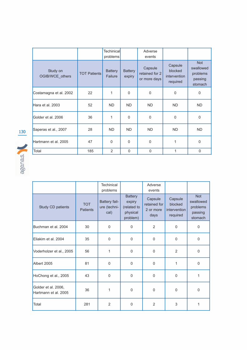
130
Techinical
problems
Adverse
events
Study CD patients TOT
Patients
Battery fail-
ure (techni-
cal)
Battery
expiry
(related to
physical
problem)
Capsule
retained for
2 or more
days
Capsule
blocked
intervention
required
Not
swallowed
problems
passing
stomach
Buchman et al. 2004 30 0 0 2 0 0
Eliakim et al. 2004 35 0 0 0 0 0
Voderholzer et al., 2005 56 1 0 0 2 0
Albert 2005 81 0 0 0 1 0
HoChong et al., 2005 43 0 0 0 0 1
Golder et al. 2006,
Hartmann et al. 200536 1 0 0 0 0
Total 281 2 0 2 3 1
Techinical
problems
Adverse
events
Study on
OGIB/WCE_othersTOT Patients
Battery
Failure
Battery
expiry
Capsule
retained for 2
or more days
Capsule
blocked
intervention
required
Not
swallowed
problems
passing
stomach
Costamagna et al. 2002 22 1 0 0 0 0
Hara et al. 2003 52 ND ND ND ND ND
Golder et al. 2006 36 1 0 0 0 0
Saperas et al., 2007 28 ND ND ND ND ND
Hartmann et al. 2005 47 0 0 0 1 0
Total 185 2 0 0 1 0

131
Techinical
problems
Adverse
events
Study on FAP
patients_WCE
TOT
Patients
Battery fail-
ure (techni-
cal)
Battery
expiry
(related to
physical
problem)
Capsule
retained for
2 or more
days
Capsule
blocked
intervention
required
Not
swallowed
problems
passing
stomach
Caspari et al 2004 20 ND ND ND ND ND
Schulman et al., 2005 40 1 0 0 2 0
Wong et al., 2006 32 0 0 0 6 0
Total 92 1 0 0 8 0

132

133
Appendix 6
List of excluded studies with reasons for exclusion
Studies excluded if not a systematic review, editorial, letter, news article, clinical guideli-nes, conference paper, comment, interview, survey or opinion piece, no author.
Adler, S. N. The probiotic agent Escherichia coli M-17 has a healing effect in patients withIBS with proximal inflammation of the small bowel, eng. Dig Liver Dis. 2006 Sep;38(9):713; ISSN: 1590-8658
Ahuja, V. Small bowel diagnostics: the taming of the shrew, eng. Trop Gastroenterol. 2006Apr-2006 Jun 30; 27(2):61-2; ISSN: 0250-636X.
Arsura, E. L. and Bobba, R. K. Small bowel exploration by wireless capsule endoscopy,eng. Am J Med. 2007 Jul; 120(7):e15; ISSN: 1555-7162 (Electronic).
Anonymous. Metachronous small bowel adenocarcinomas detected by capsule endoscopyin a patient with hereditary nonpolyposis colorectal cancer, eng. Dig Dis Sci. 2007 Apr;52(4):1134-6; ISSN: 0163-2116
Anonymous. What we have learned from 5 cases of permanent capsule retention, eng.Gastrointest Endosc. 2006 Aug; 64(2):283-7; ISSN: 0016-5107
Banerjee, R. and Reddy, D. N. Endoscopy-assisted wireless intragastric pH monitoring,eng. Gastrointest Endosc. 2007 Jan; 65(1):182; ISSN: 0016-5107
Cao, X. C.; Wang, B. M., and Han, Z. C. Wireless capsule endoscopic finding in Cronkhite-Canada syndrome, eng. Gut. 2006 Jun; 55(6):899-900; ISSN: 0017-5749
Cass, O. W. Is half-knowledge worse than ignorance?, eng. Gastrointest Endosc. 2006 Oct;64(4):542-3; ISSN: 0016-5107
Caunedo Alvarez, A.; Garcia-Montes, J. M., and Herrerias, J. M. Capsule endoscopy revie-wed by a nurse: is it here to stay?, eng. Dig Liver Dis. 2006 Aug; 38(8):603-4; ISSN: 1590-8658
De Franchis, R. and Dell’Era, A. Diagnosis and therapy of esophageal vascular disorders.Curr. Opin. Gastroenterol. 2007; 23(4):422-427; ISSN: 0267-1379.
Anonymous. Using capsule endoscopy to diagnose graft-versus-host disease: seeing isbelieving?, eng. Gastrointest Endosc. 2007 Mar; 65(3):410-1; ISSN: 0016-5107
Eliakim, R. The impact of capsule endoscopy on Crohn’s disease, eng. Dig Liver Dis. 2007Feb; 39(2):154-5; ISSN: 1590-8658
Anonymous. The impact of wireless capsule endoscopy on gastrointestinal diseases, eng.South Med J. 2007 Mar; 100(3):235-6; ISSN: 0038-4348
Anonymous Who would I consider for capsule endoscopy?—Part I, eng. Can JGastroenterol. 2006 Aug; 20(8):517-8; ISSN: 0835-7900
Erber, W. F. and Erber, J. A. Meta-analysis of the yield of capsule endoscopy in patientswith Crohn’s disease, eng. Am J Gastroenterol. 2006 Nov; 101(11):2669; ISSN: 0002-9270

134
Anonymous. Nonsteroidal antiinflammatory drugs and the small intestine. Curr. Opin.Gastroenterol. 2007; 23(2):134-141; ISSN: 0267-1379.
Frieling, T. The clinical impact of capsule endoscopy: to believe or not to believe, eng. EurJ Gastroenterol Hepatol. 2006 Aug; 18(8):873-4; ISSN: 0954-691X
Anonymous. Clinical application of wireless capsule endoscopy in pediatric patients forsuspected small bowel diseases, eng. Eur J Pediatr. 2007 Aug; 166(8):825-9; ISSN: 0340-6199
Gurudu, S. R. and Fleischer, D. E. From small-bowel bleeding to anemia: a wider range ofindications for the capsule endoscope?, eng. Endoscopy. 2006 Nov; 38(11):1144-5; ISSN:0013-726X
Hirano, I. and Richter, J. E. ACG practice guidelines: esophageal reflux testing, eng. Am JGastroenterol. 2007 Mar; 102(3):668-85; ISSN: 0002-9270
Hur, C. Esophageal capsule endoscopy for Barrett’s esophagus screening: a hard pill toswallow?, eng. Clin Gastroenterol Hepatol. 2007 Mar; 5(3):307-9; ISSN: 1542-7714
Lacy, B. E.; O’Shana, T.; Hynes, M.; Kelley, M. L. Jr; Weiss, J. E.; Paquette, L., andRothstein, R. I. Safety and tolerability of transoral Bravo capsule placement after transna-sal manometry using a validated conversion factor, eng. Am J Gastroenterol. 2007 Jan;102(1):24-32; ISSN: 0002-9270
Lashner, B. A. Sensitivity-specificity trade-off for capsule endoscopy in IBD: is it worth it?,eng. Am J Gastroenterol. 2006 May; 101(5):965-6; ISSN: 0002-9270
Leong, R. W. L. and Chan, F. K. L. Drug-induced side effects affecting the gastrointestinaltract. Expert Opin. Drug Saf. 2006; 5(4):585-592; ISSN: 1474-0338.
Lorenzo-Zuniga, V.; Moreno de Vega, V.; Manosa, M.; Domenech, E., and Boix, J. The uti-lity of wireless capsule endoscopy, as compared with barium contrast study, in a case ofPeutz-Jeghers syndrome, eng. Acta Gastroenterol Belg. 2006 Oct-2006 Dec 31; 69(4):423;ISSN: 0001-5644
Mishkin, D. S.; Chuttani, R.; Croffie, J.; Disario, J.; Liu, J.; Shah, R.; Somogyi, L.; Tierney,W.; Song, L. M., and Petersen, B. T. ASGE Technology Status Evaluation Report: wirelesscapsule endoscopy, eng. Gastrointest Endosc. 2006 Apr; 63(4):539-45; ISSN: 0016-5107
Anonymous. Enteroscopy and capsule endoscopy, eng. Endoscopy. 2006 Nov;38(11):1079-86; ISSN: 0013-726X
Rey, J. F.; Ladas, S.; Alhassani, A., and Kuznetsov, K. European Society of GastrointestinalEndoscopy (ESGE). Video capsule endoscopy: update to guidelines (May 2006), eng.Endoscopy. 2006 Oct; 38(10):1047-53; ISSN: 0013-726X
Rondonotti, E. and de Franchis, R. Diagnosing coeliac disease: is the videocapsule a suita-ble tool?, eng. Dig Liver Dis. 2007 Feb; 39(2):145-7; ISSN: 1590-8658
Rufo, P. A. and Bousvaros, A. Challenges and progress in pediatric inflammatory boweldisease. Curr. Opin. Gastroenterol. 2007; 23(4):406-412; ISSN: 0267-1379.
Shin, J. I. and Lee, J. S. Indications of capsule endoscopy in Henoch-Schonlein syndromewith gastrointestinal symptoms, eng. Am J Med. 2007 Jun; 120(6):e13; ISSN: 1555-7162

135
Sidhu, R.; Sanders, D. S.; McAlindon, M. E., and Kapur, K. Capsule endoscopy for the eva-luation of nonsteroidal anti-inflammatory drug-induced enteropathy: United Kingdom pilotdata, eng. Gastrointest Endosc. 2006 Dec; 64(6):1035; ISSN: 0016-5107
Sidhu, R.; Sanders, D. S.; Sakellariou, V. P., and McAlindon, M. E. Capsule endoscopy andobscure gastrointestinal bleeding: are transfusion dependence and comorbidity further riskfactors to predict a diagnosis?, eng. Am J Gastroenterol. 2007 Jun; 102(6):1329-30; ISSN:0002-9270
Spada, C.; Riccioni, M. E., and Costamagna, G. Patients with known small bowel strictureor with symptoms of small bowel obstruction secondary to Crohn’s disease should not per-form video capsule endoscopy without being previously tested for small bowel patency,eng. Am J Gastroenterol. 2007 Jul; 102(7):1542-3; author reply 1543-4; ISSN: 0002-9270
Anonymous. Rapid((R)) Access Real-Time device and Rapid Access software: new tools inthe armamentarium of capsule endoscopy, ENG. Expert Rev Med Devices. 2007 Jul;4(4):431-435; ISSN: 1743-4440
Tanaka, S.; Mitsui, K.; Shirakawa, K.; Tatsuguchi, A.; Nakamura, T.; Hayashi, Y.; Sakamoto,C., and Terano, A. Successful retrieval of video capsule endoscopy retained at ileal steno-sis of Crohn’s disease using double-balloon endoscopy, eng. J Gastroenterol Hepatol. 2006May; 21(5):922-3; ISSN: 0815-9319
Vanderveldt, H. S. and Barkin, J. S. Can findings on wireless capsule endoscopy predict fur-ther bleeding?, eng. Nat Clin Pract Gastroenterol Hepatol. 2007 Mar; 4(3):132-3; ISSN:1743-4386 (Electronic).
Viazis, N. and Karamanolis, D. G. Indeterminate colitis—the role of wireless capsule endo-scopy, eng. Aliment Pharmacol Ther. 2007 Apr 1; 25(7):859; author reply 860; ISSN: 0269-2813
Anonymous. Wireless esophageal pH monitoring is better tolerated than the catheter-basedtechnique: Results from a randomized cross-over trial. Am. J. Gastroenterol. 2007;102(2):239-245; ISSN: 0002-9270.
Balzola, F.; Barbon, V.; Repici, A.; Rizzetto, M.; Clauser, D.; Gandione, M., and Sapino, A.Panenteric IBD-like disease in a patient with regressive autism shown for the first time bythe wireless capsule enteroscopy: another piece in the jigsaw of this gut-brain syndrome?,eng. Am J Gastroenterol. 2005 Apr; 100(4):979-81; ISSN: 0002-9270
Bizzarri, B.; Fornaroli, F.; Cannizzaro, R.; de’ Angelis, N.; Vincenzi, F.; Maffini, V., and de’Angelis, G. L. Endoscopic placement of video capsule in a pediatric population, eng.Gastrointest Endosc. 2005 Dec; 62(6):991; ISSN: 0016-5107
Dalla Valle, R.; Fornaroli, F.; de’Angelis, N.; Bizzarri, B., and de’Angelis, G. L. Clinical use-fulness of the endoscopic video capsule in patients with obscure gastrointestinal bleeding,eng. Endoscopy. 2005 Aug; 37(8):780; author reply 781; ISSN: 0013-726X (Print).
Dubner, S.; Goldin, E., and Dubner, Y. Re: Interference between pacemaker and video cap-sule endoscopy, eng. Pacing Clin Electrophysiol. 2005 Mar; 28(3):261; ISSN: 0147-8389
Ford, R. M.; Affronti, J.; Cohen, R.; Baumgarten, D. A., and Cai, Q. Zenker’s diverticulum:a contraindication for wireless capsule endoscopy?, eng. J Clin Gastroenterol. 2005 Mar;39(3):257; ISSN: 0192-0790
136
Marrero, R. J. and Barkin, J. S. Wireless capsule endoscopy and portal hypertensive inte-stinal vasculopathy, eng. Gastrointest Endosc. 2005 Oct; 62(4):535-7; ISSN: 0016-5107
Nakamura, T. When is the optimal timing for performing video capsule endoscopy forobscure gastrointestinal bleeding?, eng. J Gastroenterol. 2005 Mar; 40(3):322-3; ISSN:0944-1174
Anonymous. Wireless capsule endoscopy and Crohn’s disease, eng. Gut. 2005 Mar;54(3):323-6; ISSN: 0017-5749
Studies excluded if not-human
No study identified
Studies excluded if not on WCE
Adebayo, D. and Bjarnason, I. Is non-steroidal anti-inflammatory drug (NSAID) enteropa-thy clinically more important than NSAID gastropathy?, eng. Postgrad Med J. 2006 Mar;82(965):186-91; ISSN: 1469-0756
Adebayo, D.; Popat, R.; Thjodleifsson, B., and Bjarnason, I. Granulomatous ileitis in apatient with ankylosing spondylitis, eng. Nat Clin Pract Gastroenterol Hepatol. 2007 Jun;4(6):347-51; ISSN: 1743-4386
Ang, D.; Luman, W., and Ooi, C. J. Early experience with double balloon enteroscopy: aleap forward for the gastroenterologist, eng. Singapore Med J. 2007 Jan; 48(1):50-60;ISSN: 0037-5675
Averbach, M.; Marques, O. W. Jr; Correa, P. A.; Cappellanes, C. A., and Hashiba, K. Small-bowel angiodysplasia and intestinal bleeding: a diagnostic challenge, eng. GastrointestEndosc. 2006 Dec; 64(6):1008; discussion 1009; ISSN: 0016-5107
Bauditz, J.; Lochs, H., and Voderholzer, W. Macroscopic appearance of intestinal angiody-splasias under antiangiogenic treatment with thalidomide, eng. Endoscopy. 2006 Oct;38(10):1036-9; ISSN: 0013-726X
Bretagne, J. F.; Manfredi, S., and Heresbach, D. [Colorectal cancer screening: present andfuture.] [Depistage de masse du cancer colorectal: present et avenir.], FRE. Presse Med.2007 Jul-2007 Aug 31; 36(7-8):1054-1063;
Bruining, D. H. and Loftus, E. V. Evolving diagnostic strategies for inflammatory boweldisease, eng. Curr Gastroenterol Rep. 2006 Dec; 8(6):478-85; ISSN: 1522-8037 (Print).
Bulfoni, A. [Primary adenocarcinoma of jejunum with obscure gastrointestinal bleeding][Adenocarcinoma primitivo del digiuno ad esteriorizzazione con enterorragia.], ita. Clin Ter.2006 Sep-2006 Oct 31; 157(5):431-4; ISSN: 0009-9074
Chiarugi, M.; Galatioto, C.; Lippolis, P.; Zocco, G., and Seccia, M. Gastrointestinal stromaltumour of the duodenum in childhood: a rare case report, eng. BMC Cancer. 2007; 779;ISSN: 1471-2407
Concha, R.; Amaro, R., and Barkin, J. S. Obscure gastrointestinal bleeding: diagnostic andtherapeutic approach, eng. J Clin Gastroenterol. 2007 Mar; 41(3):242-51; ISSN: 0192-0790

137
Corona, E.; Hatlebakk, J. G., and Galmiche, J.-P. Novel developments in non-erosive anderosive gastroesophageal reflux disease. Curr. Opin. Gastroenterol. 2006; 22(4):399-405;ISSN: 0267-1379.
Croese, J. and Speare, R. Intestinal allergy expels hookworms: seeing is believing, eng.Trends Parasitol. 2006 Dec; 22(12):547-50; ISSN: 1471-4922
Das, A. and Leighton, J. A. Is double balloon enteroscopy the best initial imaging methodfor obscure gastrointestinal bleeding?, eng. Nat Clin Pract Gastroenterol Hepatol. 2007 Mar;4(3):120-1; ISSN: 1743-4386
De Palma, G. D.; Aprea, G.; Rega, M.; Masone, S.; Simeoli, I.; Cutolo, P., and Persico, G.Polypoid vascular malformation of the small intestine, eng. Gastrointest Endosc. 2007 Feb;65(2):328-9; discussion 329; ISSN: 0016-5107
Eisen, G. M. and Schreiner, M. Small-bowel endoscopy. Endoscopy. 2007; 39(2):113-117;ISSN: 0013-726X.
Fernandez-Urien, I.; Carretero, C.; Sola, J. J.; Munoz-Navas, M.; Betes, M.; Subtil, J. C., andArmendariz, R. Refractory Whipple’s disease, eng. Gastrointest Endosc. 2007 Mar;65(3):521-2, discussion 522; ISSN: 0016-5107
Fortun, P. and Hawkey, C. J. Drug-induced gastrointestinal disorders. Medicine (GBR).2007; 35(4):210-215; ISSN: 1357-3039.
Giovannini, I.; Chiarla, C.; Murazio, M.; Clemente, G.; Giuliante, F., and Nuzzo, G. An extre-me case of Heyde syndrome, eng. Dig Surg. 2006; 23(5-6):387-8; ISSN: 0253-4886
Gomez, B. J.; Caunedo Alvarez, A.; Redondo, L.; Esteban, J.; Saenz-Dana, M.; Blasco, M.;Hergueta, P.; Rodriguez-Tellez, M.; Romero, R.; Pellicer, F. J., and Herrerias, J. M.Modification of pepsinogen I levels and their correlation with gastrointestinal injury afteradministration of dexibuprofen, ibuprofen or diclofenac: A randomized, open-label, control-led clinical trial. Int. J. Clin. Pharmacol. Ther. 2006; 44(4):154-162; ISSN: 0946-1965.
Gralnek, I. M. Endoscopic research: funding opportunities and mechanisms from the ASGEand beyond.., eng. Gastrointest Endosc. 2006 Dec; 64(6 Suppl):S16-8; ISSN: 0016-5107
Heidt, J.; Langers, A. M.; van der Meer, F. J., and Brouwer, R. E. Thalidomide as treatmentfor digestive tract angiodysplasias, eng. Neth J Med. 2006 Dec; 64(11):425-8; ISSN: 0300-2977
Heine, G. D.; Al-Toma, A.; Mulder, C. J., and Jacobs, M. A. Milestone in gastrointestinalendoscopy: double-balloon enteroscopy of the small bowel, eng. Scand J GastroenterolSuppl. 2006 May; (243):32-8; ISSN: 0085-5928
Hohl, C.; Muhlenbruch, G.; Schmidt, T.; Haage, P., and Gunther, R. W. [Bowel Imaging - AReevaluation, Part 1.] [Bildgebende Diagnostik des Darms - eine Neubewertung, Teil 1.],GER. Rofo. 2007 Jul; 179(7):683-692; ISSN: 1438-9029
Jakobs, R.; Hartmann, D.; Benz, C.; Schilling, D.; Weickert, U.; Eickhoff, A.; Schoenleben,K., and Riemann, J. F. Diagnosis of obscure gastrointestinal bleeding by intra-operativeenteroscopy in 81 consecutive patients, eng. World J Gastroenterol. 2006 Jan 14;12(2):313-6; ISSN: 1007-9327
Jewell, D. P. Crohn’s disease. Medicine (GBR). 2007; 35(5):283-289; ISSN: 1357-3039.
Karagiannis, S.; Kosmadakis, G.; Goulas, S.; Boletis, J., and Mavrogiannis, C. Education and
138
Imaging. Gastrointestinal: cytomegalovirus enterocolitis, eng. J Gastroenterol Hepatol.2007 Apr; 22(4):594; ISSN: 0815-9319
Krauss, N. and Schuppan, D. Monitoring Nonresponsive Patients Who Have Celiac Disease.Gastrointest. Endosc. Clin. North Am. 2006; 16(2):317-327; ISSN: 1052-5157.
Leighton, J. A. and Wallace, M. B. Update on small bowel imaging, eng. Gastroenterology.2007 May; 132(5):1651-4; ISSN: 0016-5085
Manabe, N.; Tanaka, S.; Fukumoto, A.; Nakao, M.; Kamino, D., and Chayama, K. Double-balloon enteroscopy in patients with GI bleeding of obscure origin, eng. GastrointestEndosc. 2006 Jul; 64(1):135-40; ISSN: 0016-5107
Martins, N. B. and Wassef, W. Upper gastrointestinal bleeding, eng. Curr OpinGastroenterol. 2006 Nov; 22(6):612-9; ISSN: 0267-1379
May, A. and Ell, C. Push-and-pull enteroscopy using the double-balloon technique/double-balloon enteroscopy, eng. Dig Liver Dis. 2006 Dec; 38(12):932-8; ISSN: 1590-8658
Modlin, I. M.; Latich, I.; Zikusoka, M.; Kidd, M.; Eick, G., and Chan, A. K. Gastrointestinalcarcinoids: the evolution of diagnostic strategies, eng. J Clin Gastroenterol. 2006 Aug;40(7):572-82; ISSN: 0192-0790.
Mrevlje, Z. and Stabuc, B. Pitfalls in diagnosing small bowel carcinoid tumors, eng. J BUON.2006 Jan-2006 Mar 31; 11(1):83-6; ISSN: 1107-0625.
Oshitani, N.; Yukawa, T.; Yamagami, H.; Inagawa, M.; Kamata, N.; Watanabe, K.; Jinno,Y.; Fujiwara, Y.; Higuchi, K., and Arakawa, T. Evaluation of deep small bowel involvementby double-balloon enteroscopy in Crohn’s disease, eng. Am J Gastroenterol. 2006 Jul;101(7):1484-9; ISSN: 0002-9270.
Perez-Cuadrado, E.; Mas, P.; Hallal, H.; Shanabo, J.; Munoz, E.; Ortega, I.; Lopez Martin,A.; Torrella, E.; Lopez Higueras, A.; Martin, A., and Carballo, F. Double-balloon enterosco-py: A descriptive study of 50 explorations: ENTEROSCOPIA DE DOBLE BALON: ESTUDIODESCRIPTIVO DE 50 EXPLORACIONES. Rev. Esp. Enferm. Dig. 2006; 98(2):73-81; ISSN:1130-0108.
Rao, R. and Shashidhar, H. Intestinal lymphangiectasia presenting as abdominal mass, eng.Gastrointest Endosc. 2007 Mar; 65(3):522-3, discussion 523; ISSN: 0016-5107
Rex, D. K.; Talley, N. J.; Katz, P. O.; Hanauer, S. B., and Sandborn, W. J. Report from theACG: Highlights of the 71st Annual Scientific Meeting of the American College ofGastroenterology, October 20-25, Las Vegas, NV. Rev. Gastroenterol. Disord. 2006;6(4):227-242; ISSN: 1533-001X.
Riemann, J. F.; Hartmann, D.; Schilling, D.; Damian, U.; Eickhoff, A., and Weickert, U.Frequency of small bowel polyps in patients with duodenal adenoma but without familialadenomatous polyposis, eng. Z Gastroenterol. 2006 Mar; 44(3):235-8; ISSN: 0044-2771.
Satsangi, J.; Silverberg, M. S.; Vermeire, S., and Colombel, J.-F. The Montreal classificationof inflammatory bowel disease: Controversies, consensus, and implications. Gut. 2006;55(6):749-753; ISSN: 0017-5749.
Spyridonos, P.; Vilarino, F.; Vitria, J.; Azpiroz, F., and Radeva, P. Anisotropic feature extrac-tion from endoluminal images for detection of intestinal contractions, eng. Med Image
139
Comput Comput Assist Interv Int Conf Med Image Comput Comput Assist Interv. 2006;9(Pt 2):161-8.
Stange, E. F.; Travis, S. P. L.; Vermeire, S.; Beglinger, C.; Kupcinskas, L.; Geboes, K.;Barakauskiene, A.; Villanacci, V.; Von Herbay, A.; Warren, B. F.; Gasche, C.; Tilg, H.;Schreiber, S. W.; Scholmerich, J., and Reinisch, W. European evidence based consensus onthe diagnosis and management of Crohn’s disease: Definitions and diagnosis. Gut. 2006;55(SUPPL. 1):i1-i15; ISSN: 0017-5749.
Suzuki, T.; Matsushima, M.; Okita, I.; Ito, H.; Gocho, S.; Tajima, H.; Tokiwa, K.; Teraoka,H.; Watanabe, K.; Shirai, T., and Mine, T. Clinical utility of double-balloon enteroscopy forsmall intestinal bleeding, eng. Dig Dis Sci. 2007 Aug; 52(8):1914-9; ISSN: 0163-2116
Van Tuyl, S. A.; Letteboer, T. G.; Rogge-Wolf, C.; Kuipers, E. J.; Snijder, R. J.; Westermann,C. J., and Stolk, M. F. Assessment of intestinal vascular malformations in patients with here-ditary hemorrhagic teleangiectasia and anemia, eng. Eur J Gastroenterol Hepatol. 2007Feb; 19(2):153-8; ISSN: 0954-691X
Verschuur, E. M. L.; Kuipers, E. J., and Siersema, P. D. Nurses working in GI and endosco-pic practice: a review. Gastrointest. Endosc. 2007; 65(3):469-479; ISSN: 0016-5107.
Villa, F.; Signorelli, C.; Rondonotti, E., and de Franchis, R. Preparations and Prokinetics.Gastrointest. Endosc. Clin. North Am. 2006; 16(2):211-220; ISSN: 1052-5157.
Wardi, J.; Shahmurov, M.; Czerniak, A., and Avni, Y. Clinical challenges and images in GI.Capillary hemangioma of small intestine, eng. Gastroenterology. 2007 May; 132(5):1656,2084; ISSN: 0016-5085.
Weersma, R. K.; Limburg, A. J.; Karrenbeld, A., and Koornstra, J. J. Editor’s quiz: iron defi-ciency anaemia 10 years after small bowel resection in infancy, eng. Gut. 2007 Apr;56(4):463, 488; ISSN: 0017-5749
Arena, A.; Boulougoura, M.; Chowdrey, H. S.; Dario, P.; Harendt, C.; Irion, K. M.;Kodogiannis, V.; Lenaerts, B.; Menciassi, A.; Puers, R.; Scherjon, C., and Turgis, D.Intracorporeal Videoprobe (IVP), eng. Stud Health Technol Inform. 2005; 114167-74;ISSN: 0926-9630.
Biering, H.; Bauditz, J.; Brenner, N.; Stein, H.; Lochs, H., and Strasburger, C. J. Primaryneuroendocrine carcinoma of inguinal lymph node, eng. Horm Res. 2005; 64(1):16-9;ISSN: 0301-0163.
De Palma GD, Rega M Masone S Persico F Siciliano S Patrone F Matantuono L Persico G.Mucosal abnormalities of the small bowel in patients with cirrhosis and portal hypertension:a capsule endoscopy study. Gastrointestinal Endoscopy. 2005; 62(4):529-34; ISSN: CN-00530599.
Fortun, P. J. and Hawkey, C. J. Nonsteroidal antiinflammatory drugs and the small intesti-ne. Curr. Opin. Gastroenterol. 2005; 21(2):169-175; ISSN: 0267-1379.
Graham, D. Y.; Opekun, A. R.; Willingham, F. F., and Qureshi, W. A. Visible small-intestinalmucosal injury in chronic NSAID users, eng. Clin Gastroenterol Hepatol. 2005 Jan; 3(1):55-9; ISSN: 1542-3565.
Grosbois, B.; Decaux, O.; Cador, B.; Cazalets, C., and Jego, P. [Human iron deficiency] [Lescarences en fer chez l’homme.], fre. Bull Acad Natl Med. 2005 Nov; 189(8):1649-63;discussion 1663-4; ISSN: 0001-4079.

140
Hatoum, O. A.; Binion, D. G.; Phillips, S. A.; O’Loughlin, C.; Komorowski, R. A.; Gutterman,D. D., and Otterson, M. F. Radiation induced small bowel “web” formation is associated withacquired microvascular dysfunction, eng. Gut. 2005 Dec; 54(12):1797-800; ISSN: 0017-5749.
Ikeda, K. and Swain, P. Future innovations and techniques: Endoscopic hemostasis for UGI,non-variceal bleeding. Tech. Gastrointest. Endosc. 2005; 7(3 SPEC. ISS.):164-170; ISSN:1096-2883.
Imbesi, J. J. and Kurtz, R. C. A multidisciplinary approach to gastrointestinal bleeding incancer patients. J. Supportive Oncol. 2005; 3(2):101-110; ISSN: 1544-6794.
Kelly, M. E.; McMahon, L. E.; Jaroszewski, D. E.; Yousfi, M. M.; De Petris, G., and Swain, J.M. Small-bowel diaphragm disease: seven surgical cases, eng. Arch Surg. 2005 Dec;140(12):1162-6; ISSN: 0004-0010
Keuchel, M. and Hagenmuller, F. Small bowel endoscopy, eng. Endoscopy. 2005 Feb;37(2):122-32; ISSN: 0013-726X
Kornbluth, A.; Colombel, J. F.; Leighton, J. A., and Loftus, E. ICCE consensus for inflamma-tory bowel disease. Endoscopy. 2005; 37(10):1051-1054; ISSN: 0013-726X.
Kovacs, T. O. G. Small bowel bleeding. Curr. Treat. Options Gastroenterol. 2005; 8(1):31-38; ISSN: 1092-8472.
Kruger, S.; Noack, F.; Blochle, C., and Feller, A. C. Primary malignant melanoma of the smallbowel: a case report and review of the literature, eng. Tumori. 2005 Jan-2005 Feb 28;91(1):73-6; ISSN: 0300-8916.
Lee, S. K. and Green, P. H. Endoscopy in celiac disease, eng. Curr Opin Gastroenterol. 2005Sep; 21(5):589-94; ISSN: 0267-1379.
Leighton, J. A. and Loftus Jr., E. V. Evolving diagnostic modalities in inflammatory boweldisease. Curr. Gastroenterol. Rep. 2005; 7(6):467-474; ISSN: 1522-8037.
Marek, T. A. Gastrointestinal bleeding. Endoscopy. 2005; 37(11):1098-1104; ISSN: 0013-726X.
Modlin, I. M.; Kidd, M.; Latich, I.; Zikusoka, M. N., and Shapiro, M. D. Current status ofgastrointestinal carcinoids. Gastroenterology. 2005; 128(6):1717-1751; ISSN: 0016-5085.
Mussi, C.; Caprotti, R.; Scaini, A.; Angelini, C.; Crippa, S.; Uggeri, F.; Uggeri, F., and Sartori,P. Management of small bowel tumors: personal experience and new diagnostic tools, eng.Int Surg. 2005 Sep-2005 Oct 31;
Pei, K.; Zemon, H.; Venbrux, A., and Brody, F. Laparoendoscopic techniques for occultgastrointestinal bleeding, eng. J Laparoendosc Adv Surg Tech A. 2005 Dec; 15(6):615-9;ISSN: 1092-6429.
Strate, L. L. Lower GI bleeding: Epidemiology and diagnosis. Gastroenterol. Clin. North Am.2005; 34(4):643-664; ISSN: 0889-8553.
Taal, B. G. and Smits, M. Developments in diagnosis and treatment of metastatic midgutcarcinoid tumours: A review. Minerva Gastroenterol. Dietol. 2005; 51(4):335-344; ISSN:0026-4776.
141
Veltzke-Schlieker, W. and Rosch, T. UEGW report, Prague 2004: Endoscopic highlights.Endoscopy. 2005; 37(4):367-377; ISSN: 0013-726X.
Yamamoto, H. and Kita, H. Enteroscopy, eng. J Gastroenterol. 2005 Jun; 40(6):555-62;ISSN: 0944-1174.
Studies excluded if the topic was not Obscure Gastrointestinal Bleeding (OGIB), Crohn’sdisease, familiar polyposis and celiac disease
Agrawal, G. A.; Johnson, P. T., and Fishman, E. K. Intermittent small bowel obstruction withretained endoscopic capsule: MDCT and plain radiographic appearance, eng. Emerg Radiol.2007 Mar; 13(6):319-21; ISSN: 1070-3004.
Antao, B.; Bishop, J.; Shawis, R., and Thomson, M. Clinical application and diagnostic yieldof wireless capsule endoscopy in children, eng. J Laparoendosc Adv Surg Tech A. 2007 Jun;17(3):364-70; ISSN: 1092-6429.
Antonietti, M.; Savoye, G.; Lecleire, S.; Hecketsweiler, P., and Ben-Soussan, E. A video cap-sule attached to a probe can be used for prolonged stationary endoscopic monitoring, eng.Endoscopy. 2006 Mar; 38(3):289; ISSN: 0013-726X .
Badran, A. M.; Vahedi, K.; Berrebi, D.; Catana, D.; De Lagausie, P.; Drouet, L.; Ferkadji, F.,and Mougenot, J. F. Pediatric ampullar and small bowel blue rubber bleb nevus syndromediagnosed by wireless capsule endoscopy, eng. J Pediatr Gastroenterol Nutr. 2007 Feb;44(2):283-6; ISSN: 1536-4801.
Balachandran, P. and Prasad, V. G. Intestinal parasites seen on capsule endoscopy, eng.Gastrointest Endosc. 2006 Oct; 64(4):651; ISSN: 0016-5107.
Bayraktar, Y.; Ersoy, O., and Sokmensuer, C. The findings of capsule endoscopy in patientswith common variable immunodeficiency syndrome, eng. Hepatogastroenterology. 2007Jun; 54(76):1034-7; ISSN: 0172-6390.
Brown, G.; Fraser, C.; Schofield, G.; Taylor, S.; Bartram, C.; Phillips, R., and Saunders, B.Video capsule endoscopy in peutz-jeghers syndrome: a blinded comparison with bariumfollow-through for detection of small-bowel polyps, eng. Endoscopy. 2006 Apr; 38(4):385-90; ISSN: 0013-726X .
Caddy, G. R.; Moran, L.; Chong, A. K.; Miller, A. M.; Taylor, A. C., and Desmond, P. V. Theeffect of erythromycin on video capsule endoscopy intestinal-transit time, eng. GastrointestEndosc. 2006 Feb; 63(2):262-6; ISSN: 0016-5107.
Calabrese, C.; Fabbri, A.; Gionchetti, P.; Rizzello, F.; Morselli, C.; Liguori, G.; Poggioli, G.;Campieri, M., and Di Febo, G. Controlled study using wireless capsule endoscopy for theevaluation of the small intestine in chronic refractory pouchitis, eng. Aliment PharmacolTher. 2007 Jun 1; 25(11):1311-6; ISSN: 0269-2813 .
Carpi, F.; Galbiati, S., and Carpi, A. Magnetic shells for gastrointestinal endoscopic capsu-les as a means to control their motion, eng. Biomed Pharmacother. 2006 Sep; 60(8):370-4; ISSN: 0753-3322.
Chamberlain, S. M.; Patel, J.; Carter Balart, J.; Gossage, J. R. Jr, and Sridhar, S. Evaluationof patients with hereditary hemorrhagic telangiectasia with video capsule endoscopy: a sin-gle-center prospective study, eng. Endoscopy. 2007 Jun; 39(6):516-20; ISSN: 1438-8812.

142
Chamouard, P.; Nehme-Schuster, H.; Simler, J. M.; Finck, G.; Baumann, R., and Pasquali, J.L. Videocapsule endoscopy is useful for the diagnosis of intestinal lymphangiectasia, eng.Dig Liver Dis. 2006 Sep; 38(9):699-703.
Chao, C. C. and Ray, M. L. Education and imaging. Gastrointestinal: Hookworm diagnosedby capsule endoscopy, eng. J Gastroenterol Hepatol. 2006 Nov; 21(11):1754; ISSN: 0815-9319.
Chen, G. C.; Lee-Henderson, M.; Mehdizadeh, S.; Goltzer, O.; Shen, J.; Sul, J., and Jutabha,R. Novel external sensor array for capsule endoscopy, eng. Endoscopy. 2006 Jul;38(7):752-4; ISSN: 0013-726X .
Chen, T. H.; Chen, T. Y.; Shyu, L. Y.; Lin, C. K., and Lin, C. C. Hookworm infestation dia-gnosed by capsule endoscopy (with video), eng. Gastrointest Endosc. 2006 Aug;64(2):277-8; ISSN: 0016-5107.
De Francesco, V.; Stoppino, G.; Tonti, P.; D’Agnessa, M. R.; Castriota, M.; Panella, C., andIerardi, E. Ileal metastasis from thoracic melanoma disclosed by video capsule endoscopy:an unusual but not extraordinary source of obscure bleeding, ENG. Endoscopy. 2007 Apr18; ISSN: 1438-8812.
Dominguez, E. P.; Choi, Y.; Raijman, I. L., and Sweeney, J. F. Laparoscopic approach for theretrieval of retained video capsule endoscopy, eng. JSLS. 2006 Oct-2006 Dec 31;10(4):496-8; ISSN: 1086-8089.
Drastich, P. [Capsule endoscopy—the past, presence, and future] [Kapslova endoskopie—minulost, pritomnost a budoucnost.], cze. Cas Lek Cesk. 2006; 145(6):432-6; ISSN: 0008-7335.
Dubner, S.; Dubner, Y.; Rubio, H., and Goldin, E. Electromagnetic interference from wire-less video-capsule endoscopy on implantable cardioverter-defibrillators, eng. Pacing ClinElectrophysiol. 2007 Apr; 30(4):472-5; ISSN: 0147-8389.
Eisen, G. M.; Eliakim, R.; Zaman, A.; Schwartz, J.; Faigel, D.; Rondonotti, E.; Villa, F.;Weizman, E.; Yassin, K., and deFranchis, R. The accuracy of PillCam ESO capsule endosco-py versus conventional upper endoscopy for the diagnosis of esophageal varices: A pro-spective three-center pilot study. Endoscopy. 2006; 38(1):31-35; ISSN: 0013-726X.
Eliakim, R.; Fireman, Z.; Gralnek, I. M.; Yassin, K.; Waterman, M.; Kopelman, Y.; Lachter,J.; Koslowsky, B., and Adler, S. N. Evaluation of the PillCam Colon capsule in the detectionof colonic pathology: results of the first multicenter, prospective, comparative study, eng.Endoscopy. 2006 Oct; 38(10):963-70; ISSN: 0013-726X.
Enns, R. Capsule endoscopy: specifics for enhanced performance—Part II, eng. Can JGastroenterol. 2006 Sep; 20(9):575-7; ISSN: 0835-7900.
Ewertsen, C.; Svendsen, C. B.; Svendsen, L. B.; Hansen, C. P.; Gustafsen, J. H., andJendresen, M. B. [Is screening of wireless capsule endoscopies by non-physicians feasible?][Er screening af kapselendoskopiske undersogelser udfort af ikkelaegeligt personale anven-delig?], dan. Ugeskr Laeger. 2006 Oct 9; 168(41):3530-3; ISSN: 1603-6824.
Fenkel, J. M.; Grasso, M. A.; Goldberg, E. M., and Feller, E. D. Capsule endoscopy is safe inpatients with pulsatile Novacor PC left ventricular assist device, eng. Gastrointest Endosc.2007 Mar; 65(3):559-60; author reply 560; ISSN: 0016-5107.

143
Floro, L.; Pak, G.; Sreter, L., and Tulassay, Z. Wireless capsule endoscopy in the diagnosisof helminthiasis, eng. Gastrointest Endosc. 2007 Jun; 65(7):1078; discussion 1079; ISSN:0016-5107.
Gardner, T. B.; Bensen, S. P.; Toor, A., and Anderson, P. B. Video capsule endoscopy to dia-gnose metastatic melanoma, eng. Dig Dis Sci. 2007 Apr; 52(4):1036-7; ISSN: 0163-2116.
Garza, A.; Patel, T., and Brady, P. Diagnosis of malignant hemangioendothelioma of thesmall intestine with capsule endoscopy, eng. Dig Dis Sci. 2007 Mar; 52(3):852-5; ISSN:0163-2116.
Ge, Z. Z.; Chen, H. Y.; Gao, Y. J.; Gu, J. L.; Hu, Y. B., and Xiao, S. D. [Clinical applicationof wireless capsule endoscopy in pediatric and adolescent patients], chi. Zhonghua Er KeZa Zhi. 2006 Sep; 44(9):676-9; ISSN: 0578-1310.
Ge, Z.-Z.; Chen, H.-Y.; Goa, Y.-J.; Hu, Y.-B., and Xiao, S.-D. The role of simeticone in small-bowel preparation for capsule endoscopy. Endoscopy. 2006; 38(8):836-840; ISSN: 0013-726X.
Gerson, L. and Lin, O. S. Cost-benefit analysis of capsule endoscopy compared with stan-dard upper endoscopy for the detection of Barrett’s esophagus, eng. Clin GastroenterolHepatol. 2007 Mar; 5(3):319-25; ISSN: 1542-7714.
Goldstein, J. L.; Eisen, G. M.; Lewis, B.; Gralnek, I. M.; Aisenberg, J.; Bhadra, P., andBerger, M. F. Small bowel mucosal injury is reduced in healthy subjects treated with cele-coxib compared with ibuprofen plus omeprazole, as assessed by video capsule endoscopy,eng. Aliment Pharmacol Ther. 2007 May 15; 25(10):1211-22; ISSN: 0269-2813.
Gonzalez-Suarez, B.; Galter, S., and Balanzo, J. [Wireless capsule endoscopy: basic princi-ples and clinical utility.] [Capsula endoscopica: fundamentos y utilidad clinica.], spa. CirEsp. 2007 Jun; 81(6):299-306; ISSN: 0009-739X .
Gralnek, I. M.; Rabinovitz, R.; Afik, D., and Eliakim, R. A simplified ingestion procedure foresophageal capsule endoscopy: initial evaluation in healthy volunteers, eng. Endoscopy.2006 Sep; 38(9):913-8; ISSN: 0013-726X .
Ho, K. K. and Joyce, A. M. Complications of capsule endoscopy, eng. Gastrointest EndoscClin N Am. 2007 Jan; 17(1):169-78, viii-ix; ISSN: 1052-5157.
Hogan, R. B.; Ahmad, N.; Hogan, R. B. 3rd; Hensley, S. D.; Phillips, P.; Doolittle, P., andReimund, E. Video capsule endoscopy detection of jejunal carcinoid in life-threateninghemorrhage, first trimester pregnancy, eng. Gastrointest Endosc. 2007 Jul; 66(1):205-7;ISSN: 0016-5107 .
Holden, J. P.; Dureja, P.; Pfau, P. R.; Schwartz, D. C.; Reichelderfer, M.; Judd, R. H.; Danko,I.; Iyer, L. V., and Gopal, D. V. Endoscopic placement of the small-bowel video capsule byusing a capsule endoscope delivery device, eng. Gastrointest Endosc. 2007 May;65(6):842-7; ISSN: 0016-5107.
Hosono, S.; Ohira, M.; Maeda, K.; Muguruma, K.; Nishihara, T.; Inoue, T.; Yashiro, M., andHirakawa, K. Synchronous adenocarcinomas of the ileum and transverse colon detected bycapsule endoscopy: report of a case, eng. Surg Today. 2006; 36(7):663-5; ISSN: 0941-1291.

144
Kong, H.; Kim, Y. S.; Hyun, J. J.; Cho, Y. J.; Keum, B.; Jeen, Y. T.; Lee, H. S.; Chun, H. J.;Um, S. H.; Lee, S. W.; Choi, J. H.; Kim, C. D.; Ryu, H. S., and Hyun, J. H. Limited ability ofcapsule endoscopy to detect normally positioned duodenal papilla, eng. GastrointestEndosc. 2006 Oct; 64(4):538-41; ISSN: 0016-5107.
Lapalus, M.-G.; Dumortier, J.; Fumex, F.; Roman, S.; Lot, M.; Prost, B.; Mion, F., andPonchon, T. Esophageal capsule endoscopy versus esophagogastroduodenoscopy for eva-luating portal hypertension: A prospective comparative study of performance and toleran-ce. Endoscopy. 2006; 38(1):36-41; ISSN: 0013-726X.
Leighton, J. A. Recent advances in endoscopic capsule imaging: See what we have beenmissing. Rev. Gastroenterol. Disord. 2006; 6(SUPPL. 1):S19-S27; ISSN: 1533-001X.
Lenaerts, B. and Puers, R. An inductive power link for a wireless endoscope, eng. BiosensBioelectron. 2007 Feb 15; 22(7):1390-5; ISSN: 0956-5663.
Lin, M. C.; Dung, L. R., and Weng, P. K. An ultra-low-power image compressor for capsuleendoscope, eng. Biomed Eng Online. 2006; 514; ISSN: 1475-925X .
Lin, O. S.; Schembre, D. B.; Mergener, K.; Spaulding, W.; Lomah, N.; Ayub, K.; Brandabur,J. J.; Bredfeldt, J.; Drennan, F.; Gluck, M.; Jiranek, G. C.; McCormick, S. E.; Patterson, D.,and Kozarek, R. A. Blinded comparison of esophageal capsule endoscopy versus conven-tional endoscopy for a diagnosis of Barrett’s esophagus in patients with chronic gastroeso-phageal reflux. Gastrointest. Endosc. 2007; 65(4):577-583; ISSN: 0016-5107.
Lykke, J.; Hansen, M. B.; Ovesen, H., and Meisner, S. [Capsule endoscopy detection ofmetastasis of a malignant melanoma in the small intestine] [Kapselendoskopisk pavisningaf malignt melanom-metastase i tyndtarmen.], dan. Ugeskr Laeger. 2006 Oct 9;168(41):3533-4; ISSN: 1603-6824 .
Magdeburg, R.; Riester, T.; Hummel, F.; Lohr, M.; Post, S., and Sturm, J. Ileus secondary towireless capsule enteroscopy, eng. Int J Colorectal Dis. 2006 Sep; 21(6):610-3; ISSN:0179-1958.
Marchese, M.; Spada, C.; Iacopini, F.; Familiari, P.; Shah, S. G.; Tringali, A., andCostamagna, G. Nonendoscopic transnasal placement of a wireless capsule for esophage-al pH monitoring: feasibility, safety, and efficacy of a manometry-guided procedure, eng.Endoscopy. 2006 Aug; 38(8):813-8; ISSN: 0013-726X .
Martines, H.; Fanciulli, E., and Menardo, G. Incidental video-capsule diagnosis of small-bowel Taenia saginata in a patient with recurrent hemorrage due to angiodysplasias, eng.Endoscopy. 2006; 38 Suppl 2E35; ISSN: 1438-8812.
Matas, J. L.; Asteinza, M.; Loscos, J. M.; Fernandez, S.; Ramirez-Armengol, J. A., and Diaz-Rubio, M. Diagnostic yield and safety of capsule endoscopy, eng. spa. Rev Esp Enferm Dig.2006 Sep; 98(9):666-73; ISSN: 1130-0108.
Maunoury, V.; Savoye, G.; Bourreille, A.; Bouhnik, Y.; Jarry, M.; Sacher-Huvelin, S.;Soussan, E. B.; Lerebours, E.; Galmiche, J. P., and Colombel, J. F. Value of wireless capsu-le endoscopy in patients with indeterminate colitis (inflammatory bowel disease typeunclassified), eng. Inflamm Bowel Dis. 2007 Feb; 13(2):152-5; ISSN: 1078-0998.
Moglia, A.; Menciassi, A.; Schurr, M. O., and Dario, P. Wireless capsule endoscopy: from dia-gnostic devices to multipurpose robotic systems, eng. Biomed Microdevices. 2007 Apr;9(2):235-43; ISSN: 1387-2176.
145
Moy, L. and Levine, J. Wireless capsule endoscopy in the pediatric age group: experienceand complications, eng. J Pediatr Gastroenterol Nutr. 2007 Apr; 44(4):516-20; ISSN: 1536-4801.
Nagri, S.; Duddempudi, S.; Anand, S., and Arya, Y. Video capsule endoscopy in the diagno-sis of gastrointestinal Kaposi’s sarcoma, ENG. Endoscopy. 2007 Mar 13; ISSN: 0013-726X .
Nakamura, M.; Niwa, Y.; Ohmiya, N.; Arakawa, D.; Honda, W.; Miyahara, R.; Matsuura, T.;Ando, T.; Maeda, O.; Itoh, A.; Hirooka, Y., and Goto, H. Small bowel tuberculosis diagno-sed by the combination of video capsule endoscopy and double balloon enteroscopy, eng.Eur J Gastroenterol Hepatol. 2007 Jul; 19(7):595-8; ISSN: 0954-691X .
Neumann, S.; Schoppmeyer, K.; Lange, T.; Wiedmann, M.; Golsong, J.; Tannapfel, A.;Mossner, J.; Niederwieser, D., and Caca, K. Wireless capsule endoscopy for diagnosis ofacute intestinal graft-versus-host disease, eng. Gastrointest Endosc. 2007 Mar; 65(3):403-9; ISSN: 0016-5107.
Patel, V. G.; Babalola, O. A.; Martin, D. M.; Fortson, J. K., and Weaver, W. L. Video capsu-le endoscopy: clinical relevance of capsule endoscopy findings, eng. Am Surg. 2006 Jun;72(6):546-51; ISSN: 0003-1348.
Pelargonio, G.; Dello Russo, A.; Pace, M.; Casella, M.; Lecca, G.; Riccioni, M. E., andBellocci, F. Use of video capsule endoscopy in a patient with an implantable cardiac defi-brillator, eng. Europace. 2006 Dec; 8(12):1062-3; ISSN: 1099-5129.
Pennazio, M. Capsule endoscopy: where are we after 6 years of clinical use?, eng. Dig LiverDis. 2006 Dec; 38(12):867-78; ISSN: 1590-8658.
Prakoso, E. and Selby, W. S. Capsule endoscopy in patients with malignant melanoma, eng.Am J Gastroenterol. 2007 Jun; 102(6):1204-8; ISSN: 0002-9270.
Preud’Homme, D. L.; Michail, S.; Hodges, C.; Milliken, T., and Mezoff, A. G. Use of wirelesscapsule endoscopy in the management of severe Henoch-Schonlein purpura, eng.Pediatrics. 2006 Sep; 118(3):e904-6; ISSN: 1098-4275.
Quentin, V.; Lermite, E.; Lebigot, J.; Marinnes, M. Z.; Arnaud, J. P., and Boyer, J. Smallbowel cavernous hemangioma: wireless capsule endoscopy diagnosis of a surgical case,eng. Gastrointest Endosc. 2007 Mar; 65(3):550-2; ISSN: 0016-5107.
Racz, I.; Janoki, M., and Kovacs, V. Measurement of small-bowel polyp size in patients withPeutz-Jeghers syndrome by using reference granules during video capsule endoscopy, ENG.Endoscopy. 2007 Feb 7; ISSN.
Riegler, G.; Esposito, I.; Esposito, P.; Bennato, R.; Bassi, M.; Ursillo, A., and Balzano, A.Wireless capsule enteroscopy (Given) in a case of Cowden syndrome, eng. Dig Liver Dis.2006 Feb; 38(2):151-2; ISSN: 1590-8658.
Riemann, J. F. and Rosenbaum, A. [Bowel imaging—capsule endoscopy] [Darmdiagnostik—Kapselendoskopie.], ger. Schweiz Rundsch Med Prax. 2006 Dec 13; 95(50):1979-82; ISSN:1013-2058.
Rivet, C.; Lapalus, M. G.; Dumortier, J.; Le Gall, C.; Budin, C.; Bouvier, R.; Ponchon, T., andLachaux, A. Use of capsule endoscopy in children with primary intestinal lymphangiectasia,eng. Gastrointest Endosc. 2006 Oct; 64(4):649-50; ISSN: 0016-5107.

146
Rubenstein, J. H.; Inadomi, J. M.; Brill, J. V., and Eisen, G. M. Cost utility of screening forBarrett’s esophagus with esophageal capsule endoscopy versus conventional upper endo-scopy, eng. Clin Gastroenterol Hepatol. 2007 Mar; 5(3):312-8; ISSN: 1542-7714.
Sanchez-Yague, A.; Caunedo-Alvarez, A.; Romero-Castro, R.; Romero-Vazquez, J.; Antunez-Infante, A.; Pellicer-Bautista, F., and Herrerias-Gutierrez, J. M. Esophageal tumor diagnosedby capsule endoscopy, eng. Endoscopy. 2006 Jul; 38(7):765; ISSN: 0013-726X .
Sapoznikov, B.; Morgenstern, S.; Raanani, P.; Aviram, A.; Rabizadeh, E.; Prokocimer, M.,and Niv, Y. Follicular lymphoma with extensive gastrointestinal tract involvement: follow-upby capsule endoscopy, eng. Dig Dis Sci. 2007 Apr; 52(4):1031-5; ISSN: 0163-2116.
Schoofs, N.; Deviere, J., and Van Gossum, A. PillCam colon capsule endoscopy comparedwith colonoscopy for colorectal tumor diagnosis: a prospective pilot study, eng. Endoscopy.2006 Oct; 38(10):971-7; ISSN: 0013-726X.
Schwartz, G. D. and Barkin, J. S. Small-bowel tumors detected by wireless capsule endo-scopy, eng. Dig Dis Sci. 2007 Apr; 52(4):1026-30; ISSN: 0163-2116.
Seidman, E. G. and Dirks, M. H. Capsule Endoscopy in the Pediatric Patient, ENG. Curr TreatOptions Gastroenterol. 2006 Sep; 9(5):416-422; ISSN: 1092-8472.
Selvasekar, C. R.; Hochman, D. J., and Larson, D. W. Capsule endoscopy: a note of cau-tion, eng. Surgery. 2007 Jan; 141(1):123-4; ISSN: 0039-6060.
Shamir, R.; Hino, B.; Hartman, C.; Berkowitz, D.; Eshach-Adiv, O., and Eliakim, R. Wirelessvideo capsule in pediatric patients with functional abdominal pain, eng. J PediatrGastroenterol Nutr. 2007 Jan; 44(1):45-50; ISSN: 1536-4801.
Shim, K. N.; Kim, Y. S.; Kim, K. J.; Kim, Y. H.; Kim, T. I.; Do, J. H.; Ryu, J. K.; Moon, J. S.;Park, S. H.; Hee Park, C.; Lee, K. M.; Lee, I. S.; Chun, H. J.; Jung, I. S., and Choi, M. G.Abdominal pain accompanied by weight loss may increase the diagnostic yield of capsuleendoscopy: a Korean multicenter study, eng. Scand J Gastroenterol. 2006 Aug; 41(8):983-8; ISSN: 0036-5521.
Shiotani, A.; Opekun, A. R., and Graham, D. Y. Visualization of the small intestine using cap-sule endoscopy in healthy subjects, eng. Dig Dis Sci. 2007 Apr; 52(4):1019-25; ISSN: 0163-2116.
Sidhu, R.; Sanders, D. S.; Kapur, K.; Hurlstone, D. P., and McAlindon, M. E. Capsule endo-scopy changes patient management in routine clinical practice, eng. Dig Dis Sci. 2007 May;52(5):1382-6; ISSN: 0163-2116.
Sidhu, R.; Sanders, D. S.; Kapur, K.; Marshall, L.; Hurlstone, D. P., and McAlindon, M. E.Capsule endoscopy: is there a role for nurses as physician extenders?, eng. GastroenterolNurs. 2007 Jan-2007 Feb 28; 30(1):45-8; ISSN: 1042-895X .
Spada, C.; Pirozzi, G. A.; Riccioni, M. E.; Iacopini, F.; Marchese, M., and Costamagna, G.Capsule endoscopy in patients with chronic abdominal pain, eng. Dig Liver Dis. 2006 Sep;38(9):696-8; ISSN: 1590-8658.
Spada, C.; Shah, S. K.; Riccioni, M. E.; Spera, G.; Marchese, M.; Iacopini, F.; Familiari, P.,and Costamagna, G. Video capsule endoscopy in patients with known or suspected smallbowel stricture previously tested with the dissolving patency capsule, eng. J ClinGastroenterol. 2007 Jul; 41(6):576-82; ISSN: 0192-0790.

147
Stancanelli, B.; Vita, A.; Vinci, M.; Magnano, A., and Purrello, F. Bleeding of small bowel inHenoch-Schonlein syndrome: the successful diagnostic role of video capsule endoscopy,eng. Am J Med. 2006 Jan; 119(1):82-4; ISSN: 1555-7162.
Stenschke, F.; Nemetz, A., and Dancygier, H. Chronic abdominal pain aggravated by eating:diagnosis by video capsule endoscopy, eng. Gut. 2006 Apr; 55(4):443, 497; ISSN: 0017-5749.
Strosberg, J. R.; Shibata, D., and Kvols, L. K. Intermittent bowel obstruction due to a retai-ned wireless capsule endoscope in a patient with a small bowel carcinoid tumour, eng. CanJ Gastroenterol. 2007 Feb; 21(2):113-5; ISSN: 0835-7900.
Sturniolo, G. C.; Di Leo, V.; Vettorato, M. G.; De Boni, M.; Lamboglia, F.; De Bona, M.;Bellumat, A.; Martines, D., and D’Inca, R. Small bowel exploration by wireless capsuleendoscopy: results from 314 procedures, eng. Am J Med. 2006 Apr; 119(4):341-7; ISSN:1555-7162.
Tabbers, M. M.; Bruin, K. F.; Taminiau, J. A.; Norbruis, O. F., and Benninga, M. A. An unex-pected finding in a child with rectal blood loss using video capsule endoscopy, eng. Eur JPediatr. 2006 Apr; 165(4):270-2; ISSN: 0340-6199.
Thomson, M.; Fritscher-Ravens, A.; Mylonaki, M.; Swain, P.; Eltumi, M.; Heuschkel, R.;Murch, S.; McAlindon, M., and Furman, M. Wireless capsule endoscopy in children: a studyto assess diagnostic yield in small bowel disease in paediatric patients, eng. J PediatrGastroenterol Nutr. 2007 Feb; 44(2):192-7; ISSN: 1536-4801.
Torroni, F.; De Angelis, P.; Caldaro, T.; Federici, G.; Pane, A.; Romano, C., and Dall’oglio, L.Video capsule diagnosis of intestinal duplication in a 15-year-old patient, eng. Endoscopy.2006; 38 Suppl 2E10; ISSN: 1438-8812.
Tremaine, W. Indeterminate colitis - The role of wireless capsule endoscopy: author’s reply,eng. Aliment Pharmacol Ther. 2007 Apr 1; 25(7):860; ISSN: 0269-2813.
Triantafyllou, K.; Kalantzis, C.; Papadopoulos, A. A.; Apostolopoulos, P.; Rokkas, T.;Kalantzis, N., and Ladas, S. D. Video-capsule endoscopy gastric and small bowel transittime and completeness of the examination in patients with diabetes mellitus, eng. Dig LiverDis. 2007 Jun; 39(6):575-80; ISSN: 1590-8658.
Trindade, A. J. and Pitchumoni, C. S. Incidental detection of Ascaris lumbricoides by wire-less capsule endoscopy, eng. J Clin Gastroenterol. 2006 Mar; 40(3):226; ISSN: 0192-0790.
Urbain, D.; De Looze, D.; Demedts, I.; Louis, E.; Dewit, O.; Macken, E., and Van Gossum,A. Video capsule endoscopy in small-bowel malignancy: a multicenter Belgian study, eng.Endoscopy. 2006 Apr; 38(4):408-11; ISSN: 0013-726X .
Van Tuyl, S. A.; van Noorden, J. T.; Timmer, R.; Stolk, M. F.; Kuipers, E. J., and Taal, B. G.Detection of small-bowel neuroendocrine tumors by video capsule endoscopy, eng.Gastrointest Endosc. 2006 Jul; 64(1):66-72; ISSN: 0016-5107.
Varela-Lema, L. and Ruano-Ravina, A. Development and use of a quality scale for asses-sing studies that analyze the diagnostic capacity of capsule endoscopy, eng. Endoscopy.2006 Dec; 38(12):1261-7; ISSN: 0013-726X .
Vignes, S. and Bellanger, J. [Videocapsule endoscopy as a useful tool to diagnose primaryintestinal lymphangiectasia] [Interet de l’enteroscopie par videocapsule dans le diagnostic
148
des lymphangiectasies intestinales primitives.], fre. Rev Med Interne. 2007 Mar; 28(3):173-5; ISSN: 0248-8663.
Wartel, F.; Maunoury, V.; Bulois, P.; Papadopoulos, S.; Filoche, B., and Colombel, J. F. Small-bowel ulcerations at wireless capsule endoscopy: go the whole way, ENG. Gut. 2007 Aug;56(8):1132; ISSN: 0017-5749.
Wenner, J.; Johnsson, F.; Johansson, J., and Oberg, S. Acid reflux immediately above thesquamocolumnar junction and in the distal esophagus: simultaneous pH monitoring usingthe wireless capsule pH system, eng. Am J Gastroenterol. 2006 Aug; 101(8):1734-41;ISSN: 0002-9270.
Wong, G. L.; Lai, L. H.; Lau, J. Y.; Sung, J. J., and Leung, W. K. Prolonged esophageal tran-sit during wireless capsule endoscopy: a series of five cases and analysis of risk factors,eng. J Clin Gastroenterol. 2006 Feb; 40(2):176; ISSN: 0192-0790.
Yagi, Y.; Vu, H.; Echigo, T.; Sagawa, R.; Yagi, K.; Shiba, M.; Higuchi, K., and Arakawa, T. Adiagnosis support system for capsule endoscopy, eng. Inflammopharmacology. 2007 Apr;15(2):78-83; ISSN: 0925-4692.
Yu, W. K. and Yang, R. D. M2A video capsule lodged in the Meckel’s diverticulum, eng.Gastrointest Endosc. 2006 Jun; 63(7):1071-2; discussion 1072; ISSN: 0016-5107.
Wireless capsule endoscopy for the diagnosis of obscure small-bowel disorders in children,ENG. Nat Clin Pract Gastroenterol Hepatol. 2005 May 12; 2(5):204-205; ISSN: 1743-4378.
Belafsky, P. C.; Godin, D. A.; Garcia, J. C., and Rahim, N. Comparison of data obtained fromsedated versus unsedated wireless telemetry capsule placement: does sedation affect theresults of ambulatory 48-hour pH testing?, eng. Laryngoscope. 2005 Jun; 115(6):1109-13;ISSN: 0023-852X .
Ben-Soussan, E.; Savoye, G.; Antonietti, M.; Ramirez, S.; Lerebours, E., and Ducrotte, P.Factors that affect gastric passage of video capsule, eng. Gastrointest Endosc. 2005 Nov;62(5):785-90; ISSN: 0016-5107.
Bhuket, T.; Takami, M., and Fisher, L. The use of wireless capsule endoscopy in clinical dia-gnostic gastroenterology, eng. Expert Rev Med Devices. 2005 May; 2(3):259-66; ISSN:1743-4440 .
Brummer, U.; Cappelli, P.; Laterza, F.; Di Liberato, L.; Sirolli, V.; Milano, A.; Mastrippolito,S.; D’Arezzo, M.; Muscianese, P.; Amoroso, L.; Neri, M., and Bonomini, M. Wireless capsu-le endoscopy in the diagnostic of small intestine angiodysplasia in chronic uremic patient,eng. ita. Minerva Urol Nefrol. 2005 Mar; 57(1):61-9; ISSN: 0393-2249.
Cave, D.; Legnani, P.; de Franchis, R., and Lewis, B. S. ICCE consensus for capsule reten-tion. Endoscopy. 2005; 37(10):1065-1067; ISSN: 0013-726X.
Chang, P. K.; Holt, E. G.; De Villiers, W. J., and Boulanger, B. R. A new complication froma new technology: what a general surgeon should know about wireless capsule endosco-py, eng. Am Surg. 2005 May; 71(5):455-8; ISSN: 0003-1348.
Delvaux, M.; Ben Soussan, E.; Laurent, V.; Lerebours, E., and Gay, G. Clinical evaluation ofthe use of the M2A patency capsule system before a capsule endoscopy procedure, inpatients with known or suspected intestinal stenosis, eng. Endoscopy. 2005 Sep;37(9):801-7; ISSN: 0013-726X.

149
Dubner, S.; Dubner, Y.; Gallino, S.; Spallone, L.; Zagalsky, D.; Rubio, H.; Zimmerman, J.,and Goldin, E. Electromagnetic interference with implantable cardiac pacemakers by videocapsule, eng. Gastrointest Endosc. 2005 Feb; 61(2):250-4; ISSN: 0016-5107.
Eisen, G. M. Capsule endoscopy: New applications. J. Fam. Pract. 2005; 54(12 SUPPL.):9-14; ISSN: 0094-3509.
Flieger, D.; Keller, R.; May, A.; Ell, C., and Fischbach, W. Capsule endoscopy in gastroente-stinal lymphomas. Endoscopy. 2005; 37(12):1174-1180; ISSN: 0013-726X.
Fry, L. C.; De Petris, G.; Swain, J. M., and Fleischer, D. E. Impaction and fracture of a videocapsule in the small bowel requiring laparotomy for removal of the capsule fragments, eng.Endoscopy. 2005 Jul; 37(7):674-6; ISSN: 0013-726X .
Gaba, R. C.; Schlesinger, P. K., and Wilbur, A. C. Endoscopic video capsules: radiologic fin-dings of spontaneous entrapment in small intestinal diverticula, eng. AJR Am J Roentgenol.2005 Oct; 185(4):1048-50; ISSN: 0361-803X .
Golder, S.; Schmidt, J.; Kolmsee, P.; Rosner, K.; Strik, M.; Mohren, W., and Weigert, N.Identification of a Meckel’s diverticulum by wireless capsule endoscopy, eng. Endoscopy.2005 Jun; 37(6):608; ISSN: 0013-726X .
Goldstein, J. L.; Eisen, G. M.; Lewis, B.; Gralnek, I. M.; Zlotnick, S., and Fort, J. G. Videocapsule endoscopy to prospectively assess small bowel injury with celecoxib, naproxen plusomeprazole, and placebo, eng. Clin Gastroenterol Hepatol. 2005 Feb; 3(2):133-41; ISSN:1542-3565.
Gonzalez Carro, P.; Picazo Yuste, J.; Fernandez Diez, S.; Perez Roldan, F., and RonceroGarcia-Escribano, O. Intestinal perforation due to retained wireless capsule endoscope,eng. Endoscopy. 2005 Jul; 37(7):684; ISSN: 0013-726X .
Guilhon de Araujo Sant’Anna, A. M.; Dubois, J.; Miron, M. C., and Seidman, E. G. Wirelesscapsule endoscopy for obscure small-bowel disorders: final results of the first pediatric con-trolled trial, eng. Clin Gastroenterol Hepatol. 2005 Mar; 3(3):264-70; ISSN: 1542-3565.
Hammond, P. A.; Ali, D., and Cumming, D. R. A system-on-chip digital pH meter for use ina wireless diagnostic capsule, eng. IEEE Trans Biomed Eng. 2005 Apr; 52(4):687-94; ISSN:0018-9294.
Heuss, L. T. [About the value of video capsule endoscopy] [Another magic bullet? ZumStellenwert der Kapselendoskopie.], ger. Schweiz Rundsch Med Prax. 2005 Nov 2;94(44):1731-4; ISSN: 1013-2058.
Hochman, J. A. and Favaloro-Sabatier, J. Tolerance and reliability of wireless pH monitoringin children, eng. J Pediatr Gastroenterol Nutr. 2005 Oct; 41(4):411-5; ISSN: 0277-2116.
Jonnalagadda, S. and Prakash, C. Intestinal strictures can impede wireless capsule entero-scopy, eng.
Kubota, K.; Nakamura, T., and Terano, A. [Capsule endoscopy], jpn. Nippon Geka GakkaiZasshi. 2005 Nov; 106(11):685-8; ISSN: 0301-4894.
Lapalus, M. G.; Hervieu, V.; Crombe-Ternamian, A.; Scoazec, J. Y.; Valette, P. J.;Stremsdoerfer, N., and Poncet, G. [Small bowel stromal tumor: a multitechnique diagnosticapproach by video-capsule and CT scan] [Tumeur stromale de l’intestin grele: une appro-che diagnostique multitechnique par video-capsule et enteroscanner.], fre. GastroenterolClin Biol. 2005 Nov; 29(11):1183-5; ISSN: 0399-8320.

150
Leighton, J. A.; Srivathsan, K.; Carey, E. J.; Sharma, V. K.; Heigh, R. I.; Post, J. K.; Erickson,P. J.; Robinson, S. R.; Bazzell, J. L., and Fleischer, D. E. Safety of wireless capsule endosco-py in patients with implantable cardiac defibrillators, eng. Am J Gastroenterol. 2005 Aug;100(8):1728-31; ISSN: 0002-9270.
Maiden, L.; Thjodleifsson, B.; Theodors, A.; Gonzalez, J., and Bjarnason, I. A quantitativeanalysis of NSAID-induced small bowel pathology by capsule enteroscopy, eng.Gastroenterology. 2005 May; 128(5):1172-8; ISSN: 0016-5085.
Melmed, G. Y. and Lo, S. K. Capsule endoscopy: practical applications, eng. ClinGastroenterol Hepatol. 2005 May; 3(5):411-22; ISSN: 1542-3565.
Mohl, W.; Schneider, A., and Lutz, M. P. Endoscopic insertion of an M2A video capsulethrough an overtube, eng. Endoscopy. 2005 Jun; 37(6):605; ISSN: 0013-726X .
Napierkowski, J. J.; Maydonovitch, C. L.; Belle, L. S.; Brand, W. T. Jr, and Holtzmuller, K. C.Wireless capsule endoscopy in a community gastroenterology practice, eng. J ClinGastroenterol. 2005 Jan; 39(1):36-41; ISSN: 0192-0790.
Niv, Y.; Niv, G.; Wiser, K., and Demarco, D. C. Capsule endoscopy - Comparison of two stra-tegies of bowel preparation. Aliment. Pharmacol. Ther. 2005; 22(10):957-962; ISSN: 0269-2813.
Payeras, G.; Piqueras, J.; Moreno, V. J.; Cabrera, A.; Menendez, D., and Jimenez, R. Effectsof capsule endoscopy on cardiac pacemakers, eng. Endoscopy. 2005 Dec; 37(12):1181-5;ISSN: 0013-726X .
Prakash, C. and Clouse, R. E. Value of extended recording time with wireless pH monito-ring in evaluating gastroesophageal reflux disease, eng. Clin Gastroenterol Hepatol. 2005Apr; 3(4):329-34; ISSN: 1542-3565.
Quan, C.; Chen, G.; Lee-Henderson, M.; Kalpakian, M.; Chan, S.; Tran, T.; Enayati, P.;Dulai, G.; Kitahara, F.; Arnott, I.; Sul, J., and Jutabha, R. Overtube-assisted placement ofwireless capsule endoscopy device, eng. Gastrointest Endosc. 2005 Jun; 61(7):914-6;ISSN: 0016-5107.
Raju, G. S. and Nath, S. K. Capsule endoscopy, eng. Curr Gastroenterol Rep. 2005 Oct;7(5):358-64; ISSN: 1522-8037.
Ramirez, F. C.; Hakim, S.; Tharalson, E. M.; Shaukat, M. S., and Akins, R. Feasibility andsafety of string wireless capsule endoscopy in the diagnosis of esophageal varices, eng. AmJ Gastroenterol. 2005 May; 100(5):1065-71; ISSN: 0002-9270
Redondo-Cerezo, E.; Perez-Sola, A.; Gomez, C.; Perez-Vigara, G.; Perez-Garcia, J. I.;Morillas, J., and Gonzalez-Martin, J. A. Oesophageal entrapment of wireless capsule endo-scopy in valvular patients, eng. Gut. 2005 Feb; 54(2):309-10; ISSN: 0017-5749
Remedios, M. L. and Appleyard, M. Capsule endoscopy: current indications and future pro-spects, eng. Intern Med J. 2005 Apr; 35(4):234-9; ISSN: 1444-0903.
Rivera, M.; Lucero, J.; Guerrero, A.; Marquez, J. L.; Montes, R.; Suner, M.; Ruiz, A.;Valdivia, M. A., and Mateos, J. [Octreotide in the treatment of angiodysplasia in patientswith advanced chronic renal failure] [Tratamiento con octreotide en pacientes con angiodi-splasia e insuficiencia renal cronica avanzada.], spa. Nefrologia. 2005; 25(3):332-5; ISSN:0211-6995.

151
Saddler, D. Wireless capsule endoscopy: two research methods, one outcome, eng.Gastroenterol Nurs. 2005 Nov-2005 Dec 31; 28(6):516-7; ISSN: 1042-895X.
Schwacha, H. [Capsule endoscopy] [Kapselendoskopie.], ger. Schweiz Rundsch Med Prax.2005 Sep 14; 94(37):1440-2; ISSN: 1013-2058.
Schwarzberg, M. N. Pro-kinetic medications as aids in imaging the small bowel by video-capsule, eng. Med Hypotheses. 2005; 64(3):602-7; ISSN: 0306-9877 pada, C.; Spera, G.;Riccioni, M.; Biancone, L.; Petruzziello, L.; Tringali, A.; Familiari, P.; Marchese, M.; Onder,G.; Mutignani, M.; Perri, V.; Petruzziello, C.; Pallone, F., and Costamagna, G. A novel dia-gnostic tool for detecting functional patency of the small bowel: the Given patency capsu-le, eng. Endoscopy. 2005 Sep; 37(9):793-800; ISSN: 0013-726X .
Sturniolo, G. C.; Di Leo, V.; Vettorato, M. G., and D’Inca, R. Clinical relevance of small-bowelfindings detected by wireless capsule endoscopy, eng. Scand J Gastroenterol. 2005 Jun;40(6):725-33; ISSN: 0036-5521.
Tabbers, M. M.; Bruin, K. F.; Taminiau, J. A.; de Ridder, L.; Norbruis, O. F., and Benninga,M. A. [Video-capsule endoscopy in children with unexplained symptoms of the small inte-stine] [Videocapsule-endoscopie bij kinderen met onbegrepen aandoeningen van de dunnedarm.], dut. Ned Tijdschr Geneeskd. 2005 Sep 17; 149(38):2119-24; ISSN: 0028-2162.
Teramoto Matsubara, O.; Zamarripa Dorsey, F., and Lopez Acosta, M. E. [Capsule endosco-py: The evolution in the diagnosis of small bowel diseases] [La capsula endoscopica: laevolucion en el diagnostico de las enfermedades del intestino delgado.], spa. RevGastroenterol Mex. 2005 Apr-2005 Jun 30; 70(2):138-42; ISSN: 0375-0906.
Wong, W.-M.; Bautista, J.; Dekel, R.; Malagon, I. B.; Tuchinsky, I.; Green, C.; Dickman, R.;Esquivel, R., and Fass, R. Feasibility and tolerability of transnasal/per-oral placement of thewireless pH capsule vs. traditional 24-h oesophageal pH monitoring - A randomized trial.Aliment. Pharmacol. Ther. 2005; 21(2):155-163; ISSN: 0269-2813.
Studies excluded if the comparator was not PE, DBE SBS, SBTS, CT, MRI
Adler, S. N.; Jacob, H.; Lijovetzky, G.; Mulder, C. J., and Zwiers, A. Positive coeliac serolo-gy in irritable bowel syndrome patients with normal duodenal biopsies: Video capsule endo-scopy findings and HLA-DQ typing may affect clinical management, eng. J GastrointestinLiver Dis. 2006 Sep; 15(3):221-5; ISSN: 1841-8724
Apostolopoulos, P.; Liatsos, C.; Gralnek, I. M.; Giannakoulopoulou, E.; Alexandrakis, G.;Kalantzis, C.; Gabriel, P., and Kalantzis, N. The role of wireless capsule endoscopy in inve-stigating unexplained iron deficiency anemia after negative endoscopic evaluation of theupper and lower gastrointestinal tract, eng. Endoscopy. 2006 Nov; 38(11):1127-32; ISSN:0013-726X .
Baichi, M. M.; Arifuddin, R. M., and Mantry, P. S. Capsule endoscopy for obscure GI blee-ding: therapeutic yield of follow-up procedures, eng. Dig Dis Sci. 2007 May; 52(5):1370-5;ISSN: 0163-2116.
Bailey, A. A.; Debinski, H. S.; Appleyard, M. N.; Remedios, M. L.; Hooper, J. E.; Walsh, A.J., and Selby, W. S. Diagnosis and outcome of small bowel tumors found by capsule endo-scopy: a three-center Australian experience, eng. Am J Gastroenterol. 2006 Oct;101(10):2237-43; ISSN: 0002-9270.

152
Banerjee, R.; Bhargav, P.; Reddy, P.; Gupta, R.; Lakhtakia, S.; Tandan, M.; Rao, V. G., andReddy, N. D. Safety and efficacy of the M2A patency capsule for diagnosis of critical inte-stinal patency: Results of a prospective clinical trial, ENG. J Gastroenterol Hepatol. 2007 Jul5; ISSN: 0815-9319.
Banka, N. H.; Dhavan, P., and Chevale, B. P. Coeliac sprue—new frontiers in diagnosis androle of capsule endoscopy, eng. J Assoc Physicians India. 2006 Dec; 54959-61; ISSN: 0004-5772..
Bar-Meir, S. Review article: capsule endoscopy - are all small intestinal lesions Crohn’sdisease?, eng. Aliment Pharmacol Ther. 2006 Oct; 24 Suppl 319-21; ISSN: 0269-2813.
Biagi, F.; Rondonotti, E.; Campanella, J.; Villa, F.; Bianchi, P. I.; Klersy, C.; De Franchis, R.,and Corazza, G. R. Video capsule endoscopy and histology for small-bowel mucosa evalua-tion: a comparison performed by blinded observers, eng. Clin Gastroenterol Hepatol. 2006Aug; 4(8):998-1003; ISSN: 1542-3565.
Biancone L, Clabrese E et all, Wireless Capsule Endoscopy and Small Intestine ContrastUltrasonography in Recurrence of Crohn’s Disease, Inflamm Bowel Dis,2007;13(10);1256:1265.
Bossa, F.; Cocomazzi, G.; Valvano, M. R.; Andriulli, A., and Annese, V. Detection of abnor-mal lesions recorded by capsule endoscopy. A prospective study comparing endoscopist’sand nurse’s accuracy, eng. Dig Liver Dis. 2006 Aug; 38(8):599-602; ISSN: 1590-8658.
Bosseckert, H. Importance of capsule endoscopy in small bowel diseases: DIE BEDEUTUNGDER KAPSELENDOSKOPIE BEI DUNNDARM-ERKRANKUNGEN. Verdauungskrankheiten.2007; 25(2):51-74; ISSN: 0174-738X.
Bourreille, A.; Jarry, M.; D’Halluin, P. N.; Ben-Soussan, E.; Maunoury, V.; Bulois, P.; Sacher-Huvelin, S.; Vahedy, K.; Lerebours, E.; Heresbach, D.; Bretagne, J. F.; Colombel, J. F., andGalmiche, J. P. Wireless capsule endoscopy versus ileocolonoscopy for the diagnosis ofpostoperative recurrence of Crohn’s disease: a prospective study, eng. Gut. 2006 Jul;55(7):978-83; ISSN: 0017-5749..
Carey, E. J.; Leighton, J. A.; Heigh, R. I.; Shiff, A. D.; Sharma, V. K.; Post, J. K., andFleischer, D. E. A single-center experience of 260 consecutive patients undergoing capsuleendoscopy for obscure gastrointestinal bleeding, eng. Am J Gastroenterol. 2007 Jan;102(1):89-95; ISSN: 0002-9270 .
Technology Insight: current status of video capsule endoscopy, eng. Nat Clin PractGastroenterol Hepatol. 2006 Mar; 3(3):158-64; ISSN: 1743-4378.
Cheifetz, A. S.; Kornbluth, A. A.; Legnani, P.; Schmelkin, I.; Brown, A.; Lichtiger, S., andLewis, B. S. The risk of retention of the capsule endoscope in patients with known orsuspected Crohn’s disease, eng. Am J Gastroenterol. 2006 Oct; 101(10):2218-22; ISSN:0002-9270 .
Daum, S.; Wahnschaffe, U.; Glasenapp, R.; Borchert, M.; Ullrich, R.; Zeitz, M., and Faiss,S. Capsule endoscopy in refractory celiac disease, eng. Endoscopy. 2007 May; 39(5):455-8; ISSN: 1438-8812 .eshpande, A.; Storch, I., and Barkin, J. Diagnosis of lymphoma inovert obscure GI bleeding aided by capsule endoscopy, eng. Gastrointest Endosc. 2007Jan; 65(1):159-60; discussion 160; ISSN: 0016-5107 .
Eliakim, A. R. Video capsule endoscopy of the small bowel (PillCam SB), eng. Curr OpinGastroenterol. 2006 Mar; 22(2):124-7; ISSN: 0267-1379 .
153
Estevez, E.; Gonzalez-Conde, B.; Vazquez-Iglesias, J. L.; de Los Angeles Vazquez-Millan,M.; Pertega, S.; Alonso, P. A.; Clofent, J.; Santos, E.; Ulla, J. L., and Sanchez, E. Diagnosticyield and clinical outcomes after capsule endoscopy in 100 consecutive patients with obscu-re gastrointestinal bleeding, eng. Eur J Gastroenterol Hepatol. 2006 Aug; 18(8):881-8;ISSN: 0954-691X .
Fidder, H. H.; Nadler, M.; Lahat, A.; Lahav, M.; Bardan, E.; Avidan, B., and Bar-Meir, S. Theutility of capsule endoscopy in the diagnosis of Crohn’s disease based on patient’s sym-ptoms, eng. J Clin Gastroenterol. 2007 Apr; 41(4):384-7; ISSN: 0192-0790.
Fix, O. K.; Simon, J. T.; Farraye, F. A.; Oviedo, J. A.; Pratt, D. S.; Chen, W. T., and Cave, D.R. Obscure gastrointestinal hemorrhage from mesenteric varices diagnosed by video cap-sule endoscopy, eng. Dig Dis Sci. 2006 Jul; 51(7):1169-74; ISSN: 0163-2116
Gaj, F.; Trecca, A.; Crispino, P., and Silano, V. [The role of capsule endoscopy in the studyof intestinal diseases. Critical analysis and observation of the first 50 cases] [Il ruolo del-l’endoscopia capsulate nello studio delle patologie intestinali Analisi critica e riflessioni suz-primi 50 casi.], ita. Chir Ital. 2007 Jan-2007 Feb 28; 59(1):69-73; ISSN: 0009-4773.
Girelli, C. M.; Porta, P.; Malacrida, V.; Barzaghi, F., and Rocca, F. Clinical outcome of patientsexamined by capsule endoscopy for suspected small bowel Crohn’s disease, eng. Dig LiverDis. 2007 Feb; 39(2):148-54; ISSN: 1590-8658 .
Griffa, B.; Basilico, V.; Feltri, M., and Griffa, A. [Submucosal jejunal lymphangioma: an unu-sual case with obscure gastrointestinal bleeding in an adult, detected by video-capsuleendoscopy and treated by laparoscopy] [Linfangioma sottomucoso del digiuno: un rarocaso di sanguinamento digestivo occulto nell’adulto, diagnosticato con video-capsula endo-scopia e trattato con tecnica laparoscopica.], ita. Minerva Chir. 2006 Dec; 61(6):529-32;ISSN: 0026-4733.
Heinrich, M.; Grgic, A.; Heckmann, M., and Uder, M. [Retention of a videoendoscopy cap-sule leading to small bowel obstruction in a patient with recurrence of rectum carcinoma][Dunndarmobstruktion durch Retention einer Videoendoskopie-Kapsel bei einem Patientenmit Rektumkarzinomrezidiv.], ger. Rontgenpraxis. 2006; 56(3):113-7; ISSN: 0035-7820.
Hopper, A. D.; Sidhu, R.; Hurlstone, D. P.; McAlindon, M. E., and Sanders, D. S. Capsuleendoscopy: an alternative to duodenal biopsy for the recognition of villous atrophy in coe-liac disease?, eng. Dig Liver Dis. 2007 Feb; 39(2):140-5; ISSN: 1590-8658 .
Karagiannis, S.; Goulas, S.; Kosmadakis, G.; Galanis, P.; Arvanitis, D.; Boletis, J.; Georgiou,E., and Mavrogiannis, C. Wireless capsule endoscopy in the investigation of patients withchronic renal failure and obscure gastrointestinal bleeding (preliminary data), eng. World JGastroenterol. 2006 Aug 28; 12(32):5182-5; ISSN: 1007-9327.
Kavin, H.; Berman, J.; Martin, T. L.; Feldman, A., and Forsey-Koukol, K. Successful wirelesscapsule endoscopy for a 2.5-year-old child: obscure gastrointestinal bleeding from mixed,juvenile, capillary hemangioma-angiomatosis of the jejunum, eng. Pediatrics. 2006 Feb;117(2):539-43; ISSN: 1098-4275.
Kim, S. G.; Choi, K. D.; Kim, J. S.; Jung, H. C., and Song, I. S. Ileal inflammatory fibroidpolyp: a rare cause of obscure gastrointestinal bleeding diagnosed by wireless capsuleendoscopy, eng. Dig Dis Sci. 2006 May; 51(5):906-8; ISSN: 0163-2116.
Kimchi, N. A.; Eliakim, R.; Suissa, A., and Scapa, E. [The contribution of capsule endosco-py to the diagnosis of small-bowel tumors in cases of obscure overt gastrointestinal blee-ding], heb. Harefuah. 2006 Nov; 145(11):811-4, 862, 861; ISSN: 0017-7768.

154
Kovacs, D. J. and Berk, T. Gastrointestinal bleeding of obscure origin undetected by multi-ple tests for fecal occult blood and diagnosed only by capsule endoscopy: a case report,eng. J Am Board Fam Med. 2006 Nov-2006 Dec 31; 19(6):641-2; ISSN: 1557-2625.
Legnani, P. and Abreu, M. T. Use of Capsule Endoscopy for Established Crohn’s Disease.Gastrointest. Endosc. Clin. North Am. 2006; 16(2):299-306; ISSN: 1052-5157.
Leighton, J. A.; Legnani, P., and Seidman, E. G. Role of capsule endoscopy in inflammato-ry bowel disease: where we are and where we are going, eng. Inflamm Bowel Dis. 2007Mar; 13(3):331-7; ISSN: 1078-0998.
Leighton, J. A.; Triester, S. L., and Sharma, V. K. Capsule Endoscopy: A Meta-Analysis forUse with Obscure Gastrointestinal Bleeding and Crohn’s Disease. Gastrointest. Endosc. Clin.North Am. 2006; 16(2):229-250; ISSN: 1052-5157.
LePane, C. A.; Barkin, J. S.; Parra, J., and Simon, T. Ulcerative jejunoileitis: a complicationof celiac sprue simulating Crohn’s disease diagnosed with capsule endoscopy (PillCam),eng. Dig Dis Sci. 2007 Mar; 52(3):698-701; ISSN: 0163-2116.
Lopes, L.; Ramada, J., and Certo, M. Blue rubber bleb nevus syndrome: capsule endosco-py in a patient with GI bleeding, eng. Gastrointest Endosc. 2007 Jan; 65(1):151-2; discus-sion 152; ISSN: 0016-5107 .
Marmo, R.; Rotondano, G.; Riccio, G.; D’Angella, R.; Rescinito, M.; Rescinito, A.; Bianco, M.A., and Cipolletta, L. Small-bowel adenocarcinoma diagnosed via capsule endoscopy in apatient found to have hereditary nonpolyposis colorectal cancer, eng. Gastrointest Endosc.2007 Mar; 65(3):524-5, discussion 525; ISSN: 0016-5107.
Mazzarolo, S. and Brady, P. Small bowel capsule endoscopy: a systematic review, eng.South Med J. 2007 Mar; 100(3):274-80; ISSN: 0038-4348.
Park, C. H.; Kim, J. O.; Choi, M. G.; Kim, K. J.; Kim, Y. H.; Kim, Y. S.; Kim, T. I.; Do, J. H.;Ryu, J. K.; Moon, J. S.; Park, S. H.; Shim, K. N.; Lee, K. M.; Lee, I. S., and Chun, H. J. Utilityof capsule endoscopy for the classification of Crohn’s disease: a multicenter study in Korea,eng. Dig Dis Sci. 2007 Jun; 52(6):1405-9; ISSN: 0163-2116.
Redondo-Cerezo, E.; Perez-Vigara, G.; Perez-Sola, A.; Gomez-Ruiz, C. J.; Chicano, M. V.;Sanchez-Manjavacas, N.; Morillas, J.; Perez-Garcia, J. I., and Garcia-Cano, J. Diagnosticyield and impact of capsule endoscopy on management of patients with gastrointestinalbleeding of obscure origin, eng. Dig Dis Sci. 2007 May; 52(5):1376-81; ISSN: 0163-2116.
Rieder, F.; Schneidewind, A.; Bolder, U.; Zorger, N.; Scholmerich, J.; Schaffler, A.; Golder,S.; Kullmann, F., and Herfarth, H. Use of anticoagulation during wireless capsule endosco-py for the investigation of recurrent obscure gastrointestinal bleeding, eng. Endoscopy.2006 May; 38(5):526-8; ISSN: 0013-726X.
Rondonotti, E.; Spada, C.; Cave, D.; Pennazio, M.; Riccioni, M. E.; De Vitis, I.; Schneider,D.; Sprujevnik, T.; Villa, F.; Langelier, J.; Arrigoni, A.; Costamagna, G., and de Franchis, R.Video Capsule Enteroscopy in the Diagnosis of Celiac Disease: A Multicenter Study, ENG.Am J Gastroenterol. 2007 Apr 24; ISSN: 0002-9270
Rotondano, G.; Riccio, G., and Marmo, R. Capsule enteroscopy us conventional proceduresin diagnosing small bowel diseases: A meta-analysis of controlled trials: ENTEROSCOPIACON VIDEO CAPSULA VS PROCEDURE CONVENZIONALI NELLA DIAGNOSI DELLE MALAT-
155
TIE DELL’INTESTINO TENUE: UN META-ANALISI DEGLI STUDI CONTROLLATI. G. Ital.Endosc. Dig. 2006; 29(4):331-337; ISSN: 0394-0225.
Tatar, E. L.; Shen, E. H.; Palance, A. L.; Sun, J. H., and Pitchumoni, C. S. Clinical utility ofwireless capsule endoscopy: experience with 200 cases, eng. J Clin Gastroenterol. 2006Feb; 40(2):140-4; ISSN: 0192-0790 .
Toth, E.; Lillienau, J.; Ekelund, M.; Alumets, J.; Olsson, R., and Thorlacius, H. Ulceratedsmall-intestine duplication cyst: an unusual source of GI bleeding revealed by wireless cap-sule endoscopy, eng. Gastrointest Endosc. 2006 Jan; 63(1):192-4; ISSN: 0016-5107.
Toth, E.; Ohlsson, B.; Ljungberg, O., and Thorlacius, H. Celiac disease diagnosed usingvideo capsule endoscopy in a patient with Crohn’s disease, eng. Endoscopy. 2006 May;38(5):548; ISSN: 0013-726X .
Voderholzer, W. A. The role of PillCam endoscopy in Crohn’s disease: the European expe-rience, eng. Gastrointest Endosc Clin N Am. 2006 Apr; 16(2):287-97, ix; ISSN: 1052-5157.
Zhang, B. L.; Li, Y. M., and Chen, C. X. [Clinical value of capsule endoscopy in obscuregastrointestinal bleeding], chi. Zhejiang Da Xue Xue Bao Yi Xue Ban. 2006 Sep; 35(5):547-50; ISSN: 1008-9292 .
Barkay, O.; Moshkowitz, M., and Reif, S. Crohn’s disease diagnosed by wireless capsuleendoscopy in adolescents with abdominal pain, protein-losing enteropathy, anemia andnegative endoscopic and radiologic findings, eng. Isr Med Assoc J. 2005 Apr; 7(4):216-8;ISSN: 1565-1088.
Boivin, M. L.; Lochs, H., and Voderholzer, W. A. Does passage of a patency capsule indica-te small-bowel patency? A prospective clinical trial?, eng. Endoscopy. 2005 Sep; 37(9):808-15; ISSN: 0013-726X.
Chao, C. C.; Ng Jao, Y. T., and Mo, L. R. Capsule endoscopy for gastrointestinal bleedingwith an obscure etiology, eng. J Formos Med Assoc. 2005 Sep; 104(9):659-65; ISSN: 0929-6646
Culliford, A.; Daly, J.; Diamond, B.; Rubin, M., and Green, P. H. The value of wireless cap-sule endoscopy in patients with complicated celiac disease, eng. Gastrointest Endosc. 2005Jul; 62(1):55-61; ISSN: 0016-5107.
Dai N, Gubler C Hengstler P Meyenberger C Bauerfeind P. Improved capsule endoscopyafter bowel preparation. Gastrointestinal Endoscopy. 2005; 61(1):28-31; ISSN: CN-00511167 otes: Publication Type: Clinical Trial; Controlled Clinical Trial; Journal Article;Research Support, Non-U.S. Gov’t
Davis, B. R.; Harris, H., and Vitale, G. C. The evolution of endoscopy: wireless capsulecameras for the diagnosis of occult gastrointestinal bleeding and inflammatory bowel disea-se, eng. Surg Innov. 2005 Jun; 12(2):129-33; ISSN: 1553-3506.
De Leusse, A.; Landi, B.; Edery, J.; Burtin, P.; Lecomte, T.; Seksik, P.; Bloch, F.; Jian, R.,and Cellier, C. Video capsule endoscopy for investigation of obscure gastrointestinal blee-ding: feasibility, results, and interobserver agreement, eng. Endoscopy. 2005 Jul;37(7):617-21; ISSN: 0013-726X .
Gal, I.; Gyenes, I.; Gerdan, J.; Plosz, J.; Kiss, G., and Szegedi, L. [Wireless capsule endo-scopy and its role in gastroenterology: our results and a review of the literature] [Kapszulas

156
endoszkopia helye a gasztroenterologiai diagnosztikaban: irodalmi attekintes sajat eredme-nyeink tukreben.], hun. Orv Hetil. 2005 Sep 25; 146(39):2023-8; ISSN: 0030-6002.
Jones, B. H.; Fleischer, D. E.; Sharma, V. K.; Heigh, R. I.; Shiff, A. D.; Hernandez, J. L., andLeighton, J. A. Yield of repeat wireless video capsule endoscopy in patients with obscuregastrointestinal bleeding, eng. Am J Gastroenterol. 2005 May; 100(5):1058-64; ISSN:0002-9270.
Kalantzis, N.; Papanikolaou, I. S.; Giannakoulopoulou, E.; Alogari, A.; Kalantzis, C.;Papacharalampous, X.; Gabriel, P.; Alexandrakis, G., and Apostolopoulos, P. Capsule endo-scopy; the cumulative experience from its use in 193 patients with suspected small boweldisease, eng. Hepatogastroenterology. 2005 Mar-2005 Apr 30; 52(62):414-9; ISSN: 0172-6390.
Kesari, A.; Bobba, R. K., and Arsura, E. L. Video capsule endoscopy and celiac disease, eng.Gastrointest Endosc. 2005 Nov; 62(5):796-7; ISSN: 0016-5107.
Knebel P, Hummel F Boecker U Antoni C Loehr M Weiss C Singer MV. [Video capsule endo-scopy: a randomised, controlled, single-blind study on preparation with Prepacol (R) ver-sus the standard preparation] [Videokapselendoskopie: eine randomisierte, kontrollierte,einfach-blinde Studie zur Vorbereitung mit Prepacol (R) versus Standardvorbereitung.].Zeitschrift Fur Gastroenterologie. 2005; 43(8):940. CODEN: RCT; ISSN: CN-00547110.
Legnani, P. and Kornbluth, A. Video capsule endoscopy in inflammatory bowel disease2005, eng. Curr Opin Gastroenterol. 2005 Jul; 21(4):438-42; ISSN: 0267-1379.
Marmo, R.; Rotondano, G.; Piscopo, R.; Bianco, M. A.; Siani, A.; Catalano, O., andCipolletta, L. Capsule endoscopy versus enteroclysis in the detection of small-bowel invol-vement in Crohn’s disease: a prospective trial, eng. Clin Gastroenterol Hepatol. 2005 Aug;3(8):772-6; ISSN: 1542-3565.
Mata, A.; Llach, J.; Castells, A.; Rovira, J. M.; Pellise, M.; Gines, A.; Fernandez-Esparrach,G.; Andreu, M.; Bordas, J. M., and Pique, J. M. A prospective trial comparing wireless cap-sule endoscopy and barium contrast series for small-bowel surveillance in hereditary GIpolyposis syndromes, eng. Gastrointest Endosc. 2005 May; 61(6):721-5; ISSN: 0016-5107.
Moreno, C.; Arvanitakis, M.; Deviere, J., and Van Gossum, A. Capsule endoscopy examina-tion of patients with obscure gastrointestinal bleeding: evaluation of clinical impact, eng.Acta Gastroenterol Belg. 2005 Jan-2005 Mar 31; 68(1):10-4; ISSN: 0001-5644.
Papadakis, K. A.; Lo, S. K.; Fireman, Z., and Hollerbach, S. Wireless capsule endoscopy inthe evaluation of patients with suspected or known Crohn’s disease, eng. Endoscopy. 2005Oct; 37(10):1018-22; ISSN: 0013-726X
Qvigstad, G.; Flottum, O., and Waldum, H. L. [Capsule endoscopy—a new method for thediagnosis of diseases of the small intestine] [Kapselendoskopi - en ny metode for diagno-stikk av sykdom i tynntarm.], nor. Tidsskr Nor Laegeforen. 2005 Jan 20; 125(2):163-6;ISSN: 0807-7096.
Schulmann, K.; Hollerbach, S.; Kraus, K.; Willert, J.; Vogel, T.; Moslein, G.; Pox, C.; Reiser,M.; Reinacher-Schick, A., and Schmiegel, W. Feasibility and diagnostic utility of video cap-sule endoscopy for the detection of small bowel polyps in patients with hereditary polypo-sis syndromes, eng. Am J Gastroenterol. 2005 Jan; 100(1):27-37; ISSN: 0002-9270.
Signorelli, C.; Villa, F.; Rondonotti, E.; Abbiati, C.; Beccari, G., and de Franchis, R.

157
Sensitivity and specificity of the suspected blood identification system in video capsuleenteroscopy, eng. Endoscopy. 2005 Dec; 37(12):1170-3; ISSN: 0013-726X.
Viazis, N.; Papaxoinis, K.; Theodoropoulos, I.; Sgouros, S.; Vlachogiannakos, J.; Pipis, P.;Markoglou, C., and Avgerinos, A. Impact of capsule endoscopy in obscure small-bowel blee-ding: defining strict diagnostic criteria for a favorable outcome, eng. Gastrointest Endosc.2005 Nov; 62(5):717-22; ISSN: 0016-5107.
Wang, X.; -H Meng, M., and Chan, Y. Physiological factors of the small intestine in designof active capsule endoscopy, eng. Conf Proc IEEE Eng Med Biol Soc. 2005; 32942-5; ISSN:1557-170X.
Ziegler, K. M.; Flamm, C. R., and Aronson, N. Wireless capsule endoscopy in patients withobscure small-intestinal bleeding, eng. J Am Coll Radiol. 2005 Oct; 2(10):818-20; ISSN:1546-1440 .
Bresci, G.; Parisi, G.; Bertoni, M.; Tumino, E., and Capria, A. The role of video capsule endo-scopy for evaluating obscure gastrointestinal bleeding: usefulness of early use, eng. JGastroenterol. 2005 Mar; 40(3):256-9; ISSN: 0944-1174.
Studies excluded because less than 10 patients received WCE
Filippone, A.; Cianci, R.; Milano, A.; Valeriano, S.; Di Mizio, V., and Storto, M. L. Obscuregastrointestinal bleeding and small bowel pathology: comparison between wireless capsu-le endoscopy and multidetector-row CT enteroclysis, ENG. Abdom Imaging. 2007 Jul 6;ISSN: 0942-8925.
Johanssen S, Mariam B, Herbert L., Winfried V., The yield of wireless capsule endoscopy inthe detection of neuroendocrine tumors in comparison with CT enteroclysis, GastroentinalEndoscopy, 2000; 63(4):660-665.

158
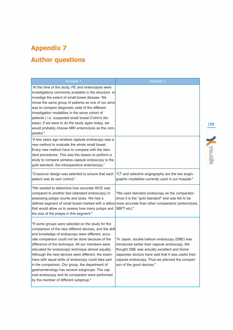
159
Appendix 7
Author questions
Answer 1 Answer 2
“At the time of the study, PE and enteroclysis were
investigations commonly available in the structure to
investige the extent of small bowel disease. We
chose the same group of patients as one of our aims
was to compare diagnostic yield of the different
investigation modalities in the same cohort of
patients ( i.e. suspected small bowel Crohn's dis-
ease). If we were to do the study again today, we
would probably choose MRI enteroclysis as the com-
parator."
“A few years ago wireless capsule endoscopy was a
new method to evaluate the whole small bowel.
Every new method have to compare with the stan-
dard procedures. This was the reason to perform a
study to compare wireless capsule endoscopy is the
gold standard, the intraoperative enteroscopy.”
“Crossover design was selected to ensure that each
patient was its own control.”
“CT and selective angiography are the two angio-
graphic modalities currently used in our hospital."
"We wanted to determine how accurate WCE was
compared to another test (standard endoscopy) in
assessing polyps counts and sizes. We had a
defined segment of small bowel marked with a tattoo
that would allow us to assess how many polyps and
the size of the polyps in this segment."
"We used standard endoscopy as the comparator
since it is the "gold standard" and was felt to be
more accurate than other comparators (enteroclysis,
SBFT etc)."
"If some groups were selected on the study for the
comparison of the new different devices, and the skill
and knowledge of endoscopy were different, accu-
rate comparison could not be done because of the
difference of the technique. All our members were
educated for endoscopic technique almost equally.
Although the new devices were different, the exam-
iners with equal skills of endoscopy could take part
in the comparison. Our group, the department of
gastroenterology has several subgroups. The cap-
sule endoscopy and its comparator were performed
by the member of different subgroup."
"In Japan, double balloon endoscopy (DBE) was
introduced earlier than capsule endoscopy. We
thought DBE was actually excellent and Some
Japanese doctors have said that it was useful than
capsule endoscopy. Thus we planned the compari-
son of the good devices."

160
"By introducing a new method it is of utmost impor-
tance to compare with other standard methods, of
course in the same group of patients."
"In our hospital, CT enteroclysis is the standard
method for diagnosing Crohn’s disease. I hope I
could answer your questions to your satisfaction."
"As any new diagnostic mean, we felt that a gold
standard was of importance to determine efficiently
the clinical usefulness of capsule endoscopy."
"Push enteroscopy was at this time the only avail-
able not so invasive although this comparator was
used by one german team. Please contact the first
writer of this email if you require further information
or clarification."

161
APPENDIX 8
Responding CentresRegion Responding Centre Manager of department In charge of
compilation
Piemonte
ASL BIELLA GIUSEPPE MALFITANA GIUSEPPE MALFITANA
ASO ORDINE MAURIZIANO RODOLFO CAVALIERE RAFFAELLO SOSTEGNI
ASL ALESSANDRIA CARLO GEMME CARLO GEMME
ASO SANTA CROCE RENZO FERRARIS RAFFAELLA PULITANO’
AO MAGGIORE DELLA CARITA’ MARIO DEL PIANO STEFANIA CARMAGNOLA
CASA DI CURA FORNACA FRANCESCO PAOLO ROSSINI FRANCESCO PAOLO ROSSINI
OSPEDALE MARIA VITTORIA ARNALDO FERRARI PAOLA SECRETO
OSPEDALE MOLINETTE MARIO RIZZETTO GIORGIO SARACCO
OSPEDALE SAN GIOVANNI AS ALBERTO FOGO MARCO PENNAZIO
OS SS TRINITA (PRESIDIO ASL13) PIETRO OCCHIPINTI SILVIA SAETTONE
OSPEDALE SAN LAZZARO GIACOMO PRANDI SABINA MARTINI
Valle d’Aosta OSPEDALE BEAUREGARA SERGIO CROTTA ROBERTO LOLLI
Lombardia
CASA DI CURA S.PIOX MARCO DAL FANTE MARCO DAL FANTE
IRCSS FONDAZIONE POLICLINICO MANAGIAGALLI
ROBERTO DE FRANCHIS EMANUELE RONDONOTTI
AO VALTELLINA E VALCHIAVENNA GIANFRANCO TRINCA CARLO PANSONI
AO SAN CARLO BORROMEO DOMINGO SPOTTI MARCO SONCINI
OSPEDALE SAN GIUSEPPE EDOARDO FESCE EDOARDO FESCE
FONDAZIONE POLIAMBULANZA ALESSANDRO PATERLINI PIETRO CESARI
AO CREMA ALESSANDRO ZAMBELLI GUIDO LUPINACCI
AO DI CIRCOLO MELEGNANO P.O MELLO IVANA RAGUZZI PASQUALE DI VITAGLIANO
OSPEDALE CIRCOLO BUSTO ARSIZIO EUGENIO LIMIDO GIRELLI
Veneto
AO VERONA LUCIANO BITI CARLO MANFRINI
AO DI PADOVA GIACOMO CARLO STURNIOLO RENATA D’INCA
ASL 5 OVEST VICENTINO ENRICO BOTTONA MAURIZIO PANTALENA
ULSS 2 FELTRE MICHELE DE BONI MICHELE DE BONI
Friuli Venezia
Giulia
CENTRO RIFERIMENTO ONCOLOGICO RENATO CANNIZZARO RENATO CANNIZZARO
IRCC BURLO GAROFALO CL PEDIATRICA STEFANO MARTELLOSSI STEFANO MARTELLOSSI
Liguria AZIENDA OSPEDALIERA SANTA CORONA ROBERTO TESTA CESARINA RAINISIO

162
Emilia Romagna
ARCISPEDALE SANTA MARIA NUOVA ROMANO SASSATELLI CRISTINA TIOLI
ASL OSP BELLARIA MAGGIORE NICOLA D’IMPERIO ANGELA ALBERANI
ARCISPEDALE SANTA ANNA SERGIO GULLINI FRANCO PEZZOLI
OSPEDALE MORGAGNI - PIERANTONI RICCI ENRICO LIONELLO GANDOLFI
Toscana
AO UNIVERSITARIA PISANA SANTINO MARCHI LINDA CECCARELLI
DIP GASTROENTEROLOGIA E MALRICAMBIO
ALFONSO CAPRIA EMANUELE TUMINO
Marche AO OSPEDALI RIUNITI IVANO LORENZINI IVANO LORENZINI
Lazio
AZIENDA OSPEDALIERA POLICLINI-CO UMBERTO I
PAOLO PAOLUZI PAOLO PAOLUZI
POLICLINICO GEMELLI GUIDO COSTAMGNA MARIA ELENA RICCIONI
AO SAN CAMILLO FORLANINI COSIMO PRANTERA TOMMASO PANTANELLA
POLICLINICO TOR VERGATA FRANCESCO PALLONE CARMELINA PETRUZIELLO
ACO SAN FILIPPO NERI LUCIO CAPURSO ROBERTO LUCCHETTI
Abruzzo ASL L’AQUILA PO SAN SALVATORE ANTONIETTA PISTOIA LORETO LOMBARDI
Molise ASR OSPEDALE S S ROSARIO TOMMASO GAGLIONE ROBERTO CHIAVERINI
Campania
OSPEDALE MERCATO SAN SEVERINOASL SA 2
ATTILIO MAURANO GELSONIMO QUAGLIARIELLO
AORN SAN G. MOSCATI GAETANO IAQUINTO SIMONE DI BELLA
OSPEDALE SAN GENNARO ASL NA1 SANTO MONASTRA GIANCARLO CAVARELLI
ASL SALERNO 3 PO POLLA RICCARDO MARMO RICCARDO MARMO
AO DI RILIEVO NAZIONALE ANTONIO CARDARELLI
GIAN BATTISTA FORTE MARIA PACELLI
Puglia
AZIENDA OSPEDALI RIUNITI DI FOGGIA
VINCENZO DI FRANCESCO VINCENZO DI FRANCESCO
OSPEDALE CIVILE UMBERTO I GIUSEPPE SCACCIANOCE GIUSEPPE SCACCIANOCE
ISTITUTO TUMORI BARI ANTONIO PELLECCHIA FRANCESCO SCOTTO
ASL1 TARANTO OSP. SANT'ANNUNZIATA
ANDREA FRUNZIO ANDREA FRUNZIO
Calabria
AO ANNUNZIATA PIETRO LEO PIETRO LEO
OSPEDALE SAN GIOVANNI DI DIO ENRICO CILIBERTO CESARE CAVALIERE
Sicilia
ASL 5 MESSINA SALVATORE OCERA SALVATORE OCERA
OSPEDALE VITTORIO EMANUELE ANTONIO MAGNANO ANTONIO MAGNANO
Sardegna AUSL 7 CARBONIA OSP. S. BARBARA SERGIO CADONI PAOLO GAUTIN

163
APPENDIX 9
Questionnaire
Wireless Capsule Endoscopy

164
Institution
Address
Region
Type of institution Public Private Private (operating within
the National Health Service)
Hospital Hospital Hospital
Outpatient Outpatient Outpatient
Other: Other: Other:
Phone
Manager of department …………………………
Phone
In charge of compilation …………………………

165
Information related to the examinations with Video Endoscopic Capsule (WCE)
1. Indicate the total number of esamination with WCE-year 2006 N° ……
2. Indicate the number of WCE for regime of health service admission N°
Hospitalization
Outpatient treatment
Day Hospital
3. Indicate the number of patients for age group N°
<30
30-50
>50
4. Incomplete/inadequate preparation:
N° %
– Repeated for inadequate preparation Real data Estimate
– Incomplete Real data Estimate
– Other
Real data Estimate
Real data Estimate
Real data Estimate
5. Diagnostic test workup pre WCE: N°
- Colonoscopy (CS)
- Rectoscopy (RT)

166
- FOBT
- EGDS
- RX small intestine
- Ileoscopy
- Double balloon enteroscopy
- Push enteroscopy
- Jejunoscopy
- Arteriography
- Other
6. Motivation of WCE: N°
- FOBT(+)
- FOBT(-)
- Familiar Polyposis
- Diagnosed IBD
- Suspected IBD
- Intestinal Neoplasm
- Angiodysplasia
- Peutz-Jeghers Syndrome
- Hepatic metastasis
- Other
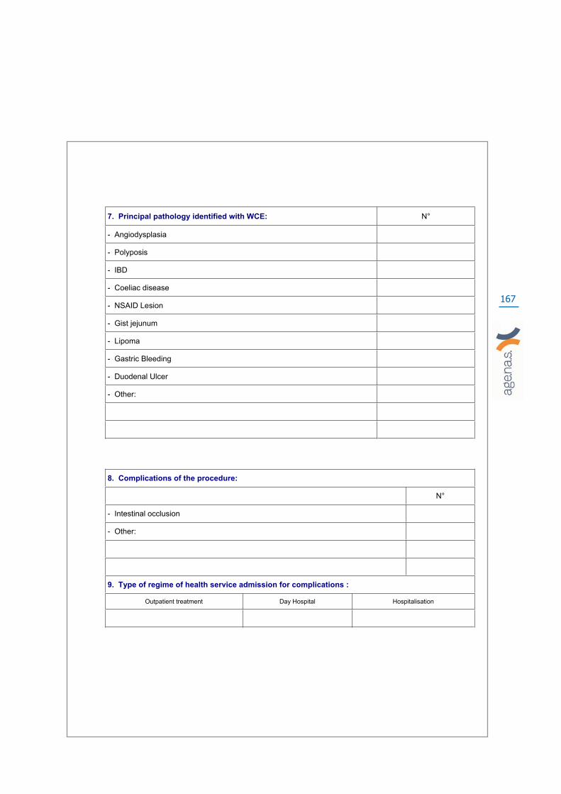
167
7. Principal pathology identified with WCE: N°
- Angiodysplasia
- Polyposis
- IBD
- Coeliac disease
- NSAID Lesion
- Gist jejunum
- Lipoma
- Gastric Bleeding
- Duodenal Ulcer
- Other:
8. Complications of the procedure:
N°
- Intestinal occlusion
- Other:
9. Type of regime of health service admission for complications :
Outpatient treatment Day Hospital Hospitalisation

168
10. Is a diet recommended before the examination? Yes No
11. If yes, specify
12. Is an enema recommend of before the examination? Yes No
13. Which solution is used for the intestinal preparation?
Poliethileneglicole (PEG)
Sodium-phosphate
Other (specify)
14. What percentage is furnished free of charge from the hospital?
– Outpatient treatment
– Day Hospital
15. Indicate the clinical or surgical decisions following the WCE:
1)
2)
3)

169
16. Are drugs used before the WCE examination?
Yes
No
17. If yes, specify:
Unit quantity Unit cost
–
–
18. Staff time (minutes) for one WCE procedure:
WCE Diagnosis Consultation
Physician
– Number
– Middle time dedicated (minutes)*
Nurse
– Number
– Average time dedicated (minutes)*
Support (auxiliaries)
– Number
– Average time dedicated (minutes)*
Administrative
– Number
– Average time dedicated (minutes)*
Total time
*for patient

170
19. Consumables for one standard WCE procedure:
Unit quantity Unit cost Manufacturer (only WCE)
Capsule Endoscopy
Test Capsule Endoscopy
Surgical gauze
Surgical gloves
Disinfectant
Sheet Disposable Reusable
Other:
Disposable Reusable
Disposable Reusable
Disposable Reusable
20. Indicate the stationery:
Unitquantity Unit cost
Toner
Cartridge
Sensitised paper
Other:
Disposable Reusable Both
Disposable Reusable Both
Disposable Reusable Both

171
21. Indicate the equipment:
Unit year Unit cost Maintenance cost Year of amortization
Hardware + software*
Tools
Software
Monitor
Printing
Computer
Equipped trolley
Other:
* Total cost

172

173
APPENDIX 10
Wireless capsule and traditional endoscopy:
patient acceptability

174
1. The ingestion of the capsule has been:
Very easy
Easy
Tolerable
Difficult
Very difficult
2. Did you experience pain during the procedure?
No pain
Mild
Moderate
Severe
Extreme
3. Did you experience pain after the procedure?
No pain
Mild
Moderate
Severe
Extreme
4. During the hours of tracking which of the following inconveniences did you have?:
A) Difficultyto move
Yes No
B Impossibility to go in some places:
Yes No
C) State of anxiety
Yes No
Minimum Minimum Minimum
Medium Medium Medium
Severe Severe Severe
Very Severe Very Severe Very Severe
5. How would appraise the overall tolerability of this type of procedure?
High
Middle
Low

175
6. Time subtracted from normal activities
0-2 hours
3-4 hours
5-6 hours
7-8 hours
> 8 hours
7. How would you evaluate the way the results were communicated?
Optimal
Good
Mean
Insufficient
Bad
8. Have you undergone other endoscopic procedures?
Colonoscopy/Ileoscopy
EGDS
The questions that follow concern the endoscopic procedure referred to in question 8
9. The procedure CS/EGDS was:
Very easy
Easy
Tolerable
Difficult
Very difficult
10. Did you experience pain during the procedure?
No pain
Mild
Moderate
Severe
Extreme
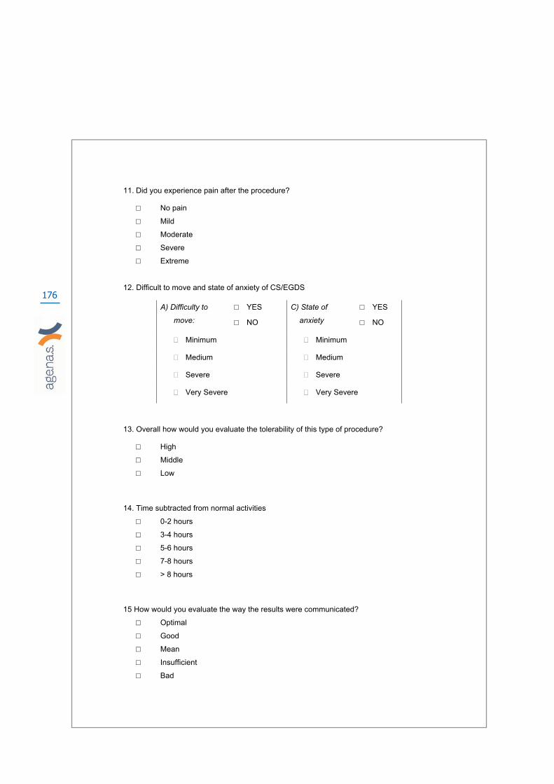
176
11. Did you experience pain after the procedure?
No pain
Mild
Moderate
Severe
Extreme
12. Difficult to move and state of anxiety of CS/EGDS
A) Difficulty to
move:
YES
NO
C) State of
anxiety
YES
NO
Minimum Minimum
Medium Medium
Severe Severe
Very Severe Very Severe
13. Overall how would you evaluate the tolerability of this type of procedure?
High
Middle
Low
14. Time subtracted from normal activities
0-2 hours
3-4 hours
5-6 hours
7-8 hours
> 8 hours
15 How would you evaluate the way the results were communicated?
Optimal
Good
Mean
Insufficient
Bad

177
Additional information
16. Age _____
17. Gender:
M
F
18. Status
Single
Married
Cohabiting
19.Education:
Primary school
Secondary school
Degree
Master
20. Occupation:
worker
Employee
Manager
Trader
Entrepreneur
Student
Housewife
Consultant
Retired
Unemployed
Other


age.na.s - Agenzia Nazionale per i Servizi Sanitari Regionali
Sezione A.G.P. - Centro Stampa
Via Puglie 23, 00187 – Roma . Tel. 06.427491 – fax. 06.42749488
www.agenas.it e-mail [email protected]