Embed Size (px)
Citation preview
HSC Biology
Maintaining a Balance
Week 5
T: (02) 8007 6824 E: [email protected]
W: www.duxcollege.com.au
DUX
Student name: ……………………………….
Class code: ……………………………………..
Teacher name: ……………………………….
Need help? Visit the student forums and ask our tutors. www.duxcollege.com.au/forums
HSC Biology 1 Maintaining a Balance
© Dux College 2015 | Do not distribute
T: (02) 8007 6824 E: [email protected]
W: www.duxcollege.com.au
DUX
WEEK 5 – THEORY
PLANTS AND ANIMALS REGULATE THE CONCENTRATION OF GASES,
WATER AND WASTE PRODUCTS OF METABOLISM IN CELLS AND IN
INTERSTITIAL FLUID
Explain why the concentration of water in cells should be maintained within a narrow
range for optimal function
ROLE OF WATER IN CELLS
Chemically, we are the product of a myriad of different metabolic reactions, and water is of vital
importance in this process:
Water is the medium in which all biochemical reactions take place – without water and its
excellent solvent properties, our metabolic reactions could never function.
Water also serves as a reactant in some chemical reactions, such as photosynthesis.
Recall that enzymes are required for adequate metabolism, and that enzyme function depends on a
number of factors including temperature, substrate concentration and pH. Interestingly, water affects
all three of these factors:
1. Substrate concentration: the concentration of substrates refers to the amount of the
substrate in a given volume of water. Enzyme function is maximised at the saturation point
i.e. where substrate concentration is such that every active site of all the enzymes are
occupied. In order to maintain this concentration of substrate, the volume of water in the
cell must be controlled – increasing the volume of water will dilute the substrate while
decreasing the volume will concentrate the substrate.
2. pH: recall that pH is actually the hydrogen ion concentration in water and that a high pH
refers to a low hydrogen ion concentration (basic) while a low pH refers to a high hydrogen
ion concentration (acidic). Since pH is a measure of concentration, it too depends on the
volume of water present in the same way that substrate concentration does.
3. Temperature: water is actually of vital importance when it comes to the maintenance of
temperature. This is because water has a high specific heat, meaning it takes a lot of heat
energy to actually increase the temperature of water. Thus, water acts as a kind of buffer,
slowing temperature changes within the cell, making it easier for the cell to maintain the
optimum temperature for enzyme function.
Water is also important in maintaining cell turgidity. This is essentially how “swollen” the cell is and is
thus important in maintaining the shape of the cell. The concentration of the outside environment
compared to the cellular contents can result in one of three situations:
Need help? Visit the student forums and ask our tutors. www.duxcollege.com.au/forums
HSC Biology 2 Maintaining a Balance
© Dux College 2015 | Do not distribute
T: (02) 8007 6824 E: [email protected]
W: www.duxcollege.com.au
DUX
1. Isotonic: where surroundings are of equal concentration to the cellular contents, so that
there is no net movement of water. The cell maintains a constant shape since the water
volume is constant. This is the situation most cells aim for.
2. Hypertonic: where the surroundings are more concentrated than the environment.
Remember that when solutions separated by a semi-permeable barrier are of different
concentrations, water will move across the barrier in order to equalise the concentrations.
Thus, in the case of hypertonic solutions, water moves out of the cell in order to try and
dilute the surroundings, making them closer to the cellular concentration. Since water is
leaving the cell, it loses turgidity and shrivels up.
3. Hypotonic: where the surroundings are less concentrated than the cellular contents. This
causes water to move into the cells to try and make the cell less concentrated/make the
surroundings more concentrated. As a result, cell volume increases and the cell swells up,
possibly bursting if enough water enters.
Explain why the removal of wastes is essential for continued metabolic activity
Metabolic wastes are any substances produced by cellular metabolism which are useless to the body.
There are 3 main reasons these wastes must be removed from cells:
Toxicity: metabolic wastes are frequently toxic in sufficiently high concentrations, often due
to their effects on the pH of cellular contents. One example is carbon dioxide, which acidifies
cell contents, while the highly toxic compound ammonia makes cell contents more basic. In
both of these cases, enzyme function may be compromised, as pH’s outside the optimum
range will reduce enzyme activity and possibly denature them. This in turn reduces metabolic
efficiency. Other toxins may directly attack the structure of cells, which, if severe enough,
can kill the cell outright.
Slowing metabolic reactions: metabolic reactions are almost invariably equilibria. What this
means is outside the scope of the course, but a buildup of the products of a metabolic
reaction (including the wastes) will slow the reaction, preventing the formation of essential
metabolites.
Space considerations: regardless of the toxic effects and effects on reactions, the presence
of large amounts of wastes occupies much needed space in the cell, obstructing other
processes.
Examples of wastes include carbon dioxide, hydrogen ions and the nitrogenous wastes (ammonia,
urea and uric acid).
Need help? Visit the student forums and ask our tutors. www.duxcollege.com.au/forums
HSC Biology 3 Maintaining a Balance
© Dux College 2015 | Do not distribute
T: (02) 8007 6824 E: [email protected]
W: www.duxcollege.com.au
DUX
Explain why the processes of diffusion and osmosis are inadequate in removing dissolved
nitrogenous wastes in some organisms
Identify the role of the kidney in the excretory system of fish and mammals
Single celled and other very small organisms are able to deal with the problem of waste accumulation
quite easily – since the wastes are only being produced inside the cell, the concentration of the waste
compounds is always going to be higher inside the cell than it is outside the cell. Hence
Wastes will move out of the cell by diffusion
Water will move into the cell by osmosis, diluting the concentration of wastes inside the cell
and hence reducing their toxicity.
The reason these processes are sufficient to cater to the needs of microbes is because over small
distances they are relatively efficient. However, since both osmosis and diffusion are products of
random particle movement (Brownian motion) molecules take a long time to travel long distances.
Thus, in larger organisms diffusion and osmosis would not be rapid enough to prevent the detrimental
effects of waste build up. This is why larger organisms have evolved more sophisticated ways of
solving the problem.
The kidney is the body’s filtration system, responsible for, among other things, removing nitrogenous
wastes from the blood. Blood enters the kidneys via the renal artery and, having been filtered, leaves
via the renal vein, where it re-enters systemic circulation.
The urine produced by the kidneys travels via the ureters to the bladder, where it is excreted from the
body through the urethra, removing nitrogenous wastes with it.
Filtration of nitrogenous wastes is not the only role of the kidney – in fact is has many, but notably it is
also a major player in the maintenance of body water and salt concentration, a role known as
osmoregulation:
Osmoregulation is the major function of the kidneys for fish, as they excrete most of their
nitrogenous wastes from their gills. Excretion in fish will be dealt with in more detail next
week.
Need help? Visit the student forums and ask our tutors. www.duxcollege.com.au/forums
HSC Biology 4 Maintaining a Balance
© Dux College 2015 | Do not distribute
T: (02) 8007 6824 E: [email protected]
W: www.duxcollege.com.au
DUX
Explain how the processes of filtration and reabsorption in the mammalian nephron
regulate body fluid composition
Distinguish between active and passive transport and relate these to processes occurring in
the mammalian kidney
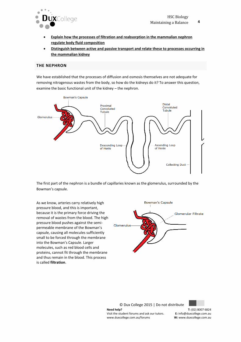
THE NEPHRON
We have established that the processes of diffusion and osmosis themselves are not adequate for
removing nitrogenous wastes from the body, so how do the kidneys do it? To answer this question,
examine the basic functional unit of the kidney – the nephron.
The first part of the nephron is a bundle of capillaries known as the glomerulus, surrounded by the
Bowman’s capsule.
As we know, arteries carry relatively high pressure blood, and this is important, because it is the primary force driving the removal of wastes from the blood. The high pressure blood pushes against the semi-permeable membrane of the Bowman’s capsule, causing all molecules sufficiently small to be forced through the membrane into the Bowman’s Capsule. Larger molecules, such as red blood cells and proteins, cannot fit through the membrane and thus remain in the blood. This process is called filtration.
Need help? Visit the student forums and ask our tutors. www.duxcollege.com.au/forums
HSC Biology 5 Maintaining a Balance
© Dux College 2015 | Do not distribute
T: (02) 8007 6824 E: [email protected]
W: www.duxcollege.com.au
DUX
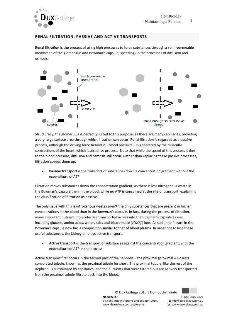
RENAL FILTRATION, PASSIVE AND ACTIVE TRANSPORTS
Renal filtration is the process of using high pressures to force substances through a semi-permeable
membrane of the glomerulus and Bowman’s capsule, speeding up the processes of diffusion and
osmosis.
Structurally, the glomerulus is perfectly suited to this purpose, as there are many capillaries, providing
a very large surface area through which filtration can occur. Renal filtration is regarded as a passive
process, although the driving force behind it – blood pressure – is generated by the muscular
contractions of the heart, which is an active process. Note that while the speed of this process is due
to the blood pressure, diffusion and osmosis still occur. Rather than replacing these passive processes,
filtration speeds them up.
Passive transport is the transport of substances down a concentration gradient without the
expenditure of ATP
Filtration moves substances down the concentration gradient, as there is less nitrogenous waste in
the Bowman’s capsule than in the blood, while no ATP is consumed at the site of transport, explaining
the classification of filtration as passive.
The only issue with this is nitrogenous wastes aren’t the only substances that are present in higher
concentrations in the blood than in the Bowman’s capsule. In fact, during the process of filtration,
many important nutrient molecules are transported across into the Bowman’s capsule as well,
including glucose, amino acids, water, salts and bicarbonate (𝐻𝐶𝑂3−) ions. As such, the filtrate in the
Bowman’s capsule now has a composition similar to that of blood plasma. In order not to lose these
useful substances, the kidney employs active transport.
Active transport is the transport of substances against the concentration gradient, with the
expenditure of ATP in the process.
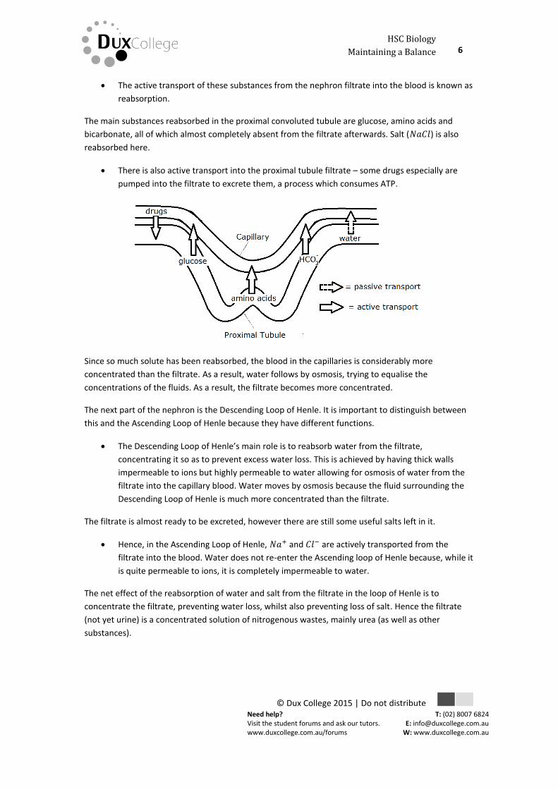
Active transport first occurs in the second part of the nephron – the proximal (proximal = closest)
convoluted tubule, known as the proximal tubule for short. The proximal tubule, like the rest of the
nephron, is surrounded by capillaries, and the nutrients that were filtered out are actively transported
from the proximal tubule filtrate back into the blood.
Need help? Visit the student forums and ask our tutors. www.duxcollege.com.au/forums
HSC Biology 6 Maintaining a Balance
© Dux College 2015 | Do not distribute
T: (02) 8007 6824 E: [email protected]
W: www.duxcollege.com.au
DUX
The active transport of these substances from the nephron filtrate into the blood is known as
reabsorption.
The main substances reabsorbed in the proximal convoluted tubule are glucose, amino acids and
bicarbonate, all of which almost completely absent from the filtrate afterwards. Salt (𝑁𝑎𝐶𝑙) is also
reabsorbed here.
There is also active transport into the proximal tubule filtrate – some drugs especially are
pumped into the filtrate to excrete them, a process which consumes ATP.
Since so much solute has been reabsorbed, the blood in the capillaries is considerably more
concentrated than the filtrate. As a result, water follows by osmosis, trying to equalise the
concentrations of the fluids. As a result, the filtrate becomes more concentrated.
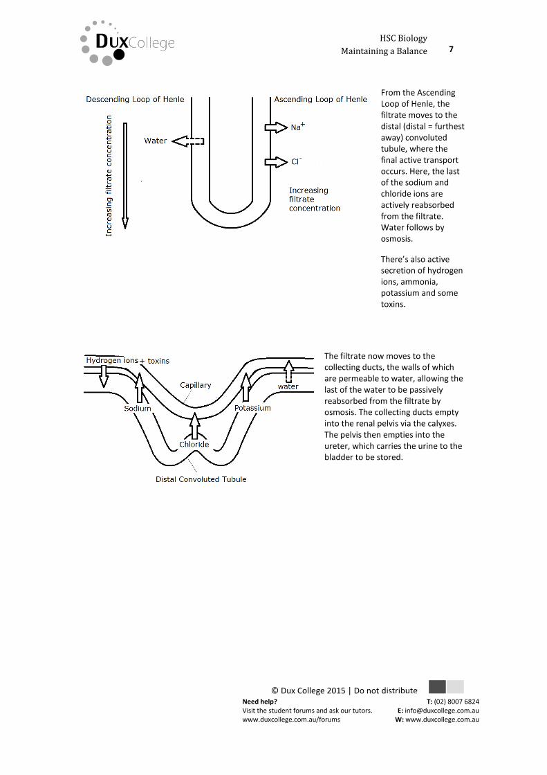
The next part of the nephron is the Descending Loop of Henle. It is important to distinguish between
this and the Ascending Loop of Henle because they have different functions.
The Descending Loop of Henle’s main role is to reabsorb water from the filtrate,
concentrating it so as to prevent excess water loss. This is achieved by having thick walls
impermeable to ions but highly permeable to water allowing for osmosis of water from the
filtrate into the capillary blood. Water moves by osmosis because the fluid surrounding the
Descending Loop of Henle is much more concentrated than the filtrate.
The filtrate is almost ready to be excreted, however there are still some useful salts left in it.
Hence, in the Ascending Loop of Henle, 𝑁𝑎+ and 𝐶𝑙− are actively transported from the
filtrate into the blood. Water does not re-enter the Ascending loop of Henle because, while it
is quite permeable to ions, it is completely impermeable to water.
The net effect of the reabsorption of water and salt from the filtrate in the loop of Henle is to
concentrate the filtrate, preventing water loss, whilst also preventing loss of salt. Hence the filtrate
(not yet urine) is a concentrated solution of nitrogenous wastes, mainly urea (as well as other
substances).
Need help? Visit the student forums and ask our tutors. www.duxcollege.com.au/forums
HSC Biology 7 Maintaining a Balance
© Dux College 2015 | Do not distribute
T: (02) 8007 6824 E: [email protected]
W: www.duxcollege.com.au
DUX
From the Ascending Loop of Henle, the filtrate moves to the distal (distal = furthest away) convoluted tubule, where the final active transport occurs. Here, the last of the sodium and chloride ions are actively reabsorbed from the filtrate. Water follows by osmosis.
There’s also active secretion of hydrogen ions, ammonia, potassium and some toxins.
The filtrate now moves to the collecting ducts, the walls of which are permeable to water, allowing the last of the water to be passively reabsorbed from the filtrate by osmosis. The collecting ducts empty into the renal pelvis via the calyxes. The pelvis then empties into the ureter, which carries the urine to the bladder to be stored.
Need help? Visit the student forums and ask our tutors. www.duxcollege.com.au/forums
HSC Biology 8 Maintaining a Balance
© Dux College 2015 | Do not distribute
T: (02) 8007 6824 E: [email protected]
W: www.duxcollege.com.au
DUX
SUMMARY
The following is a summary of the movement of substances throughout the nephron:
Region of Nephron Substances Transported Type of Transport
Glomerulus Glucose, amino acids, salts, bicarbonate, urea, uric acid, potassium, calcium
Passive transport –filtration
Proximal Convoluted Tubule Glucose, amino acids, bicarbonate, salts Drugs Water
Active transport – reabsorption
Active transport – secretion Passive transport - Osmosis
Descending Loop of Henle Water Passive transport - Osmosis
Ascending Loop of Henle Sodium, Chloride, Potassium Active transport – reabsorption
Distal Convoluted Tubule Sodium, Chloride, Potassium Hydrogen ions, Potassium, Ammonia
Active transport – reabsorption Active transport – secretion
Collecting ducts Water Passive transport - osmosis
Outline the role of the hormones, aldosterone and ADH (anti-diuretic hormone) in the
regulation of water and salt levels in blood
Present information to outline the general use of hormone replacement therapy in people
who cannot secrete aldosterone
ROLE OF ADH AND ALDOSTERONE
Since they control salt and water balance in the body, the kidneys is very important in homeostasis
and, as with all homeostatic systems, they are subject to feedback which alters their function.
Specifically, the function of the kidneys is controlled by the pituitary and the adrenal glands.
The pituitary is located in the brain and is the controller of virtually all the body’s endocrine functions
through the release of one of many hormones. The hormone we are concerned with is anti-diuretic
hormone (ADH) or vasopressin. As the name suggests (dieresis = urination), anti-diuretic hormone
reduces the need and frequency of urination by reducing urine output. The way it does so is by acting
on the collecting ducts of the kidney. The main functions of the collecting duct are reabsorption of
water and to carry urine to the ureter.
Need help? Visit the student forums and ask our tutors. www.duxcollege.com.au/forums
HSC Biology 9 Maintaining a Balance
© Dux College 2015 | Do not distribute
T: (02) 8007 6824 E: [email protected]
W: www.duxcollege.com.au
DUX
ADH affects the former by increasing the permeability of the collecting duct walls.
This in turn increases the amount of water reabsorbed from the urine. Thus
ADH serves to maintain body fluids by reabsorbing water from the urine, concentrating it in
the process.
This means that when the body’s fluid balance is low, or the blood salt concentration is high, ADH is
released to increase the amount of water reabsorbed from the urine, reducing water loss and
increasing blood volume.
The other controllers of kidney function are the adrenal glands – small glands which sit on top of the
kidneys and which secrete, among other hormones, adrenaline and aldosterone.
Aldosterone is very important in osmoregulation, because of its effects on the distal tubule of the
kidney.
Aldosterone increases the reabsorption of sodium, decreases the reabsorption of potassium
in the distal convoluted tubules.
Thus, aldosterone release is increased when low salt concentrations are detected, however this is not
the only effect of aldosterone. With increased sodium reabsorption, the blood in the capillaries
becomes more concentrated than the filtrate in the distal convoluted tubule. As such, water moves by
osmosis to try and equalise the concentrations.
Thus, aldosterone also increases water reabsorption and blood volume.
Interestingly, both ADH and aldosterone increase the efficiency of kidney function by increasing blood
volume. An increase in blood volume causes a consequent increase in blood pressure – the driving
force behind the process of filtration.
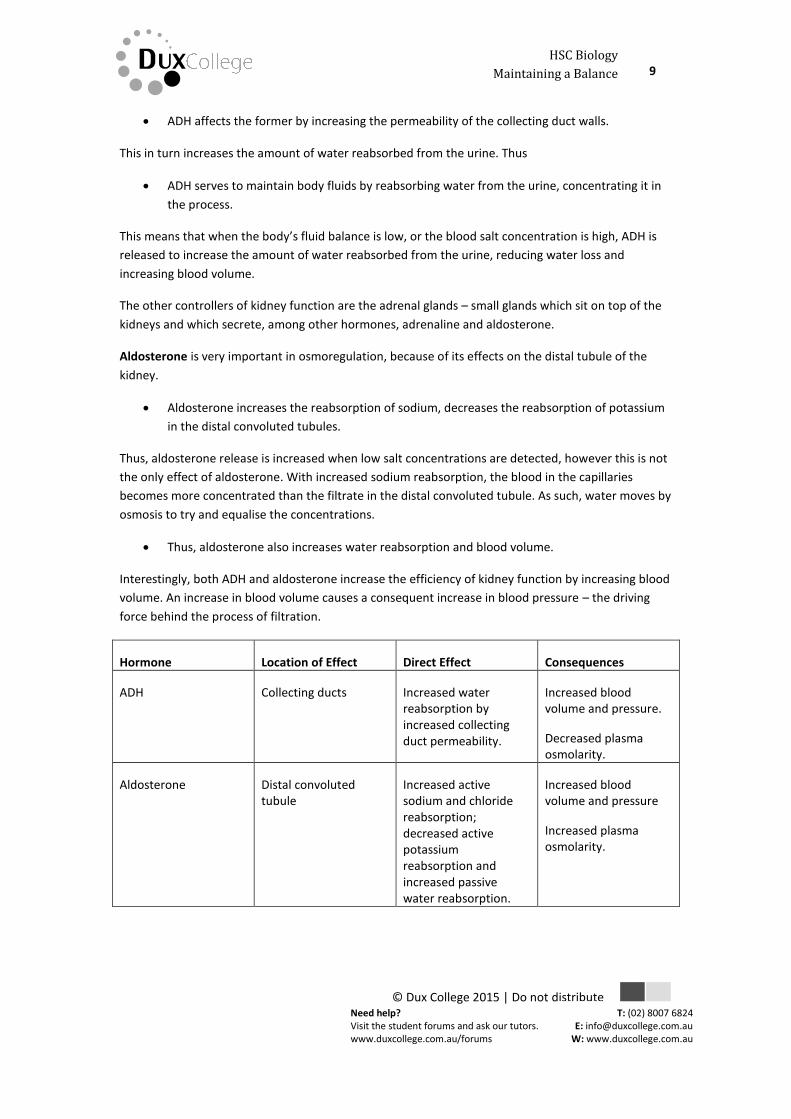
Hormone Location of Effect Direct Effect Consequences
ADH Collecting ducts Increased water reabsorption by increased collecting duct permeability.
Increased blood volume and pressure.
Decreased plasma osmolarity.
Aldosterone Distal convoluted tubule
Increased active sodium and chloride reabsorption; decreased active potassium reabsorption and increased passive water reabsorption.
Increased blood volume and pressure
Increased plasma osmolarity.
Need help? Visit the student forums and ask our tutors. www.duxcollege.com.au/forums
HSC Biology 10 Maintaining a Balance
© Dux College 2015 | Do not distribute
T: (02) 8007 6824 E: [email protected]
W: www.duxcollege.com.au
DUX
HORMONE REPLACEMENT THERAPY
The importance these hormones can be illustrated by examining those who don’t have them.
Addison’s disease is characterized by hypoaldosteronism, or abnormally low aldosterone levels. Due
to this lack of aldosterone and the consequent lack of water and salt reabsorption, sufferers of
Addison’s disease tend to have very low blood pressure (due to low blood volume) and consequent
fatigue and light headedness, as well as a host of other symptoms, notably including a strong craving
for salty foods, due to the low salt content of the blood.
This possibly life threatening disease is caused by a lack of cortisol (which stimulates the adrenal
glands to produce aldosterone) or an adrenal gland dysfunction which prevents the formation of
aldosterone. Thus, there are two types of hormone replacement therapy available for sufferers:
Hydrocortisone or prednisone to replace the cortisol, stimulating the adrenal glands to
produce more aldosterone.
Fludrocortisone to replace the aldosterone for those who can’t manufacture aldosterone at
all.
These treatments are very effective and once put in place, sufferers generally live perfectly normal
lives, however care should be exercised to prevent excess water retention leading to high blood
pressures and possible heart attack.
Gather, process and analyse information from secondary sources to compare the process
of renal dialysis with the function of the kidney
It should be clear by now that the kidneys are essential for normal body function, not just for the
removal of wastes, but also in the maintenance of body water and salt homeostasis. This being said,
kidney failure is quite obviously a very serious condition which, if untreated, invariably leads to death
in a short time. The long term treatment of people with kidney failure is to perform a kidney
transplant, however in the short term they need to be kept alive while they wait for the transplant.
Renal dialysis is the method used to keep patients with renal failure alive.
Renal dialysis is a process which approximates the function of the kidney and there are two main
types:
Haemodialysis
Peritoneal dialysis
Need help? Visit the student forums and ask our tutors. www.duxcollege.com.au/forums
HSC Biology 11 Maintaining a Balance
© Dux College 2015 | Do not distribute
T: (02) 8007 6824 E: [email protected]
W: www.duxcollege.com.au
DUX
HAEMODIALYSIS
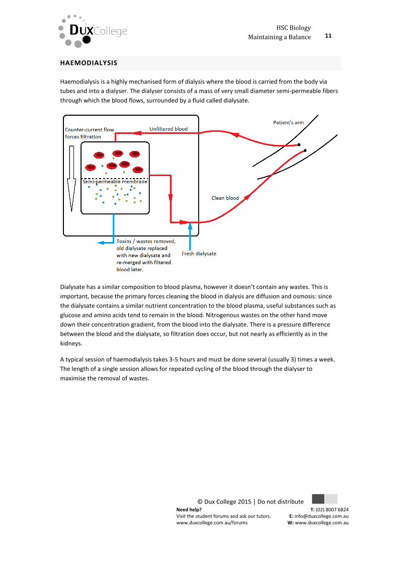
Haemodialysis is a highly mechanised form of dialysis where the blood is carried from the body via
tubes and into a dialyser. The dialyser consists of a mass of very small diameter semi-permeable fibers
through which the blood flows, surrounded by a fluid called dialysate.
Dialysate has a similar composition to blood plasma, however it doesn’t contain any wastes. This is
important, because the primary forces cleaning the blood in dialysis are diffusion and osmosis: since
the dialysate contains a similar nutrient concentration to the blood plasma, useful substances such as
glucose and amino acids tend to remain in the blood. Nitrogenous wastes on the other hand move
down their concentration gradient, from the blood into the dialysate. There is a pressure difference
between the blood and the dialysate, so filtration does occur, but not nearly as efficiently as in the
kidneys.
A typical session of haemodialysis takes 3-5 hours and must be done several (usually 3) times a week.
The length of a single session allows for repeated cycling of the blood through the dialyser to
maximise the removal of wastes.
Need help? Visit the student forums and ask our tutors. www.duxcollege.com.au/forums
HSC Biology 12 Maintaining a Balance
© Dux College 2015 | Do not distribute
T: (02) 8007 6824 E: [email protected]
W: www.duxcollege.com.au
DUX
PERITONEAL DIALYSIS
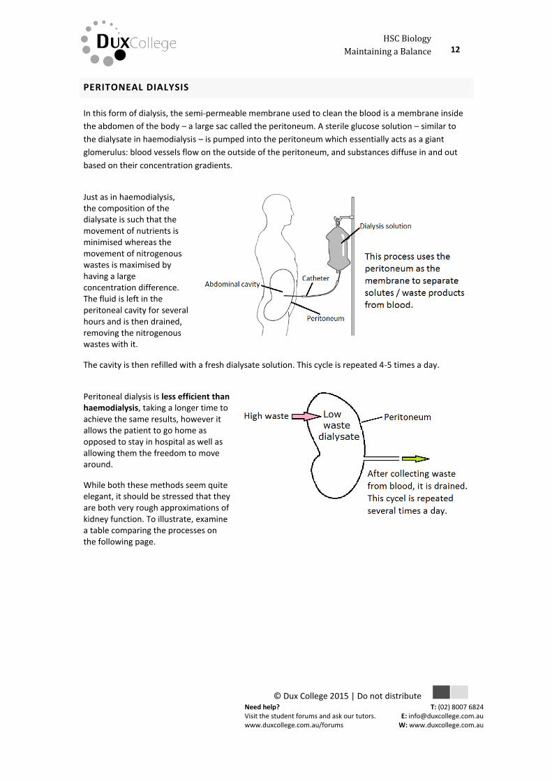
In this form of dialysis, the semi-permeable membrane used to clean the blood is a membrane inside
the abdomen of the body – a large sac called the peritoneum. A sterile glucose solution – similar to
the dialysate in haemodialysis – is pumped into the peritoneum which essentially acts as a giant
glomerulus: blood vessels flow on the outside of the peritoneum, and substances diffuse in and out
based on their concentration gradients.
Just as in haemodialysis, the composition of the dialysate is such that the movement of nutrients is minimised whereas the movement of nitrogenous wastes is maximised by having a large concentration difference. The fluid is left in the peritoneal cavity for several hours and is then drained, removing the nitrogenous wastes with it.
The cavity is then refilled with a fresh dialysate solution. This cycle is repeated 4-5 times a day.
Peritoneal dialysis is less efficient than haemodialysis, taking a longer time to achieve the same results, however it allows the patient to go home as opposed to stay in hospital as well as allowing them the freedom to move around.
While both these methods seem quite elegant, it should be stressed that they are both very rough approximations of kidney function. To illustrate, examine a table comparing the processes on the following page.
Need help? Visit the student forums and ask our tutors. www.duxcollege.com.au/forums
HSC Biology 13 Maintaining a Balance
© Dux College 2015 | Do not distribute
T: (02) 8007 6824 E: [email protected]
W: www.duxcollege.com.au
DUX
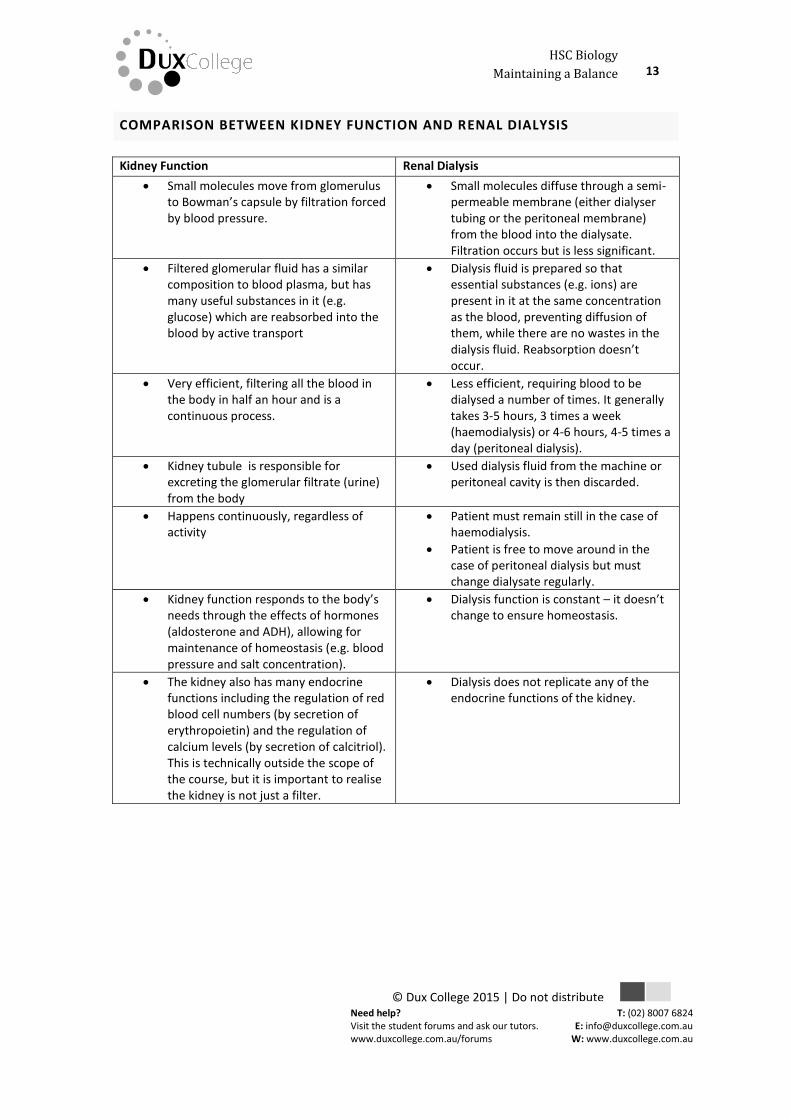
COMPARISON BETWEEN KIDNEY FUNCTION AND RENAL DIALYSIS
Kidney Function Renal Dialysis
Small molecules move from glomerulus to Bowman’s capsule by filtration forced by blood pressure.
Small molecules diffuse through a semi-permeable membrane (either dialyser tubing or the peritoneal membrane) from the blood into the dialysate. Filtration occurs but is less significant.
Filtered glomerular fluid has a similar composition to blood plasma, but has many useful substances in it (e.g. glucose) which are reabsorbed into the blood by active transport
Dialysis fluid is prepared so that essential substances (e.g. ions) are present in it at the same concentration as the blood, preventing diffusion of them, while there are no wastes in the dialysis fluid. Reabsorption doesn’t occur.
Very efficient, filtering all the blood in the body in half an hour and is a continuous process.
Less efficient, requiring blood to be dialysed a number of times. It generally takes 3-5 hours, 3 times a week (haemodialysis) or 4-6 hours, 4-5 times a day (peritoneal dialysis).
Kidney tubule is responsible for excreting the glomerular filtrate (urine) from the body
Used dialysis fluid from the machine or peritoneal cavity is then discarded.
Happens continuously, regardless of activity
Patient must remain still in the case of haemodialysis.
Patient is free to move around in the case of peritoneal dialysis but must change dialysate regularly.
Kidney function responds to the body’s needs through the effects of hormones (aldosterone and ADH), allowing for maintenance of homeostasis (e.g. blood pressure and salt concentration).
Dialysis function is constant – it doesn’t change to ensure homeostasis.
The kidney also has many endocrine functions including the regulation of red blood cell numbers (by secretion of erythropoietin) and the regulation of calcium levels (by secretion of calcitriol). This is technically outside the scope of the course, but it is important to realise the kidney is not just a filter.
Dialysis does not replicate any of the endocrine functions of the kidney.
Need help? Visit the student forums and ask our tutors. www.duxcollege.com.au/forums
HSC Biology 14 Maintaining a Balance
© Dux College 2015 | Do not distribute
T: (02) 8007 6824 E: [email protected]
W: www.duxcollege.com.au
DUX
WEEK 5 – HOMEWORK
Explain why the concentration of water in cells should be maintained within a narrow
range for optimal function
1. A cell is placed in solution and shrivels up. Account for this observation. [3 marks]
………………………………………………………………………………………………………………………………………………………………
………………………………………………………………………………………………………………………………………………………………
………………………………………………………………………………………………………………………………………………………………
………………………………………………………………………………………………………………………………………………………………
………………………………………………………………………………………………………………………………………………………………
………………………………………………………………………………………………………………………………………………………………
Explain why the removal of wastes is essential for continued metabolic activity
1. Carbon dioxide is a metabolic waste.
a. Identify one metabolic reaction which produces carbon dioxide as a waste. [1 mark]
………………………………………………………………………………………………………………………………………………………………
b. Predict the effect of a build up of carbon dioxide in a cell, justifying your answer. [2
marks]
………………………………………………………………………………………………………………………………………………………………
………………………………………………………………………………………………………………………………………………………………
………………………………………………………………………………………………………………………………………………………………
………………………………………………………………………………………………………………………………………………………………
2. Outline THREE reasons why metabolic wastes should not be allowed to accumulate in cells.
[3 marks]
………………………………………………………………………………………………………………………………………………………………
………………………………………………………………………………………………………………………………………………………………
………………………………………………………………………………………………………………………………………………………………
………………………………………………………………………………………………………………………………………………………………
………………………………………………………………………………………………………………………………………………………………
………………………………………………………………………………………………………………………………………………………………
Need help? Visit the student forums and ask our tutors. www.duxcollege.com.au/forums
HSC Biology 15 Maintaining a Balance
© Dux College 2015 | Do not distribute
T: (02) 8007 6824 E: [email protected]
W: www.duxcollege.com.au
DUX
Explain why the processes of diffusion and osmosis are inadequate in removing dissolved
nitrogenous wastes in some organisms
1. Discuss the statement “No organism could survive if its waste removal system consisted
solely of diffusion and osmosis.” [4 marks]
………………………………………………………………………………………………………………………………………………………………
………………………………………………………………………………………………………………………………………………………………
………………………………………………………………………………………………………………………………………………………………
………………………………………………………………………………………………………………………………………………………………
………………………………………………………………………………………………………………………………………………………………
………………………………………………………………………………………………………………………………………………………………
………………………………………………………………………………………………………………………………………………………………
………………………………………………………………………………………………………………………………………………………………
Identify the role of the kidney in the excretory system of fish and mammals
Explain how the processes of filtration and reabsorption in the mammalian nephron
regulate body fluid composition
Distinguish between active and passive transport and relate these to processes occurring in
the mammalian kidney
1. Compare and contrast the role of the kidneys in mammals with the role of the kidneys in fish.
[2 marks]
………………………………………………………………………………………………………………………………………………………………
………………………………………………………………………………………………………………………………………………………………
………………………………………………………………………………………………………………………………………………………………
………………………………………………………………………………………………………………………………………………………………
………………………………………………………………………………………………………………………………………………………………
Need help? Visit the student forums and ask our tutors. www.duxcollege.com.au/forums
HSC Biology 16 Maintaining a Balance
© Dux College 2015 | Do not distribute
T: (02) 8007 6824 E: [email protected]
W: www.duxcollege.com.au
DUX
2. Draw a labelled diagram of a kidney (not dissected) showing all vessels entering and leaving
the kidney. [3 marks]
3. Draw a fully labelled diagram of a nephron. In your diagram, identify the sites where: [4
marks]
Filtration occurs (label these area(s) F)
Reabsorption occurs (label these area(s) R)
Secretion occurs (label these area(s) S)
Need help? Visit the student forums and ask our tutors. www.duxcollege.com.au/forums
HSC Biology 17 Maintaining a Balance
© Dux College 2015 | Do not distribute
T: (02) 8007 6824 E: [email protected]
W: www.duxcollege.com.au
DUX
Outline the role of the hormones, aldosterone and ADH (anti-diuretic hormone) in the
regulation of water and salt levels in blood
Present information to outline the general use of hormone replacement therapy in people
who cannot secrete aldosterone
1.
a. Identify the name of the condition characterised by hypoaldosteronism. [1 mark]
………………………………………………………………………………………………………………………………………………………………
b. Describe ONE symptom found in people suffering this condition [2 marks]
………………………………………………………………………………………………………………………………………………………………
………………………………………………………………………………………………………………………………………………………………
………………………………………………………………………………………………………………………………………………………………
………………………………………………………………………………………………………………………………………………………………
c. Outline the treatment of this condition. [2 marks]
………………………………………………………………………………………………………………………………………………………………
………………………………………………………………………………………………………………………………………………………………
………………………………………………………………………………………………………………………………………………………………
………………………………………………………………………………………………………………………………………………………………
Need help? Visit the student forums and ask our tutors. www.duxcollege.com.au/forums
HSC Biology 18 Maintaining a Balance
© Dux College 2015 | Do not distribute
T: (02) 8007 6824 E: [email protected]
W: www.duxcollege.com.au
DUX
Gather, process and analyse information from secondary sources to compare the process
of renal dialysis with the function of the kidney
1. Compare and contrast renal dialysis with normal kidney function and assess its effectiveness
treating patients with kidney failure. [6 marks]
Renal Dialysis Kidney function
………………………………………………………………………………………………………………………………………………………………
………………………………………………………………………………………………………………………………………………………………
………………………………………………………………………………………………………………………………………………………………
………………………………………………………………………………………………………………………………………………………………
………………………………………………………………………………………………………………………………………………………………
………………………………………………………………………………………………………………………………………………………………
End of homework