Embed Size (px)
Citation preview
Human Papillomavirus Episome Stability Is Reduced by Aphidicolinand Controlled by DNA Damage Response Pathways
Terri G. Edwards,a Michael J. Helmus,a Kevin Koeller,b James K. Bashkin,a,b Chris Fishera
NanoVir, Kalamazoo, Michigan, USAa; University of Missouri—St. Louis, Department of Chemistry & Biochemistry, Center for Nanoscience, St. Louis, Missouri, USAb
A highly reproducible quantitative PCR (Q-PCR) assay was used to study the stability of human papillomavirus (HPV) in undif-ferentiated keratinocytes that maintain viral episomes. The term “stability” refers to the ability of episomes to persist with littlecopy number variation in cells. In investigating the mechanism of action of PA25, a previously published compound that desta-bilizes HPV episomes, aphidicolin was also found to markedly decrease episome levels, but via a different pathway from that ofPA25. Since aphidicolin is known to activate DNA damage response (DDR) pathways, effects of inhibitors and small interferingRNAs (siRNAs) acting within DDR pathways were investigated. Inhibitors of Chk1 and siRNA directed against ataxia-telangiec-tasia mutated (ATM) and ataxia-telangiectasia Rad3-related (ATR) pathways significantly reduced viral episomes, suggestingthat these pathways play a role in maintaining HPV episome stability. Inhibitors of Chk2 and DNA-PK had no effect on episomelevels. Pharmacological inhibition of ATM proteins had no effect on episome levels, but ATM knockdown by siRNA significantlyreduced episome levels, suggesting that ATM proteins are playing an important role in HPV episome stability that does not re-quire kinase activity. These results outline two pathways that trigger episome loss from cells and suggest the existence of a little-understood mechanism that mediates viral DNA elimination. Together, our results also indicate that HPV episomes have a sta-bility profile that is remarkably similar to that of fragile sites; these similarities are outlined and discussed. This closecorrespondence may influence the preference of HPV for integration into fragile sites.
Human papillomaviruses (HPVs) are a large family of DNAviruses that infect stratified squamous epithelia of the skin,
oropharynx, urogenital tract, and anus. A subset of high-risk vi-ruses are essential, although not sufficient, to cause cancer (1). Thesmall, approximately 8-kb viral genome is maintained as a circularextrachromosomal element, or episome, which encodes a limitednumber of proteins that are required for its establishment, repli-cation, and maintenance. These proteins include E1, which, incooperation with E2, binds and licenses the ori for DNA replica-tion by forming a hexameric helicase to unwind viral DNA andinitiate the loading of host cell replication factors (2). E2 is a pro-tein with multiple functions that include transcriptional repres-sion of the viral oncogenes E6 and E7 (3). E6 and E7 are requiredfor maintenance of the viral genome, including assisting unsched-uled rounds of DNA synthesis in differentiating keratinocytes insupport of the productive phase of viral replication (4, 5).
A major risk factor for carcinogenic progression following in-fection by high-risk HPV is viral persistence (6). Episomal DNApersistence is thought to lead to viral DNA integration. Almost allcervical cancers exhibit integration at a single chromosomal locus,lending support for both the idea of a clonal origin of cervicalcancer and the view that integration is key to carcinogenic pro-gression (7). Integration occurs widely throughout the host ge-nome but with a preference for fragile sites (8, 9). It has long beenrecognized that most HPV-associated cancers have integratedHPV sequences that do not retain the E1/E2 genes. The loss of E2leads to upregulation of E6 and E7, genomic instability, and car-cinogenic progression (10).
Cells maintaining HPV have been isolated from low-gradeHPV lesions (11, 12) and created in the laboratory from primarycells transfected with HPV genomes (13–15). In general, high-riskHPV episomes are necessary for establishment of stable, episome-maintaining cell lines. This is most likely due to the ability ofhigh-risk HPVs to influence keratinocyte phenotypes such as ex-
tension of cell life span, immortalization, circumvention of innateimmunity, and promotion of DNA synthesis, which are requiredfor both episome maintenance and the prolonged life span of thehost cell line (4, 16, 17).
Keratinocytes maintaining high-risk HPV genomes in vitro,whether derived from clinical samples or engineered in the labo-ratory, generally maintain a constant number of episomes per cell(15, 18–20). The E2 protein and E8Ê2C have been implicated asplaying an important role in HPV episome copy number controlvia their role in modulating both E1-dependent replication andtranscriptional repression (21). Specific viral genes have beenshown to be required for episome maintenance (6, 13, 22, 23).Control of viral episome levels has also been studied with respectto long-term cell population dynamics following viral DNA inte-gration, and with respect to the productive replication phase of theviral life cycle during keratinocyte differentiation (15, 24, 25). It isknown that significant episome loss follows interferon treatmentin the HPV16 episome-maintaining cell line W12, and that theeffect is not dependent upon the cytolytic loss of cells (25, 26).However, the cellular pathways responsible for episome elimina-tion in response to interferon are unknown.
DNA viruses have a close association with DNA damage re-sponse (DDR) pathways, which are increasingly recognized as afirst line of cellular antiviral defense (27, 28). Viruses represent athreat to genomic stability, and as such, the host cell acts to min-imize genome damage and eliminate the foreign DNA. The virus
Received 18 December 2012 Accepted 19 January 2013
Published ahead of print 30 January 2013
Address correspondence to Chris Fisher, [email protected].
Copyright © 2013, American Society for Microbiology. All Rights Reserved.
doi:10.1128/JVI.03473-12
April 2013 Volume 87 Number 7 Journal of Virology p. 3979–3989 jvi.asm.org 3979
Dow
nloa
ded
from
http
s://j
ourn
als.
asm
.org
/jour
nal/j
vi o
n 19
Feb
ruar
y 20
22 b
y 22
0.11
6.20
2.13
5.
employs strategies to circumvent detection and elimination, oftenredirecting the host DDR machinery to achieve these goals. Theataxia-telangiectasia mutated (ATM) and ATM and Rad3-related(ATR) serine/threonine protein kinases are primary sensors ofDNA damage (29). ATM is chiefly involved with sensing and trig-gering the response to double-stranded DNA breaks. ATR has awider function in organizing the response to a variety of DNAinsults, including stalled replication forks and exposure of single-stranded regions of DNA. Acting downstream of ATM and ATRare the Chk2 and Chk1 kinases, respectively, which phosphorylatea host of substrates to coordinate the DDR (30). Elements of boththe ATM and ATR pathways are activated in HPV-positive cells,and a role for ATM activation has been implicated in productiveHPV DNA replication (31–34).
Stable maintenance, defined as the ability of cells to maintain aconstant copy number of viral episomes over many cell passages invitro, is well studied. However, little is known of the factors thatcontribute to episome stability over short periods of time in cul-ture. We employ a quantitative PCR (Q-PCR) assay for HPV epi-somes that permits the rapid and accurate examination of changesin episome levels over short periods of time in culture (15, 19).Two DNA binding compounds, polyamide 1 (PA1) and poly-amide 25 (PA25), targeting HPV were recently reported to cause alarge, rapid loss of episomes in monolayer and organotypic cul-tures (19). In investigating the mechanism by which PA25 acts todestabilize episomes, we discovered that aphidicolin by itself alsoelicits episome loss from cells. We further found that ATR andChk1, but not Chk2, play a role in stabilizing the HPV episome.ATM small interfering RNA (siRNA), but not ATM kinase inhib-itor, also causes significant episome loss. The results reported herebegin to reveal a role for DDR pathways in episome stability andsuggest close parallels between HPV episomes and fragile sites.These results may help to provide insight into HPV persistenceand integration.
MATERIALS AND METHODSCells and cell culture. Tissue culture of normal foreskin keratinocytes(Ker-4) and keratinocytes maintaining HPV16 (W12E cells), HPV18(Ker4-18 cells), or HPV31 was previously described (15, 19). Cells werecultured on mitomycin C-treated J2 3T3 cells in media containing threeparts Dulbecco’s modified Eagle medium (DMEM) and one part F12medium. Culture media were supplemented with 0.4 �g/ml hydrocorti-sone, 10 ng/ml cholera toxin, 5 �g/ml insulin, 24 �g/ml adenine, 5 �g/mltransferrin, 20 pM 3,3=,5-triiodo-thyronine (T3), 5 ng/ml epidermalgrowth factor (EGF), 100 U/ml penicillin,100 �g/ml streptomycin, and5% fetal bovine serum (FBS). Cells were passaged at 70% confluenceusing a split ratio of 1:10 (Ker-4, W12E, and HPV31 cells) or 1:20(Ker4-18).
Inhibitors and siRNAs. Pharmacological inhibitors were used at con-centrations that had been previously reported to be effective in cells andagainst their enzymatic targets (35–39). Inhibitors were dissolved as stocksolutions in 100% dimethyl sulfoxide (DMSO) and added to cell culturesat the indicated concentrations with a final vehicle concentration of 0.1%DMSO. Hydroxyurea was prepared in water. Chemical inhibitors (withsources and concentrations indicated in parentheses) used in the studyincluded aphidicolin (Sigma catalog no. A0781; 0.1 �M to 25 �M), no-codazole (Sigma catalog no. M1404; 250 nM), hydroxyurea (Sigma cata-log no. 8627; 2 mM), ATM inhibitor KU55933 (Tocris; 10 �M), Chk1inhibitors PF477736 (Axon Medchem catalog no. 1379; 200 nM) andChir124 (Axon Medchem catalog no. 1636; 200 nM), Chk2 inhibitor IIhydrate (Sigma catalog no. C3742; 5 �M), and DNA-PK inhibitorNU7441 (Tocris; 1 �M).
All siRNAs were ON-TARGETplus SMARTpools purchased fromDharmacon, including Chk1 (catalog no. J-003255), Chk2 (catalog no.J-003256), ATR (catalog no. J-003202), ATM (catalog no. J-003201), andNontargeting Control 2 (catalog no. D-001810). The siRNA was delivered72 h prior to assay at a concentration of 100 nM using DharmaFect 1transfection reagent (Dharmacon; catalog no. T-2001) according to themanufacturer’s recommendations.
For ATM- or ATR-specific quantitative reverse transcription-PCR(Q-RT-PCR), RNA was extracted and reverse transcribed using a MaximaFirst-Strand cDNA synthesis kit from Fermentas (catalog no. K164125).cDNA (2.5 ng) was amplified using PCR primer sets obtained from Inte-grated DNA Technologies (IDT; San Jose, CA) at a final concentration of300 nM. The ATR primer sequences were as follows: for the reverseprimer, 5=-CCCAGACAAGCATGATCCAG-3=; and for the forwardprimer, 5=-GAAGATGATGACCACACTGAGA-3=. The ATM primerswere as follows: for the reverse primer, 5=-CCTCAACACTTCTGACCATCT-3=; and for the forward primer, 5=-GTGCCTAAACAAAGCTCTCAG-3=. PCR conditions included 40 cycles of 95°C for 10 s, 60°C for 10 s,and 72°C for 10 s.
HPV episome stability assay. HPV-maintaining cells were split into24-well plates. The following day, inhibitors were added to replicate wellsand cells cultured for 48 h. Total DNA was extracted by the use of DNAzolreagent (Invitrogen; catalog no. 10503-027), and quantitative PCR (Q-PCR) was performed using TaqMan probes on an ABI PRISM 7700 Se-quence Detector (Applied Biosystems, Foster City, CA) as previously de-scribed (15, 19). The probes (IDT) were labeled with the 5= reporter dyeFAM (6-carboxyfluorescein) and the 3= quencher dye TAMRA (6-car-boxytetramethylrhodamine). Q-PCR mixtures contained 20 ng totalDNA, a final concentration of 1� JumpStart Taq ReadyMix (Sigma; cat-alog no. D7440), 200 nM each primer (IDT), and 250 nM probe (IDT) ina reaction volume of 25 �l. Standard curves were generated using clonedHPV16, HPV18, and HPV31 DNA. The HPV episome copy number wascalculated from the standard curve, and the effects of inhibitors wereplotted as a percentage of the vehicle-treated control value.
Southern blotting. Total cellular DNA was extracted by lysing the cellswith 20 mM Tris (pH 8), 100 mM EDTA, 150 mM sodium chloride, and1% sodium dodecyl sulfate (SDS). Proteinase K (Invitrogen; catalog no.100005393) (50 �g/ml) was added, and lysates were incubated overnightat 37°C. Samples were extracted with phenol-chloroform-isoamyl alcohol(25:24:1 [vol/vol]) until the interphase was clear followed by 2 rounds ofchloroform extraction. Total DNA was precipitated with 2 volumes ofethanol and incubated overnight at �20°C. Pellets were resuspended in0.5 ml Tris-EDTA (TE) buffer (pH 8.0) and sheared by 10 passagesthrough an 18-gauge needle, and 50 �g/ml RNase A (Sigma; catalog no.R4642) was added for 1 h at 37°C. DNA was again phenol-chloroformextracted and ethanol precipitated. DNA pellets were resuspended in TEbuffer, and 5 �g was digested with BamHI or HindIII (New EnglandBioLabs). DNA was subjected to electrophoresis at 5 V/cm for 18 h, trans-ferred onto positively charged nylon membranes (Roche), and probedwith HPV16 genomic DNA that had been gel purified and labeled with[32P]dCTP using a Random Primed DNA Labeling kit (Roche) accordingto the manufacturer’s directions. Membranes were exposed to phosphorscreens and imaged with a Molecular Dynamics Phosphorimager.
Western blotting. Cells were washed with ice-cold phosphate-buff-ered saline (PBS) and lysed in radioimmunoprecipitation assay (RIPA)buffer (25 mM Tris-HCl [pH 7.5], 150 mM NaCl, 1% NP-40, 2% sodiumdeoxycholate, 0.1% SDS) supplemented with 2� Halt Protease & Phos-phatase Inhibitor Cocktail (ThermoScientific; catalog no. 1861284). Ly-sates were incubated with 150 U/ml DNase I (ThermoScientific; catalogno. 89835) for 30 min at room temperature with mixing. Protein concen-trations were determined by a bicinchoninic acid (BCA) assay (Thermo-Scientific; catalog no. 23227), and 50 �g of protein was subjected to elec-trophoresis in Tris-glycine (4% to 20%) gels (NuPAGE). Proteins weretransferred onto polyvinylidene difluoride (PVDF) membranes using aniBlot system (Invitrogen), and the membranes were blocked with 5%
Edwards et al.
3980 jvi.asm.org Journal of Virology
Dow
nloa
ded
from
http
s://j
ourn
als.
asm
.org
/jour
nal/j
vi o
n 19
Feb
ruar
y 20
22 b
y 22
0.11
6.20
2.13
5.
nonfat milk–TBST buffer (20 mM Tris-HCl [pH 7.5], 150 mM NaCl,0.1% Tween 20) overnight at 4°C. Membranes were then incubated atroom temperature for 2 h with one of the following antibodies: ATR(Santa Cruz; catalog no. sc-28901), pATR (Santa Cruz; catalog no. sc-109912), ATM (Abcam; catalog no. ab78), pATM (Abcam; catalog no.ab81292), Chk1 (Cell Signaling; catalog no. 2G1D5), pChk1 (Cell Signal-ing; catalog no. 133D3), Chk2 (Cell Signaling; catalog no. 1C12), pChk2(Cell Signaling; catalog no. C13C1), Rad9 (Santa Cruz; catalog no. sc-32489), pRad9 (Santa Cruz; catalog no. sc-130213), RPA32 (Abcam;catalog no. ab2175), or �-actin (Abcam; catalog no. ab6276). Primaryantibodies were detected with goat anti-rabbit or goat anti-mouse poly-horseradish peroxidase (poly-HRP) (Pierce; catalog no. 32230 and 32260)(1:25,000) or donkey anti-goat IgG-HRP (Santa Cruz; catalog no. sc-2033) (1:5,000) secondary antibody.
Fluorescence-activated cell sorter (FACS) analysis. W12E cells weretreated for 24 h with aphidicolin, nocodazole, or hydroxyurea and har-vested for cell cycle analysis by trypsinization. Cells were washed twice in4°C phosphate-buffered saline (PBS); 1 ml of modified Videlov’s pro-pidium iodide staining solution was added, and each sample was sub-jected to a vigorous vortex procedure. Samples were stained for a mini-mum of 1 h at 4°C and analyzed using a Becton Dickinson FACSCalibur4-color flow cytometer (Becton Dickinson). Data analysis was performedusing BD CellQUEST Pro v6.0.1 software.
RESULTS
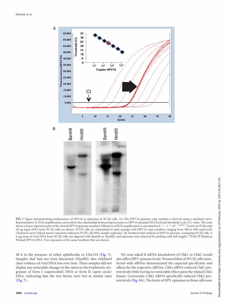
W12E cells maintaining approximately 300 to 500 HPV16 epi-some copies per cell and exhibiting no evidence of viral DNAintegration were first studied. Viral DNA in Southern blots wasassociated predominantly with episomal bands, and no additionalbands indicative of integration were detected (Fig. 1). Initial ex-periments queried whether cell cycle progression was required forthe antiviral effect of PA25. Three agents that activate cell cyclecheckpoints were initially tested for their ability to block PA25-mediated loss of HPV16 episomes. Cell cycle status had no mea-surable effect upon the antiviral activity of PA25. Aphidicolin,nocodazole, and hydroxyurea all acted to block cells within the cellcycle as anticipated (Fig. 2). Arrest of W12E cells in G2/M bypretreatment of cells with nocodazole (Fig. 3A) or at the G1/S-phase boundary by treatment with hydroxyurea (Fig. 3B) oraphidicolin (Fig. 3C) did not prevent the loss of episomes follow-ing treatment with PA25. Nocodazole by itself had no effect onepisome levels as measured by Q-PCR (Fig. 3A), while hy-droxyurea treatment caused a small apparent decrease in episomelevels that was not statistically significant (Fig. 3B). In contrast,aphidicolin alone, at a concentration of 4 �M, elicited a robustdecrease in HPV16 episome levels in W12E cells (Fig. 3C).
These results were surprising because a variety of pharmaceu-tically active and toxic compounds, including etoposide andpodophyllotoxin (data not shown), in addition to hydroxyureaand nocodazole (Fig. 3), have little or no significant effect on HPVepisome levels. Furthermore, aphidicolin contributed to HPV ep-isome loss in an additive manner when it was provided prior totreatment with multiple, increasing PA25 doses (Fig. 3C). Thisobservation indicated that aphidicolin and PA25 were causing ep-isome loss via different cellular pathways.
The effects of aphidicolin on two additional HPV genotypes,HPV18 and HPV31, were then also tested. Aphidicolin, at a con-centration of 4 �M, resulted in a statistically significant loss ofapproximately 40% of HPV31 episomes and 50% of HPV18 epi-somes after 48 h (Fig. 3D), showing that the effect was not limitedto a single HPV genotype or cell type.
An aphidicolin dose-response analysis conducted with W12E
cells showed that increasing amounts of the inhibitor to 10 �Mresulted in a greater loss of episomes, with 80% of the viral DNAeliminated after 48 h at the highest dose (Fig. 4). The loss of epi-somes appeared to correlate with aphidicolin effects on the cellcycle (Fig. 2). The lowest doses (0.1 and 0.4 �M), which hadsmaller effects on HPV episome copy number, allowed cells toenter S phase, where they stalled and accumulated. On the otherhand, the higher doses, which caused greater episome loss,blocked S-phase entry, resulting in congregation of cells at theG1/S boundary (Fig. 2).
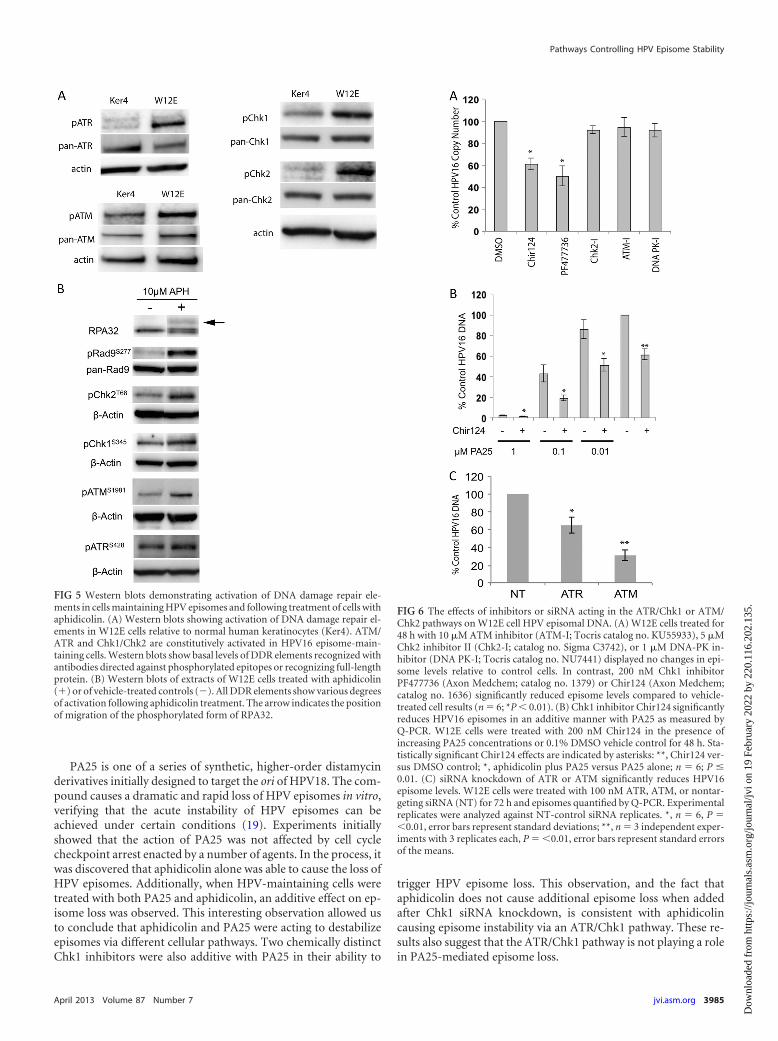
Aphidicolin is known to activate a DDR in the ATR/Chk1pathway due to stalling of replication forks. It can also activate theATM/Chk2 pathway due to collapse of replication forks and gen-eration of double-stranded DNA (dsDNA) breaks. The presenceof HPV episomes can also cause an elevation of cellular DDRsignaling, and in fact our own experiments found that a variety ofDDR elements were activated in cells that maintain HPV episomesrelative to normal keratinocytes (Fig. 5A). In addition, we foundthat aphidicolin activates both the ATM and ATR pathways inHPV16-positive cells (Fig. 5B). For this reason, we set out to testthe hypothesis that DDR elements contribute to HPV episomestability in undifferentiated keratinocytes.
We first tested inhibitors acting within the ATR/Chk1 pathwayfor their effects on HPV episome stability. Similar to the aphidi-colin results, the Chk1 kinase inhibitor Chir124 produced a sta-tistically significant 40% reduction in episome levels (Fig. 6A).Chir124 also reduced HPV episomes in an additive manner whencoadministered with increasing doses of PA25 (Fig. 6B). This re-sult suggested that, like aphidicolin (Fig. 3), Chir124 acts to desta-bilize episomes via a pathway different from that of PA25.PF477736, a second Chk1 inhibitor chemically distinct fromChir124, also caused a statistically significant 50% decline inHPV16 episome numbers (Fig. 6A). Additional inhibitors actingon other DDR pathway elements were also tested: KU55933, anATM-specific kinase inhibitor, Chk2-specific inhibitor II, and theDNA-PK inhibitor NU7441 all had no effect on episome levels(Fig. 6A).
There were no available specific chemical ATR inhibitors ofwhich we were aware, so we employed a siRNA strategy to targetATR for downregulation. ATR siRNA reduced ATR transcriptlevels by 78% relative to nontargeting control levels as measuredby Q-RT-PCR. Q-PCR of the ATR siRNA-treated W12E cellsdemonstrated a 40% loss of HPV16 episomal DNA at 48 h(Fig. 6C). As a control for this experiment, we also tested theeffects of siRNA directed against ATM. ATM siRNA caused a 64%reduction in ATM mRNA levels after 72 h of treatment. Surpris-ingly, ATM siRNA also caused a highly reproducible 70% decreasein HPV16 episome levels. The data shown represent a summary ofthree independent experiments, each having multiple replicates(Fig. 6C). The specificity of both ATR and ATM siRNAs was con-firmed in a matrix of 22 siRNAs chosen from DDR pathways (datanot shown).
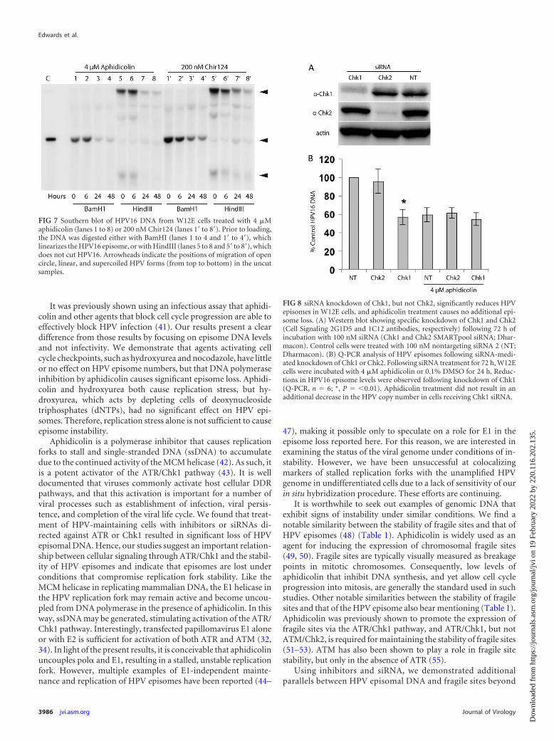
The effects of aphidicolin and Chir124 on viral episome main-tenance were also investigated by Southern blotting in order toconfirm and extend the Q-PCR results (Fig. 7). Loss of HPV16DNA following treatment with either inhibitor was time depen-dent. As expected, the samples digested with either BamHI orHindIII showed no evidence of viral DNA integration. Samplesdigested with BamHI, which linearizes HPV16 episomes, yieldedthe expected 7.9-kb HPV16 DNA that was progressively lost over
Pathways Controlling HPV Episome Stability
April 2013 Volume 87 Number 7 jvi.asm.org 3981
Dow
nloa
ded
from
http
s://j
ourn
als.
asm
.org
/jour
nal/j
vi o
n 19
Feb
ruar
y 20
22 b
y 22
0.11
6.20
2.13
5.
48 h in the presence of either aphidicolin or Chir124 (Fig. 7).Samples that had not been linearized (HindIII) also exhibitedclear evidence of viral DNA loss over time. These samples did notdisplay any noticeable changes in the ratios or electrophoretic mi-gration of form I (supercoiled) DNA or form II (open circle)DNA, indicating that the two forms were lost at similar rates(Fig. 7).
We next asked if siRNA knockdown of Chk1 or Chk2 wouldalso affect HPV episome levels. Western blots of W12E cells trans-fected with siRNAs demonstrated the expected specificities andeffects for the respective siRNAs. Chk1 siRNA reduced Chk1 pro-tein levels while having no noticeable effect upon the related Chk2kinase. Conversely, Chk2 siRNA specifically reduced Chk2 pro-tein levels (Fig. 8A). The levels of HPV episomes in these cells were
FIG 1 Figure demonstrating maintenance of HPV16 as episomes in W12E cells. (A) The HPV16 episome copy number is derived using a standard curve.Representative Q-PCR amplification curves show the relationship between log increases in HPV16 plasmid DNA level and threshold cycle (Ct) value. The insetshows a linear regression plot of the cloned HPV16 genome standard. Efficiency of PCR amplification is calculated as E � �1 � 10(�1/slope). Twelve Q-PCRs with20 ng input DNA from W12E cells are shown. W12E cells are maintained at early passage with HPV16 copy numbers ranging from 300 to 500 copies/cell.Clustered curves (block arrow) represent unknown W12E cell DNA sample replicates. (B) Southern blot analysis of HPV16 episome-containing W12E cells. A5-�g mass of total DNA from W12E cells was digested with BamHI or HindIII, and episomes were detected by probing with full-length [32P]dCTP RandomPrimed HPV16 DNA. Two exposures of the same Southern blot are shown.
Edwards et al.
3982 jvi.asm.org Journal of Virology
Dow
nloa
ded
from
http
s://j
ourn
als.
asm
.org
/jour
nal/j
vi o
n 19
Feb
ruar
y 20
22 b
y 22
0.11
6.20
2.13
5.
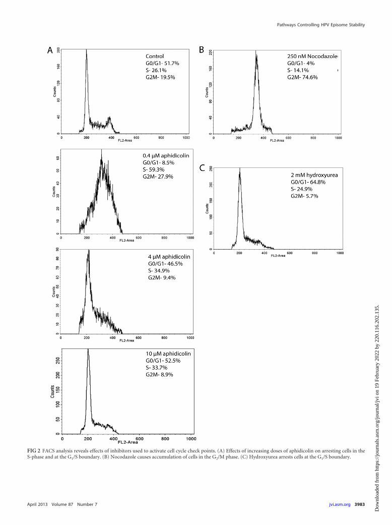
FIG 2 FACS analysis reveals effects of inhibitors used to activate cell cycle check points. (A) Effects of increasing doses of aphidicolin on arresting cells in theS-phase and at the G1/S boundary. (B) Nocodazole causes accumulation of cells in the G2/M phase. (C) Hydroxyurea arrests cells at the G1/S boundary.
Pathways Controlling HPV Episome Stability
April 2013 Volume 87 Number 7 jvi.asm.org 3983
Dow
nloa
ded
from
http
s://j
ourn
als.
asm
.org
/jour
nal/j
vi o
n 19
Feb
ruar
y 20
22 b
y 22
0.11
6.20
2.13
5.
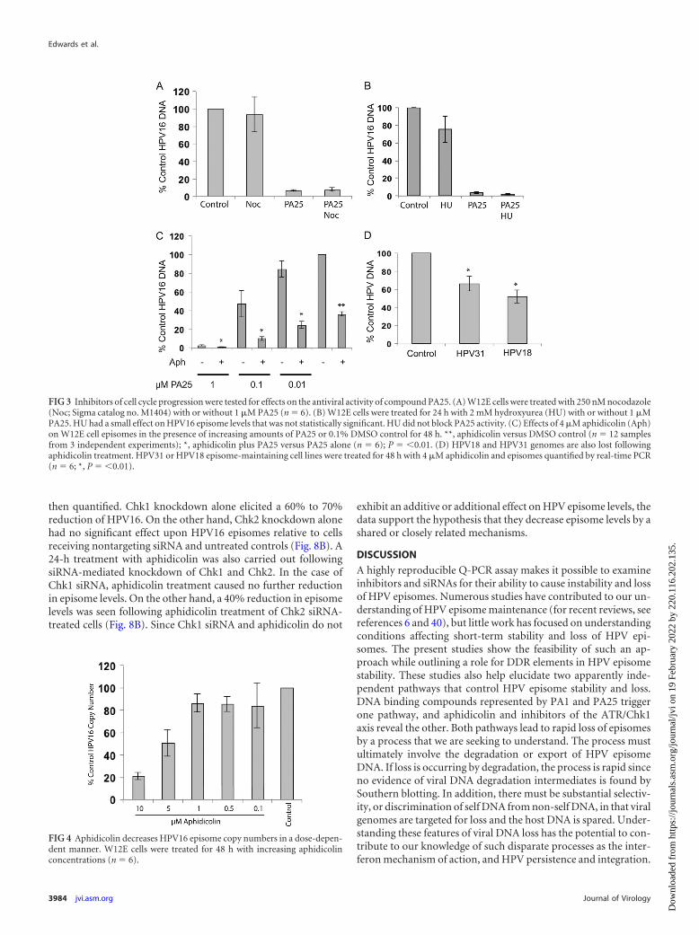
then quantified. Chk1 knockdown alone elicited a 60% to 70%reduction of HPV16. On the other hand, Chk2 knockdown alonehad no significant effect upon HPV16 episomes relative to cellsreceiving nontargeting siRNA and untreated controls (Fig. 8B). A24-h treatment with aphidicolin was also carried out followingsiRNA-mediated knockdown of Chk1 and Chk2. In the case ofChk1 siRNA, aphidicolin treatment caused no further reductionin episome levels. On the other hand, a 40% reduction in episomelevels was seen following aphidicolin treatment of Chk2 siRNA-treated cells (Fig. 8B). Since Chk1 siRNA and aphidicolin do not
exhibit an additive or additional effect on HPV episome levels, thedata support the hypothesis that they decrease episome levels by ashared or closely related mechanisms.
DISCUSSION
A highly reproducible Q-PCR assay makes it possible to examineinhibitors and siRNAs for their ability to cause instability and lossof HPV episomes. Numerous studies have contributed to our un-derstanding of HPV episome maintenance (for recent reviews, seereferences 6 and 40), but little work has focused on understandingconditions affecting short-term stability and loss of HPV epi-somes. The present studies show the feasibility of such an ap-proach while outlining a role for DDR elements in HPV episomestability. These studies also help elucidate two apparently inde-pendent pathways that control HPV episome stability and loss.DNA binding compounds represented by PA1 and PA25 triggerone pathway, and aphidicolin and inhibitors of the ATR/Chk1axis reveal the other. Both pathways lead to rapid loss of episomesby a process that we are seeking to understand. The process mustultimately involve the degradation or export of HPV episomeDNA. If loss is occurring by degradation, the process is rapid sinceno evidence of viral DNA degradation intermediates is found bySouthern blotting. In addition, there must be substantial selectiv-ity, or discrimination of self DNA from non-self DNA, in that viralgenomes are targeted for loss and the host DNA is spared. Under-standing these features of viral DNA loss has the potential to con-tribute to our knowledge of such disparate processes as the inter-feron mechanism of action, and HPV persistence and integration.
FIG 3 Inhibitors of cell cycle progression were tested for effects on the antiviral activity of compound PA25. (A) W12E cells were treated with 250 nM nocodazole(Noc; Sigma catalog no. M1404) with or without 1 �M PA25 (n � 6). (B) W12E cells were treated for 24 h with 2 mM hydroxyurea (HU) with or without 1 �MPA25. HU had a small effect on HPV16 episome levels that was not statistically significant. HU did not block PA25 activity. (C) Effects of 4 �M aphidicolin (Aph)on W12E cell episomes in the presence of increasing amounts of PA25 or 0.1% DMSO control for 48 h. **, aphidicolin versus DMSO control (n � 12 samplesfrom 3 independent experiments); *, aphidicolin plus PA25 versus PA25 alone (n � 6); P � �0.01. (D) HPV18 and HPV31 genomes are also lost followingaphidicolin treatment. HPV31 or HPV18 episome-maintaining cell lines were treated for 48 h with 4 �M aphidicolin and episomes quantified by real-time PCR(n � 6; *, P � �0.01).
FIG 4 Aphidicolin decreases HPV16 episome copy numbers in a dose-depen-dent manner. W12E cells were treated for 48 h with increasing aphidicolinconcentrations (n � 6).
Edwards et al.
3984 jvi.asm.org Journal of Virology
Dow
nloa
ded
from
http
s://j
ourn
als.
asm
.org
/jour
nal/j
vi o
n 19
Feb
ruar
y 20
22 b
y 22
0.11
6.20
2.13
5.
PA25 is one of a series of synthetic, higher-order distamycinderivatives initially designed to target the ori of HPV18. The com-pound causes a dramatic and rapid loss of HPV episomes in vitro,verifying that the acute instability of HPV episomes can beachieved under certain conditions (19). Experiments initiallyshowed that the action of PA25 was not affected by cell cyclecheckpoint arrest enacted by a number of agents. In the process, itwas discovered that aphidicolin alone was able to cause the loss ofHPV episomes. Additionally, when HPV-maintaining cells weretreated with both PA25 and aphidicolin, an additive effect on ep-isome loss was observed. This interesting observation allowed usto conclude that aphidicolin and PA25 were acting to destabilizeepisomes via different cellular pathways. Two chemically distinctChk1 inhibitors were also additive with PA25 in their ability to
trigger HPV episome loss. This observation, and the fact thataphidicolin does not cause additional episome loss when addedafter Chk1 siRNA knockdown, is consistent with aphidicolincausing episome instability via an ATR/Chk1 pathway. These re-sults also suggest that the ATR/Chk1 pathway is not playing a rolein PA25-mediated episome loss.
FIG 5 Western blots demonstrating activation of DNA damage repair ele-ments in cells maintaining HPV episomes and following treatment of cells withaphidicolin. (A) Western blots showing activation of DNA damage repair el-ements in W12E cells relative to normal human keratinocytes (Ker4). ATM/ATR and Chk1/Chk2 are constitutively activated in HPV16 episome-main-taining cells. Western blots show basal levels of DDR elements recognized withantibodies directed against phosphorylated epitopes or recognizing full-lengthprotein. (B) Western blots of extracts of W12E cells treated with aphidicolin(�) or of vehicle-treated controls (�). All DDR elements show various degreesof activation following aphidicolin treatment. The arrow indicates the positionof migration of the phosphorylated form of RPA32.
FIG 6 The effects of inhibitors or siRNA acting in the ATR/Chk1 or ATM/Chk2 pathways on W12E cell HPV episomal DNA. (A) W12E cells treated for48 h with 10 �M ATM inhibitor (ATM-I; Tocris catalog no. KU55933), 5 �MChk2 inhibitor II (Chk2-I; catalog no. Sigma C3742), or 1 �M DNA-PK in-hibitor (DNA PK-I; Tocris catalog no. NU7441) displayed no changes in epi-some levels relative to control cells. In contrast, 200 nM Chk1 inhibitorPF477736 (Axon Medchem; catalog no. 1379) or Chir124 (Axon Medchem;catalog no. 1636) significantly reduced episome levels compared to vehicle-treated cell results (n � 6; *P � 0.01). (B) Chk1 inhibitor Chir124 significantlyreduces HPV16 episomes in an additive manner with PA25 as measured byQ-PCR. W12E cells were treated with 200 nM Chir124 in the presence ofincreasing PA25 concentrations or 0.1% DMSO vehicle control for 48 h. Sta-tistically significant Chir124 effects are indicated by asterisks: **, Chir124 ver-sus DMSO control; *, aphidicolin plus PA25 versus PA25 alone; n � 6; P �0.01. (C) siRNA knockdown of ATR or ATM significantly reduces HPV16episome levels. W12E cells were treated with 100 nM ATR, ATM, or nontar-geting siRNA (NT) for 72 h and episomes quantified by Q-PCR. Experimentalreplicates were analyzed against NT-control siRNA replicates. *, n � 6, P ��0.01, error bars represent standard deviations; **, n � 3 independent exper-iments with 3 replicates each, P � �0.01, error bars represent standard errorsof the means.
Pathways Controlling HPV Episome Stability
April 2013 Volume 87 Number 7 jvi.asm.org 3985
Dow
nloa
ded
from
http
s://j
ourn
als.
asm
.org
/jour
nal/j
vi o
n 19
Feb
ruar
y 20
22 b
y 22
0.11
6.20
2.13
5.
It was previously shown using an infectious assay that aphidi-colin and other agents that block cell cycle progression are able toeffectively block HPV infection (41). Our results present a cleardifference from those results by focusing on episome DNA levelsand not infectivity. We demonstrate that agents activating cellcycle checkpoints, such as hydroxyurea and nocodazole, have littleor no effect on HPV episome numbers, but that DNA polymeraseinhibition by aphidicolin causes significant episome loss. Aphidi-colin and hydroxyurea both cause replication stress, but hy-droxyurea, which acts by depleting cells of deoxynucleosidetriphosphates (dNTPs), had no significant effect on HPV epi-somes. Therefore, replication stress alone is not sufficient to causeepisome instability.
Aphidicolin is a polymerase inhibitor that causes replicationforks to stall and single-stranded DNA (ssDNA) to accumulatedue to the continued activity of the MCM helicase (42). As such, itis a potent activator of the ATR/Chk1 pathway (43). It is welldocumented that viruses commonly activate host cellular DDRpathways, and that this activation is important for a number ofviral processes such as establishment of infection, viral persis-tence, and completion of the viral life cycle. We found that treat-ment of HPV-maintaining cells with inhibitors or siRNAs di-rected against ATR or Chk1 resulted in significant loss of HPVepisomal DNA. Hence, our studies suggest an important relation-ship between cellular signaling through ATR/Chk1 and the stabil-ity of HPV episomes and indicate that episomes are lost underconditions that compromise replication fork stability. Like theMCM helicase in replicating mammalian DNA, the E1 helicase inthe HPV replication fork may remain active and become uncou-pled from DNA polymerase in the presence of aphidicolin. In thisway, ssDNA may be generated, stimulating activation of the ATR/Chk1 pathway. Interestingly, transfected papillomavirus E1 aloneor with E2 is sufficient for activation of both ATR and ATM (32,34). In light of the present results, it is conceivable that aphidicolinuncouples pol� and E1, resulting in a stalled, unstable replicationfork. However, multiple examples of E1-independent mainte-nance and replication of HPV episomes have been reported (44–
47), making it possible only to speculate on a role for E1 in theepisome loss reported here. For this reason, we are interested inexamining the status of the viral genome under conditions of in-stability. However, we have been unsuccessful at colocalizingmarkers of stalled replication forks with the unamplified HPVgenome in undifferentiated cells due to a lack of sensitivity of ourin situ hybridization procedure. These efforts are continuing.
It is worthwhile to seek out examples of genomic DNA thatexhibit signs of instability under similar conditions. We find anotable similarity between the stability of fragile sites and that ofHPV episomes (48) (Table 1). Aphidicolin is widely used as anagent for inducing the expression of chromosomal fragile sites(49, 50). Fragile sites are typically visually measured as breakagepoints in mitotic chromosomes. Consequently, low levels ofaphidicolin that inhibit DNA synthesis, and yet allow cell cycleprogression into mitosis, are generally the standard used in suchstudies. Other notable similarities between the stability of fragilesites and that of the HPV episome also bear mentioning (Table 1).Aphidicolin was previously shown to promote the expression offragile sites via the ATR/Chk1 pathway, and ATR/Chk1, but notATM/Chk2, is required for maintaining the stability of fragile sites(51–53). ATM has also been shown to play a role in fragile sitestability, but only in the absence of ATR (55).
Using inhibitors and siRNA, we demonstrated additionalparallels between HPV episomal DNA and fragile sites beyond
FIG 8 siRNA knockdown of Chk1, but not Chk2, significantly reduces HPVepisomes in W12E cells, and aphidicolin treatment causes no additional epi-some loss. (A) Western blot showing specific knockdown of Chk1 and Chk2(Cell Signaling 2G1D5 and 1C12 antibodies, respectively) following 72 h ofincubation with 100 nM siRNA (Chk1 and Chk2 SMARTpool siRNA; Dhar-macon). Control cells were treated with 100 nM nontargeting siRNA 2 (NT;Dharmacon). (B) Q-PCR analysis of HPV episomes following siRNA-medi-ated knockdown of Chk1 or Chk2. Following siRNA treatment for 72 h, W12Ecells were incubated with 4 �M aphidicolin or 0.1% DMSO for 24 h. Reduc-tions in HPV16 episome levels were observed following knockdown of Chk1(Q-PCR, n � 6; *, P � �0.01). Aphidicolin treatment did not result in anadditional decrease in the HPV copy number in cells receiving Chk1 siRNA.
FIG 7 Southern blot of HPV16 DNA from W12E cells treated with 4 �Maphidicolin (lanes 1 to 8) or 200 nM Chir124 (lanes 1= to 8=). Prior to loading,the DNA was digested either with BamHI (lanes 1 to 4 and 1= to 4=), whichlinearizes the HPV16 episome, or with HindIII (lanes 5 to 8 and 5= to 8=), whichdoes not cut HPV16. Arrowheads indicate the positions of migration of opencircle, linear, and supercoiled HPV forms (from top to bottom) in the uncutsamples.
Edwards et al.
3986 jvi.asm.org Journal of Virology
Dow
nloa
ded
from
http
s://j
ourn
als.
asm
.org
/jour
nal/j
vi o
n 19
Feb
ruar
y 20
22 b
y 22
0.11
6.20
2.13
5.
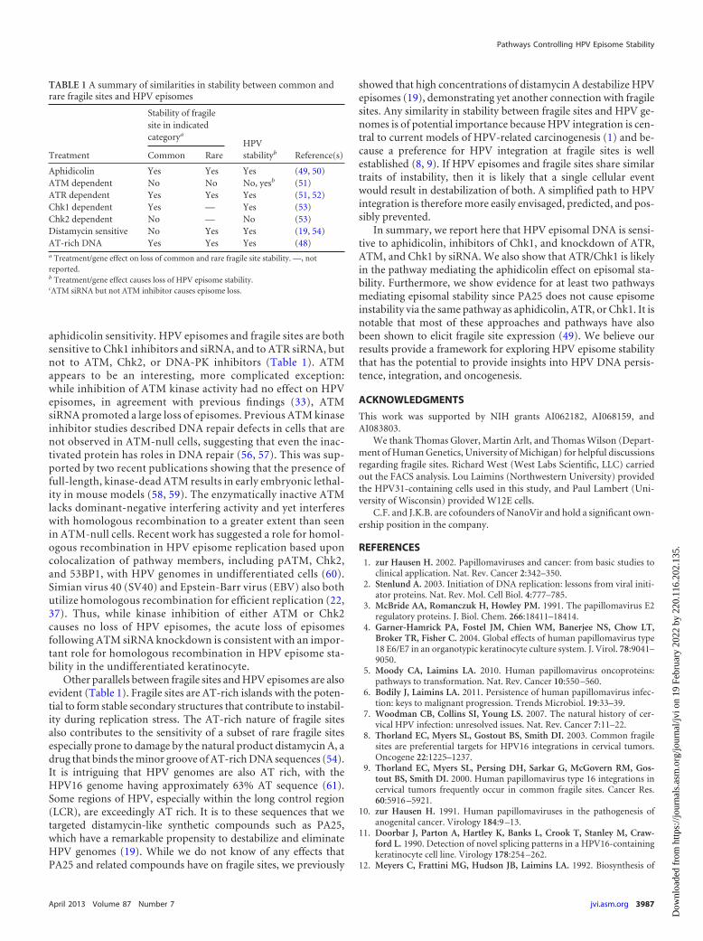
aphidicolin sensitivity. HPV episomes and fragile sites are bothsensitive to Chk1 inhibitors and siRNA, and to ATR siRNA, butnot to ATM, Chk2, or DNA-PK inhibitors (Table 1). ATMappears to be an interesting, more complicated exception:while inhibition of ATM kinase activity had no effect on HPVepisomes, in agreement with previous findings (33), ATMsiRNA promoted a large loss of episomes. Previous ATM kinaseinhibitor studies described DNA repair defects in cells that arenot observed in ATM-null cells, suggesting that even the inac-tivated protein has roles in DNA repair (56, 57). This was sup-ported by two recent publications showing that the presence offull-length, kinase-dead ATM results in early embryonic lethal-ity in mouse models (58, 59). The enzymatically inactive ATMlacks dominant-negative interfering activity and yet interfereswith homologous recombination to a greater extent than seenin ATM-null cells. Recent work has suggested a role for homol-ogous recombination in HPV episome replication based uponcolocalization of pathway members, including pATM, Chk2,and 53BP1, with HPV genomes in undifferentiated cells (60).Simian virus 40 (SV40) and Epstein-Barr virus (EBV) also bothutilize homologous recombination for efficient replication (22,37). Thus, while kinase inhibition of either ATM or Chk2causes no loss of HPV episomes, the acute loss of episomesfollowing ATM siRNA knockdown is consistent with an impor-tant role for homologous recombination in HPV episome sta-bility in the undifferentiated keratinocyte.
Other parallels between fragile sites and HPV episomes are alsoevident (Table 1). Fragile sites are AT-rich islands with the poten-tial to form stable secondary structures that contribute to instabil-ity during replication stress. The AT-rich nature of fragile sitesalso contributes to the sensitivity of a subset of rare fragile sitesespecially prone to damage by the natural product distamycin A, adrug that binds the minor groove of AT-rich DNA sequences (54).It is intriguing that HPV genomes are also AT rich, with theHPV16 genome having approximately 63% AT sequence (61).Some regions of HPV, especially within the long control region(LCR), are exceedingly AT rich. It is to these sequences that wetargeted distamycin-like synthetic compounds such as PA25,which have a remarkable propensity to destabilize and eliminateHPV genomes (19). While we do not know of any effects thatPA25 and related compounds have on fragile sites, we previously
showed that high concentrations of distamycin A destabilize HPVepisomes (19), demonstrating yet another connection with fragilesites. Any similarity in stability between fragile sites and HPV ge-nomes is of potential importance because HPV integration is cen-tral to current models of HPV-related carcinogenesis (1) and be-cause a preference for HPV integration at fragile sites is wellestablished (8, 9). If HPV episomes and fragile sites share similartraits of instability, then it is likely that a single cellular eventwould result in destabilization of both. A simplified path to HPVintegration is therefore more easily envisaged, predicted, and pos-sibly prevented.
In summary, we report here that HPV episomal DNA is sensi-tive to aphidicolin, inhibitors of Chk1, and knockdown of ATR,ATM, and Chk1 by siRNA. We also show that ATR/Chk1 is likelyin the pathway mediating the aphidicolin effect on episomal sta-bility. Furthermore, we show evidence for at least two pathwaysmediating episomal stability since PA25 does not cause episomeinstability via the same pathway as aphidicolin, ATR, or Chk1. It isnotable that most of these approaches and pathways have alsobeen shown to elicit fragile site expression (49). We believe ourresults provide a framework for exploring HPV episome stabilitythat has the potential to provide insights into HPV DNA persis-tence, integration, and oncogenesis.
ACKNOWLEDGMENTS
This work was supported by NIH grants AI062182, AI068159, andAI083803.
We thank Thomas Glover, Martin Arlt, and Thomas Wilson (Depart-ment of Human Genetics, University of Michigan) for helpful discussionsregarding fragile sites. Richard West (West Labs Scientific, LLC) carriedout the FACS analysis. Lou Laimins (Northwestern University) providedthe HPV31-containing cells used in this study, and Paul Lambert (Uni-versity of Wisconsin) provided W12E cells.
C.F. and J.K.B. are cofounders of NanoVir and hold a significant own-ership position in the company.
REFERENCES1. zur Hausen H. 2002. Papillomaviruses and cancer: from basic studies to
clinical application. Nat. Rev. Cancer 2:342–350.2. Stenlund A. 2003. Initiation of DNA replication: lessons from viral initi-
ator proteins. Nat. Rev. Mol. Cell Biol. 4:777–785.3. McBride AA, Romanczuk H, Howley PM. 1991. The papillomavirus E2
regulatory proteins. J. Biol. Chem. 266:18411–18414.4. Garner-Hamrick PA, Fostel JM, Chien WM, Banerjee NS, Chow LT,
Broker TR, Fisher C. 2004. Global effects of human papillomavirus type18 E6/E7 in an organotypic keratinocyte culture system. J. Virol. 78:9041–9050.
5. Moody CA, Laimins LA. 2010. Human papillomavirus oncoproteins:pathways to transformation. Nat. Rev. Cancer 10:550 –560.
6. Bodily J, Laimins LA. 2011. Persistence of human papillomavirus infec-tion: keys to malignant progression. Trends Microbiol. 19:33–39.
7. Woodman CB, Collins SI, Young LS. 2007. The natural history of cer-vical HPV infection: unresolved issues. Nat. Rev. Cancer 7:11–22.
8. Thorland EC, Myers SL, Gostout BS, Smith DI. 2003. Common fragilesites are preferential targets for HPV16 integrations in cervical tumors.Oncogene 22:1225–1237.
9. Thorland EC, Myers SL, Persing DH, Sarkar G, McGovern RM, Gos-tout BS, Smith DI. 2000. Human papillomavirus type 16 integrations incervical tumors frequently occur in common fragile sites. Cancer Res.60:5916 –5921.
10. zur Hausen H. 1991. Human papillomaviruses in the pathogenesis ofanogenital cancer. Virology 184:9 –13.
11. Doorbar J, Parton A, Hartley K, Banks L, Crook T, Stanley M, Craw-ford L. 1990. Detection of novel splicing patterns in a HPV16-containingkeratinocyte cell line. Virology 178:254 –262.
12. Meyers C, Frattini MG, Hudson JB, Laimins LA. 1992. Biosynthesis of
TABLE 1 A summary of similarities in stability between common andrare fragile sites and HPV episomes
Treatment
Stability of fragilesite in indicatedcategorya
HPVstabilityb Reference(s)Common Rare
Aphidicolin Yes Yes Yes (49, 50)ATM dependent No No No, yesb (51)ATR dependent Yes Yes Yes (51, 52)Chk1 dependent Yes — Yes (53)Chk2 dependent No — No (53)Distamycin sensitive No Yes Yes (19, 54)AT-rich DNA Yes Yes Yes (48)a Treatment/gene effect on loss of common and rare fragile site stability. —, notreported.b Treatment/gene effect causes loss of HPV episome stability.cATM siRNA but not ATM inhibitor causes episome loss.
Pathways Controlling HPV Episome Stability
April 2013 Volume 87 Number 7 jvi.asm.org 3987
Dow
nloa
ded
from
http
s://j
ourn
als.
asm
.org
/jour
nal/j
vi o
n 19
Feb
ruar
y 20
22 b
y 22
0.11
6.20
2.13
5.
human papillomavirus from a continuous cell line upon epithelial differ-entiation. Science 257:971–973.
13. Flores ER, Allen-Hoffmann BL, Lee D, Lambert PF. 2000. The humanpapillomavirus type 16 E7 oncogene is required for the productive stage ofthe viral life cycle. J. Virol. 74:6622– 6631.
14. Frattini MG, Lim HB, Laimins LA. 1996. In vitro synthesis of onco-genic human papillomaviruses requires episomal genomes for differ-entiation-dependent late expression. Proc. Natl. Acad. Sci. U. S. A.93:3062–3067.
15. Garner-Hamrick PA, Fisher C. 2002. HPV episomal copy number closelycorrelates with cell size in keratinocyte monolayer cultures. Virology 301:334 –341.
16. Hebner CM, Laimins LA. 2006. Human papillomaviruses: basic mecha-nisms of pathogenesis and oncogenicity. Rev. Med. Virol. 16:83–97.
17. Münger K, Howley PM. 2002. Human papillomavirus immortalizationand transformation functions. Virus Res. 89:213–228.
18. Bedell MA, Hudson JB, Golub TR, Turyk ME, Hosken M, WilbanksGD, Laimins LA. 1991. Amplification of human papillomavirus ge-nomes in vitro is dependent on epithelial differentiation. J. Virol. 65:2254 –2260.
19. Edwards TG, Koeller KJ, Slomczynska U, Fok K, Helmus M, BashkinJK, Fisher C. 2011. HPV episome levels are potently decreased by pyrrole-imidazole polyamides. Antiviral Res. 91:177–186.
20. Jeon S, Allen-Hoffmann BL, Lambert PF. 1995. Integration of humanpapillomavirus type 16 into the human genome correlates with a selectivegrowth advantage of cells. J. Virol. 69:2989 –2997.
21. Stubenrauch F, Hummel M, Iftner T, Laimins LA. 2000. The E8E2Cprotein, a negative regulator of viral transcription and replication, is re-quired for extrachromosomal maintenance of human papillomavirus type31 in keratinocytes. J. Virol. 74:1178 –1186.
22. Bodily JM, Mehta KP, Cruz L, Meyers C, Laimins LA. 2011. The E7 openreading frame acts in cis and in trans to mediate differentiation-dependentactivities in the human papillomavirus type 16 life cycle. J. Virol. 85:8852–8862.
23. Wilson R, Fehrmann F, Laimins LA. 2005. Role of the E1–E4 protein inthe differentiation-dependent life cycle of human papillomavirus type 31.J. Virol. 79:6732– 6740.
24. Chang YE, Pena L, Sen GC, Park JK, Laimins LA. 2002. Long-term effectof interferon on keratinocytes that maintain human papillomavirus type31. J. Virol. 76:8864 – 8874.
25. Herdman MT, Pett MR, Roberts I, Alazawi WO, Teschendorff AE,Zhang XY, Stanley MA, Coleman N. 2006. Interferon-beta treatment ofcervical keratinocytes naturally infected with human papillomavirus 16episomes promotes rapid reduction in episome numbers and emergenceof latent integrants. Carcinogenesis 27:2341–2353.
26. Chang YE, Laimins LA. 2000. Microarray analysis identifies interferon-inducible genes and Stat-1 as major transcriptional targets of human pap-illomavirus type 31. J. Virol. 74:4174 – 4182.
27. Turnell AS, Grand RJ. 2012. DNA viruses and the cellular DNA-damageresponse. J. Gen. Virol. 93:2076 –2097.
28. Weitzman MD, Lilley CE, Chaurushiya MS. 2010. Genomes in conflict:maintaining genome integrity during virus infection. Annu. Rev. Micro-biol. 64:61– 81.
29. Harper JW, Elledge SJ. 2007. The DNA damage response: ten years after.Mol. Cell 28:739 –745.
30. Stracker TH, Usui T, Petrini JH. 2009. Taking the time to make impor-tant decisions: the checkpoint effector kinases Chk1 and Chk2 and theDNA damage response. DNA Repair (Amst) 8:1047–1054.
31. Banerjee NS, Wang HK, Broker TR, Chow LT. 2011. Human papil-lomavirus (HPV) E7 induces prolonged G2 following S phase reentryin differentiated human keratinocytes. J. Biol. Chem. 286:15473–15482.
32. Fradet-Turcotte A, Bergeron-Labrecque F, Moody CA, Lehoux M,Laimins LA, Archambault J. 2011. Nuclear accumulation of the papillo-mavirus E1 helicase blocks S-phase progression and triggers an ATM-dependent DNA damage response. J. Virol. 85:8996 –9012.
33. Moody CA, Laimins LA. 2009. Human papillomaviruses activate theATM DNA damage pathway for viral genome amplification upon differ-entiation. PLoS Pathog. 5:e1000605. doi:10.1371/journal.ppat.1000605.
34. Sakakibara N, Mitra R, McBride A. 2011. The papillomavirus E1 helicaseactivates a cellular DNA damage response in viral replication foci. J. Virol.85:8981– 8995.
35. Arienti KL, Brunmark A, Axe FU, McClure K, Lee A, Blevitt J, Neff
DK, Huang L, Crawford S, Pandit CR, Karlsson L, Breitenbucher JG.2005. Checkpoint kinase inhibitors: SAR and radioprotective proper-ties of a series of 2-arylbenzimidazoles. J. Med. Chem. 48:1873–1885.
36. Ashwell S, Zabludoff S. 2008. DNA damage detection and repair path-ways—recent advances with inhibitors of checkpoint kinases in cancertherapy. Clin. Cancer Res. 14:4032– 4037.
37. Hickson I, Zhao Y, Richardson CJ, Green SJ, Martin NM, Orr AI,Reaper PM, Jackson SP, Curtin NJ, Smith GC. 2004. Identification andcharacterization of a novel and specific inhibitor of the ataxia-telangiectasia mutated kinase ATM. Cancer Res. 64:9152–9159.
38. Tavecchio M, Munik JM, Cano C, Newell DR, Curtin NJ. 2012. Furthercharacterisation of the cellular activity of the DNA-PK inhibitor, NU7441,reveals potential cross-talk with homologous recombination. Cancer Che-mother. Pharmacol. 69:155–164.
39. Tse AN, Rendahl KG, Sheikh T, Cheema H, Aardalen K, Embry M, MaS, Moler EJ, Ni ZJ, Lopes de Menezes DE, Hibner B, Gesner TG,Schwartz GK. 2007. CHIR-124, a novel potent inhibitor of Chk1, poten-tiates the cytotoxicity of topoisomerase I poisons in vitro and in vivo. Clin.Cancer Res. 13:591– 602.
40. McBride AA, Sakakibara N, Stepp WH, Jang MK. 2012. Hitchhiking onhost chromatin: how papillomaviruses persist. Biochim. Biophys. Acta1819:820 – 825.
41. Pyeon D, Pearce SM, Lank SM, Ahlquist P, Lambert PF. 2009. Estab-lishment of human papillomavirus infection requires cell cycle progres-sion. PLoS Pathog. 5:e1000318. doi:10.1371/journal.ppat.1000318.
42. Byun TS, Pacek M, Yee MC, Walter JC, Cimprich KA. 2005. Functionaluncoupling of MCM helicase and DNA polymerase activities activates theATR-dependent checkpoint. Genes Dev. 19:1040 –1052.
43. Smith J, Tho LM, Xu N, Gillespie DA. 2010. The ATM-Chk2 andATR-Chk1 pathways in DNA damage signaling and cancer. Adv. CancerRes. 108:73–112.
44. Angeletti PC, Kim K, Fernandes FJ, Lambert PF. 2002. Stable replicationof papillomavirus genomes in Saccharomyces cerevisiae. J. Virol. 76:3350 –3358.
45. Egawa N, Nakahara T, Ohno S, Narisawa-Saito M, Yugawa T, Fujita M,Yamato K, Natori Y, Kiyono T. 2012. The E1 protein of human papillo-mavirus type 16 is dispensable for maintenance replication of the viralgenome. J. Virol. 86:3276 –3283.
46. Kim K, Lambert PF. 2002. E1 protein of bovine papillomavirus 1 is notrequired for the maintenance of viral plasmid DNA replication. Virology293:10 –14.
47. Pittayakhajonwut D, Angeletti PC. 2010. Viral trans-factor independentreplication of human papillomavirus genomes. Virol. J. 7:123. doi:10.1186/1743-422X-7-123.
48. Durkin SG, Glover TW. 2007. Chromosome fragile sites. Annu. Rev.Genet. 41:169 –192.
49. Glover TW, Arlt MF, Casper AM, Durkin SG. 2005. Mechanisms ofcommon fragile site instability. Hum. Mol. Genet. 14(Suppl 2):R197–R205.
50. Mrasek K, Schoder C, Teichmann AC, Behr K, Franze B, Wilhelm K,Blaurock N, Claussen U, Liehr T, Weise A. 2010. Global screening andextended nomenclature for 230 aphidicolin-inducible fragile sites, includ-ing 61 yet unreported ones. Int. J. Oncol. 36:929 –940.
51. Casper AM, Nghiem P, Arlt MF, Glover TW. 2002. ATR regulates fragilesite stability. Cell 111:779 –789.
52. Kumari D, Somma V, Nakamura AJ, Bonner WM, D’Ambrosio E,Usdin K. 2009. The role of DNA damage response pathways in chro-mosome fragility in Fragile X syndrome. Nucleic Acids Res. 37:4385–4392.
53. Durkin SG, Arlt MF, Howlett NG, Glover TW. 2006. Depletion ofCHK1, but not CHK2, induces chromosomal instability and breaks atcommon fragile sites. Oncogene 25:4381– 4388.
54. Hori T, Takahashi E, Murata M. 1988. Nature of distamycin A-induciblefragile sites. Cancer Genet. Cytogenet. 34:189 –194.
55. Ozeri-Galai E, Schwartz M, Rahat A, Kerem B. 2008. Interplay betweenATM and ATR in the regulation of common fragile site stability. Onco-gene 27:2109 –2117.
56. Gamper AM, Choi S, Matsumoto Y, Banerjee D, Tomkinson AE,Bakkenist CJ. 2012. ATM protein physically and functionally interactswith proliferating cell nuclear antigen to regulate DNA synthesis. J. Biol.Chem. 287:12445–12454.
Edwards et al.
3988 jvi.asm.org Journal of Virology
Dow
nloa
ded
from
http
s://j
ourn
als.
asm
.org
/jour
nal/j
vi o
n 19
Feb
ruar
y 20
22 b
y 22
0.11
6.20
2.13
5.
57. White JS, Choi S, Bakkenist CJ. 2010. Transient ATM kinase inhibitiondisrupts DNA damage-induced sister chromatid exchange. Sci. Signal.3:ra44. doi:10.1126/scisignal.2000758.
58. Daniel JA, Pellegrini M, Lee BS, Guo Z, Filsuf D, Belkina NV, You Z,Paull TT, Sleckman BP, Feigenbaum L, Nussenzweig A. 2012. Loss ofATM kinase activity leads to embryonic lethality in mice. J. Cell Biol.198:295–304.
59. Yamamoto K, Wang Y, Jiang W, Liu X, Dubois RL, Lin CS, Ludwig
T, Bakkenist CJ, Zha S. 2012. Kinase-dead ATM protein causesgenomic instability and early embryonic lethality in mice. J. Cell Biol.198:305–313.
60. Gillespie KA, Mehta KP, Laimins LA, Moody CA. 2012. Humanpapillomaviruses recruit cellular DNA repair and homologous recom-bination factors to viral replication centers. J. Virol. 86:9520 –9526.
61. Seedorf K, Krammer G, Durst M, Suhai S, Rowekamp WG. 1985.Human papillomavirus type 16 DNA sequence. Virology 145:181–185.
Pathways Controlling HPV Episome Stability
April 2013 Volume 87 Number 7 jvi.asm.org 3989
Dow
nloa
ded
from
http
s://j
ourn
als.
asm
.org
/jour
nal/j
vi o
n 19
Feb
ruar
y 20
22 b
y 22
0.11
6.20
2.13
5.