Embed Size (px)
Citation preview
Nicola Sverzellati
How to diagnose UIP
University Hospital of Parma, Italy Fondazione IRCCS Istituto Nazionale dei Tumori, Milan, Italy
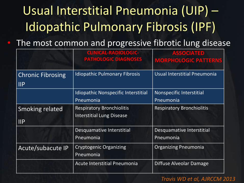
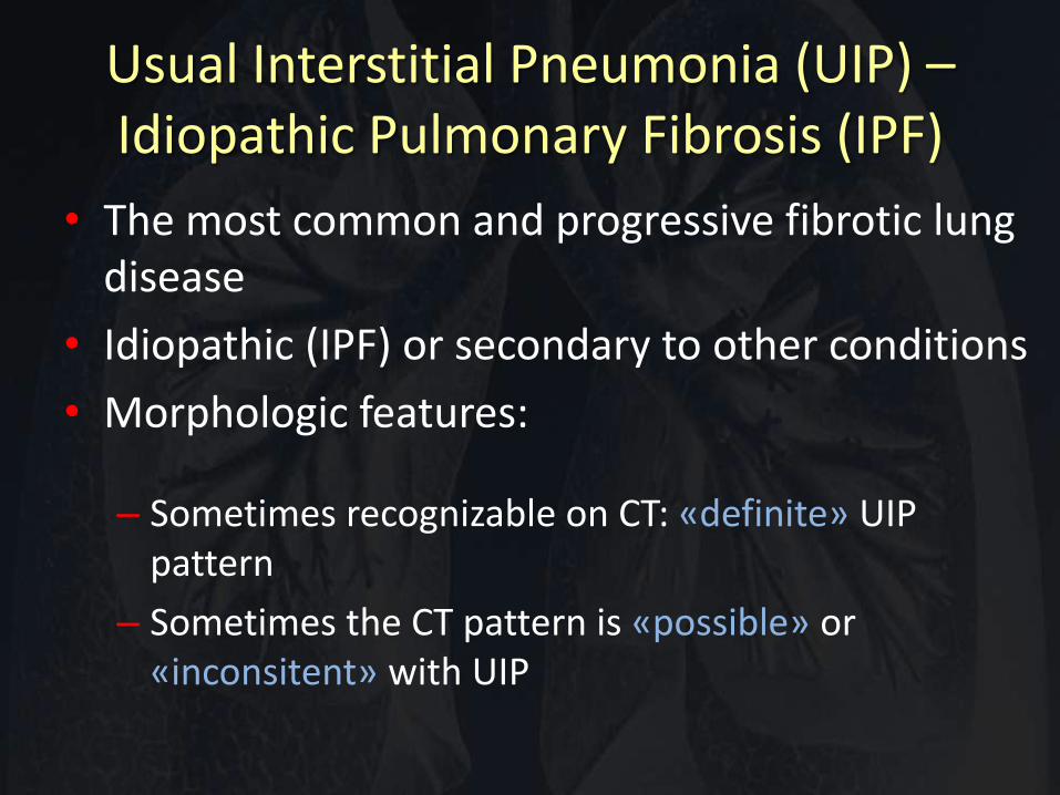
Usual Interstitial Pneumonia (UIP) – Idiopathic Pulmonary Fibrosis (IPF)
• The most common and progressive fibrotic lung disease
CLINICAL-RADIOLOGIC- PATHOLOGIC DIAGNOSES
ASSOCIATED MORPHOLOGIC PATTERNS
Chronic Fibrosing
IIP
Idiopathic Pulmonary Fibrosis Usual Interstitial Pneumonia
Idiopathic Nonspecific Interstitial
Pneumonia
Nonspecific Interstitial
Pneumonia
Smoking related
IIP
Respiratory Bronchiolitis
Interstitial Lung Disease
Respiratory Bronchiolitis
Desquamative Interstitial
Pneumonia
Desquamative Interstitial
Pneumonia
Acute/subacute IP Cryptogenic Organizing
Pneumonia
Organizing Pneumonia
Acute Interstitial Pneumonia Diffuse Alveolar Damage
Travis WD et al, AJRCCM 2013
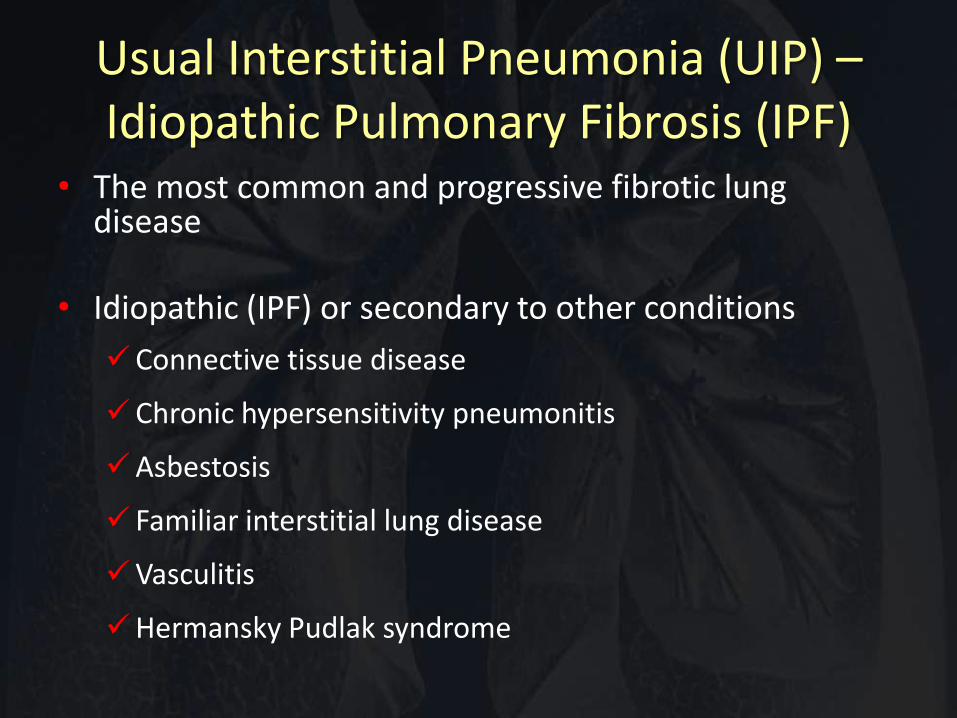
Usual Interstitial Pneumonia (UIP) – Idiopathic Pulmonary Fibrosis (IPF)
• The most common and progressive fibrotic lung disease
• Idiopathic (IPF) or secondary to other conditions
Connective tissue disease
Chronic hypersensitivity pneumonitis
Asbestosis
Familiar interstitial lung disease
Vasculitis
Hermansky Pudlak syndrome
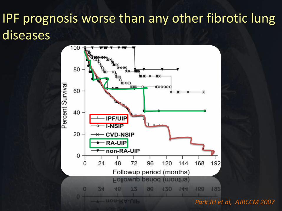
IPF prognosis worse than any other fibrotic lung diseases
Park JH et al, AJRCCM 2007
Usual Interstitial Pneumonia (UIP) – Idiopathic Pulmonary Fibrosis (IPF)
• The most common and progressive fibrotic lung disease
• Idiopathic (IPF) or secondary to other conditions
• Morphologic features:
– Sometimes recognizable on CT: «definite» UIP pattern
– Sometimes the CT pattern is «possible» or «inconsitent» with UIP
Learning objectives:
• Key principles for radiologic assessment of UIP
• Identify the CT features of UIP
• Differential diagnosis
• Longitudinal evaluation
– Acute complications, comorbidities
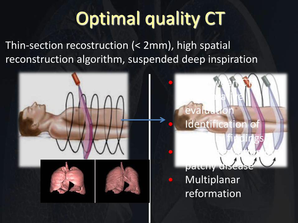
Optimal quality CT Thin-section recostruction (< 2mm), high spatial reconstruction algorithm, suspended deep inspiration
• In between CTs comparative evaluation
• Identification of ancillary findings
• Characterization of patchy disease
• Multiplanar reformation
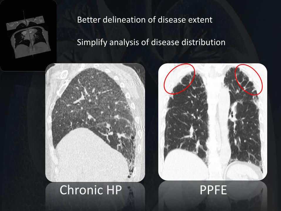
Chronic HP PPFE
Better delineation of disease extent Simplify analysis of disease distribution
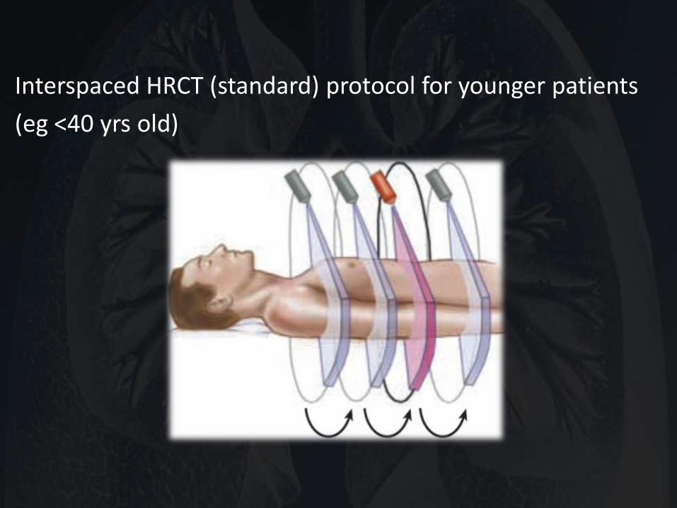
Interspaced HRCT (standard) protocol for younger patients
(eg <40 yrs old)
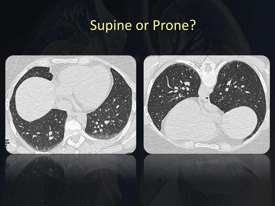
Supine or Prone?
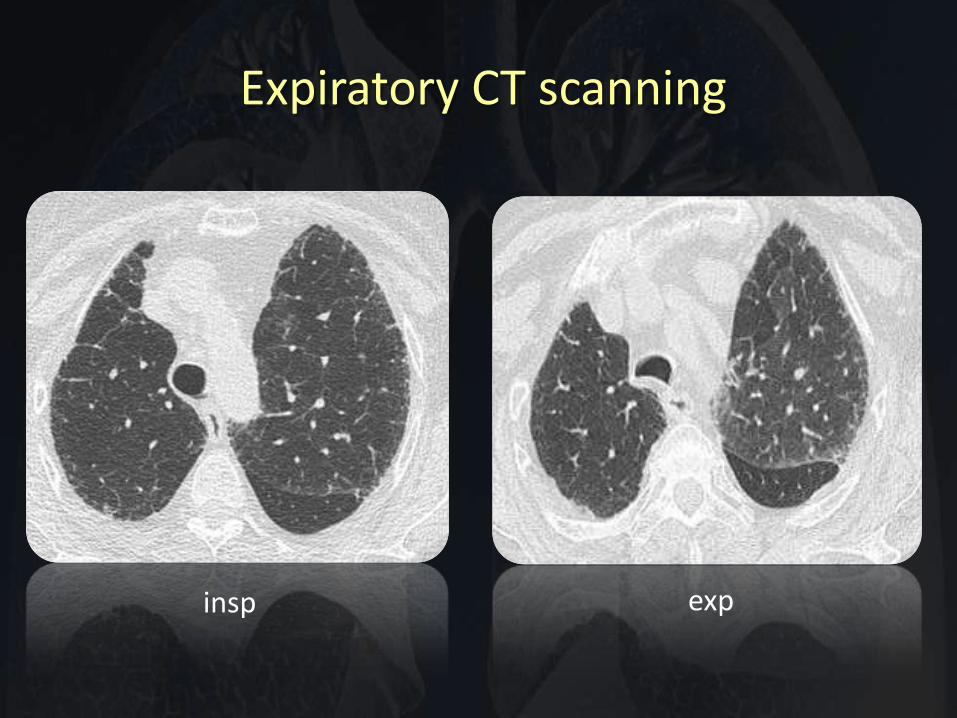
Expiratory CT scanning
insp exp
Systematic approach to CT
• Evaluation of image quality
• Precise description of specific disease features using standard terminology
• Disease distribution
• Is it a fibrotic ILD or non-fibrotic ILD?
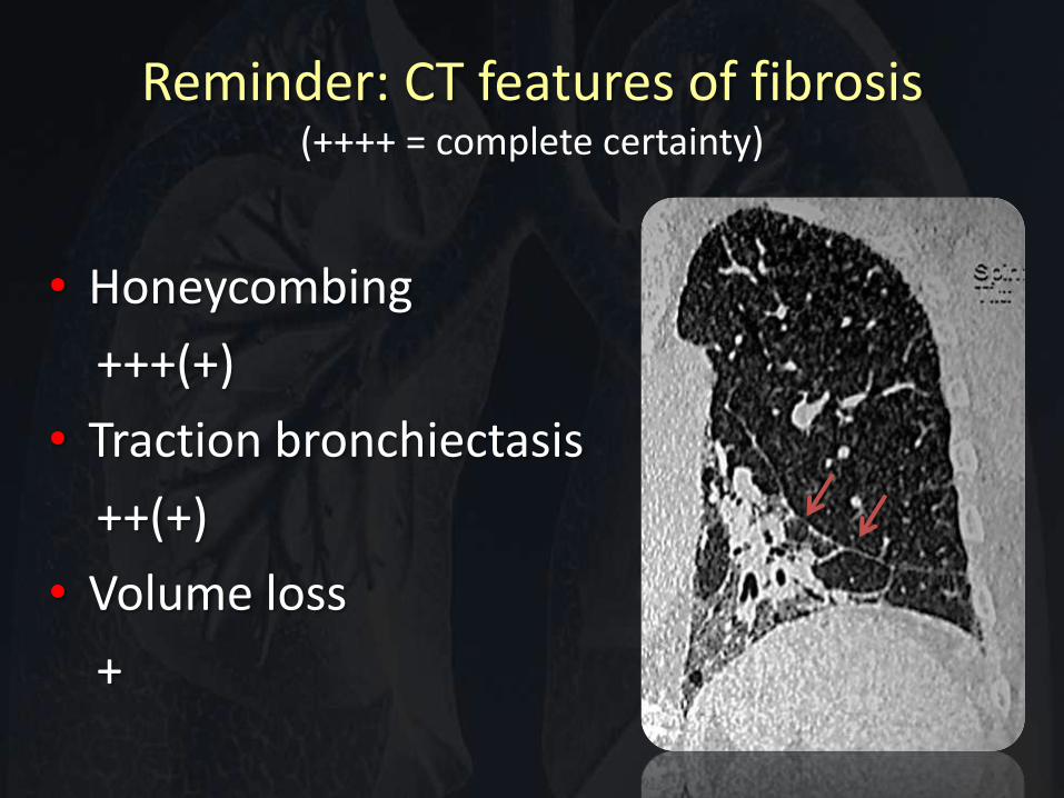
Reminder: CT features of fibrosis (++++ = complete certainty)
• Honeycombing
+++(+)
• Traction bronchiectasis
++(+)
• Volume loss
+
Systematic approach to CT
• Evaluation of image quality
• Precise description of specific disease features using standard terminology
• Disease distribution
• Is it a fibrotic ILD or non-fibrotic ILD? – If so, is it definite UIP?
– If no, is possible or inconsistent?
– what are the alternatives (e.g. fibrotic sarcoid, PPFE etc.)?
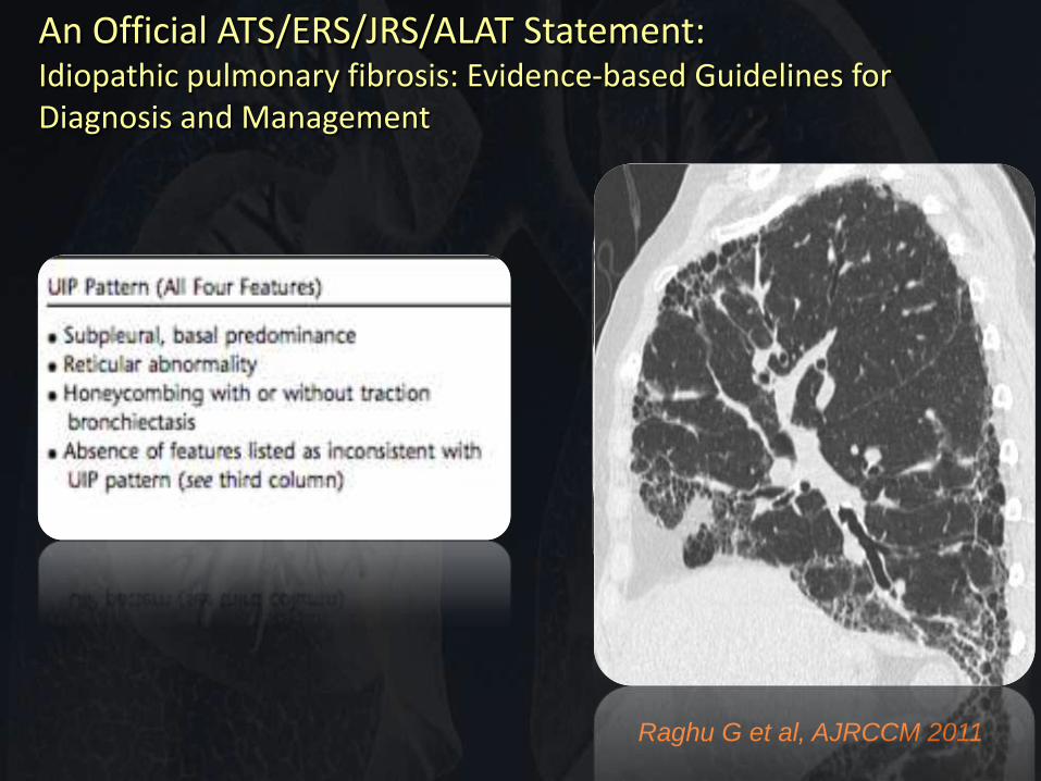
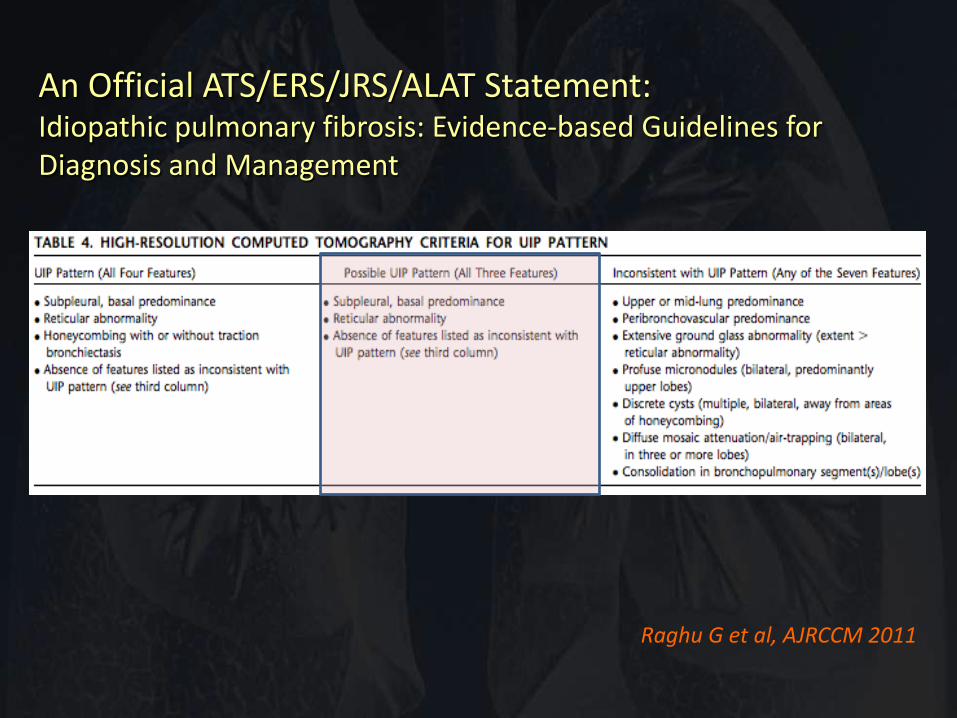
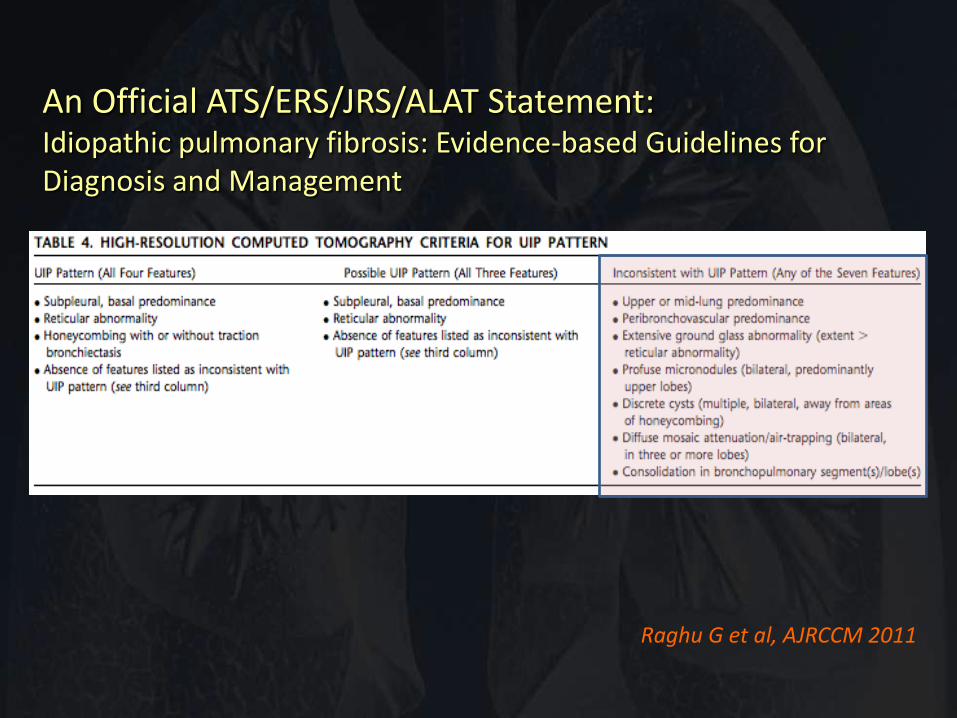
Raghu G et al, AJRCCM 2011
An Official ATS/ERS/JRS/ALAT Statement: Idiopathic pulmonary fibrosis: Evidence-based Guidelines for Diagnosis and Management
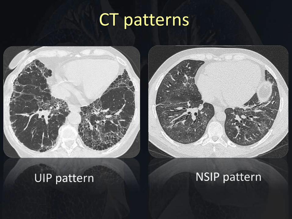
UIP pattern NSIP pattern
CT patterns
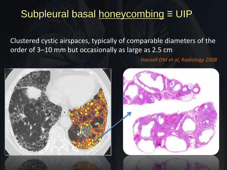
Clustered cystic airspaces, typically of comparable diameters of the order of 3–10 mm but occasionally as large as 2.5 cm
Hansell DM et al, Radiology 2008
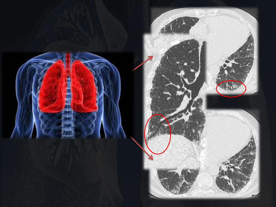
Subpleural basal honeycombing ≡ UIP
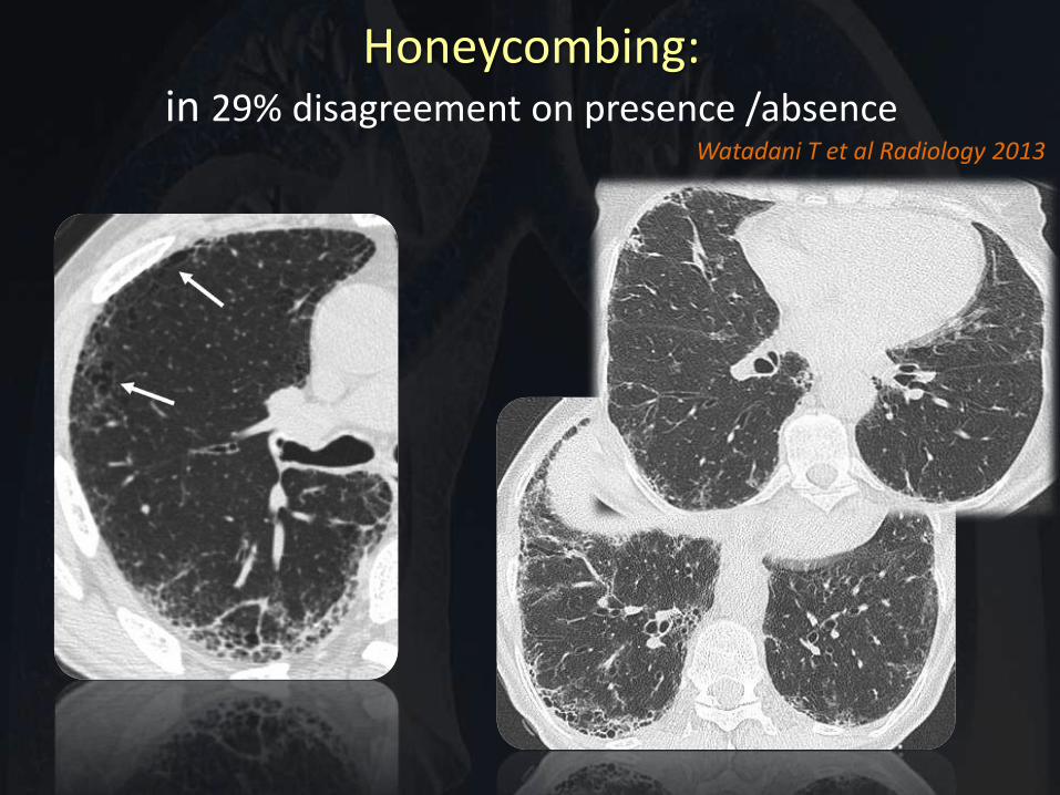
Honeycombing: in 29% disagreement on presence /absence
Watadani T et al Radiology 2013
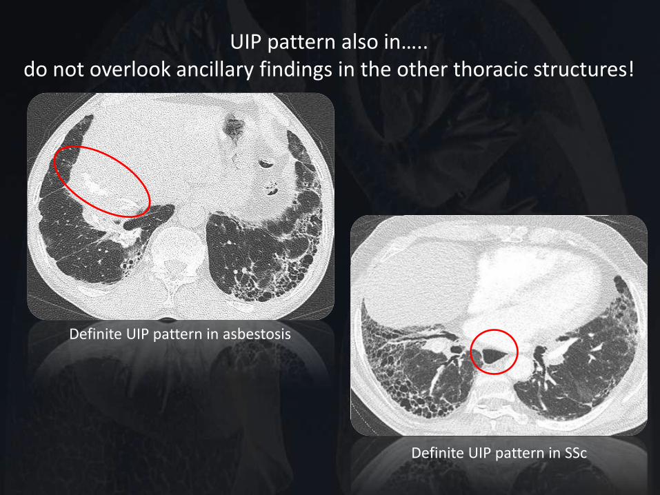
UIP pattern also in….. do not overlook ancillary findings in the other thoracic structures!
Definite UIP pattern in asbestosis
Definite UIP pattern in SSc
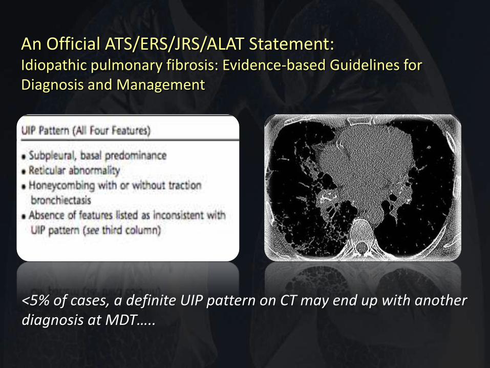
An Official ATS/ERS/JRS/ALAT Statement: Idiopathic pulmonary fibrosis: Evidence-based Guidelines for Diagnosis and Management
<5% of cases, a definite UIP pattern on CT may end up with another diagnosis at MDT…..
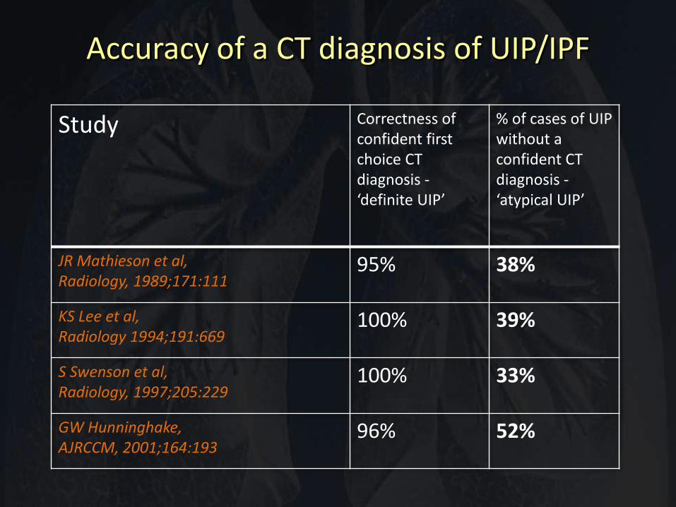
Accuracy of a CT diagnosis of UIP/IPF
Study Correctness of confident first choice CT diagnosis - ‘definite UIP’
% of cases of UIP without a confident CT diagnosis - ‘atypical UIP’
JR Mathieson et al, Radiology, 1989;171:111
95% 38%
KS Lee et al, Radiology 1994;191:669
100% 39%
S Swenson et al, Radiology, 1997;205:229
100% 33%
GW Hunninghake, AJRCCM, 2001;164:193
96% 52%
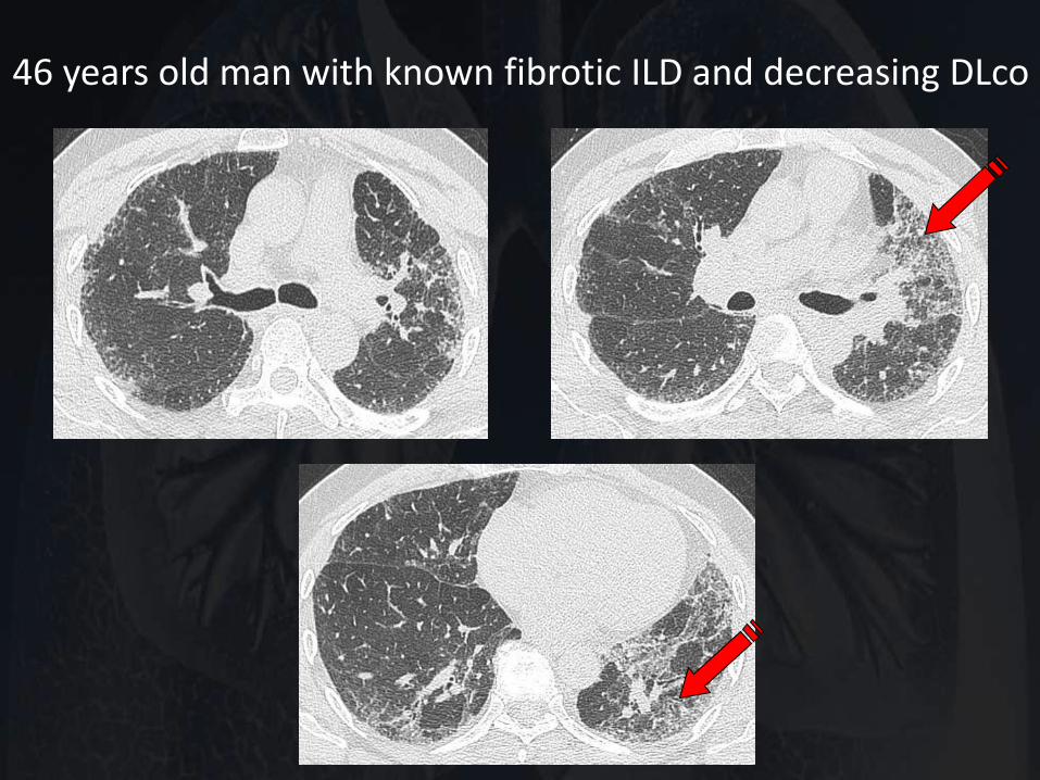
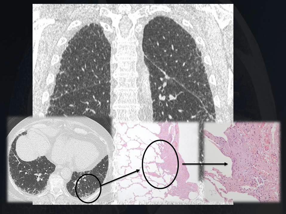
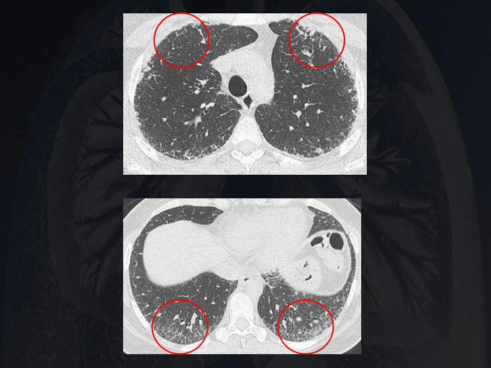
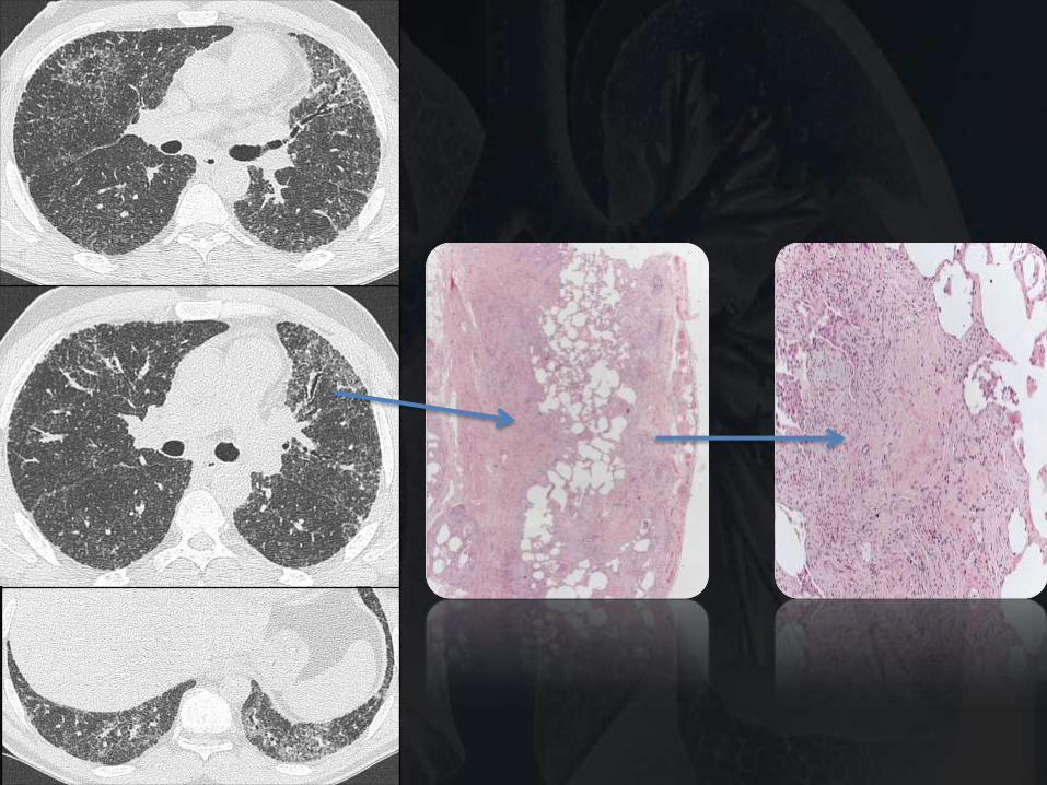
46 years old man with known fibrotic ILD and decreasing DLco
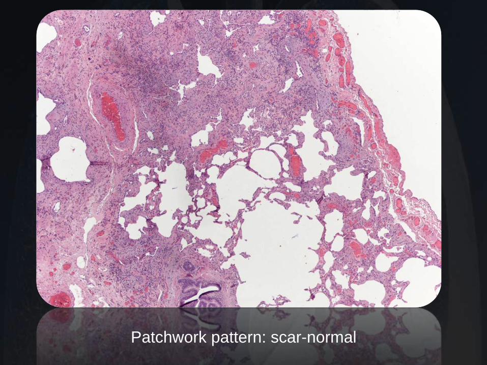
Patchwork pattern: scar-normal
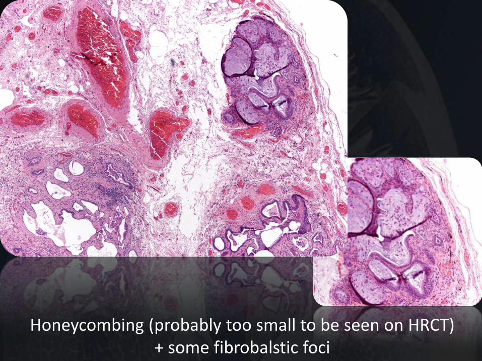
Honeycombing (probably too small to be seen on HRCT) + some fibrobalstic foci
Raghu G et al, AJRCCM 2011
An Official ATS/ERS/JRS/ALAT Statement: Idiopathic pulmonary fibrosis: Evidence-based Guidelines for Diagnosis and Management
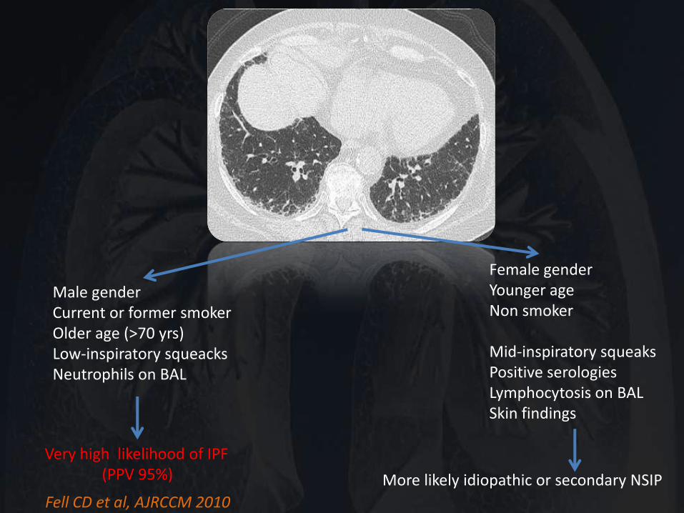
Male gender Current or former smoker Older age (>70 yrs) Low-inspiratory squeacks Neutrophils on BAL
Very high likelihood of IPF (PPV 95%)
Female gender Younger age Non smoker Mid-inspiratory squeaks Positive serologies Lymphocytosis on BAL Skin findings More likely idiopathic or secondary NSIP
Fell CD et al, AJRCCM 2010
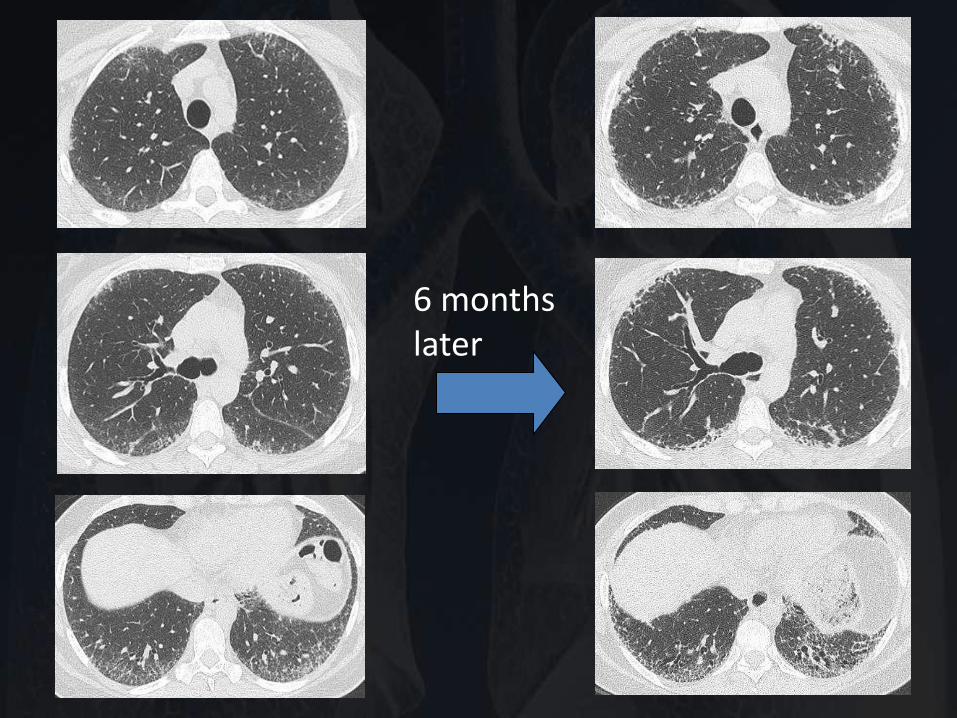
6 months later
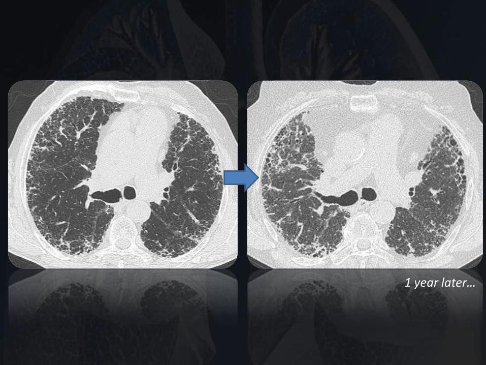
1 year later…
Raghu G et al, AJRCCM 2011
An Official ATS/ERS/JRS/ALAT Statement: Idiopathic pulmonary fibrosis: Evidence-based Guidelines for Diagnosis and Management
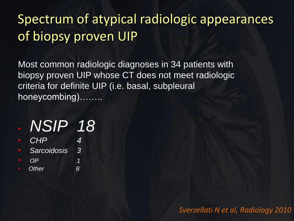
Most common radiologic diagnoses in 34 patients with
biopsy proven UIP whose CT does not meet radiologic
criteria for definite UIP (i.e. basal, subpleural
honeycombing)……..
• NSIP 18 • CHP 4
• Sarcoidosis 3
• OP 1
• Other 8
Sverzellati N et al, Radiology 2010
Spectrum of atypical radiologic appearances of biopsy proven UIP
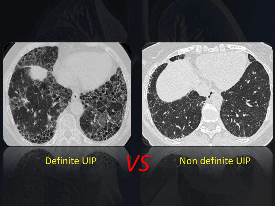
VS Definite UIP Non definite UIP
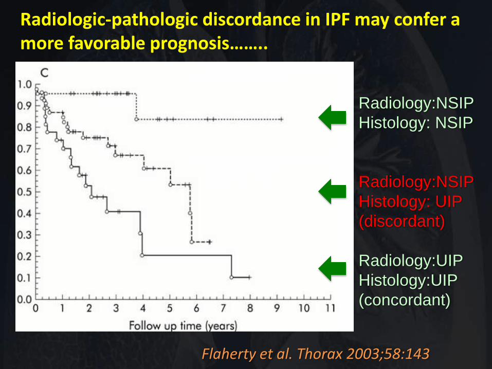
Flaherty et al. Thorax 2003;58:143
Radiologic-pathologic discordance in IPF may confer a more favorable prognosis……..
Radiology:NSIP
Histology: NSIP
Radiology:NSIP
Histology: UIP
(discordant)
Radiology:UIP
Histology:UIP
(concordant)
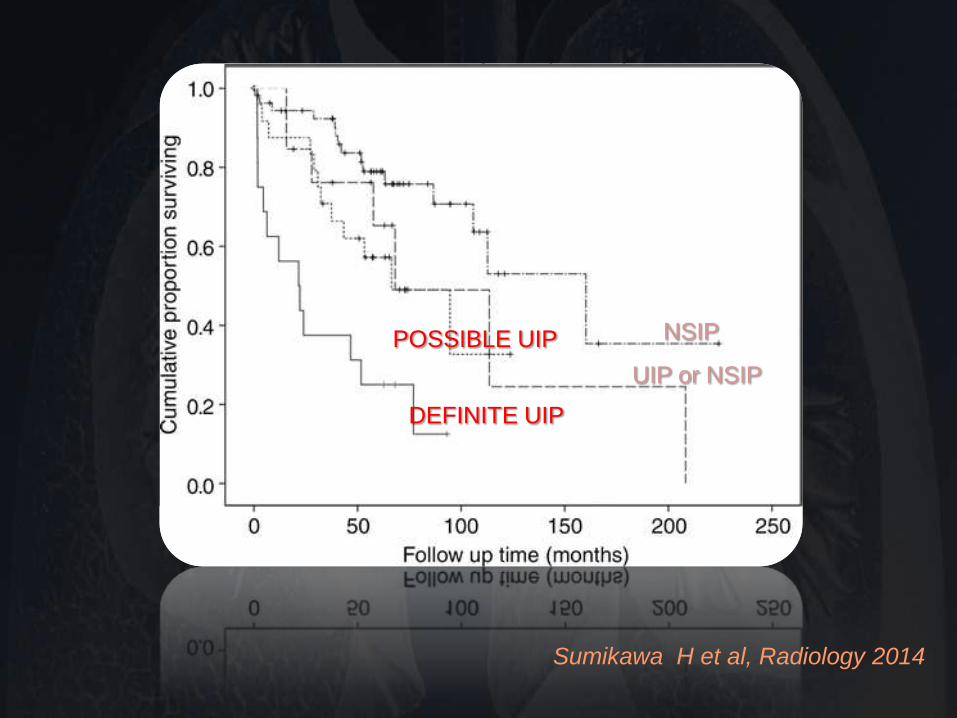
Sumikawa H et al, Radiology 2014
DEFINITE UIP
POSSIBLE UIP
UIP or NSIP
NSIP
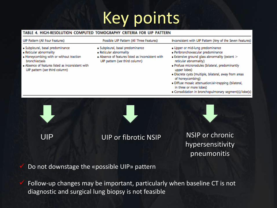
UIP UIP or fibrotic NSIP NSIP or chronic hypersensitivity
pneumonitis
Key points
Do not downstage the «possible UIP» pattern
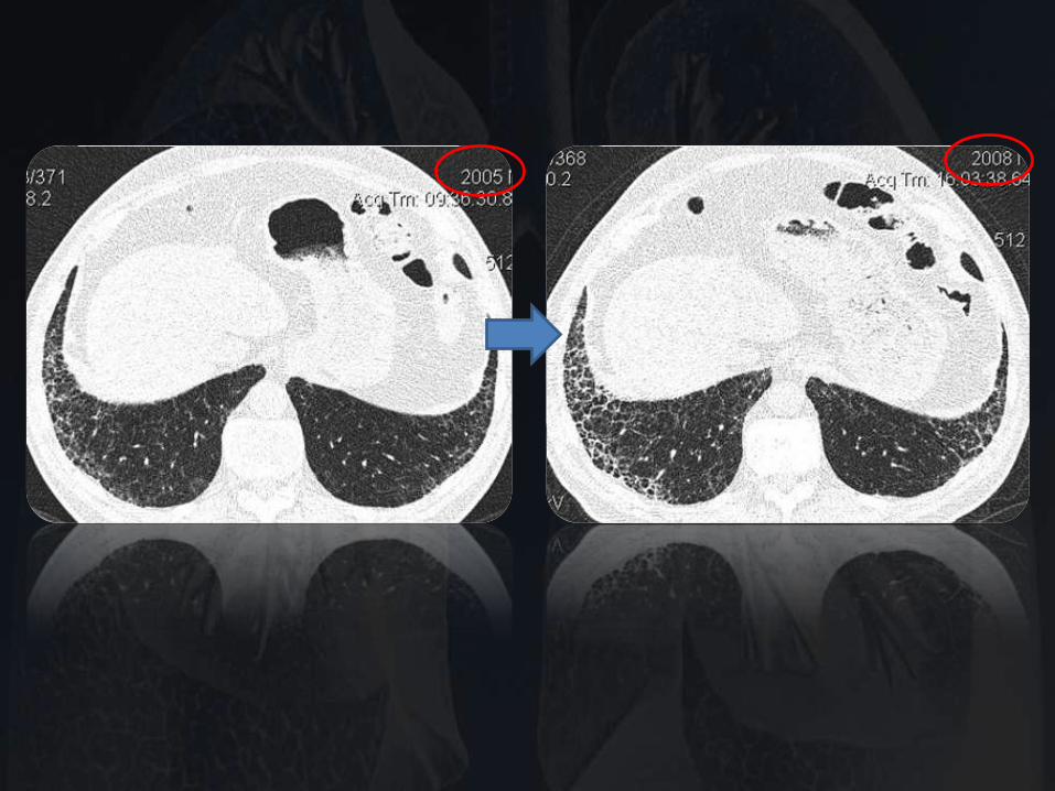
Follow-up changes may be important, particularly when baseline CT is not diagnostic and surgical lung biopsy is not feasible
![Performant TCP for Low-Power Wireless Networks · 0 20 40 60 80 100 120 uIP [ZAJ11] uIP [AMR+11] uIP [HDIV15] BLIP [KIL+15] Arch Rock [HC08] This Paper s) Single-Hop Multi-Hop Making](https://img.pdfslide.us/doc/110x75/5f84307eb2a981198c522c1c/performant-tcp-for-low-power-wireless-networks-0-20-40-60-80-100-120-uip-zaj11.jpg)