Embed Size (px)
DESCRIPTION
How to deal with suspected arrhythmia CCG Educational Evening 17.7.14. Dr Sanjay Kumar Lead Consultant Cardiologist Dr Ravi Kamdar Consultant Cardiologist in Devices and Arrhythmia Croydon Health Services NHS Trust. Objectives. Brief overview of new suspected arrhythmia pathway - PowerPoint PPT Presentation
Citation preview

How to deal with suspected arrhythmia
CCG Educational Evening 17.7.14
Dr Sanjay KumarLead Consultant Cardiologist
Dr Ravi KamdarConsultant Cardiologist in Devices and Arrhythmia
Croydon Health Services NHS Trust
© your company name. All rights reserved.

Objectives
• Brief overview of new suspected arrhythmia pathway
• Things to do before referral
• Case studies
• Questions

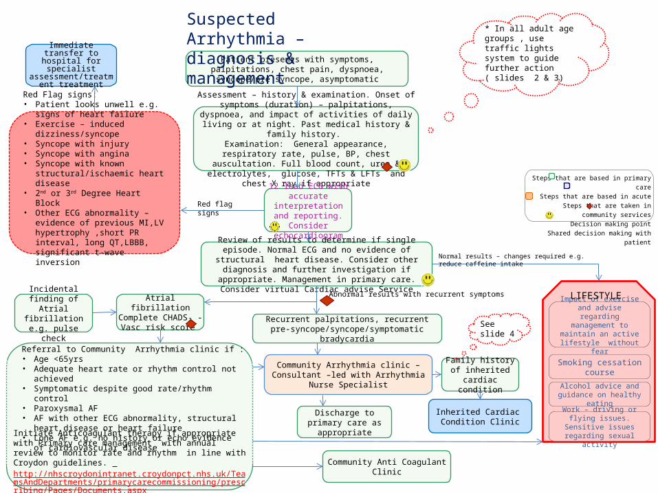
Initiate Anticoagulant therapy if appropriate with Primary care management with annual review to monitor rate and rhythm in line with Croydon guidelines. http://nhscroydonintranet.croydonpct.nhs.uk/TeamsAndDepartments/primarycarecommissioning/prescribing/Pages/Documents.aspx
Immediate transfer to hospital for specialist
assessment/treatment treatment
Patient presents with symptoms, palpitations, chest pain, dyspnoea, syncope/pre syncope, asymptomatic
Assessment – history & examination. Onset of symptoms (duration) – palpitations, dyspnoea, and impact of activities of daily living or at night. Past medical history & family history.
Examination: General appearance, respiratory rate, pulse, BP, chest auscultation. Full blood count, urea & electrolytes, glucose,
TFTs & LFTs and chest X ray if appropriate
Suspected Arrhythmia – diagnosis & management
Review of results to determine if single episode. Normal ECG and no evidence of structural heart disease. Consider other diagnosis and further investigation if appropriate. Management in primary care.
Consider virtual Cardiac advise Service
Atrial fibrillationComplete CHADS₂ - Vasc
risk score Recurrent palpitations, recurrent pre-syncope/syncope/symptomatic bradycardia
Community Arrhythmia clinic – Consultant –led with Arrhythmia Nurse Specialist
* In all adult age groups , use traffic lights system to guide further action ( slides 2 & 3)
Steps that are based in primary careSteps that are based in acute
Steps that are taken in community servicesDecision making point
Shared decision making with patient
12 lead ECG with accurate interpretation and reporting. Consider
echocardiogram
Family history of inherited cardiac
condition
Inherited Cardiac Condition Clinic
Community Anti Coagulant Clinic
Impact of exercise and advise regarding
management to maintain an active lifestyle without
fear
Smoking cessation course
Alcohol advice and guidance on healthy eating
Work – driving or flying issues. Sensitive issues
regarding sexual activity
LIFESTYLE
Red Flag signs:• Patient looks unwell e.g. signs of heart
failure• Exercise – induced dizziness/syncope• Syncope with injury• Syncope with angina• Syncope with known
structural/ischaemic heart disease• 2nd or 3rd Degree Heart Block• Other ECG abnormality – evidence of
previous MI,LV hypertrophy ,short PR interval, long QT,LBBB, significant t-wave inversion
Incidental finding of Atrial fibrillation e.g.
pulse check
Red flag signs
Normal results – changes required e.g. reduce caffeine intake
Abnormal results with recurrent symptoms
Discharge to primary care as appropriate
See slide 4
Referral to Community Arrhythmia clinic if :• Age <65yrs• Adequate heart rate or rhythm control not achieved• Symptomatic despite good rate/rhythm control• Paroxysmal AF• AF with other ECG abnormality, structural heart disease or
heart failure• Lone AF e.g. no history or echo evidence of cardiovascular
disease


Palpitations - What to do before referral (1)
Identify specific patients….high risk of adverse outcomes? Can intervention improve QoL?
What does the patient mean? ‘Abnormally perceived heartbeat’ – tachycardia, bradycardia, irregularity, pounding, ‘missed beats’, ‘extra beats’, ‘fluttering’Do they mean chest discomfort?Duration, frequency, Onset/offset, Precipitating factors – nocturnal? exertional? Emotional? Mode of termination – vagal?
What else happens? Pre-syncope or syncope, acute-onset sustained SOB, chest pain
What’s making it worse? Mental health – anxiety, depression, somatisation disordersCaffeine, alcoholRecreational drugs – amphetamines, cocaineMedications – β agonist, theophylline, T4, calcium antagonists, class I anti-arrhythmics (e.g. flecainide), drugs prolonging
QTc
Family history? Evidence of early-onset AF, heart failure or premature CAD in close relative?Sudden cardiac or unexplained death <40yrs old? RTA? Drowning?Potential misdiagnosis of epilepsy? Sudden death?

Palpitations - What to do before referral (2)Examination & simple tests
Physical evidence of structural heart disease (HF, abnormal HS, murmurs)?Thyrotoxicosis? Anaemia?12 lead ECG – AF, high-grade heart block, old MI, LVH, LBBB, ischaemic ST/t-wave change, short PR, long QTc
Sources of advice for patientsCUH arrhythmia nurse / clinicCharities: Arrhythmia Alliance: www.heartrhythmcharity.org.uk
Atrial Fibrillation Association: www.atrialfibrillation.org.ukSudden Adult Death Trust: www.sadsuk.orgCardiac Risk in the Young: www.c-r-y.org.uk
Occupation / driving: DVLA rulesIf arrhythmia identified that causes (or is likely to cause) incapacity, stop drivingDoctors’ responsibility to inform patientIf no diagnosis (or awaiting specialist assessment), but disabling symptoms, advise patient to stop drivingWorking at height?Control of potentially dangerous machinery?


How would you manage these patients?Case Study 1 64yr old ladyIrregular pulse on recent pulse check12-lead ECG: Atrial fibrillation, heart rate 120bpm, otherwise NAD
Many years’ minor (NYHA class 2) exertional breathlessness, otherwise no symptomsHypertension, type 2 diabetes, 30U alcohol/weeko/e Looks well, BP 150/90, RR 12, no cardiac murmursFBC, U&E, TFT normalCXR – no parenchymal lung abnormality, borderline cardiomegaly
ActionAdvice? Reduce alcohol intake. Avoid vigorous effort until rate-controlled. Treat co-morbidity e.g. salt/BP
Drugs? Rate control with β blocker (exercise-induced tachycardia) or rate-limiting CCB (BP); anti-coagulate with warfarin
Investigation? Echo (LV function, LA size, valves, evidence of CAD); 24hr Holter after initiating rate-limiters (HR control)
Onward referral? Suspected arrhythmia clinic – <65, rate or rhythm control? Diagnose underlying cause?

Patients (%)
36.1%
30.1%
27%
6.9%
The CHADS2 index has been routinely used as an initial, rapid, and easy-to-remember means of assessing stroke risk1–4
CHADS2 criteria Score
Congestive HF 1
Hypertension 1
Age ≥75 years 1
Diabetes 1
Stroke or TIA (previous history) 2
Sum
*Adjusted stroke rate = expected stroke rate per 100 patient-years from exponential survival model, assuming ASA not taken
CHADS2 Adjusted stroke rate*
(95% CI)
6 18.2 (10.5 to 27.4)
5 12.5 (8.2 to 17.5)
4 8.5 (6.3 to 11.1)
3 5.9 (4.6 to 7.3)
2 4.0 (3.1 to 5.1)
1 2.8 (2.0 to 3.8)
0 1.9 (1.2 to 3.0)
33.6% with CHADS2
0 or 1
1. Gage et al. JAMA 2001;285:2864–28702. Gage et al. Circulation 2004;110:2287–2292
3. Camm et al. Eur Heart J 2010;31:2369–24294. Nieuwlaat et al. Eur Heart J 2006;27:3018–3026

CHADS2 Score CHA2DS2-VASc Score
Congestive heart failure 1 Congestive heart failure/left ventricular dysfunction 1
Hypertension 1 Hypertension 1
Aged ≥75 years 1 Aged ≥75 years 2
Diabetes mellitus 1 Diabetes mellitus 1
Stroke/TIA/TE 2 Stroke/TIA/TE 2
Maximum score 6 Vascular disease (prior MI, PAD, or aortic plaque) 1
Aged 65–74 years 1
Sex category (i.e. female gender) 1
Maximum score 9
The CHA2DS2-VASc scheme was adopted by the ESCto complement the CHADS2 scoring system
CHA2DS2-VASc: In patients with a CHADS2 score of 0–1, or When a more detailed stroke risk assessment is indicated
Camm et al. Eur Heart J 2010;31:2369–429.

Letter Clinical characteristic Points awarded
H Hypertension 1
AAbnormal renal and liver function (1 point each) 1 or 2
S Stroke 1
B Bleeding 1
L Labile INRs 1
E Elderly (e.g. age >65 years) 1
D Drugs or alcohol (1 point each) 1 or 2
Maximum 9 points
The 2010/2012 ESC guidelines recommend use of a simple bleeding risk score: HAS-BLED
HAS-BLED ≥3: Indicates ”high risk”, and Some caution and regular review of the patient is needed following
the initiation of antithrombotic therapy, whether with OAC or aspirin
Camm et al. Eur Heart J 2010;31:2369–429.Camm et al. Eur Heart J 2012;e-published August 2012, doi:10.1093/eurheartj/ehs253.Pisters et al. Chest 2010;138:1093–100.

How would you manage these patients?Case Study 2 27yr old female medico-legal solicitorc/o 1 episode of severe dizziness while standing in a long post-office queue, nearly lost consciousnessNo chest pain, SOBNo recurrent symptoms
PMH: 2 previous similar episodes aged 16 and 18, while studying for examsNo medication, no alcohol, occasional cannabis useFH: mother died suddenly aged 35, cause unknowno/e normal – no cardiac murmurs
FBC, U&E, TFT normal12-lead ECG: sinus rhythm 70bpm, no heart block, normal QTc
Action
Advice? Identify & avoid triggers e.g. prolonged standing, pain/cough/laugh, review/minimise work stresses; keep well-hydrated; curtail cannabis use
Drugs? Water
Investigation? Echocardiogram (subclinical cardiomyopathy, severe valvular heart disease)
Onward referral? One-stop arrhythmia clinic, GP advice service
What if no FHx? Re-assure likely vasovagal – advice, referral only if symptoms not controlled by conservative means

How would you manage these patients?Case Study 3 79yr old manc/o 4 episodes of self-terminating palpitation, over a period of 6 weeksEach episode approximately 1-minute long, associated with minor SOB
Minor (NYHA class 2) exertional SOB, no angina pectoris, no syncopeMI 10 years ago, hypertension, hypercholesterolaemia, current smokerRx: Aspirin, lisinopril, simvastatin (beta blocker not tolerated – dizziness)o/e normal – no cardiac murmurs
FBC, U&E, TFT normal12-lead ECG: sinus rhythm 60bpm, first degree heart block, LBBBSimilar ECG appearances 3 years ago
Action
Advice? Stop smoking (IHD)
Drugs? Avoid rate-limiting medication
Investigation? CXR (HF), echo (LVSD, LVH, scar, substrate for AF/VT)
Onward referral? A&E (red flag)