Embed Size (px)
Citation preview

iGEM 2016
How To
Bacillus subtilis
A Collaboration
Bonn – Freiburg
This manual should serve as an assistance while starting to work with the
versatile bacterium Bacillus subtilis. The iGEM team 2016 Bonn and
Freiburg both struggled with some experiments concerning Bacillus
subtilis, so we decided to create a manual for future teams and some
additional tips and protocols which may help with future work. At this
point, we also like to thank the different persons who helped us working
with Bacillus subtilis: BGSC, Munich iGEM 2012, department of
Pharmaceutical Biotechnology of the Greifswald University (Germany),
Guido de Boer.

What is Bacillus subtilis?
Bacillus subtilis has a rod shaped appearance and belongs to the family of the gram-
positive bacteria.
Apart from being widely present in nature, it is also a part of the microbial gut flora.
The US Food and Drug Administration (FDA) classifies Bacillus subtilis as a GRAS
organism. That means it is generally recognized as safe and can be used problem-free
in S1 laboratories. Bacillus subtilis colonies have an irregular, large size with undulate
margin. They have a white and dull colour and a dry texture. (figure 1)
Figure 1. Colonies of Bacillus subtilis. The figure shows the structure of colonies formed by Bacillus subtilis. They have an irregular, large size
with undulate margin. Their colour is white and dull and they have a dry texture.
The prefered medium of Bacillus subtilis is LB medium. The growth follows the typical
four phases of bacterial growth (figure 2) and with reaching the exponential growth
phase, Bacillus subtilis has a doubling time of 30 minutes under ideal conditions. This
can be calculated with the slope of the exponential growth phase. In our calculation,
the doubling time is 28 minutes.
Figure 2. Growth curve of the Bacillus subtilis
strain WT168.
A sample of Bacillus subtilis WT168 was diluted to
an OD600 of 0.1/ml in LB. The OD600 as
measured in a plate reader (CLARIOstar, BMG
labtech) for 6 hours. The graph represents a
typical growth curve of a bacterium.
Figure 3. Exponential growth phase of Bacillus
subtilis.
The graph shows the exponential growth
phase of Bacillus subtilis. This phase can be
used to calculate the doubling time of bacteria.
In this case, the doubling time was around
28mins.
Bacillus subtilis is also able to form endospores under distress (see sporulation
protocol), which is one of the most efficient adaptations to lack of nutrients. These
endospores are a highly stable form of the bacterium which are resistant to heat, UV-
light and pressure. Under normal conditions they are able to re-enter their normal life
cycle.
Helpful tips to get started with Bacillus subtilis
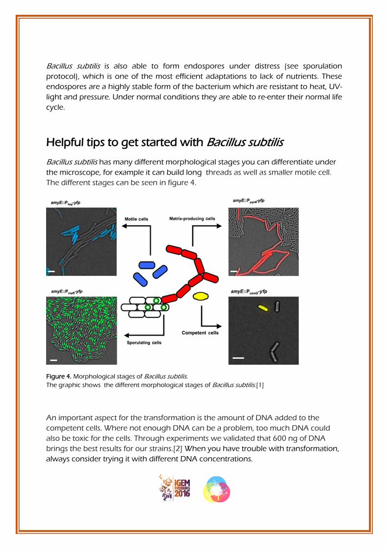
Bacillus subtilis has many different morphological stages you can differentiate under
the microscope, for example it can build long threads as well as smaller motile cell.
The different stages can be seen in figure 4.
Figure 4. Morphological stages of Bacillus subtilis.
The graphic shows the different morphological stages of Bacillus subtilis.[1]
An important aspect for the transformation is the amount of DNA added to the
competent cells. Where not enough DNA can be a problem, too much DNA could
also be toxic for the cells. Through experiments we validated that 600 ng of DNA
brings the best results for our strains.[2] When you have trouble with transformation,
always consider trying it with different DNA concentrations.
Competent cells
Competent #1 - Chemo-Competent Bacillus subtilis
1. Inoculate a culture of Bacillus subtilis in 4 ml of LB-Medium
2. Let them grow overnight at 37°C, 200 rpm
3. Measure the OD600 of your overnight culture and dilute it in MNGE
Medium to an OD600 of around 0.1-0.2/ml in 10 ml LB
4. Let the cells grow to an OD600 of 1.0-1.3/ml (37°C, 200 rpm)
5. Cells should be competent now (very motile)
6. Aliquot 400 μl of the samples in different tubes (each tube stands for one
transformation)
7. You can either use the competent cells directly or add glycerol to dilute to a
final concentration of 10% and freeze them in -80°C
(Protocol from iGEM team munich 2012)
Competent #2 - Electro-Competent Bacillus subtilis
1. Inoculate a liquid culture of Bacillus subtilis and let it grow overnight
2. Vortex the culture gently and give 500 μl each in 3x 20 ml competency
medium
3. Grow the bacteria in the flasks at 37°C 250 rpm shaking till you reach an OD600
between 0.5 and 0.7/ml
4. Add 1 ml of a 20% glycine solution to the first 1.25 ml to the second and 1,5
ml to the third flask to reach total glycine concentrations of 1%, 1.25% and
1.5%
5. Keep shaking for 1 h (because of the glycine the optical density should not
change significantly)
6. Cool down the cells on ice for 15 mins (if not done already, transfer cells in 50
ml Falcon tubes)
7. Centrifuge at 8500 rpm for 10 min at 4°C for getting bacteria pellets

8. Pour off the supernatant and wash the cells three times with ice-cold washing-
buffer (20 ml/10 ml/5 ml). Centrifuge the cells down at 8500 rpm for 10 min at
4°C between each washing procedure and decant the supernatant
9. Resuspend the cells in 1 ml ice cold washing-buffer (all three different cultures
are supposed to get in it!)
10. Make aliquots (recommended: 120 μl → enough for two transformations)
11. Freeze in liquid nitrogen and store at -80°C
(Protocol submitted by department of Pharmaceutical Biotechnology of the Greifswald University)
Results:
Figure 5. Electroporation efficiency for Bacillus subtilis regarding glycine concentration. The electroporation worked best with a higher glycine concentration. Glycine works as a weakening agent, making the cell wall looser by replacing the alanine in the cell wall [3]. The highest colony amount was spotted by using a mixture of all three glycine concentrations (1%,1.25% and 1.5%) for the preparation of electrocompetent cells.

Transformation of Bacillus subtilis
Transformation #1
Using fresh, competent B. subtilis cells:
1. Add 600 ng of DNA and incubate at 37°C (200 x g) for 1 h
2. Add 100 μl of expression mix to each sample
3. Incubate at 37°C for another hour
4. Plate 400 μl the samples on selective agar
Note: if you use the frozen aliquots you can also just add the DNA to the sample, don't centrifuge the competent cells!
(Protocol from iGEM team Munich 2012)
Transformation #2 - Electroporation
Electroporation is a transformation method that relies on sending an electrical current through the cell and creating holes or pores in the cell membrane. Through those pores, the plasmid will enter the cell before they are getting closed. For doing this transformation, special equipment is needed:
1. Electrocompetent cells
2. Competency Medium
3. Plasmid you want to transform
4. Electroporator
5. Electroporation cuvettes
Procedure: 1. Mix electrocompetent cells with the plasmid and acquire a total volume
of 60 μl with a final DNA concentration of 10 ng/μl
2. Place the cell-plasmid-suspension, the electroporation-cuvettes and
competency-medium on ice for 10 min (you need 1 ml competency-
medium & 1 electroporation cuvette per transformation, but it is
recommend to take more)
3. Pipet the cold cell-plasmid-suspension in the prechilled electroporation-
cuvette and tap the cuvette multiple times (this way you get rid of
bubbles and spread your suspension equally)
4. Make sure that the electroporation-cuvette is dry (take care that you
don’t touch the metal sides anymore!)
5. Electroporate at 2100 Volt (the electroporator will give out a “time-
constant”. A time-constant from 3.0 to 5.5 is a positive indicator
although the transformation could also be successful at a lower time-
constant)
6. Flush the electroporated mixture out of the electroporation-cuvette
with 1 ml of competency-medium
7. Let the cells grow for 3 h at 37°C 300 rpm shaking
8. Pellet cells by centrifugation at RT 5 min and decant the supernatant
(do not throw away)
9. Resuspend the pellet in 100 μl of the supernatant
10. Plate on selective agar
(Protocol from Greifswald)
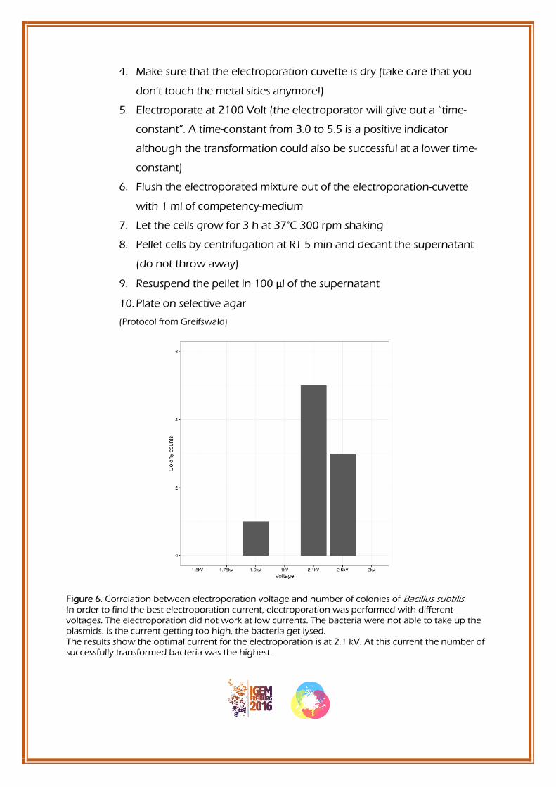
Figure 6. Correlation between electroporation voltage and number of colonies of Bacillus subtilis. In order to find the best electroporation current, electroporation was performed with different voltages. The electroporation did not work at low currents. The bacteria were not able to take up the plasmids. Is the current getting too high, the bacteria get lysed. The results show the optimal current for the electroporation is at 2.1 kV. At this current the number of successfully transformed bacteria was the highest.

Table 1. Electroporation efficiency for Bacillus subtilis regarding DNA-concentration. The table shows the number of formed Bacillus subtilis colonies after the transformation using electroporation with different amounts of DNA-concentrations. It shows that a higher amount of DNA
(10 ng/μl) leads to a higher number of colonies (11). (de Boer, 2015)
DNA-concentration (ng/μl) Number of colonies
10 11
5 3
1 1
0 0
In order to find the optimal DNA-concentration for the transformation the electroporation was performed with different DNA-amounts. The higher the DNA-concentration the higher the number of successfully transformed bacteria. Therefore, it is recommended to use a higher DNA-
concentration. 10 ng/μl is a sufficient amount.

Sporulation
1. Overnight culture
● Inoculate your culture of Bacillus subtilis in 4 ml LB-medium
● Let them grow overnight at 37°C, 200 rpm
2. Exponential growth
● Measure the OD600 of your overnight culture and dilute it in LB-medium to an OD600 off around 0.1-0.2/ml in 10 ml LB
● Let the cells grow to an OD600 of 0,8/ml (37°C, 200 rpm)
3. Sporulation
● Centrifuge 10 ml of the cells at 13,000 x g for 1 minute
● Wash the pellet with 1 x PBS
● Re-suspend the pellet in 5 ml DSM (Difco Sporulation Medium)
● Let the cells grow for 24 hours at 37°C (200 rpm)
4. Lysozyme treatment (additional for spore purification)
● Treat the samples with lysozyme (15 mg/ml) at a dilution of 1:6
● Incubate for 1 h at room temperature
● Wash 6 times with 1 x PBS
5. Additional:
→ count spores using a Neubauer improved counting chamber and make aliquots
with a defined number of spores per aliquot (e.g. 100 Million spores per 500 μl)
(Note: always use fresh DSM since the FeSO4 is rusting when it is in dilution)

Data from the purification:
In forward and side scatter you can easily distinct the difference between the purified
and unpurified samples (figure 5). Purification leads to a better and solid read out in
further experiments and applications.
Figure 7. Bacillus subtilis spore purification.
(A) The spores of Bacillus subtilis WT168 were analyzed using flow cytometry. The set gate for spores
shows that 75.5% of the sample consisted of spores. (B) The spores of Bacillus subtilis WT168 were
treated with lysozyme for 1 h. The flow cytometry analysis shows that after purification the amount of
spores is considerably higher with 91.9%.
Antibiotics
Table 2: The concentrations of antibiotics used for Bacillus subtilis.
The table shows the minimal concentrations of different antibiotics used for the selection of
antibiotic resistant Bacillus subtilis cells.
Antibiotic Stock solution Dilution for B. subtilis
Spectinomycin (Spec) 100 mg/ml 1 to 1.000
Chloramphenicol (Cml) 25 mg/ml 1 to 5.000
Erythromycin (Erm) 10 mg/ml 1 to 1.000
Ampicillin (Amp) 100 mg/ml 1 to 1.000
Kanamycin (Kan) 50 mg/ml 1 to 1.000

Media
DSM – Sporulation Medium
8 g Nutrient Broth 1 g KCl 1 ml MgSO4 (1 M) 1 ml MnCl2 (10 mM) 1000 ml Aqua bidest
Autoclave and add:
0.5 ml CaCl2 (1 M) 1 ml FeSO4 (1 M)
10x MN Medium (1 Liter)
136 g K2HPO4*H2O
60 g KH2PO4
10 g Na-citrat * 2H2O
MNGE-Medium (100 ml)
9.2 ml 10x MN-Medium
82.8 ml Sterile water
10 ml Glucose (20% filtered)
500 µl K-Glutamate (40%)
500 µl Fe[III]-ammonium-citrate (2,2 mg/ml)
1 ml Tryptophan (5 mg/ml)
300 µl MgSO4 (1 M)
(1 ml Threonine (5 mg/ml) only for selective agar after the transformation)

Expression mix 10,5 ml
5 ml Yeast extract (5%)
2.5 ml Casamino-acids (CAA)(10%)
2.5 ml Sterile water
500 µl Tryptophan (5 mg/ml)
Competency Medium 500 ml: 10 g LB
45.5 g Sorbitol
→ autoclave
Washing-Buffer 500 ml : 45.5 g Sorbitol (total 0.5 M) 45.5 g Mannitol (total 0.5 M) 50 ml Glycerol (total 10%)
→ autoclave
References
[1] (Lopez D., Vlamakis H. &Kolter R Generation of multiple cell types in Bacillus subtilis. FEMS
Microbiology Reviews. Jan 2009)
[2] http://www.askabiologist.org.uk/answers/viewtopic.php?id=9446 – 16.10.2016
[3] Zhi Zhang, Development of an Efficient Electroporation Method for Iturin A-Producing
Bacillus subtilis ZK, April 2015

B. subtilis manual
Made by the iGEM Teams
Bonn and Freiburg
Danja Steinberg
Jonas Gockel
Nathalie Wagner
Katharina Ostmann