Embed Size (px)
Citation preview
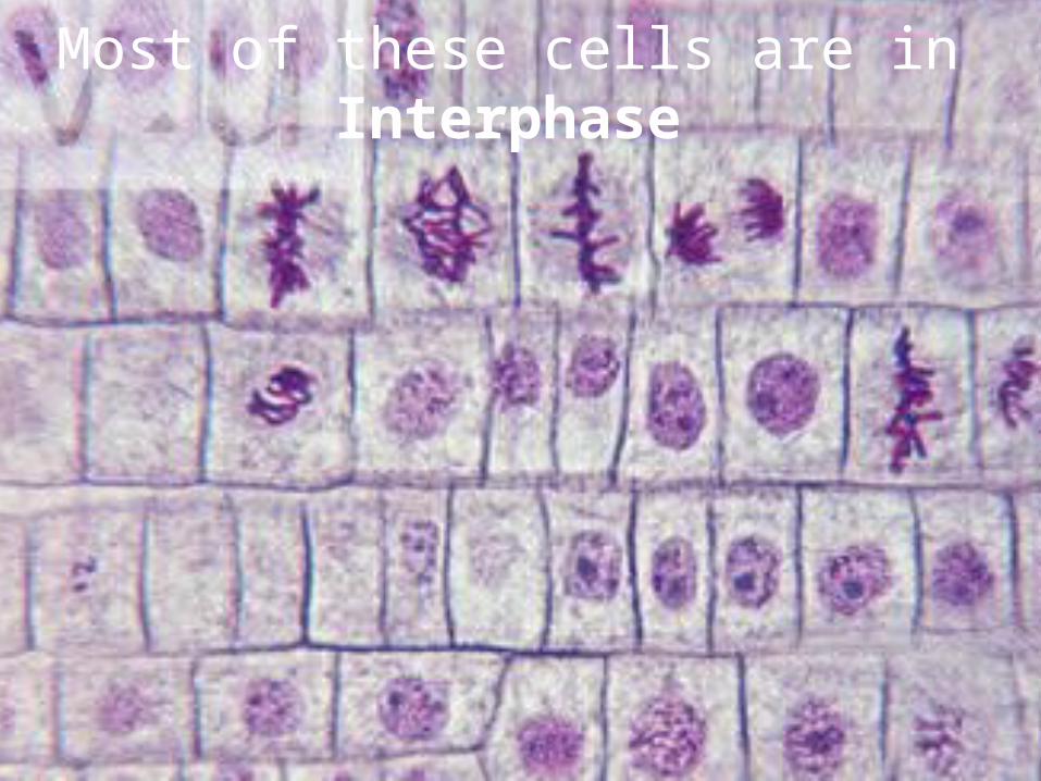
How many stages of cell division do you see in this image?
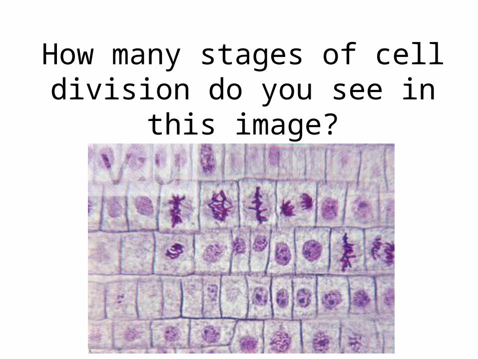
Cells reproduce by a cycle of growing and dividing called the cell cycle.
)
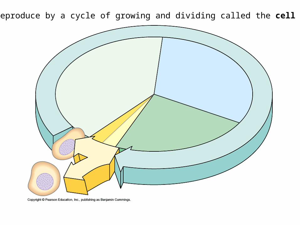
Cell Cycle
Mitosis will be our main focus.
First, we will look at Interphase…
This graph is NOT to scale!!
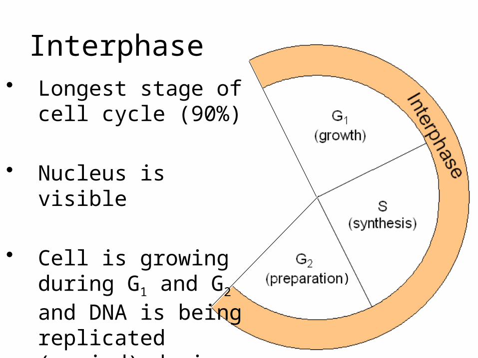
Interphase
)
• Longest stage of cell cycle (90%)
• Nucleus is visible
• Cell is growing during G1 and G2 and DNA is being replicated (copied) during S.
Interphase:3 Phases
)
1. G1: Cell is Growing
2. S: DNA is Synthesized (DNA Replication)
3. G2: Cell growth; Organelles duplicate in preparation for division
Most of these cells are in Interphase
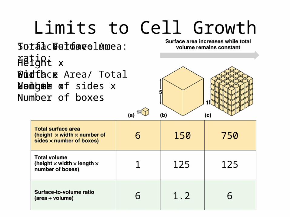
Limits to Cell Growth
6 150 750
1 125 125
6 61.2
Total Surface Area:
Height x Width x Number of sides x Number of boxes
Total Volume:
Height x Width x Length x Number of boxes
Surface-to-volume ratio:
Surface Area/ Total Volume
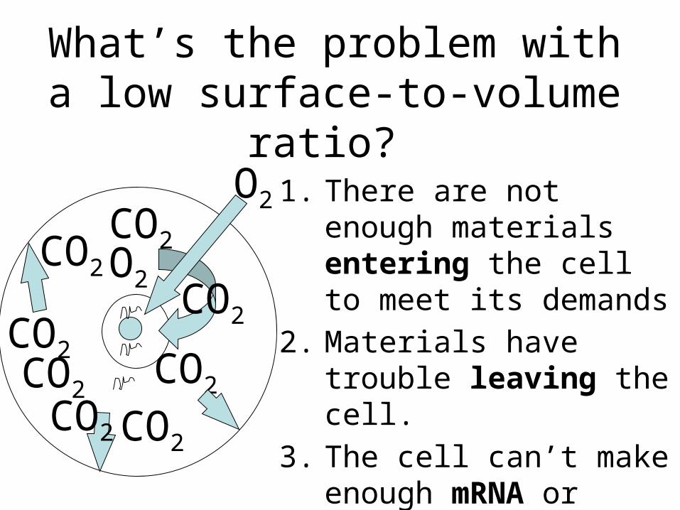
What’s the problem with a low surface-to-volume ratio?
1. There are not enough materials entering the cell to meet its demands
2. Materials have trouble leaving the cell.
3. The cell can’t make enough mRNA or proteins to meet the demands of the cell.
O2
O2
CO2
CO2CO2CO2
CO2
CO2
CO2
CO2
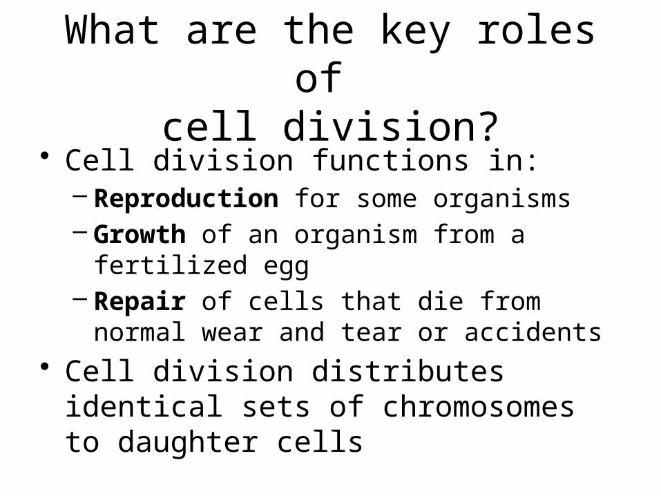
What are the key roles of cell division?
• Cell division functions in:– Reproduction for some organisms– Growth of an organism from a fertilized egg– Repair of cells that die from normal wear and
tear or accidents• Cell division distributes identical sets of
chromosomes to daughter cells
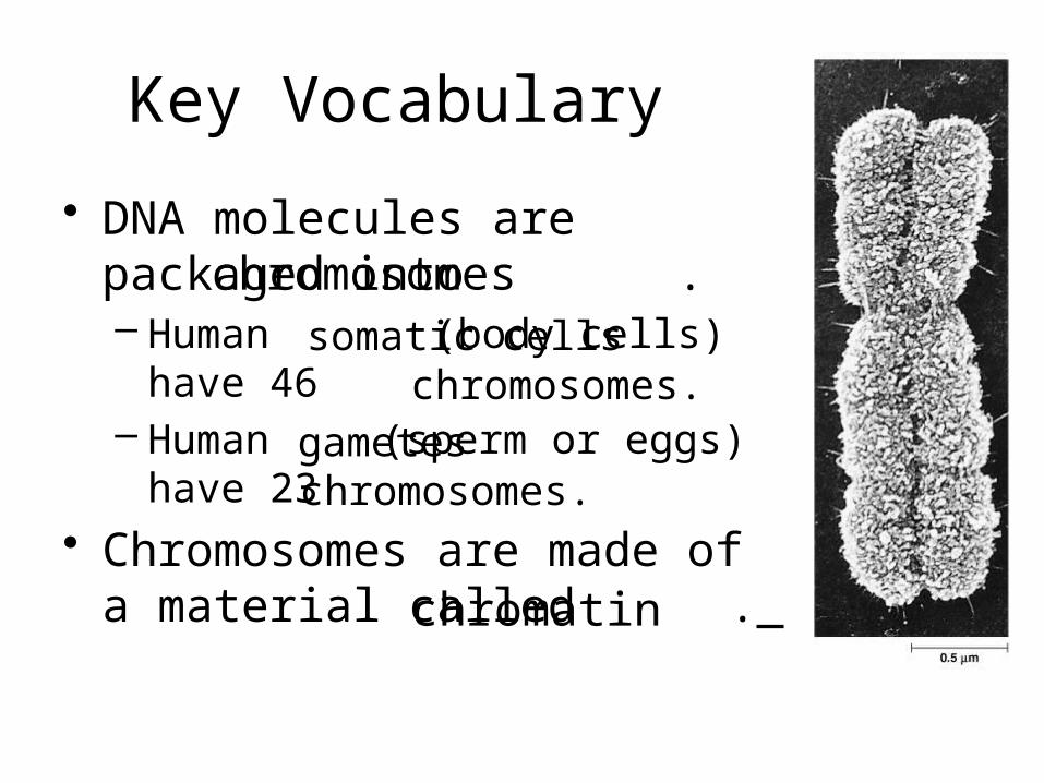
Key Vocabulary
• DNA molecules are packaged into .– Human (body cells)
have 46 – Human (sperm or eggs)
have 23• Chromosomes are made of a
material called .
chromosomessomatic cells
gametes
chromosomes.
chromosomes.
chromatin
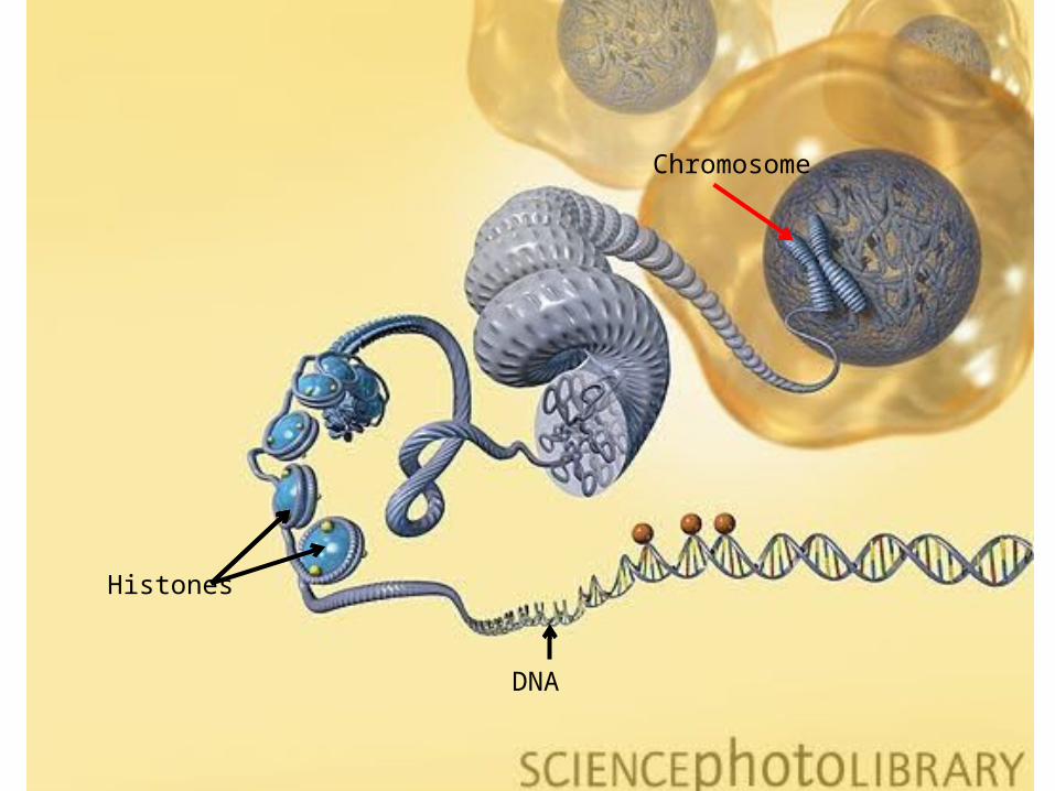
Key Vocabulary
• Chromatin is composed of and .
• DNA is supercoiled around proteins called .
• Together the DNA and histone proteins form bead-like structures called
DNAproteins
histones
nucleosomesHistones
DNA
Chromosome
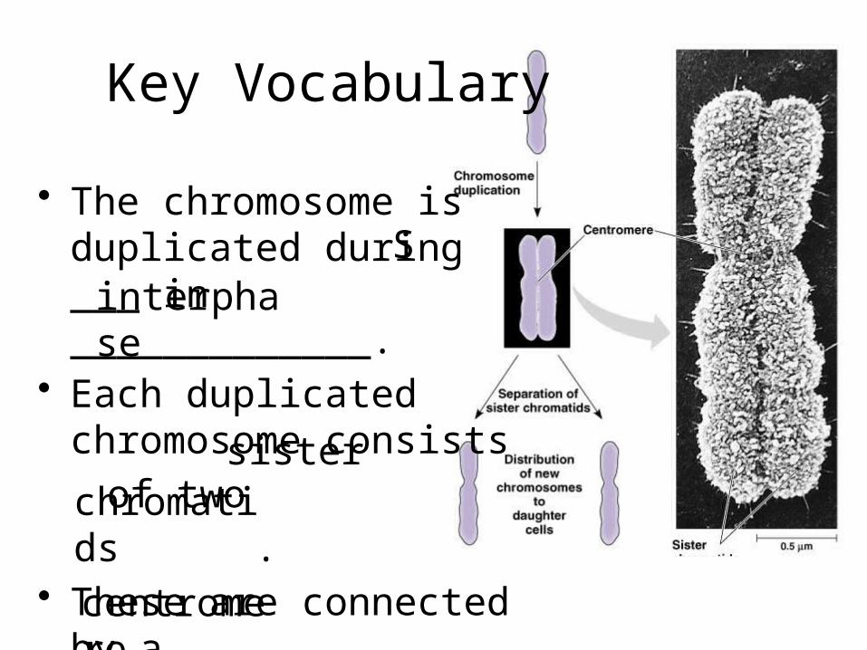
Key Vocabulary
sisterchromatids
centromere
Sinterphase
• The chromosome is duplicated during ___ in _____________.
• Each duplicated chromosome consists
of two
.• These are connected by a
.
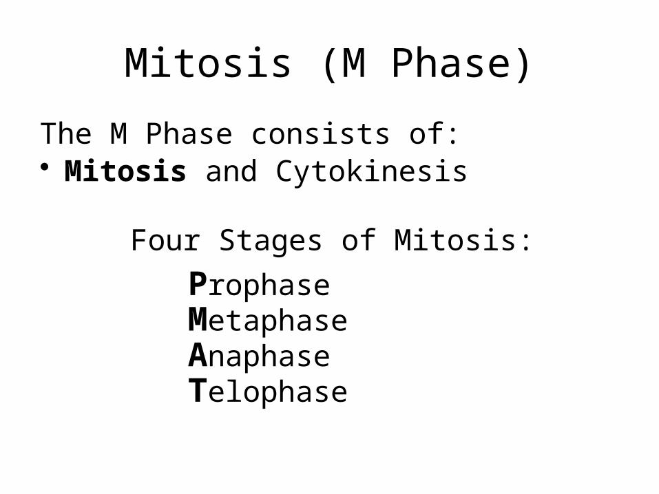
Mitosis (M Phase)
The M Phase consists of:• Mitosis and Cytokinesis
Four Stages of Mitosis:
Prophase MetaphaseAnaphaseTelophase
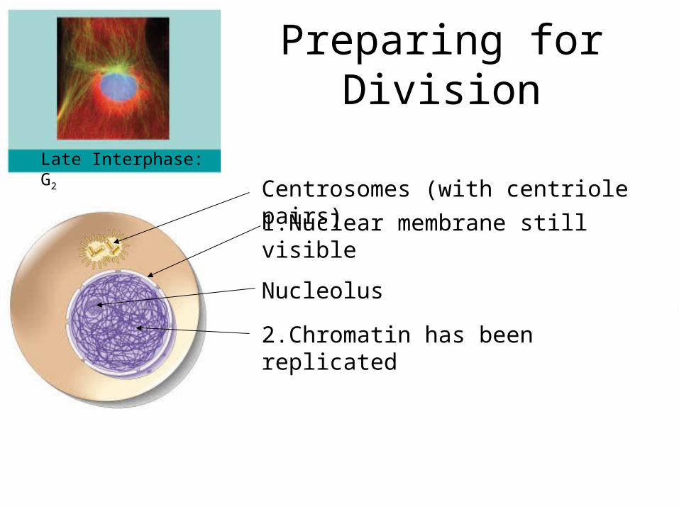
Late Interphase: G2
Centrosomes (with centriole pairs)
Preparing for Division
1.Nuclear membrane still visible
Nucleolus
2.Chromatin has been replicated
Late Interphase: G2
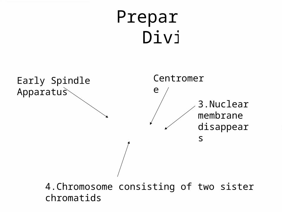
Preparing for Division
4.Chromosome consisting of two sister chromatids
CentromereEarly Spindle Apparatus
Prophase
3.Nuclear membrane disappears
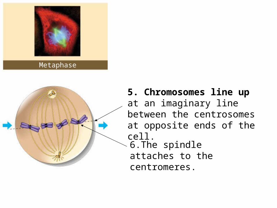
Metaphase
5. Chromosomes line up at an imaginary line between the centrosomes at opposite ends of the cell.
6.The spindle attaches to the centromeres.
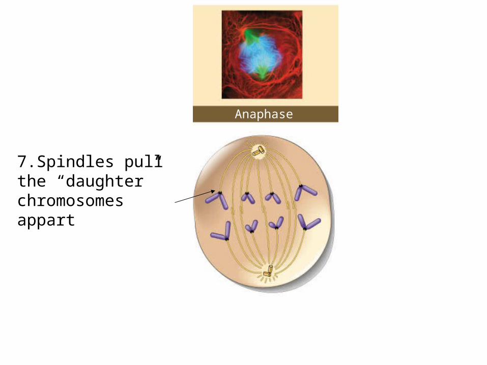
Anaphase
7.Spindles pull the “daughter” chromosomes appart
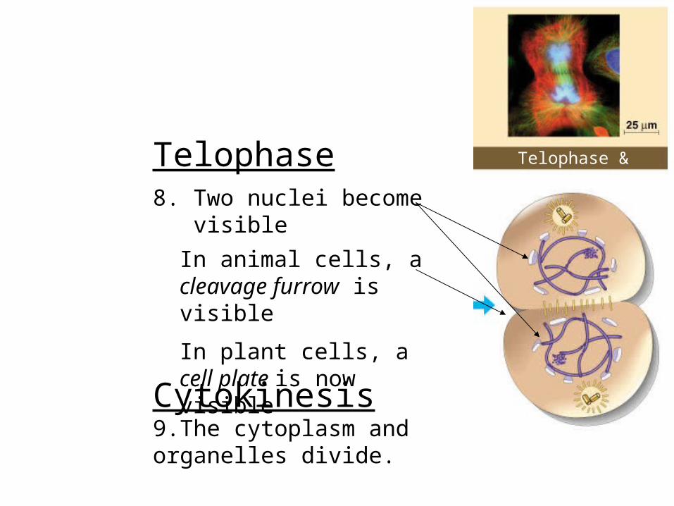
Telophase & CytokinesisTelophase8. Two nuclei become visible
In animal cells, a cleavage furrow is visible
In plant cells, a cell plate is now visible
Cytokinesis9.The cytoplasm and organelles divide.
Interphase:3 Phases
)
1. G1: Cell is Growing
2. S: DNA is Synthesized (DNA Replication)
3. G2: Organelles duplicate in preparation for division
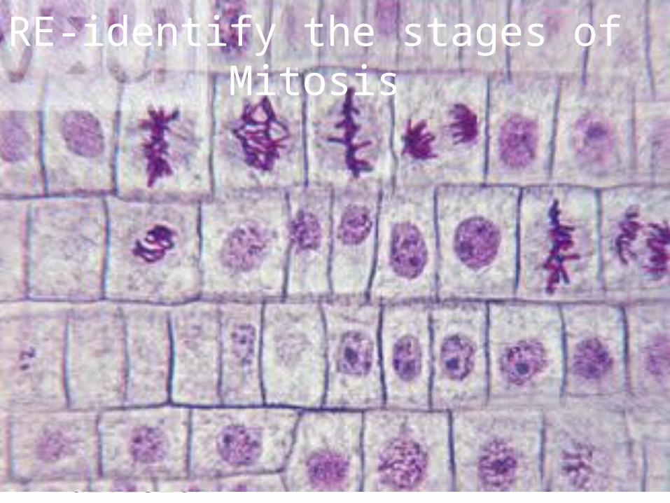
RE-identify the stages of Mitosis
What’s the end product?
• Two identical daughter cells ready to start the cycle again… or not.
• Some cells (like your nerve and muscle cells) do not undergo division.
• For those that do (your skin and the lining of your intestinal tract) how would you know how quickly division takes place?