Embed Size (px)
Citation preview
HOW I DO IT ARTICLE Open Access
How I do it: judging appropriateness for TTEand TEERicardo Fonseca and Thomas H Marwick*
Abstract
The increasing cost of healthcare is a widespread international problem to which the cost of imaging has been animportant contributor. Some imaging tests are ordered inappropriately and contribute to wasted use of resources.Appropriate use criteria have been developed in the USA in order to guide test selection, but there are a numberof problems, including the evidence base for these criteria and the steps that can be taken to change physicianpractice. A restrictive approach to test ordering is difficult to fit to the nuances of clinical presentation and maycompromise patient care. We propose an alternative approach to physician guidance based on the most commonmarkers of inappropriate testing.
Keywords: Appropriate use, Transthoracic echocardiography, Transoesophageal echocardiography
No management decisions in medical practice are ex-empt from a concept that is difficult to measure: appro-priateness. In common parlance, an appropriate choiceis one that which is suitable or proper in the circum-stances, but this is surprisingly different from the med-ical definitions. The concept of appropriateness definedby the RAND/UCLA methodology in the 1980’s was thecornerstone for developing the first attempt at appropri-ate use criteria (AUC). That concept suggested that “anappropriate procedure in one in which the expectedhealth benefit (e.g, increased life expectancy) exceeds theexpected negative consequences (e.g., mortality, morbid-ity, anxiety, pain, time lost from work) by a sufficientlywide margin that the procedure is worth doing, exclusiveof cost” [1,2].The adaption of this concept to cardiac imaging led to
an appropriate test being defined as “one in which theexpected incremental information, combined with clin-ical judgement, exceeds the expected negative conse-quences (risks of the procedure i.e. radiation or contrastexposure and the downstream impact of poor test per-formance, such as delay in diagnostic (false negatives) orinappropriate diagnosis (false positives)) by a sufficientlywide margin for specific indication that the procedure isgenerally considered acceptable care and a reasonable
approach for the indication” [3]. Because of the low riskof imaging, there are many circumstances in where thisdefinition seems to be insufficient – the risk is almostzero so the balance of benefit and risk is positive, butthe information obtained is still inadequate to justifyperformance of the test. A new definition overcomesthese concerns by framing the decision in the context ofa consensus about “reasonable care” [4], and resourceutilization “The concept of appropriateness, as appliedto health care, balances risk and benefit of a treatment,test, or procedure in the context of available resourcesfor an individual patient with specific characteristics” [5].Importantly, it is now acknowledged that AUC shouldprovide guidance to supplement the clinician’s judgment,rather than being prescriptive.
Motivations to the definition of appropriate usecriteriaWhile the risk of harm with inappropriate interventionwas an important motivator to the application of AUC,the focus on appropriate use in imaging is mainly rootedin resource utilization and medical expenditure. Thecontribution of imaging to the medical budget startedto be highlighted in the United States >20 years ago. Atthis time, the Medicare Payment Advisory Commission(MedPAC) showed a 10%/year increase of spending forcardiac imaging between 1999 and 2002, when the aver-age growth per year of all services was 5.2% [6]. This
* Correspondence: [email protected] Research Institute Tasmania, 17 Liverpool St, Hobart, Tasmania 7000,Australia
CARDIOVASCULAR ULTRASOUND
© 2014 Fonseca and Marwick; licensee BioMed Central Ltd. This is an Open Access article distributed under the terms of theCreative Commons Attribution License (http://creativecommons.org/licenses/by/4.0), which permits unrestricted use,distribution, and reproduction in any medium, provided the original work is properly credited. The Creative Commons PublicDomain Dedication waiver (http://creativecommons.org/publicdomain/zero/1.0/) applies to the data made available in thisarticle, unless otherwise stated.
Fonseca and Marwick Cardiovascular Ultrasound 2014, 12:22http://www.cardiovascularultrasound.com/content/12/1/22
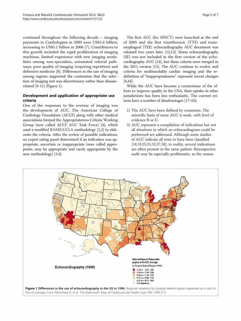
continued throughout the following decade – imagingpayments to Cardiologists in 2000 were US$1.6 billion,increasing to US$5.1 billion in 2006 [7]. Contributors tothis growth included the rapid proliferation of imagingmachines, limited experience with new imaging moda-lities among non-specialists, automated referral path-ways, poor quality of imaging (requiring repetition) anddefensive medicine [8]. Differences in the use of imagingamong regions supported the contention that the selec-tion of imaging test was discretionary rather than disease-related [9-11] (Figure 1).
Development and application of appropriate usecriteriaOne of the responses to the overuse of imaging wasthe development of AUC. The American College ofCardiology Foundation (ACCF) along with other medicalassociations formed the Appropriateness Criteria WorkingGroup (now called ACCF AUC Task Force) [4], whichused a modified RAND/UCLA methodology [1,2] to elab-orate the criteria. After the review of possible indications,an expert rating panel determined if an indication was ap-propriate, uncertain or inappropriate (now called appro-priate, may be appropriate and rarely appropriate by thenew methodology) [3,4].
The first AUC (for SPECT) were launched at the endof 2005 and the first transthoracic (TTE) and trans-esophageal (TEE) echocardiography AUC document wasreleased two years later [12,13]. Stress echocardiography(SE) was not included in the first version of the echo-cardiography AUC [14], but these criteria were merged inthe 2011 version [15]. The AUC continue to evolve, andcriteria for multimodality cardiac imaging and the re-definition of “inappropriateness” represent recent changes[4,16].While the AUC have become a cornerstone of the ef-
forts to improve quality in the USA, their uptake in otherjurisdictions has been less enthusiastic. The current cri-teria have a number of disadvantages [17-43];
1) The AUC have been defined by consensus. Thescientific basis of some AUC is weak, with level ofevidence B or C.
2) AUC represent a compilation of indications but notall situations in which an echocardiogram could beperformed are addressed. Although some studiesof AUC indicate all tests to have been classified[18,19,25,31,32,37,38], in reality, several indicationsare often present in the same patient. Retrospectiveaudit may be especially problematic, as the reason
Figure 1 Differences in the use of echocardiography in the US in 1996. Regional variations by hospital referral region, expressed as a ratio tothe US average. From Wennberg D, et al. The Dartmouth Atlas of Cardiovascular Health Care. P65. 1999 [11].
Fonseca and Marwick Cardiovascular Ultrasound 2014, 12:22 Page 2 of 7http://www.cardiovascularultrasound.com/content/12/1/22
for requesting an echocardiogram is ofteninadequately detailed in the medical records.
3) Conversely, several recommendations forechocardiography in current practice guidelines (notjust in echocardiography but for disease entities)lack counterparts in the AUC. For example, a class Irecommendation is given for follow-up or surveil-lance after surgery of masses known to have a highlikelihood of recurrence (eg myxoma [44]). TheAUC classification of “suspected cardiac mass” – oreven screening – does not cover the describedscenario.
4) The application of AUC to patient selection may beproblematic as an audit tool. When an appropriateindication is required to order the test at point-of-service, the referring clinician may list a co-existingappropriate indication rather than the actualclinical problem (which may be of inappropriate).This is particularly likely when the proportion ofinappropriate tests is assessed as part of theechocardiography accreditation process.
After 7 years of using the AUC for echocardiography(TTE and TEE), there are concerns about the real im-pact of the AUC on physician ordering behaviour [45].The literature seems to show a similar proportion of in-appropriate testing, in spite of experience, educationalcampaigns and close follow-up. Moreover, the correla-tion between appropriateness and clinical impact has notbeen well studied [31].
Application of AUC in daily practiceWe do not favour the use of AUC as a “gatekeeper” toechocardiography. Rather, we see the AUC provide a yard-stick to permit three means of improving appropriateness -education, guidance at point-of-care and laboratory-basedaudit;
i). Education: Although educational interventionsseem to be a logical approach, the results ofheterogeneous attempts have been contradictory.On the one hand, for instance, an educationalcampaign consisted in lectures, a pocket card withthe AUC and feedback showed encouraging resultsas one of the successful tools for improvingappropriateness [23]. On the other hand, similarprojects focused in physician education andfeedback [46,47], did not show improvement. TheAUC are an excellent starting point in this respect.Essential parts of educational campaigns includelectures, pocket cards and feedback.
ii). Control in point-of-care: The use of priorauthorisation protocols through a Radiology BenefitManager (RBM) is widely used to control access to
expensive tests of limited availability, such aspositron emission tomography and cardiac magneticresonance, although its efficiency and effectivenesshave been questioned [47]. The use of AUC at pointof care involves ordering physicians in the attemptto decrease inappropriate tests. In order to facilitatethis, friendly electronic tools have been invented tohelp clinicians to choose “appropriately” at thepoint-of-order [24]. Recent work has proposed thatthis practice is of equivalent efficacy to the use ofthe RBM [48], with greater efficiency and betterpreservation of the autonomy of the attendingphysician. Incorporation with an electronicordering process can inform the clinician aboutappropriateness when the test is requested. Therisk of both AUC and RBM are that otherappropriate (but inactive) clinical problems thatcan be used to have a test approved to addressan inappropriate question.
iii). Laboratory-based audit: We have focused on thisbecause of the limitations of the above twomethods. Laboratories are potentially moremotivated than requestors because of thereputational and economic risk of high levels ofinappropriate use. While we acknowledge that theaudit process can be problematic in privatepractice, as the locus of control is with thereferring doctor, it is important to consider thatthe laboratory will be held responsible for theperformance of inappropriate tests and theconsequence of more inappropriate tests will beless reimbursement. In this setting, it seems likelythat some investment into auditing this processwill be reasonable. Inevitably, urgentechocardiograms and communication problemsrepresent scenarios where the process ischallenging, but if appropriateness is to be audited,we would suggest that defining the “at risk” studyfor inappropriateness (see below) is a means ofimproving the efficiency of this process fromneeding to audit 100% of requests to audit ofthe ~15% of requests that are included in this list.The additional scrutiny given to these requestsdoes not necessitate individual contact with thereferring physician in all cases.
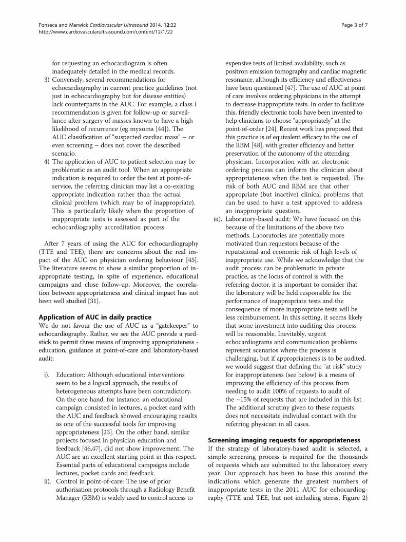
Screening imaging requests for appropriatenessIf the strategy of laboratory-based audit is selected, asimple screening process is required for the thousandsof requests which are submitted to the laboratory everyyear. Our approach has been to base this around theindications which generate the greatest numbers ofinappropriate tests in the 2011 AUC for echocardiog-raphy (TTE and TEE, but not including stress, Figure 2)
Fonseca and Marwick Cardiovascular Ultrasound 2014, 12:22 Page 3 of 7http://www.cardiovascularultrasound.com/content/12/1/22
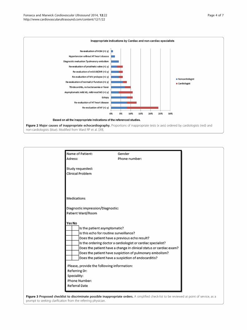
Figure 2 Major causes of inappropriate echocardiography. Proportions of inappropriate tests (x axis) ordered by cardiologists (red) andnon-cardiologists (blue). Modified from Ward RP et al. [39].
Figure 3 Proposed checklist to discriminate possible inappropriate orders. A simplified check-list to be reviewed at point of service, as aprompt to seeking clarification from the referring physician.
Fonseca and Marwick Cardiovascular Ultrasound 2014, 12:22 Page 4 of 7http://www.cardiovascularultrasound.com/content/12/1/22
[15]. These are related to routine surveillance, evalua-tion of symptoms without other symptoms/signs of car-diac disease and low pretest probability of endocarditis[18,20,21,23,33,34,36,41]. Other situations include a sus-picion of pulmonary embolism, when the exam wouldnot change management, and when a test is ordered bynon-cardiologists.Routine surveillance is the most common inappropri-
ate indication for TTE. The most common situations ofinappropriate repeat imaging of ventricular function in-clude assessment in patients with known CAD and nochange in clinical status or cardiac exam [34,41], syste-mic hypertension without symptoms or signs of hyper-tensive heart disease [20], and within a year of previoustesting in heart failure (systolic or diastolic) when thereis no change in clinical status or cardiac exam [20,34]).A very common situation in patients with nonspecificsymptoms includes patients with lightheadedness/presyn-cope without other symptoms) [23,41]. Common valve-related indications include <3 year after prosthetic valveimplantation in the absence of known or suspected valvedysfunction [33], and evaluation of infective endocarditiswhen there is transient fever without evidence of bac-teremia [23] or new murmur or transient bacteraemiawith a pathogen not typically associated with endocar-ditis. For transoesophageal echocardiography, the mostcommon inappropriate indications are related to endo-carditis with low pretest probability and routine use ofTEE when a diagnostic TTE is reasonably anticipated toresolve all concerns [21].The availability of this information on the characteris-
tics of inappropriate tests has enabled the developmentof a checklist to identify studies where a discussion re-garding the merits of testing can be initiated from thelaboratory (Figure 3).
ConclusionsJudging appropriateness in echocardiography is a processbased on knowledge, experience, information, resourcesand the real desire to provide an adequate service to thepatient. It does not sit well with formulaic approachesbased on uncritical application of AUC. Importantly,it is now acknowledged that AUC should provide guid-ance to supplement the clinician’s judgment, rather thanbeing prescriptive [5].Although the audit process described above helps to
strengthen the application of the AUC, it is difficult tocontrol the problems associated with self-referral and theveracity on the part of ordering physicians. In our opinion,the optimal approach requires dialogue between treatingphysicians, cardiologists and sonographers. The perfecttool has not yet been designed, but a process that flags dis-cussion at the point of imaging may be more effective thana gatekeeper at the point of ordering the test.
AbbreviationsACCF: American college of cardiology foundation; AUC: Appropriate usecriteria; CAD: Coronary artery disease; MedPAC: Medicare payment advisorycommission; RAND/UCLA: RAND corporation/university of California LosAngeles; RBM: Radiology benefit manager; SE: Stress echocardiography;SPECT: Single photon emission computed tomography; TTE: Transthoracicechocardiography; TEE: Transesophageal echocardiography.
Competing interestsThe authors declare that they have no competing interests.
Authors’ contributionsRF; contributed to conception/design, gathered and interpreted data andwrote the paper, approved final version and agrees to be accountable forcontent. THM; contributed to conception/design, gathered and interpreteddata, edited the paper, approved final version and agrees to be accountablefor content.
Received: 20 March 2014 Accepted: 5 June 2014Published: 24 June 2014
References1. Fitch K, Bernstein S, Aguilar MD, Burnand B, La Calle JR, Lazaro P, Loo M,
McDonnell J, Vader JP, Kaham JP: The RAND/UCLA Appropriateness Methoduser's manual. Santa Monica (CA): RAND Corporation; 2001.
2. Brook RH, Chassin MR, Fink A, Solomon DH, Kosecoff J, Park RE: A Methodfor the Detailed Assessment of the Appropriateness of MedicalTechnologies. Int J Technol Assess Health Care 1986, 2(01):53–63.
3. Patel MR, Spertus JA, Brindis RG, Hendel RC, Douglas PS, Peterson ED,Wolk MJ, Allen JM, Raskin IE: ACCF proposed method for evaluating theappropriateness of cardiovascular imaging. J Am Coll Cardiol 2005,46(8):1606–1613.
4. Hendel RC, Patel MR, Allen JM, Min JK, Shaw LJ, Wolk MJ, Douglas PS,Kramer CM, Stainback RF, Bailey SR, Doherty JU, Brindis RG: AppropriateUse of Cardiovascular Technology 2013 ACCF Appropriate Use CriteriaMethodology Update: A Report of the American College of CardiologyFoundation Appropriate Use Criteria Task Force. J Am Coll Cardiol 2013,61(12):1305–1317.
5. Carr JJ, Hendel RC, White RD, Patel MR, Wolk MJ, Bettmann MA, Douglas P,Rybicki FJ, Kramer CM, Woodard PK, Shaw LJ, Yucel EK, Writing G:Appropriate Utilization of Cardiovascular Imaging A Methodology for theDevelopment of Joint Criteria for the Appropriate Utilization ofCardiovascular Imaging by the American College of CardiologyFoundation and American College of Radiology. J Am Coll Radiol 2013,10(6):456–463.
6. Medicare Payment Advisory Commission (MedPAC): Report to thecongress: Medicare payment policy March 2005. [http://www.medpac.gov/documents/Mar05_EntireReport.pdf]
7. Shaw LJ, Marwick TH, Zoghbi WA, Hundley WG, Kramer CM, Achenbach S,Dilsizian V, Kern MJ, Chandrashekhar Y, Narula J: Why all the focus oncardiac imaging? JACC Cardiovasc Imaging 2010, 3(7):789–794.
8. Medicare Payment Advisory Commission (MedPAC): Report to thecongress: New approaches in Medicare. 2004 [http://www.medpac.gov/documents/June04_Entire_Report.pdf]
9. Medicare Payment Advisory Commission (MedPAC): Report to thecongress: Medicare payment policy March 2003. [http://www.medpac.gov/documents/Mar03_Entire_report.pdf]
10. Lucas FL, Sirovich BE, Gallagher PM, Siewers AE, Wennberg DE: Variation incardiologists’ propensity to test and treat: Is it associated with regionalvariation in utilization? Circ Cardiovasc Qual Outcomes 2010, 3:253–260.
11. The Dartmouth Atlas of Cardiovascular Health Care. In 115F. [http://www.dartmouthatlas.org/downloads/atlases/cardiovascular_atlas.pdf]
12. Brindis RG, Douglas PS, Hendel RC, Peterson ED, Wolk MJ, Allen JM, PatelMR, Raskin IE, Bateman TM, Cerqueira MD, Gibbons RJ, Gillam LD, GillespieJA, Iskandrian AE, Jerome SD, Krumholz HM, Messer JV, Spertus JA, StowersSA: ACCF/ASNC appropriateness criteria for single-photon emissioncomputed tomography myocardial perfusion imaging (SPECT MPI): areport of the American College of Cardiology Foundation QualityStrategic Directions Committee Appropriateness Criteria Working Groupand the American Society of Nuclear Cardiology endorsed by theAmerican Heart Association. J Am Coll Cardiol 2005, 46(8):1587–1605.
Fonseca and Marwick Cardiovascular Ultrasound 2014, 12:22 Page 5 of 7http://www.cardiovascularultrasound.com/content/12/1/22
13. Douglas PS, Khandheria B, Stainback RF, Weissman NJ, Brindis RG, Patel MR,Alpert JS, Fitzgerald D, Heidenreich P, Martin ET, Messer JV, Miller AB, PicardMH, Raggi P, Reed KD, Rumsfeld JS, Steimle AE, Tonkovic R, VijayaraghavanK, Yeon SB, Hendel RC, Peterson E, Wolk MJ, Allen JM: ACCF/ASE/ACEP/ASNC/SCAI/SCCT/SCMR 2007 Appropriateness Criteria for Transthoracicand Transesophageal Echocardiography: A Report of the AmericanCollege of Cardiology Foundation Quality Strategic DirectionsCommittee Appropriateness Criteria Working Group, American Society ofEchocardiography, American College of Emergency Physicians, AmericanSociety of Nuclear. J Am Coll Cardiol 2007, 50(2):187–204.
14. Douglas PS, Khandheria B, Stainback RF, Weissman NJ, Peterson ED,Hendel RC, Blaivas M, Des Prez RD, Gillam LD, Golash T, Hiratzka LF,Kussmaul WG, Labovitz AJ, Lindenfeld J, Masoudi FA, Mayo PH,Porembka D, Spertus JA, Wann LS, Wiegers SE, Brindis RG, Patel MR,Wolk MJ, Allen JM: ACCF/ASE/ACEP/AHA/ASNC/SCAI/SCCT/SCMR 2008appropriateness criteria for stress echocardiography. Circulation 2008,117(11):1478–1497.
15. Douglas PS, Garcia MJ, Haines DE, Lai WW, Manning WJ, Patel AR, PicardMH, Polk DM, Ragosta M, Ward RP, Weiner RB, Bailey SR, Alagona P,Anderson JL, DeCara JM, Dolor RJ, Fazel R, Gillespie JA, Heidenreich PA,Leykum LK, Marine JE, Mishkel GJ, Pellikka PA, Raff GL, Vijayaraghavan K,Weissman NJ, Wu KC, Wolk MJ, Hendel RC, Kramer CM, et al.: ACCF/ASE/AHA/ASNC/HFSA/HRS/SCAI/SCCM/SCCT/SCMR 2011 Appropriate UseCriteria for Echocardiography: A Report of the American College ofCardiology Foundation Appropriate Use Criteria Task Force, AmericanSociety of Echocardiography, American Heart Association, AmericanSociety of Nuclear Cardiology, Heart Failure Society of America,Heart Rhythm Society, Society for Cardiovascular Angiographyand Interventions, Society of Critical Care Medicine, Society ofCardiovascular Computed Tomography, and Society for CardiovascularMagnetic Resonance (vol 57, pg 1126, 2011). J Am Coll Cardiol 2011,57(9):1167–1168.
16. Wolk MJ, Bailey SR, Doherty JU, Douglas PS, Hendel RC, Kramer CM, Min JK,Patel MR, Rosenbaum L, Shaw LJ, Stainback RF, Allen JM, Brindis RG,Kramer CM, Shaw LJ, Cerqueira MD, Chen J, Dean LS, Fazel R, HundleyWG, Itchhaporia D, Kligfield P, Lockwood R, Marine JE, McCully RB,Messer JV, O'Gara PT, Shemin RJ, Wann LS, Wong JB, et al.: ACCF/AHA/ASE/ASNC/HFSA/HRS/SCAI/SCCT/SCMR/STS 2013 MultimodalityAppropriate Use Criteria for the Detection and Risk Assessmentof Stable Ischemic Heart DiseaseA Report of the American Collegeof Cardiology Foundation Appropriate Use Criteria Task Force,American Heart Association, American Society of Echocardiography,American Society of Nuclear Cardiology, Heart Failure Society ofAmerica, Heart Rhythm Society, Society for CardiovascularAngiography and Interventions, Society of Cardiovascular ComputedTomography, Society for Cardiovascular Magnetic Resonance,and Society of Thoracic Surgeons. J Am Coll Cardiol 2014,63(4):380–406.
17. Aggarwal NR, Wuthiwaropas P, Karon BL, Miller FA, Pellikka PA: Applicationof the appropriateness criteria for echocardiography in an academicmedical center. J Am Soc Echocardiogr 2010, 23(3):267–274.
18. Alqarqaz M, Koneru J, Mahan M, Ananthasubramaniam K: Applicability,limitations and downstream impact of echocardiography utilizationbased on the Appropriateness Use Criteria for transthoracic andtransesophageal echocardiography. Int J Cardiovasc Imaging 2012,28(8):1951–1958.
19. Bailey SA, Mosteanu I, Tietjen PA, Petrini JR, Alexander J, Keller AM: Theuse of transthoracic echocardiography and adherence to appropriateuse criteria at a regional hospital. J Am Soc Echocardiogr 2012,25(9):1015–1022.
20. Ballo P, Bandini F, Capecchi I, Chiodi L, Ferro G, Fortini A, Giuliani G,Landini G, Laureano R, Milli M, Nenci G, Pizzarelli F, Santoro GM, Vannelli P,Cappelletti C, Zuppiroli A, American College of Cardiology Foundation,American Society of Echocardiography: Application of 2011 AmericanCollege of Cardiology Foundation/American Society ofEchocardiography appropriateness use criteria in hospitalized patientsreferred for transthoracic echocardiography in a community setting.J Am Soc Echocardiogr 2012, 25(6):589–598.
21. Bhatia RS, Carne DM, Picard MH, Weiner RB: Comparison of the 2007 and2011 appropriate use criteria for transesophageal echocardiography.J Am Soc Echocardiogr 2012, 25(11):1170–1175.
22. Bhatia RS, Carne DM, Picard MH, Weiner RB: Comparison of the 2007 and2011 appropriate use criteria for transthoracic echocardiography invarious clinical settings. J Am Soc Echocardiogr 2012, 25(11):1162–1169.
23. Bhatia RS, Milford CE, Picard MH, Weiner RB: An educational interventionreduces the rate of inappropriate echocardiograms on an inpatientmedical service. JACC Cardiovasc Imaging 2013, 6(5):545–555.
24. Bhave NM, Mansour IN, Veronesi F, Razi RR, Lang RM, Ward RP: Use of aweb-based application of the American College of Cardiology Foundation/American Society of Echocardiography appropriateness use criteria fortransthoracic echocardiography: A pilot study. J Am Soc Echocardiogr 2011,24(3):271–276.
25. Dharmarajan L, Hale TM, Velastegui Z, Castillo E, Kanna B: Utility of two-dimensional echocardiography in pregnancy and post-partum periodand impact on management in an inner city hospital. J Perinat Med 2009,37(6):663–668.
26. Ghatak A, Pullatt R, Vyse S, Silverman DI: Appropriateness criteria are animprecise measure for repeat echocardiograms. Echocardiography 2011,28(2):131–135.
27. Grewal GK, Klosterman TB, Shrestha K, Yarmohammadi H, Zurick AO,Varr BC, Tang WH, Lindsay BD, Klein AL: Indications for TEE beforecardioversion for atrial fibrillation: implications for appropriatenesscriteria. JACC Cardiovasc Imaging 2012, 5(6):641–648.
28. Kirkpatrick JN, Ky B, Rahmouni HW, Chirinos JA, Farmer SA, Fields AV,Ogbara J, Eberman KM, Ferrari VA, Silvestry FE, Keane MG, Opotowsky AR,Sutton MS, Wiegers SE: Application of Appropriateness Criteria inOutpatient Transthoracic Echocardiography. J Am Soc Echocardiogr 2009,22(1):53–59.
29. Mansour IN, Razi RR, Bhave NM, Ward RP: Comparison of the updated2011 appropriate use criteria for echocardiography to the originalcriteria for transthoracic, transesophageal, and stress echocardiography.J Am Soc Echocardiogr 2012, 25(11):1153–1161.
30. Martin NM, Picard MH: Use and appropriateness of transthoracicechocardiography in an academic medical center: a pilot observationalstudy. J Am Soc Echocardiogr 2009, 22(1):48–52.
31. Matulevicius SA, Rohatgi A, Das SR, Price AL, Deluna A, Reimold SC:Appropriate use and clinical impact of transthoracic echocardiography.JAMA Intern Med 2013, 173(17):1600–1607.
32. Ogbara J, Logani S, Ky B, Chirinos JA, Silvestry FE, Eberman K, Moss JD,Ferrari VA, Keane MG, Sutton MSJ, Wiegers SE, Kirkpatrick JN: The Utilityof Prescreening Transesophageal Echocardiograms: A ProspectiveStudy. Echocardiography-J Cardiovasc Ultrasound Allied Tech 2011,28(7):767–773.
33. Parikh PB, Asheld J, Kort S: Does the revised appropriate use criteria forechocardiography represent an improvement over the initial criteria? Acomparison between the 2011 and the 2007 appropriateness use criteriafor echocardiography. J Am Soc Echocardiogr 2012, 25(2):228–233.
34. Patil HR, Coggins TR, Kusnetzky LL, Main ML: Evaluation of appropriate useof transthoracic echocardiography in 1,820 consecutive patients usingthe 2011 revised appropriate use criteria for echocardiography. Am JCardiol 2012, 109(12):1814–1817.
35. Rahimi AR, York M, Gheewala N, Markson L, Hauser TH, Manning WJ: Trendsin outpatient transthoracic echocardiography: impact of appropriatenesscriteria publication. Am J Med 2011, 124(8):740–746.
36. Rao G, Sajnani N, Kusnetzky LL, Main ML: Appropriate use of transthoracicechocardiography. Am J Cardiol 2010, 105(11):1640–1642.
37. Rao GA, Sajnani NV, Kusnetzky LL, Main ML: Appropriate utilization oftransesophageal echocardiography. Am J Cardiol 2009, 103(5):727–729.
38. Silverman GP, Vyse S, Silverman DI: Inappropriately OrderedEchocardiograms Are Related to Socioeconomic Status. Am J Med Qual2012, 27(6):487–493.
39. Ward RP, Mansour IN, Lemieux N, Gera N, Mehta R, Lang RM: Prospectiveevaluation of the clinical application of the American College ofCardiology Foundation/American Society of EchocardiographyAppropriateness Criteria for transthoracic echocardiography. JACCCardiovasc Imaging 2008, 1(5):663–671.
40. Willens HJ, Gómez-Marín O, Heldman A, Chakko S, Postel C, Hasan T,Mohammed F: Adherence to Appropriateness Criteria for TransthoracicEchocardiography: Comparisons Between a Regional Department ofVeterans Affairs Health Care System and Academic Practice andBetween Physicians and Mid-Level Providers. J Am Soc Echocardiogr 2009,22(7):793–799.
Fonseca and Marwick Cardiovascular Ultrasound 2014, 12:22 Page 6 of 7http://www.cardiovascularultrasound.com/content/12/1/22
41. Willens HJ, Hendel RC, Inhaber FR, Chakko SC, Postel C, Hasan T,Mohammed F: Appropriateness use criteria for transthoracicechocardiography: relationship with radiology benefit managerspreauthorization determination and comparison of the new (2010)criteria to the original (2007) criteria. Am Heart J 2011, 162(4):772–779.
42. Bhattacharyya S, Kamperidis V, Nalin Shah B, Roussin I, Chahal N, Li W,Khattar R, Senior R: Clinical utility and prognostic value ofappropriateness criteria in stress echocardiography for the evaluation ofvalvular heart disease. JACC Cardiovasc Imaging 2013, 6(9):987–992.
43. Schmitz L, Mori N, Khandheria BK, Gupta A: Appropriateness criteria forstress echocardiography in patients with acute chest pain: Are wechoosing wisely? Int J Cardiol 2013, 165(2):387–388.
44. Cheitlin MD, Armstrong WF, Aurigemma GP, Beller GA, Bierman FZ, Davis JL,Douglas PS, Faxon DP, Gillam LD, Kimball TR, Kussmaul WG, Pearlman AS,Philbrick JT, Rakowski H, Thys DM, Antman EM, Smith JSC, Alpert JS,Gregoratos G, Anderson JL, Hiratzka LF, Hunt SA, Fuster V, Jacobs AK,Gibbons RJ, Russell RO: ACC/AHA/ASE 2003 Guideline Update for theClinical Application of Echocardiography: Summary Article: A Report ofthe American College of Cardiology/American Heart Association TaskForce on Practice Guidelines (ACC/AHA/ASE Committee to Update the1997 Guidelines for the Clinical Application of Echocardiography).Circulation 2003, 108(9):1146–1162.
45. Fonseca R, Negishi K, Marwick T: Nothing has changed after six years ofappropriate use criteria. A systematic review of appropriate use intransthoracic and transesophageal echocardiography [abstract]. J Am SocEchocariogr 2014, 27:B96.
46. Gibbons RJ, Askew JW, Hodge D, Kaping B, Carryer DJ, Miller T: Appropriateuse criteria for stress single-photon emission computed tomographysestamibi studies: A quality improvement project. Circulation 2011,123(5):499–503.
47. Willens HJ, Nelson K, Hendel RC: Appropriate use criteria for stressechocardiography: Impact of updated criteria on appropriatenessratings, correlation with pre-authorization guidelines, and effect oftemporal trends and an educational initiative on utilization.JACC Cardiovasc Imaging 2013, 6(3):297–309.
48. Mitchell JM, LaGalia RR: Controlling the Escalating Use of AdvancedImaging: The Role of Radiology Benefit Management Programs. Med CareRes Rev 2009, 66(3):339–351.
doi:10.1186/1476-7120-12-22Cite this article as: Fonseca and Marwick: How I do it: judgingappropriateness for TTE and TEE. Cardiovascular Ultrasound 2014 12:22.
Submit your next manuscript to BioMed Centraland take full advantage of:
• Convenient online submission
• Thorough peer review
• No space constraints or color figure charges
• Immediate publication on acceptance
• Inclusion in PubMed, CAS, Scopus and Google Scholar
• Research which is freely available for redistribution
Submit your manuscript at www.biomedcentral.com/submit
Fonseca and Marwick Cardiovascular Ultrasound 2014, 12:22 Page 7 of 7http://www.cardiovascularultrasound.com/content/12/1/22