Embed Size (px)
Citation preview

How Cells Are Put Together
Chapter 3
Cell Theory
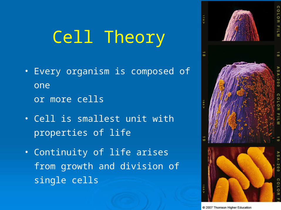
• Every organism is composed of one
or more cells
• Cell is smallest unit with properties of
life
• Continuity of life arises from growth
and division of single cells

• Smallest unit of life
• Is highly organized for metabolism
• Senses and responds to environment
• Has potential to reproduce
Cell

Structure of Cells
All start out life with:– Plasma membrane – Region where DNA
is stored– Cytoplasm
Two types:– Prokaryotic– Eukaryotic

Overview of cells

Common eukaryotic organelles
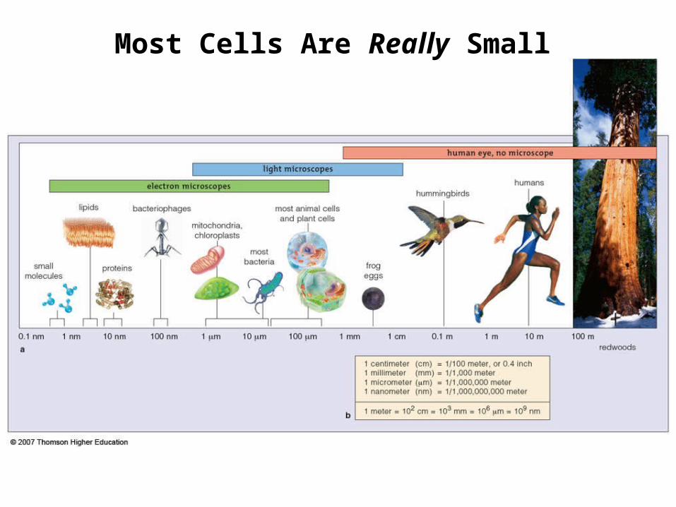
Fig. 3-4, p.41
Most Cells Are Really Small

Surface-to-Volume Ratio
• Bigger cell, less surface area per unit
volume
• Above a certain size, material cannot
move in or out of cell fast enough

0.5 1.0 1.5
0.79
0.06
3.14 7.07
0.52 1.77
diameter (cm):
surface area (cm2):
volume (cm3):
surface- to-volume ratio: 13.17:1 6.04:1 3.99:1
Fig. 3-5, p.41

• Create detailed images of something that is too small to see
• Light microscopes– Simple or compound
• Electron microscopes– Transmission EM or Scanning EM
Microscopes
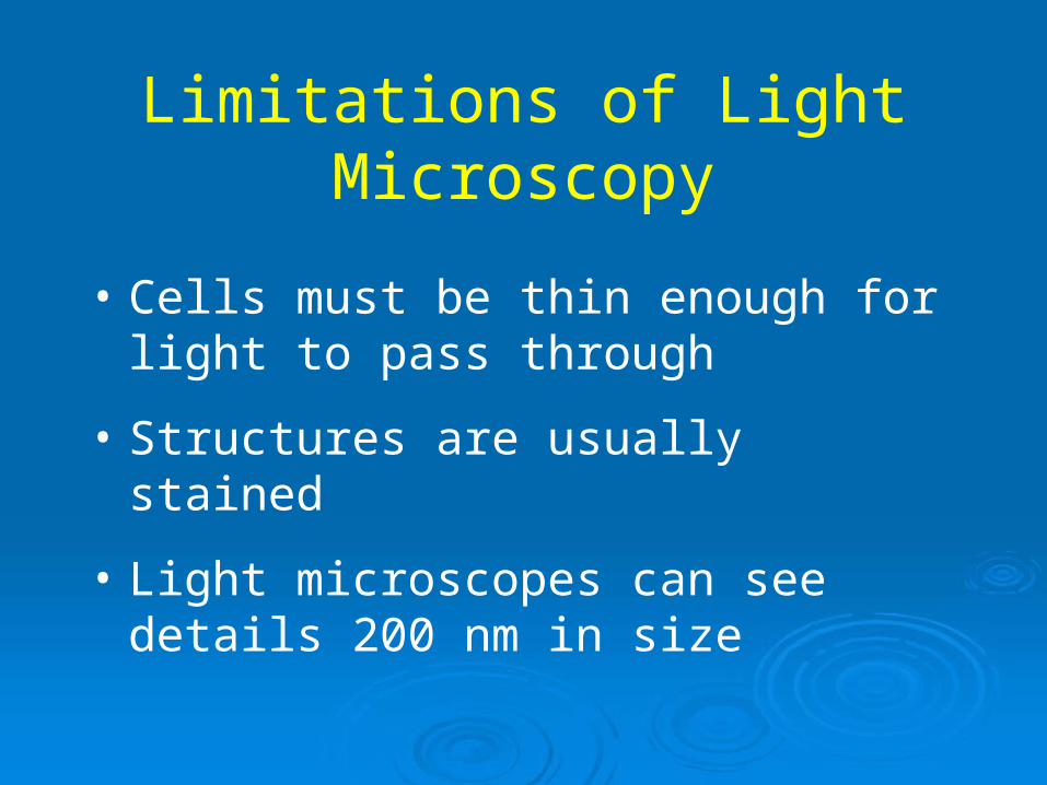
Limitations of Light Microscopy
• Cells must be thin enough for light to pass through
• Structures are usually stained
• Light microscopes can see details 200 nm in size

Electron Microscopy
• Uses beams of electrons rather than light
• Electrons are focused by magnets rather than glass lenses
• Can resolve structures down to 0.5 nm

Fig. 3-2a, p.40
Microscopes

Structure of Cell Membranes
• Fluid mosaic model
• Mixed composition:– Phospholipid bilayer – Glycolipids– Sterols– Proteins

Fig. 3-6b, p.42
one layer of lipids
one layer of lipids
Phospholipids

Lipid bilayer organization

Membrane Proteins
Adhesion proteins
Communication proteins
Receptor proteins
Recognition proteins
Passive transporters
Active transporters

Cell membranes

• Archaebacteria and eubacteria
• DNA is not enclosed in nucleus
• Generally the smallest, simplest cells
Prokaryotic Cells

Typical prokaryotic cell

Eukaryotic Cells
• Have a nucleus and other organelles
• Eukaryotic organisms– Plants– Animals– Protistans– Fungi

Eukaryotic Cell Features
• Plasma membrane• Nucleus• Endoplasmic
reticulum• Golgi body• Vesicles• Mitochondria• Ribosomes• Cytoskeleton
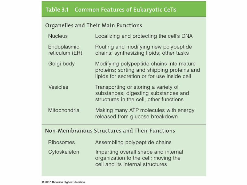
Table. 3-1, p.45

• Keeps the DNA molecules separated from metabolic machinery of cytoplasm
• Makes it easier to organize DNA and to copy it
The Nucleus

Components:Nuclear envelope NucleoplasmChromatin Nucleolus
The Nucleus

Fig. 3-9a, p.46
DNA in nucleus
rough ER
smooth ER
Golgi body
chromatin nucleolusnuclear envelope(two lipid bilayers)
cytoplasm
pore
the cell nucleus
RNA messages
The Nucleus

• Related organelles where lipids are assembled and new polypeptide chains modified
• Sorts and ships products to various destinations
• Consists of endoplasmic reticulum, Golgi bodies, vesicles
Endomembrane System

The endomembrane system

Endoplasmic Reticulum
• Starts at nuclear membrane and
extends throughout cytoplasm
• Rough ER: ribosome covered,
processes proteins
• Smooth ER: no ribosomes, builds lipids

Fig. 3-9d, p.46
smooth ER channel, cross-section
smooth ER
Endoplasmic Reticulum

Golgi Body
• Puts finishing touches on proteins and
lipids that arrive from ER
• Packages finished material for shipment to
final destinations
• Material arrives and leaves in vesicles

Fig. 3-9e-f, p.46
budding vesicle
Golgi body
plasma membrane
Secretory pathway ends.
Endocytic pathway begins.
Golgi Body

Vesicles
• Membranous sacs that
move through cytoplasm
• Lysosomes
• Peroxisomes

• ATP-producing powerhouses
• Membranes form two distinct
compartments
• ATP-making machinery embedded
in inner mitochondrial membrane
Mitochondria

Fig. 3-10, p.48
outer membrane
outer compartment
inner compartment
inner membrane

Chloroplasts
• Convert sunlight energy to ATP through photosynthesis
• Found in plants and some protistans

Fig. 3-11, p.48
two outermembranes
thylakoids(inner membranesystem folded intoflattened disks)

Organelle Origins
• Nucleus and ER– Infolding of membranes formed
compartments
• Mitochondria and chloroplasts– Endosymbiosis
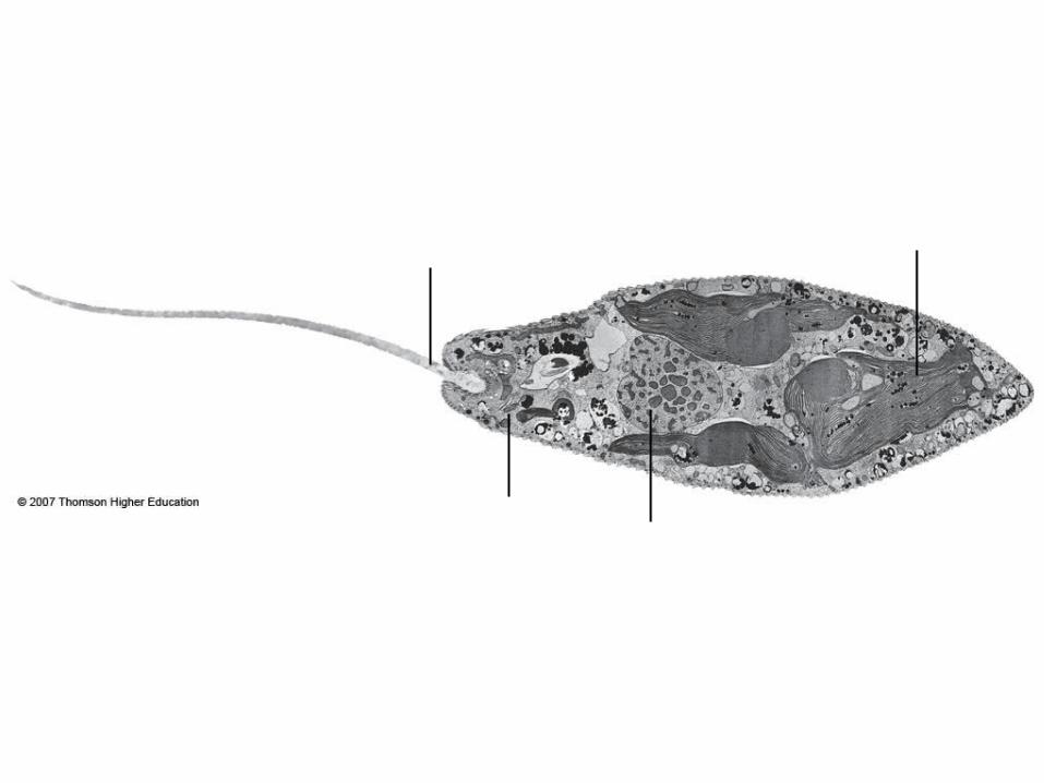
Fig. 3-14d, p.50
flagellum
mitochondrionnucleus
chloroplast

Fig. 3-15a, p.51
DNA
infolding of plasma membrane
Infolding Bacterial Membranes

• Present in all eukaryotic cells
• Cell shape and internal organization
• Allows organelle movement within cells and, in some cases, cell motility
Cytoskeleton

Microtubules
• Largest elements
• Composed of tubulin
• Involved in shape, motility,
cell division
tubulinsubunit

Microfilaments
• Thinnest elements
• Composed of actin
• Take part in movement,
formation, and
maintenance of cell
shape
actinsubunit
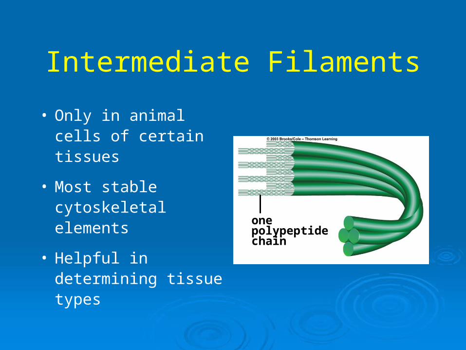
Intermediate Filaments
• Only in animal cells of certain tissues
• Most stable cytoskeletal elements
• Helpful in determining tissue types
onepolypeptidechain

Cilia, Flagellum, and Psuedopod

Plant Cell Walls

Plant Vs Animal

Table. 3-2, p.57