Embed Size (px)
Citation preview
TECHNO BYTES Q & A
How can I eliminate noise in the dark areaswhen scanning radiographs or slides?Demetrios J. Halazonetis
Athens, GreeceNoise is produced when a charge-coupled deviceregisters light when none exists. This happensparticularly in dark areas, because the incident
light there is of a very low intensity, and noise artifactsbecome more apparent. Charge-coupled devices areparticularly prone to noise, and high-end scanners havesophisticated electronics to minimize the effect. If youhave problems with noise, here are some solutions.
1. Get a better scanner. This is a radical solution butworth considering if you scan frequently and noiseis a major issue. Aim for a scanner with a highoptical density.
2. Get a scanner with the multi-scan feature. Suchscanners scan the original more than once and calcu-late the average of the acquired images. Because noiseis random, the average will tend to minimize it, whileleaving the true image pixels intact.
3. Multi-scan the image yourself. If your scanner doesnot support multi-scanning, you can simulate theprocedure by scanning the original 3 or 4 times and
Assistant professor, Orthodontic Department, University of Athens DentalSchool, Athens, Greece.Reprint requests to: D. Halazonetis, 6 Menandrou St, Kifissia 145 61, Greece;e-mail, [email protected] and accepted, November 2003.Am J Orthod Dentofacial Orthop 2005;127:83–40889-5406/$30.00Copyright © 2005 by the American Association of Orthodontists.
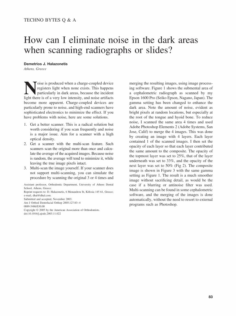
doi:10.1016/j.ajodo.2003.11.022merging the resulting images, using image process-ing software. Figure 1 shows the submental area ofa cephalometric radiograph as scanned by myEpson 1600 Pro (Seiko Epson, Nagano, Japan). Thegamma setting has been changed to enhance thedark area. Note the amount of noise, evident asbright pixels at random locations, but especially atthe root of the tongue and hyoid bone. To reducenoise, I scanned the same area 4 times and usedAdobe Photoshop Elements 2 (Adobe Systems, SanJose, Calif) to merge the 4 images. This was doneby creating an image with 4 layers. Each layercontained 1 of the scanned images. I then set theopacity of each layer so that each layer contributedthe same amount to the composite. The opacity ofthe topmost layer was set to 25%, that of the layerunderneath was set to 33%, and the opacity of thenext layer was set to 50% (Fig 2). The compositeimage is shown in Figure 3 with the same gammasetting as Figure 1. The result is a much smootherimage without sacrificing detail, as would be thecase if a blurring or antinoise filter was used.Multi-scanning can be found in some cephalometricsoftware, and the merging of the images is doneautomatically, without the need to resort to externalprograms such as Photoshop.
83

Figure 1.
American Journal of Orthodontics and Dentofacial OrthopedicsJanuary 2005
84 Halazonetis
Fig 1. Hyoid bone area, scanned once, with scanner ofoptical density 3.3. Note amount of random noise.Image has been manipulated by changing gamma
value, so that dark areas are enhanced.Fig 2. Four images arranged as layers in Photoshop.Opacity of each layer has been set so that each imagecontributes equally to final image. Layer 1 has opacity at50%, layer 2 has opacity at 33%, and layer 3 has
opacity at 25%. Background layer is at 100%.Fig 3. Composite image, at same gamma setting as