Embed Size (px)
Citation preview
Proc. Nati. Acad. Sci. USAVol. 87, pp. 1633-1637, March 1990Biochemistry
Host regulation of the cauliflower mosaic virus multiplication cycle(minichromosome/gene regulation/reverse transcription/Brassica susceptibility)
SIMON N. COVEY*, DAVID S. TURNER, ANDREW P. Lucy, AND KEITH SAUNDERS
Department of Virus Research, Agricultural and Food Research Council Institute of Plant Science Research, John Innes Institute, Colney Lane, Norwich NR47UH, United Kingdom
Communicated by Myron K. Brakke, December 12, 1989 (received for review August 20, 1989)
ABSTRACT The DNA genome of cauliflower mosaic virus(CaMV) replicates in the cytoplasm of infected plant cells byreverse transcription of an RNA template. Viral RNA isgenerated in the nucleus by transcription of an episomalminichromosome containing supercoiled DNA. We have as-sessed the relative activities of the nuclear and cytoplasmicphases of the CaMV multiplication cycle by monitoring unen-capsidated viral DNA forms and polyadenylylated RNAs indifferent organs of one host plant and in different host species.Systemically infected leaves of a highly susceptible host, turnip(Brassica rapa), contained abundant 35S RNA and 19S RNAtranscripts and unencapsidated reverse transcription productsbut relatively little supercoiled DNA. In contrast, supercoiledDNA accumulated in roots and other tissues of turnip plantsbut without significant amounts of steady-state viral RNA.Infected but asymptomatic leaves of a less susceptible CaMVhost, kohlrabi (Brassica oleracea), contained supercoiled DNAalmost exclusively but negligible viral RNA and DNA productsof reverse transcription. An allotetraploid species, rape (Bras-sica napus), exhibited infection characteristics and minichro-mosome expression levels intermediate between the other twospecies from which it was derived. We conclude that expressionof the CaMV minichromosome is a key phase of the virusmultiplication cycle, which is regulated differentially in organsof a highly susceptible host species. Furthermore, this regula-tion exhibits genetic variation among different Brassica speciesand controls host susceptibility to CaMV infection.
Cauliflower mosaic virus (CaMV) has a double-stranded DNAgenome generated by reverse transcription of an RNA inter-mediate (for reviews, see refs. 1-3). Virion DNA containssite-specific discontinuities resulting from the replicative pro-cess. At a relatively early stage in the CaMV multiplicationcycle, the discontinuities are repaired, and a supercoiled (SC)molecular form is synthesized, probably in the nucleus whereit is found. Association of the SC DNA with histone-likeproteins to produce a minichromosome precedes transcriptiondirected by host RNA polymerase II (4). Two majorRNAs aretranscribed from the minichromosome (5-9). 35S RNA is thetemplate for reverse transcription and a possible polycistronicmRNA. 19S RNA is themRNA for the CaMV gene VI protein.Synthesis of these RNAs is controlled by viral transcriptionpromoters that have characteristics typical ofother eukaryoticpromoters recognized by RNA polymerase II. Isolated CaMVpromoter fragments have been used to drive the expression ofmany different foreign genes in a variety ofnonhost transgenicplants in a manner often referred to as constitutive (for review,see ref. 10).
Transcription ofthe CaMV genome in the nucleus precedesreplication in the cytoplasm. Reverse transcription generatesa complex population of DNA forms that accumulate asunencapsidated molecules (11-21). Many of these molecular
forms have been characterized; they can be purified frominfected tissue by phenol extraction in the absence of virionDNA because CaMV virions are very stable in the presenceof phenol (13). A variety ofCaMV DNA forms accumulate ininfected turnip leaves, including single-stranded DNAs, avariety of hairpin molecules, and linear double-strandedDNA forms with single-strand extensions of the minus strand(19, 21), structures consistent with their being generated byreverse transcription. The SC DNA of the CaMV minichro-mosome is present as a relatively minor component of theunencapsidated DNA fraction from leaves actively engagedin virus multiplication. However, the SC DNA form accu-mulates in callus derived from infected leaf tissue grown invitro in which there is relatively little virus replication,implying that not all host plant tissues can support thecomplete CaMV multiplication cycle (22). We report here aninvestigation of the composition of viral RNA and DNAforms isolated from different plant organs of a highly sus-ceptible CaMV host, Brassica rapa (turnip) and from lesssusceptible Brassica species. We show that the patterns ofCaMV transcription and reverse transcription are not thesame in all organs of one species or in different host species.Our results suggest that host regulation of the CaMV multi-plication cycle is imposed upon the viral minichromosome,possibly through specific transcription factors that exhibitspecies variation, influencing host susceptibility to infection.
MATERIALS AND METHODSPlants and Virus. The following plant species representing
three major groups of Brassica (23) were used to propagateCaMV: Brassica rapa, L. (genome descriptor: aa) subspeciesrapifera (turnip) cv. Just Right; Brassica oleracea, L. (ge-nome descriptor: cc) subspecies gongylodes (kohlrabi) cv.Purple Vienna; Brassica napus (genome descriptor: aacc)subspecies oleifera (spring rape) cv. Brutor. Plants weremechanically inoculated with CaMV isolate Cabb B-JI at the3- to 4-leaf stage and grown under greenhouse conditions at18-220C in a 16-hr photoperiod. Plant parts were harvested 25days postinoculation. Leaves of 3-10 cm were harvestedfrom plants of each species and taken at equivalent stages ofdevelopment. Leaves from infected turnip plants 25 dayspostinoculation were processed for callus culture, exactly asdescribed by Rollo and Covey (22). Stem samples were takenfrom plants grown in rosette form and included the stem-proximal parts of the leaf petioles. Root samples were takenfrom the main tap root.
Nucleic Acid Analysis. Total CaMV DNA was determinedby dot-blot analysis, as described by Rollo and Covey (22).Cellular nucleic acid was isolated by phenol/chloroformextraction from various plant tissues as described by Hull andCovey (13). This procedure effects isolation of total hostnucleic and unencapsidated viral DNA and RNA. CaMV
Abbreviations: CaMV, cauliflower mosaic virus; SC, supercoiledDNA; hp, hairpin.*To whom reprint requests should be addressed.
1633
The publication costs of this article were defrayed in part by page chargepayment. This article must therefore be hereby marked "advertisement"in accordance with 18 U.S.C. §1734 solely to indicate this fact.
Dow
nloa
ded
by g
uest
on
Janu
ary
16, 2
022
Proc. Natl. Acad. Sci. USA 87 (1990)
virions are stable in phenol, and so virion DNA does notoccur in this fraction. Polyadenylylated RNA was separatedfrom the DNA and other cellular RNAs as described by Plantet al. (24). Viral unencapsidated DNA forms were analyzedby blot hybridization after two-dimensional agarose gel elec-trophoresis (19, 21). Cellular nucleic acid samples of 10 or 20,ug were loaded into a corner circular well of 1.1% agaroseslab gels. Electrophoresis at 1.25 V/cm for 20 hr was in 25mM Trisphosphoric acid buffer, pH 7.6, in the first dimen-sion and in the second dimension, at 90° orientation to thefirst, in alkaline medium (30 mM NaOH/2 mM EDTA). Gelswere depurinated, alkali treated, and neutralized beforeSouthern blotting. Gels for comparison contained equivalentamounts of cellular total DNA, and exposure of autoradio-grams was regulated by comparing the intensity of standardquantities of size-marker DNA, unless otherwise stated in thetext. Gel electrophoresis of equal amounts (2 gg) of cellulartotal polyadenylylated RNA and Northern blotting of CaMV-specific polyadenylylated RNA was done as described byCovey et al. (11). Southern and Northern (RNA) blots wereprobed for CaMV-specific sequences by hybridization with32P-labeled full-length cloned CaMV virion DNA.
RESULTSThe Composition of CaMV Replication Cycle Intermediates
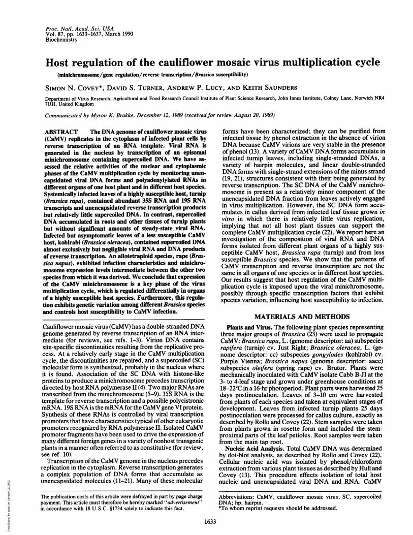
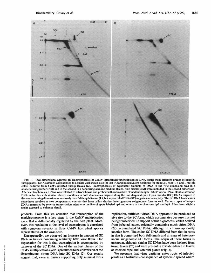
Differs in Organs of Infected Turnip Plants. The structuralcomposition of unencapsidated CaMV DNA forms isolatedfrom different organs of infected turnip plants was analyzedby two-dimensional agarose gel electrophoresis and Southernblotting. This technique resolved DNAs from leaves (Fig. lA)that we have previously identified (19, 21) as open circularand SC forms and DNA products of reverse transcription,including genome-length and subgenomic linear forms, com-plete hairpin DNAs of various sizes (hpl), and moleculespartially single- and partially double-stranded comigratingwith nested sets of hairpin forms produced by interruptedreverse transcription (hp2 and hp3). Leaves showing sys-temic vein-clearing symptoms at 25 days postinoculationcontained relatively little SC DNA, a component of theCaMV minichromosome but did contain abundant DNAforms generated by reverse transcription (Fig. 1A).
In nucleic acid preparations isolated from stem tissue at 25days postinoculation, an increased level of SC DNA relativeto that in leaves was seen, and all viral reverse transcriptionproducts were less abundant (Fig. 1B). In the unencapsidatedDNA preparation taken from roots at the same time (25 dayspostinoculation), even greater levels of SC DNA were de-tected, compared with those in leaves or stems but withnegligible reverse transcription products (compare Fig. 1Cwith A and B). Callus derived from turnip leaf discs takenfrom CaMV-infected plants at 25 days postinoculation andthen cultured in vitro for 1 mo also contained very little of theDNA forms considered to be products of replication byreverse transcription but did contain considerable SC DNA(Fig. 1D). Furthermore, the SC DNA comprised both ge-nome-length molecules and a range of subgenomic forms ofvarious sizes. The smallest SC DNA we detected from callustissue was -1 kilobase (kb) (observed after extended auto-radiogram exposure; data not shown).
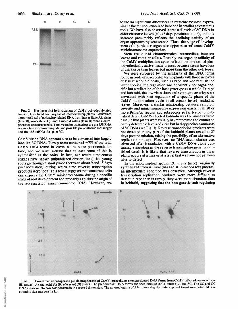
Differences in CaMV Transcripts Suggest Organ-SpecificExpression in Infected Turnip Plants. Because the CaMVreplication cycle appeared to be interrupted in some turniporgans and tissues, as indicated by the paucity ofDNA formsgenerated by reverse transcription, we wished to determinewhether this effect was at the level of transcript accumula-tion. Steady-state levels ofCaMV transcripts in turnip organswere measured by Northern hybridization analysis of cellulartotal polyadenylylated RNA (Fig. 2). Leaves contained sig-nificant amounts of the two major CaMV species, 35S RNA
and 19S RNA, together with a characteristic background ofheterogeneous-sized molecules (Fig. 2, lane A). The RNAisolated from stems was qualitatively similar to that fromleaves, although there was less of it (Fig. 2, lane B). How-ever, negligible CaMV-specific polyadenylylated RNA wasfound in roots (Fig. 2, lane C) or in callus (Fig. 2, lane D),despite the abundance of CaMV SC DNA compared withleaves (see Fig. 1). On long exposure of autoradiograms, verylow levels of largely heterogeneous CaMV RNA were foundin RNA preparations from infected roots and callus (data notshown).CaMV Replication Cycle Intermediates Also Differ in Bras-
sica Species Less Susceptible to Infection. From a survey of arange of Brassica species we have determined that hostresponse to CaMV infection falls into three broad categories(unpublished work). B. rapa (aa) variants exhibit very severesymptoms of leaf chlorosis and plant stunting (28) and yielda relatively high titer of total CaMV DNA ('400 ng of totalCaMV DNA per g of tissue at 20 days postinoculation). Incontrast, B. oleracea (cc) accumulates relatively little virus(-2 ng of total CaMV DNA per g of tissue at 20 dayspostinoculation) and exhibits very mild symptoms or nosymptoms at all depending upon the subspecies. The allo-tetraploid species B. napus (aacc) shows an intermediateresponse to CaMV infection.
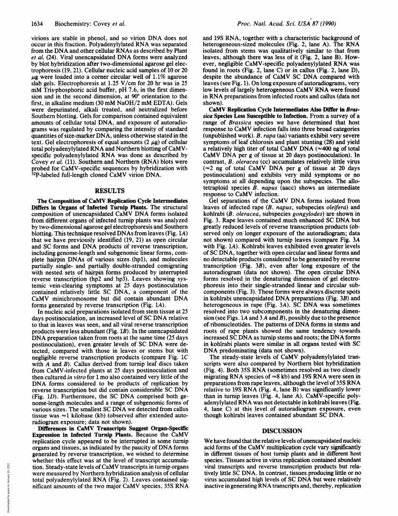
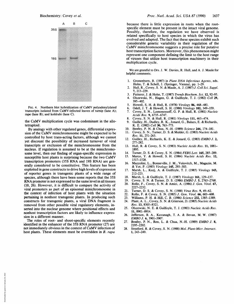
Gel separations of the CaMV DNA forms isolated fromleaves of infected rape (B. napus, subspecies oleifera) andkohlrabi (B. oleracea, subspecies gongylodes) are shown inFig. 3. Rape leaves contained much enhanced SC DNA butgreatly reduced levels of reverse transcription products (ob-served only on longer exposure of the autoradiogram; datanot shown) compared with turnip leaves (compare Fig. 3Awith Fig. 1A). Kohlrabi leaves exhibited even greater levelsof SC DNA, together with open circular and linear forms andno detectable products considered to be generated by reversetranscription (Fig. 3B), even after long exposure of theautoradiogram (data not shown). The open circular DNAforms resolved in the denaturing dimension of gel electro-phoresis into their single-stranded linear and circular sub-components (Fig. 3). These forms were always discrete spotsin kohlrabi unencapsidated DNA preparations (Fig. 3B) andheterogeneous in rape (Fig. 3A). SC DNA was sometimesresolved into two subcomponents in the denaturing dimen-sion (see Figs. 1A and 3A and B), possibly due to the presenceof ribonucleotides. The patterns ofDNA forms in stems androots of rape plants showed the same tendency towardsincreased SC DNA as turnip stems and roots; the DNA formsin kohlrabi plants were similar in all organs tested with SCDNA predominating (data not shown).The steady-state levels of CaMV polyadenylylated tran-
scripts were also compared by Northern blot hybridization(Fig. 4). Both 35S RNA (sometimes resolved as two closelymigrating RNA species of '8 kb) and 19S RNA were seen inpreparations from rape leaves, although the level of 35S RNArelative to 19S RNA (Fig. 4, lane B) was significantly lowerthan in turnip leaves (Fig. 4, lane A). CaMV-specific poly-adenylylated RNA was not detectable in kohlrabi leaves (Fig.4, lane C) at this level of autoradiogram exposure, eventhough kohlrabi leaves contained abundant SC DNA.
DISCUSSIONWe have found that the relative levels ofunencapsidated nucleicacid forms of the CaMV multiplication cycle vary significantlyin different tissues of host turnip plants and in different hostspecies. Tissues active in virus replication contained abundantviral transcripts and reverse transcription products but rela-tively little SC DNA. In contrast, tissues producing little or novirus accumulated high levels of SC DNA but were relativelyinactive in generating RNA transcripts and, thereby, replication
1634 Biochemistry: Covey et al.
Dow
nloa
ded
by g
uest
on
Janu
ary
16, 2
022
Proc. Natl. Acad. Sci. USA 87 (1990) 1635
A0
ocM I
k88.0
B
i ,SC
4 hpl
5.4 f
3.8 *
Den
C
2.6
.. 7
D
8.0 _
5.4
3.8 q'
2.6 4 A
ROOT CALLUS
FIG. 1. Two-dimensional agarose gel electrophoresis of CaMV intracellular unencapsidated DNA forms from different organs of infectedturnip plants. DNA samples were applied to a single well shown as o for leaf (A) and in equivalent positions for stem (B), root (C), and 1-mo-oldcallus cultured from CaMV-infected turnip leaves (D). Electrophoresis of equivalent amounts of DNA in the first dimension was in anondenaturing buffer (Nat) and in the second in a denaturing alkaline medium (Den). Size markers (M) were included in the second dimension.After electrophoresis, DNAs were blotted to nitrocellulose and probed with radioactive cloned full-length CaMV virion DNA. Double-strandedDNA molecules with similar relative mobilities in both dimensions migrate along the unit diagonal (ud). Open circular (OC) DNAs migrate inthe nondenaturing dimension more slowly than full-length linears (L), but supercoiled DNA (SC) migrates more rapidly. The SC DNA from leavessometimes resolves as two components, whereas that from callus also has heterogeneous subgenomic form as well. Various types of hairpinDNAs generated by reverse transcription migrate in the line of spots labeled hpl and others in the chevrons hp2 and hp3. B has been slightlyunder-exposed to enhance detail.
products. From this we conclude that transcription of theminichromosome is a key stage in the CaMV multiplicationcycle that is differentially regulated by the host plant. More-over, this regulation at the level of transcription is correlatedwith symptom severity in three CaMV host plant speciesrepresentative of the Brassicae.
Unexpectedly, we observed an increase in amount of SCDNA in tissues containing relatively little viral RNA. Oneexplanation for this is that transcription is accompanied byturnover of the SC DNA. One of the earliest phases of theCaMV multiplication cycle is presumed to be conversion ofthediscontinuous virion DNA into SC DNA (2). Our resultssuggest that, even in tissues supporting only minimal virus
replication, sufficient virion DNA appears to be produced togive rise to the SC form, which accumulates because it is notbeing transcribed. In support ofthis hypothesis, callus derivedfrom infected leaves, originally containing much virion DNA(22), accumulated SC DNA, although in a transcriptionallyinactive form. The callus SC DNA differed from that in rootsin that it comprised both full-length and a range of heteroge-neous subgenomic SC forms. The origin of these forms isunknown, although similar SC DNAs have been isolated fromturnip leaves (25) and were present in low abundance in leavesof infected rape and kohlrabi plants (Fig. 3).We presume that virus particles enter roots of infected
plants as a fortuitous consequence of systemic spread where
Biochemistry: Covey et al.
LO
IMP
4:
0,011sc0. A'O'
........
Dow
nloa
ded
by g
uest
on
Janu
ary
16, 2
022
Proc. Natl. Acad. Sci. USA 87 (1990)
A B C D
35S
19S _n
FIG. 2. Northern blot hybridization of CaMV polyadenylylatedtranscripts isolated from organs of infected turnip plants. Equivalentamounts (2 pg) ofpolyadenylylated RNA from leaves (lane A), stems(lane B), roots (lane C), and 1 mo-old callus (lane D) were electro-phoresed on agarose gels. The two major transcripts are the 35S RNAreverse transcription template and possible polycistronic messengerand the 19S mRNA for gene VI.
CaMV virion DNA appears also to be converted into largelyinactive SC DNA. Turnip roots contained -7% of the totalCaMV DNA found in leaves at the same postinoculationtime, and we must assume that at least some of this issynthesized in the roots. In fact, our recent time-coursestudies have shown (unpublished observations) that youngroots go through a short phase (between about 9 and 15 dayspostinoculation) during which time reverse transcriptionproducts were seen. This result suggests that some root cellscan express the CaMV minichromosome during a specificstage of root development and probably explains the origin ofthe accumulated minichromosome DNA. However, we
A0
M8.0
oc L1
found no significant differences in minichromosome expres-sion in the tap root examined here and in smaller adventitiousroots. We have also observed increased levels of SC DNA inolder chlorotic leaves (40-45 days postinoculation), and thisincrease presumably reflects the declining activity of anorgan approaching senescence. Thus, the stage of develop-ment of a particular organ also appears to influence CaMVminichromosome expression.Stem tissue had characteristics intermediate between
leaves and roots or callus. Possibly the organ specificity ofthe CaMV multiplication cycle reflects the amount of pho-tosynthetically active tissue present because stems have lessof this tissue than leaves but more than the other cell types.We were surprised by the similarity of the DNA forms
found in roots-of susceptible turnip plants with those in leavesof less susceptible hosts, such as rape and kohlrabi. In thelatter species, the regulation was apparently not organ spe-cific but a reflection of the host genotype as a whole. In rapeand kohlrabi, the low virus titers and symptom severity werecorrelated with host regulation of a specific phase of theCaMV multiplication cycle in all organs tested, includingleaves. Moreover, a similar relationship- between symptomseverity and minichromosome expression exists in all 20 ormore Brassica species and subspecies so far tested (unpub-lished data). CaMV-infected kohlrabi was the most extremecase, in that plants were usually asymptomatic and containedbarely detectable levels of virus but had appreciable amountsof SC DNA (see Fig. 3). Reverse transcription products werenot detected in any part of the kohlrabi plants tested at 25days postinoculation, raising the possibility of an alternativereplication strategy. However, no DNA accumulation wasobserved after inoculation with a CaMV DNA clone con-taining a mutation in the reverse transcriptase gene (unpub-lished data). It is likely that reverse transcription in theseplants occurs at a time or at a level that we have not yet beenable to detect.
In the allotetraploid species B. napus (aacc), originallysynthesized from B. rapa (aa) and B. oleracea (cc) parents,an intermediate condition was observed. Although reversetranscription replication products were more difficult todetect in rape than in turnip, they were more abundant thanin kohlrabi, suggesting that the' host genetic trait regulating
B
oc L SC
\ II /,sc.-1,S M
5.4 05:
3.8
De
n
2.6 9
ud
RAPE KOHL RABI
FIG. 3. Two-dimensional agarose gel electrophoresis of CaMV intracellular unencapsidated DNA forms from CaMV-infected leaves of rape(B. napus) (A) and kohlrabi (B. oleracea) (B) plants. The predominant DNA forms are open circular (OC), linear (L), and SC. The SC and OCDNAs resolve into two components in the second dimension. The autoradiogram ofB has been slightly underexposed to enhance detail. M lanecontains size markers in kb.
1636 Biochemistry: Covey et al.
Nate.
Dow
nloa
ded
by g
uest
on
Janu
ary
16, 2
022
Proc. Natl. Acad. Sci. USA 87 (1990) 1637
A B C
35S o_
19S _0- %W
FIG. 4. Northern blot hybridization of CaMV polyadenylylatedtranscripts isolated from CaMV-infected leaves of turnip (lane A);rape (lane B); and kohlrabi (lane C).
the CaMV multiplication cycle was codominant in the allo-tetraploid.By analogy with other regulated genes, differential expres-
sion of the CaMV minichromosome might be expected to becontrolled by host trans-acting factors, although we cannotyet discount the possibility of increased turnover of viraltranscripts or exclusion of the minichromosome from thenucleus. If regulation is assumed to be at the minichromo-some level, then our finding of organ-specific expression insusceptible host plants is surprising because the two CaMVtranscription promoters (35S RNA and 19S RNA) are gen-erally considered to be constitutive. This feature has beenexploited in gene constructs to drive high levels ofexpressionof reporter genes in transgenic plants of a wide range ofspecies, although there have been some reports that the 35SRNA promoter is not expressed to the same level in all tissues(10, 26). However, it is difficult to compare the activity ofviral promoters as part of an episomal minichromosome inthe context of infection of host plants with the situationpertaining in nonhost transgenic plants. In producing suchconstructs for transgenic plants, a viral DNA fragment isremoved from other possible viral regulatory elements, in-serted into the nuclear genome where positional effects andnonhost transcription factors are likely to influence expres-sion in a different manner.The roles of root- and shoot-specific elements recently
identified in the enhancer of the 35S RNA promoter (27) arenot immediately obvious in the context of CaMV infection ofhost plants. These elements must be overridden in B. rapa
because there is little expression in roots when the root-specific element must be present in the intact viral genome.Possibly, therefore, the regulation we have observed isrelated specifically to host species in which the virus hasevolved and adapted. The fact that these species exhibit suchconsiderable genetic variability in their regulation of theCaMV minichromosome suggests a precise role for putativehost transcription factors. Moreover, this phenomenon mightrepresent one component defining the limit to the host rangeof viruses that utilize host transcription machinery in theirmultiplication cycle.
We are grateful to Drs. J. W. Davies, R. Hull, and A. J. Maule forhelpful comments.
1. Gronenborn, B. (1987) in Plant DNA Infectious Agents, eds.Hohn, T. & Schell, J. (Springer, Vienna), pp. 1-29.
2. Hull, R., Covey, S. N. & Maule, A. J. (1987) J. Cell Sci. Suppl.7, 213-229.
3. Futterer, J. & Hohn, T. (1987) Trends Biochem. Sci. 12, 92-95.4. Olszewski, N., Hagen, G. & Guilfoyle, T. J. (1982) Cell 29,
395-402.5. Howell, S. H. & Hull, R. (1978) Virology 86, 468-481.6. Odell, J. T. & Howell, S. H. (1980) Virology 102, 349-359.7. Covey, S. N., Lomonossoff, G. P. & Hull, R. (1981) Nucleic
Acids Res. 9, 6735-6747.8. Covey, S. N. & Hull, R. (1981) Virology 111, 463-474.9. Guilley, H., Dudley, R. K., Jonard, G., Balazs, E. & Richards,
K. E. (1982) Cell 30, 763-773.10. Benfey, P. N. & Chua, N.-H. (1989) Science 244, 174-181.11. Covey, S. N., Turner, D. S. & Mulder, G. (1983) Nucleic Acids
Res. 11, 251-263.12. Guilley, H., Richards, K. E. & Jonard, G. (1983) EMBO J. 2,
277-282.13. Hull, R. & Covey, S. N. (1983) Nucleic Acids Res. 11, 1881-
1895.14. Turner, D. S. & Covey, S. N. (1984) FEBS Lett. 165, 285-289.15. Marco, Y. & Howell, S. H. (1984) Nucleic Acids Res. 12,
1517-1528.16. Mazzolini, L., Bonneville, J. M., Volovitch, M., Magazin, M.
& Yot, P. (1985) Virology 145, 293-303.17. Marsh, L., Kuzj, A. & Guilfoyle, T. J. (1985) Virology 143,
212-223.18. Marsh, L. & Guilfoyle, T. J. (1987) Virology 161, 129-137.19. Covey, S. N. & Turner, D. S. (1986) EMBO J. 5, 2763-2768.20. Rollo, F., Covey, S. N. & Amici, A. (1986) J. Gen. Virol. 67,
2227-2232.21. Turner, D. S. & Covey, S. N. (1988) Virus Res. 9, 49-62.22. Rollo, F. & Covey, S. N. (1985) J. Gen. Virol. 66, 603-608.23. Williams, P. H. & Hill, C. B. (1986) Science 232, 1385-1389.24. Plant, A. L., Covey, S. N. & Grierson, D. (1985) Nucleic Acids
Res. 13, 8305-8321.25. Olszewski, N. E. & Guilfoyle, T. J. (1983) Nucleic Acids Res.
11, 8901-8914.26. Jefferson, R. A., Kavanagh, T. A. & Bevan, M. W. (1987)
EMBO J. 6, 3901-3907.27. Benfey, P. N., Ren, L. & Chua, N.-H. (1989) EMBO J. 8,
2195-2202.28. Stratford, R. & Covey, S. N. (1988) Mol. Plant-Micr. Interact.
1, 243-249.
Biochemistry: Covey et al.
Dow
nloa
ded
by g
uest
on
Janu
ary
16, 2
022