Embed Size (px)
Citation preview

7/23/2019 Hormones_2014-131
http://slidepdf.com/reader/full/hormones2014-131 1/9
Gonadotropin secreting pituitary adenoma associated
with erythrocytosis: case report and literature review
Filippo Ceccato,1 Gianluca Occhi,1 Daniela Regazzo,1 Maria Luigia Randi,2
Diego Cecchin,3 Marina Paola Gardiman,4 Renzo Manara,5 Giuseppe Lombardi,6
Luca Denaro,7 Franco Mantero,1 Carla Scaroni1
1Endocrinology Unit, 2 Internal Medicine, 3 Nuclear Medicine Unit, 4 Surgical Pathology and Cytopathology Unit,Department of Medicine DIMED; University of Padua Medical School; 5 Neuroradiologic Unit, University Hospital of
Padua; 6 Medical Oncology 1, Venetian Oncology Institute - IRCCS; 7 Neurosurgery Division, University Hospital of
Padua; Padua, Italy
ABSTRACT
BACKGROUND: Most pituitary adenomas with FSH- or LH-positive immunohistochemistry
are endocrinologically silent, and neurological symptoms due to their large volume are the first
clinical signs; they are rarely reported to be secreting gonadotropins, this usually occurring in
cases with clinical endocrine findings. Gonadotropinomas are often treated surgically because they
are unresponsive to conventional medical therapies. Temozolomide was recently recommended
for non-responder aggressive pituitary adenoma management. CASE REPORT: A 43-year-old
male with a history of 5 years of erythrocytosis presented with severe headache, orthostatic diz-
ziness, and difficulty walking. MRI documented a giant pituitary adenoma and high uptake of111In-pentetreotide indicated somatostatin receptor (SSR) expression. Biochemical tests revealed a
secreting gonadotropinoma. Therapy with somatostatin analogs and dopamine agonists improved
the patient’s headache, achieved partial hormone control, slightly reduced the size of the adenoma,
and controlled erythrocytosis. Six months after the diagnosis, hormone escape occurred despite
therapy, thus neurosurgery was performed. After the procedure the patient died of untreatable
intracranial hypertension. The surgical specimen revealed SSR 2 and 3 expression, and temo-
zolomide did not induce apoptosis in primary cell culture. REVIEW OF LITERATURE: Among
gonadotropinomas, female gender (77%), macroadenoma (84%), young age at diagnosis (28 ±
12 years), delay from first symptoms to diagnosis (up to 15 years), and ovarian cysts/menstrual
disorders in females or macro-orchidism in males were the foremost clinical and neuroimagingfeatures. CONCLUSIONS: Male gonadotropin-secreting pituitary adenomas may have a variable
clinical expression secondary to testosterone excess. Somatostatin analogs, dopamine agonists
or temozolomide may have a role that needs to be assessed case by case.
Key words: Erythrocytosis, Giant pituitary adenoma, Gonadotroph adenoma, Somatostatin
analog therapy, Temozolomide
HORMONES 2014, 13(1):131-139
Address for correspondence:Carla Scaroni, MD, Endocrinology Unit, Department of Medicine DIMED, 35128 Padova, Italy, Tel.: +39 049 8211323;Fax: +39 049 657391; e-mail: [email protected]
Received 01-02-2013, Accepted 13-05-2013
Case report

7/23/2019 Hormones_2014-131
http://slidepdf.com/reader/full/hormones2014-131 2/9
132 F. CECCATO ET AL
INTRODUCTION
Pituitary adenomas are being identified increas-
ingly often in the general population due to a better
clinical approach to patients’ clinical symptoms andto the widespread diffusion of neuro-imaging tech-
niques.1,2 Although most clinically non-functioning
pituitary adenomas reveal LH- or FSH-positive im-
munohistochemistry, gonadotropin secreting pituitaryadenomas are still rarely diagnosed3,4 because of
clinical signs relating to their hormone over-secretion,as for example in ovarian cysts in females or macro-
orchidism in young male patients.3-26
As for their treatment, surgery is the first choice,
but little is still known about the efficacy of somato-
statin analogs (SSA)3,25 or dopamine agonists.15,24,27 Temozolomide (TMZ) has recently been recom-
mended in pituitary macroadenomas characterized
by an invasive growth and resistance to conventionaltherapies (i.e. neurosurgery, radiotherapy or other
medical treatments), particularly those secreting
ACTH or prolactin.28,29 The outcome of TMZ treat-
ment may depend at least to some degree on the
expression of O-6 methylguanine DNA methyltrans-ferase (MGMT), a DNA repair enzyme that has the
potential to interfere with TMZ;30,31 a recent study
revealed that TMZ reduced cell viability in gonado-troph pituitary adenoma cell lines.32
CASE REPORT
Clinical features
A 43-year-old Caucasian male had been referred
to our Department of Medicine 5 years earlier with
facial plethora, arterial hypertension (160/105 mmHg),a high red blood cell (RBC) count, i.e. 6.5 x 10⁹ /L
(normal value [n.v.] 4.3-5.1 x 10¹² /L), high hematocrit
61.1% (n.v. 36-46), high hemoglobin 200 g/L (n.v.123-153), and a normal platelet count (160 x 109 /L
n.v. 150-450). A full work-up for erythrocytosis hadbeen completed in accordance with the WHO criteria200133: red cell volume was found increased (46 ml/
kg) and no cause of secondary erythrocytosis was
detected (no evidence of familial erythrocytosis or
elevated erythropoietin: 8 U/L, n.v. 11-20). Bone
marrow biopsy revealed prominent erythroid pro-
liferation and a mild increase in reticulin; a normal
male karyotype was documented, with no evidence
of bcr/abl rearrangement. No mutations of the JAK2gene (both V617F and exon 12), the erythropoietin
receptor gene (EPO-R) or the genes on the oxygen-
sensing pathway (HIF-1 alpha, PHD2, VHL) cameto light. A diagnosis of polycythemia vera was ruled
out: the patient underwent regular phlebotomies andstarted antihypertensive and antiaggregant therapy.
CT of the brain was performed when he com-
plained of mood swings, acute and severe headache,
diplopia, orthostatic dizziness, and difficulty walking,revealing a giant mass in the skull base. The patient
was referred to the Endocrinology Unit and brain
MRI confirmed the presence of a giant cystic and solidpituitary adenoma in the skull base, 68x64x60 mm in
size, with peritumoral edema. The pituitary adenomainvaded the posterior cranial fossa through the clivus;the anterior cerebral arteries were encased in the le-sion, without stenosis (Figure 1). Whole-body single
photon emission tomography/computed tomography(SPET/CT) was performed after the intravenous
administration of 111In-pentetreotide (Octreoscan),
showing a very high uptake in the pituitary lesion
consistent with the presence of somatostatin recep-
tors (SSR).
We diagnosed a gonadotropinoma on the basis ofthe patient’s hormone secretion profile: FSH 106.6
U/L (n.v. 1-14, LH 19.3 U/L (n.v. 1.5-9.2), α-subunit
6.2 U/L (n.v. 1-14), prolactin 17.3 µg/L (n.v. 5-15),
inhibin-B 13 ng/L (n.v. 42-213), total testosterone
52.05 nmol/L (n.v. 10-29), SHBG 25 nmol/L (n.v.
13-71); the other pituitary hormones were all normaland any ACTH deficiency was ruled out by the resultsfor cortisol using the 1 µg corticotropin test. Scrotal
ultrasound documented testicles of normal size and
structure, with a left varicocele. An automatic visual
field evaluation revealed left temporal hemianopsia.
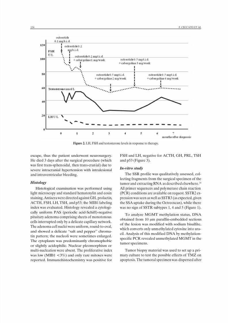
We documented a drop in FSH and LH levels
after a short (8-hour) test with 0.1 mg octreotide
injection, therefore therapy with octreotide 0.1 mg
sc. twice daily was started. Two weeks later, FSH,
LH, and total testosterone levels were still high, so
we increased the dose of SSA to 0.2 mg three times
a day, combined with cabergoline, up to a maximum
dose of octreotide 0.5 mg three times a day and caber-goline 4 mg/week, depending on the patient’s FSH,
LH and testosterone levels (Figure 2). We opted to

7/23/2019 Hormones_2014-131
http://slidepdf.com/reader/full/hormones2014-131 3/9
Aggressive giant gonadotropinoma 133
try medical treatment first because of the high surgi-cal risk and intercurrent retinal thrombosis (treated
with low-molecular-weight heparin).
After 4 months of therapy, another MRI scan showeda slight shrinkage of the pituitary lesion (59x46x60 mm)and a significant reduction in the peritumoral edema
(Figure 1). The SSA+cabergoline therapy resolved the
headaches and restored normal testosterone (24.42
nmol/L) and LH (5.3 U/L) levels, while FSH remainedhigh (FSH 50.9 U/L), but lower than at the baseline.
The patient’s RBC count also improved to 5×10¹² /L
and the phlebotomies were discontinued.
Six months after diagnosis, we documented a
worsening of headache and diplopia with hormone
Figure 1. Gadolinium-enhanced T1-weighted MRI images: axial (a), coronal (b) and sagittal (c) views obtained at diagnosis; axial (d),
coronal (e) and sagittal (f) MRI scan performed 4 months after medical therapy. 111In-pentetreotide (Octreoscan) SPET/CT: fusedtransaxial (g), coronal (h) and sagittal (i) images. In the box: PCR expression of somatostatin receptors 2 and 3.
7/23/2019 Hormones_2014-131
http://slidepdf.com/reader/full/hormones2014-131 4/9
134 F. CECCATO ET AL
escape, thus the patient underwent neurosurgery.
He died 3 days after the surgical procedure (which was first trans-sphenoidal, then trans-cranial) due tosevere intracranial hypertension with intralesional
and intraventricular bleeding.
Histology
Histological examination was performed using
light microscopy and standard hematoxylin and eosinstaining. Antisera were directed against GH, prolactin,
ACTH, FSH, LH, TSH, and p53; the MIB1 labeling
index was evaluated. Histology revealed a cytologi-
cally uniform PAS (periodic acid-Schiff)-negativepituitary adenoma comprising sheets of monotonouscells interrupted only by a delicate capillary network.The adenoma cell nuclei were uniform, round-to-oval,and showed a delicate “salt and pepper” chroma-
tin pattern; the nucleoli were sometimes enlarged.
The cytoplasm was predominantly chromophobic
or slightly acidophilic. Nuclear pleomorphism or
multi-nucleation were absent. The proliferative index was low (MIB1 <3%) and only rare mitoses were
reported. Immunohistochemistry was positive for
FSH and LH, negative for ACTH, GH, PRL, TSH
and p53 (Figure 3).
In-vitro study
The SSR profile was qualitatively assessed, col-
lecting fragments from the surgical specimen of the
tumor and extracting RNA as described elsewhere.34 All primer sequences and polymerase chain reaction(PCR) conditions are available on request. SSTR2 ex-pression was seen as well as SSTR3 (as expected, giventhe SSA uptake during the Octreoscan), while there
was no sign of SSTR subtypes 1, 4 and 5 (Figure 1).
To analyze MGMT methylation status, DNAobtained from 10 µm paraffin-embedded sections
of the lesion was modified with sodium bisulfite,
which converts only unmethylated cytosine into ura-cil. Analysis of this modified DNA by methylation-
specific PCR revealed unmethylated MGMT in the
tumor specimens.
Tumor biopsy material was used to set up a pri-
mary culture to test the possible effects of TMZ on
apoptosis. The tumoral specimen was dispersed after
Figure 2. LH, FSH and testosterone levels in response to therapy.

7/23/2019 Hormones_2014-131
http://slidepdf.com/reader/full/hormones2014-131 5/9
Aggressive giant gonadotropinoma 135
enzymatic and mechanical treatment, incubated in
DMEM supplemented with collagenase 10,000 units/ ml, 1 mg/ml hyaluronidase, 0.1 mg/ml trypsin inhibitor,and 40 mg/ml BSA for 1 hour at 37°C under gentle
shaking. The cells were washed once in supplemented
culture medium and their number and viability (al- ways higher than 90%) were estimated using the
trypan blue exclusion test. Then 105 cells/well were
seeded in a polylysine-coated 48-well plate. After 24
hours, the medium was removed and the cells were
treated with TMZ 100 µM for 24 hours. The primarycell cultures were then tested for apoptosis with the
ApopNexinAnnexin V FITC Apoptosis Kit (Merck-Millipore, Germany). The cell cycle distribution wasassessed using propidium iodide staining followed
by cytofluorimetric analysis. Apoptosis rate in the
TMZ-treated tumor cell culture was no higher than
for the control cells.
LITERATURE REVIEW
The literature published in the PubMed database
from 1982 to May 2012 on gonadotroph secreting
pituitary adenoma was searched systematically, using
the “related articles” function to identify any relevant
publications. We looked for case reports with a full
dataset as concerns sex, age, size of lesion, and the mostimportant signs or symptoms prompting the suspicionof LH- or FSH-secreting pituitary adenoma. We did notconsider gonadotroph pituitary adenomas diagnosed
as a result of the presence of a pituitary mass and
causing symptoms such as headache, deterioration of
Figure 3. Biopsy specimen with hematoxylin and eosin staining (a), proliferative index with MIB-1 labeling under 3% (b), positiveimmunohistochemistry for FSH (c) and LH (d).

7/23/2019 Hormones_2014-131
http://slidepdf.com/reader/full/hormones2014-131 6/9
136 F. CECCATO ET AL
visual field, or hypopituitarism. Including the patientdescribed here, we identified 31 case reports, most ofthem involving females (24/31, 77%) with macroadeno-
mas (4 micro- and 21 macro-pituitary adenomas); themean age at diagnosis was similar for the two genders(28 ± 12 years) and there was a considerable delay fromthe onset of the first symptoms to diagnosis (rangingfrom 6 months to 15 years). The most important signs
were ovarian cysts and menstrual disorders in femalesand macro-orchidism in males (Table 1).
DISCUSSION
Pituitary adenomas account for nearly 15% of all
intracranial neoplasms, the majority being prolactino-
mas;1,2 gonadotroph adenomas are difficult to diagnose
because they are usually non-secreting, or they secretebiologically inactive peptides with no clinical effects,
and they classically grow silently until neurological
symptoms develop. Clinical signs or symptoms ofgonadotropin hypersecretion are very rarely reported,involving a few premenopausal women with ovarian
hyperstimulation syndrome and men with macro-
orchidism.3-26 In recent series, a large proportion of theadult patients undergoing surgery for non-functioningpituitary adenoma had a silent gonadotroph adenoma:the definitive diagnosis can only be established from
a positive FSH/LH immunoreactivity.35
Here we present the unusual case, not previously
reported in the literature, in which a pituitary ad-
enoma was diagnosed due to its mass effect, though
Table 1. Gonadotroph cell adenoma diagnosed due to signs/symptoms of sex hormone hypersecretion in the literature
n, sex Age (yr) Duration (yr) Size (mm) Symptoms Reference
2, F 8,10 NA macro Precocious puberty DiRocco, 19825
4, M NA NA macro Macro-orchidism Heseltine, 19896
1, F 13 NA 30 Metrorrhagia, ovarian cysts Etzrodt, 19907
1, F 31 1 14 Ovarian cysts Djerassi, 19958
1, F 34 6 40 Ovarian cysts Christin-Maitre, 19989
1, F 28 1 14 Ovarian cysts Valimaki MJ, 199910
1, F 10 1 macro Ovarian cysts Tashiro, 199911
1, F 23 12 18 Oligomenorrhea, ovarian cysts Pentz-Vidovic, 200012
1, F 30 3 21 Galactorrhea, amenorrhea Saveanu, 20013
1, F 28 NA 30 Ovarian cysts Shimon, 200113
1, F 35 2 13 Oligomenorrhea, ovarian cysts Castelbaum, 200214
1, F 29 NA 7 Infertility, ovarian cysts Murata, 200315
1, F 21 NA NA Ovarian cysts Murakami, 200416
3, F 31, 30, 43 4/2/NA 9/17/61 Ovarian cysts Mor, 200517
1, F 40 NA micro Ovarian cysts Maruyama, 200518
1, F 27 NA macro Ovarian cysts Kihara, 200619
1, F 30 NA NA Ovarian cysts Ghayuri, 200720
1, F 40 15 27 Galactorrhea, ovarian cysts Cooper, 200821
1, F 31 NA 20 Galactorrhea, ovarian cysts Castelo-Branco, 200922
1, M 56 3 33 Macro-orchidism Dahlqvist, 201023
1, F 13 0.5 20 Ovarian cysts Gryngarten, 20104
1, M 12 1 9 Macro-orchidism Clemente, 201124
1, F 37 0.5 28 Metrorrhagia, ovarian cysts Karapanou, 201225
1, F 26 3 25 Oligomenorrhea, ovarian cysts Garmes, 201226
1, M 43 5 68 Polycythemia This case
NA: not available.

7/23/2019 Hormones_2014-131
http://slidepdf.com/reader/full/hormones2014-131 7/9
Aggressive giant gonadotropinoma 137
the diagnosis could have been reached 5 years earlier,considering that erythrocytosis may be secondary to
testosterone excess. In fact, when appropriate medi-
cal therapy was effective in reducing the patient’s LHand FSH levels, the consequent drop in testosteronelevels led to a normal erythropoiesis and bloodlettingtherapy was no longer necessary. Erythropoiesis is a
process induced hormonally by erythropoietin and
testosterone, which takes effect directly on bone
marrow at polychromatophilic erythroblast level andenhances the synthesis of ribosomal RNA.36 Some
recent publications have reported that administra-
tion of testosterone is associated with serum hepcidinsuppression: 37 hepcidin is a liver-derived peptide thatbinds to and degrades the iron channel ferroportin,
and low hepcidin is associated with increased iron
absorption and systemic transport, stimulating eryth-ropoiesis. In this paper, Bachman et al reported thathigh testosterone levels resulted in a 60% suppressionof serum hepcidin levels in a dose- and age-dependentmanner within 1 week, though the mechanisms by
which testosterone suppresses hepcidin remain as yetunknown. It is common knowledge that hypogonadismleads to anemia in men, that excessive erythrocytosisis the most common serious adverse event associated
with testosterone replacement therapy and that the
anabolic use of androgens induces an excessivelyhigh RBC count in athletes, but little is known aboutendogenous testosterone excess.37,38
In our case we found subnormal serum levels
of inhibin-B, as already in reported in three out of
five adults with macro-orchidism by Heseltine and
Dahlqvist,4,23 while Clemente described high inhibin-Bin one male adolescent with macro-orchidism.24 More-over, all of six previous males described presented
with macro-orchidism,4,23,24 whereas our patients
showed testicles of normal size and structure (leadingprobably to diagnostic delay). Testicular volume andinhibin-B secretion both depend on FSH levels, whichstimulates seminiferous tubules.6,24 In our cases the
biological effects of FSH were not clinically evident,
suggesting a secretion of less biologically active FSHor a reduced sensitivity to FSH, whereas LH was ef-
fective in increasing testosterone levels, leading to
erythrocytosis.
For large pituitary adenoma one of the first treat-
ment options is surgery. It was not the first choice for
our patient because of the high surgical risk, given
that the mass invaded the surrounding structures
and there was an intercurrent retinal complication
(for these same reasons we did not consider radio-surgery). Medical therapy was therefore attempted
first, although only a few authors have reported on
SSA 3,25 or dopamine agonist15,24,27 treatments improvingthe signs or symptoms of hormone hypersecretion ingonadotropinomas. SSA uptake during the Octreoscanand the LH/FSH response to the acute octreotide
test led us to suspect a finding of SSR in our patient
(subsequently confirmed by the SSR 2 and 3 found inthe tumor tissue by PCR), so we started SSA therapy;
we then needed to add cabergoline to attain a bet-
ter control. We did not consider androgen receptor
antagonist in order to avoid a positive feedback on
LH/FSH secretion. Moreover, we chose not to use
gonadotropin-releasing hormone (GnRH) agonist
or antagonist, because there is lack of expertise in
male subjects: there is only one report describing thefact that GnRH agonist could induce a paradoxical
increase in serum estradiol levels and may exacerbateovarian follicular cysts,14 and there are few experi-
ences in woman with ovarian hyperstimulation 8,26,39-41 treated with GnRH antagonist with contradictory
results. The patient’s retinal thrombosis was believed
to be secondary to the brain mass and erythrocytosisin combination, and it disappeared completely in 3
months.
Pituitary carcinomas are usually identified by thepresence of pituitary metastases rather than by theirmalignant histological features.42 Similarly, aggressivepituitary adenomas may exhibit a relatively benignhistological appearance despite their local invasion,malignant growth patterns, encasement of vascularstructures, and rapid enlargement. In addition, thereis a subset of tumors classified as “atypical adenomas”
accounting for 3-15% of cases in surgical series35,43 that have been identified as potentially at risk of amore aggressive growth or malignant deterioration:these tumors are characterized by a MIB-1 above 3%,p53 immunoreactivity and a high mitotic index.42 Inour biopsy specimen, the proliferative index was low(MIB-1 <3%), immunohistochemistry was negativefor p53, and we found only rare mitoses, meaning
that the mass grew slowly year by year, leading firstto hormonal conditions (erythrocytosis, arterial hy-pertension, mood swings).

7/23/2019 Hormones_2014-131
http://slidepdf.com/reader/full/hormones2014-131 8/9
138 F. CECCATO ET AL
Recent studies have shown that TMZ is effective
in patients with pituitary carcinoma and aggressive
pituitary adenoma failing to respond to conventional
treatments.29
The cytotoxic effect of TMZ relies onmethylation of the guanine in the O-6 position in theDNA, which damages the DNA. MGMT is a DNA
repair enzyme that removes the alkyl group adducts
from the O-6 position, inducing TMZ resistance as
a result.30,31 MGMT expression in pituitary adenomashould not to be taken as a reason to deny treatment,and, according to a recent review, the best hormone
and tumor response is seen in ACTH- and prolactin-secreting pituitary adenomas despite any MGMT
expression.29 In our in-vitro study, the tumor cell
culture treated with TMZ revealed no higher rate
of apoptosis than in control cells, correlating with
unmethylated MGMT in the tumor specimens.
To sum up, gonadotropin-secreting pituitary ad-
enoma in adult males may present with symptoms
such as plethora due to erythrocytosis secondary to
testosterone excess, as in the case described here.
Combined therapy with SSA and dopamine agonistsproved effective in terms of our patient’s clinical
picture and, to some degree, in reducing the volume
of the mass, whereas using TMZ in this case would
have been clinically ineffectual. A multi-disciplinary
approach is needed for cases of aggressive pituitary
macroadenoma with a view to ensuring a prompt
diagnosis and effective treatment.
ACKNOWLEDGMENTS
This work was supported by the Italian Ministry
for the University and Research (MIUR) PRIN GrantNo. 2009YJTBAZ-003.
DISCLOSURE STATEMENT
The authors have nothing to disclose.
REFERENCES
1. Daly AF, Rixhon M, Adam C, Dempegioti A, Tichomi-
rowa MA, Beckers A, 2006 High prevalence of pituitary
adenomas: a cross-sectional study in the province of
Liege, Belgium. J Clin Endocrinol Metab 91: 4769-4775.
2. Fernandez A, Karavitaki N, Wass JA, 2010 Prevalence of
pituitary adenomas: a community-based, cross-sectional
study in Banbury (Oxfordshire, UK). Clin Endocrinol
72: 377-382.
3. Saveanu A, Morange-Ramos I, Gunz G, Dufour H,
Enjalbert A, Jaquet P, 2001 A luteinizing hormone,
alpha-subunit- and prolactin-secreting pituitary adenoma
responsive to somatostatin analogs: in vivo and in vitrostudies. Eur J Endocrinol 145: 35-41.
4. Gryngarten MG, Braslavsky D, Ballerini MG, Ledesma
J, Ropelato MG, Escobar ME, 2010 Spontaneous ovar-
ian hyperstimulation syndrome caused by a follicle-
stimulating hormone-secreting pituitary macroadenoma
in an early pubertal girl. Horm Res Paediatr 73: 293-298.
5. Di Rocco C, Maira G, Borrelli P, 1982 Pituitary micro-
adenomas in children. Childs Brain 9: 165-178.
6. Heseltine D, White MC, Kendall-Taylor P, De Kretser
DM, Kelly W, 1989 Testicular enlargement and elevated
serum inhibin concentrations occur in patients with
pituitary macroadenomas secreting follicle stimulating
hormone. Clin Endocrinol 31: 411-423.7. Etzrodt A, Etzrodt H, Hey O, 1990 FSH-producing
hypophyseal tumor in a 13-year-old girl. Geburtshilfe
Frauenheilkd 50: 73-75.
8. Djerassi A, Coutifaris C, West VA, et al, 1995 Gonado-
troph adenoma in a premenopausal woman secreting
follicle-stimulating hormone and causing ovarian hy-
perstimulation. J Clin Endocrinol Metab 80: 591-594.
9. Christin-Maitre S, Rongières-Bertrand C, Kottler ML,
et al, 1998 A spontaneous and severe hyperstimulation
of the ovaries revealing a gonadotroph adenoma. J Clin
Endocrinol Metab 83: 3450-3453.
10. Välimäki MJ, Tiitinen A, Alfthan H, et al, 1999 Ovar-
ian hyperstimulation caused by gonadotroph adenomasecreting follicle-stimulating hormone in 28-year-old
woman. J Clin Endocrinol Metab 84: 4204-4208.
11. Tashiro H, Katabuchi H, Ohtake H, Kaku T, Ushio Y, Oka-
mura H, 1999 A follicle-stimulating hormone-secreting
gonadotroph adenoma with ovarian enlargement in a
10-year-old girl. Fertil Steril 72: 158-160.
12. Pentz-Vidovíc I, Skorić T, Grubisić G, et al, 2000 Evo-
lution of clinical symptoms in a young woman with a
recurrent gonadotroph adenoma causing ovarian hyper-
stimulation. Eur J Endocrinol 143: 607-614.
13. Shimon I, Rubinek T, Bar-Hava I, et al, 2001 Ovarian
hyperstimulation without elevated serum estradiol
associated with pure follicle-stimulating hormone-
secreting pituitary adenoma. J Clin Endocrinol Metab
86: 3635-3640.
14. Castelbaum AJ, Bigdeli H, Post KD, Freedman MF,
Snyder PJ, 2002 Exacerbation of ovarian hyperstimula-
tion by leuprolide reveals a gonadotroph adenoma. Fertil
Steril 78: 1311-1313.
15. Murata Y, Ando H, Nagasaka T, et al, 2003 Successful
pregnancy after bromocriptine therapy in an anovula-
tory woman complicated with ovarian hyperstimulation
caused by follicle-stimulating hormone-producing plu-
rihormonal pituitary microadenoma. J Clin Endocrinol
Metab 88: 1988-1993.

7/23/2019 Hormones_2014-131
http://slidepdf.com/reader/full/hormones2014-131 9/9
Aggressive giant gonadotropinoma 139
16. Murakami T, Higashitsuji H, Yoshinaga K, Terada Y,
Ito K, Ikeda H, 2004 Management of ovarian hyper-
stimulation due to follicle-stimulating hormone-secreting
gonadotroph adenoma. BJOG 111: 1297-1300.
17. Mor E, Rodi IA, Bayrak A, Paulson RJ, Sokol RZ,2005 Diagnosis of pituitary gonadotroph adenomas in
reproductive-aged women. Fertil Steril 84: 757 e15-e20.
18. Maruyama T, Masuda H, Uchida H, Nagashima T, Yo-
shimura Y, 2005 Follicle stimulating hormone-secreting
pituitary microadenoma with fluctuating levels of ovar-
ian hyperstimulation. Obstet Gynecol 105: 1215-1218.
19. Kihara M, Sugita T, Nagai Y, Saeki N, Tatsuno I, Seki K,
2006 Ovarian hyperstimulation caused by gonadotroph
cell adenoma: a case report and review of the literature.
Gynecol Endocrinol 22: 110-113.
20. Ghayuri M, Liu JH, 2007 Ovarian hyperstimulation
syndrome caused by pituitary gonadotroph adenoma
secreting follicle-stimulating hormone. Obstet Gynecol109: 547-549.
21. Cooper O, Geller JL, Melmed S, 2008 Ovarian hy-
perstimulation syndrome caused by an FSH-secreting
pituitary adenoma. Nat Clin Pract Endocrinol Metab
4: 234-238.
22. Castelo-Branco C, del Pino M, Valladares E, 2009 Ovarian
hyperstimulation, hyperprolactinaemia and LH gonado-
troph adenoma. Reprod Biomed Online 19: 153-155.
23. Dahlqvist P, Koskinen LO, Brännström T, Hägg E, 2010
Testicular enlargement in a patient with a FSH-secreting
pituitary adenoma. Endocrine 37: 289-293.
24. Clemente M, Caracseghi F, Gussinyer M, et al, 2011
Macroorchidism and panhypopituitarism: two differentforms of presentation of FSH-secreting pituitary adeno-
mas in adolescence. Horm Res Paediatr 75: 225-230.
25. Karapanou O, Tzanela M, Tamouridis N, Tsagarakis S,
2012 Gonadotroph pituitary macroadenoma inducing
ovarian hyperstimulation syndrome: successful response
to octreotide therapy. Hormones 11: 199-202.
26. Garmes HM, Grassiotto OR, Fernandes YB, et al, 2012
A pituitary adenoma secreting follicle-stimulating hor-
mone with ovarian hyperstimulation: treatment using
a gonadotropin-releasing hormone antagonist. Fertil
Steril 97: 231-234.
27. Paoletti AM, Depau GF, Mais V, Guerriero S, Ajossa S,
Melis GB, 1994 Effectiveness of cabergoline in reducingfollicle-stimulating hormone and prolactin hypersecretion
from pituitary macroadenoma in an infertile woman.
Fertil Steril 62: 882-885.
28. Syro LV, Ortiz LD, Scheithauer BW, et al, 2011 Treatment
of pituitary neoplasms with temozolomide: a review.
Cancer 117: 454-462.
29. Raverot G, Castinetti F, Jouanneau E, et al, 2012 Pituitary
carcinomas and aggressive pituitary tumours: merits and
pitfalls of temozolomide treatment. Clin Endocrinol
76: 769-775.
30. Mollemann M, Wolter M, Felsberg J, Collins VP, Reifen-
berger G, 2005 Frequent promoter hypermethylation and
low expression of the MGMT gene in oligodendroglial
tumors. Int J Cancer 113: 379–385.
31. Levin N, Lavon I, Zelikovitsh B, et al, 2006 Progres-
sive low-grade oligodendrogliomas: response to te-mozolomide and correlation between genetic profile
and O6-methylguanine DNA methyltransferase protein
expression. Cancer 106: 1759-1765.
32. Ma S, Liu X, Yao Y, et al, 2011 Effect of temozolomide
on cell viability in gonadotroph adenoma cell lines.
Oncol Rep 26: 543-550.
33. Jaffe ES, Harris NL, Stein H, Vardiman JW 2001 World
Health Organization Classification of Tumors; Pathology
and Genetics. Tumors of haemopoietic and lymphoid
tissues. Lyon, France: IARC.
34. Occhi G, Albiger N, Berlucchi S, et al, 2007 Peroxisome
proliferator-activated receptor gamma in the human pitu-
itary gland: expression and splicing pattern in adenomasversus normal pituitary. J Neuroendocrinol 19: 552-559.
35. Saeger W, Lüdecke DK, Buchfelder M, Fahlbusch R,
Quabbe HJ, Petersenn S, 2007 Pathohistological clas-
sification of pituitary tumors: 10 years of experience with
the German Pituitary Tumor Registry. Eur J Endocrinol
156: 203-216.
36. Gonzales GF, Gasco M, Tapia V, Gonzales-Castañeda C,
2009 High serum testosterone levels are associated with
excessive erythrocytosis of chronic mountain sickness in
men. Am J Physiol Endocrinol Metab 296: E1319-E1325.
37. Bachman E, Feng R, Travison T, et al, 2010 Testosterone
suppresses hepcidin in men: a potential mechanism for
testosterone-induced erythrocytosis. J Clin EndocrinolMetab 95: 4743-4747.
38. Zitzmann M, 2008 Effects of testosterone replacement
and its pharmacogenetics on physical performance and
metabolism. Asian J Androl 10: 364-372.
39. Daneshdoost L, Pavlou SN, Molitch ME, et al, 1990
Inhibition of follicle-stimulating hormone secretion from
gonadotroph adenomas by repetitive administration of
a gonadotropin-releasing hormone antagonist. J Clin
Endocrinol Metab 71: 92-97.
40. McGrath GA, Goncalves RJ, Udupa JK, et al, 1993 New
technique for quantitation of pituitary adenoma size: use
in evaluating treatment of gonadotroph adenomas with
a gonadotropin-releasing hormone antagonist. J Clin
Endocrinol Metab 1993 76: 1363-1368.
41. Chanson P, Lahlou N, Warnet A, et al, 1994 Responses to
gonadotropin releasing hormone agonist and antagonist
administration in patients with gonadotroph cell adeno-
mas. J Endocrinol Invest 17:91-98.
42. De Lellis RA, Lloyd RV, Heitz PU, Eng C (eds) 2004
World Health Organization Classification of Tumours:
Tumours of endocrine organs, Lyon, France: IARC.
43. Zada G, Woodmansee WW, Ramkissoon S, Amadio J,
Nose V, Laws ER Jr, 2011 Atypical pituitary adenomas:
incidence, clinical characteristics, and implications. J
Neurosurg 114: 336-344.


![[XLS] · Web view867 51 131 853 15 9052 849 17 131 854 15 9057 850 18 131 855 15 9128 857 52 131 856 14 8952 840 48 131 857 14 8967 841 13 131 858 15 9393 879 42 130 859 15 9430 883](https://img.pdfslide.us/doc/110x75/5ab8e4147f8b9ad3038d5d22/xls-view867-51-131-853-15-9052-849-17-131-854-15-9057-850-18-131-855-15-9128-857.jpg)