Embed Size (px)
Citation preview

Hormonal Regulation of MicroRNA Expression in SteroidProducing Cells of the Ovary, Testis and Adrenal GlandZhigang Hu1,2, Wen-Jun Shen1,2, Yuan Cortez1, Xudong Tang1,2, Li-Fen Liu1,2, Fredric B. Kraemer1,2,
Salman Azhar1,3*
1Geriatric Research, Education and Clinical Center, VA Palo Alto Health Care System, Palo Alto, California, United States of America, 2Division of Endocrinology, Stanford
University, Stanford, California, United States of America, 3Division of Gastroenterology and Hepatology, Stanford University, Stanford, California, United States of
America
Abstract
Background: Given the emerging roles of miRNAs as potential posttranscriptional/posttranslational regulators of thesteroidogenic process in adrenocortical and gonadal cells, we sought to determine miRNA profiles in rat adrenals fromanimals treated with vehicle, ACTH, 17a-E2 or dexamethasone. Key observations were also confirmed using hormone(Bt2cAMP)-treated mouse Leydig tumor cells, MLTC-1, and primary rat ovarian granulosa cells.
Methodology: RNA was extracted from rat adrenal glands and miRNA profiles were established using microarray andconfirmed with qRT-PCR. The expression of some of the hormone-sensitive miRNAs was quantified in MLTC-1 and granulosacells after stimulation with Bt2cAMP. Targets of hormonally altered miRNAs were explored by qRT-PCR and Western blottingin adrenals and granulosa cells.
Results: Adrenals from ACTH, 17a-E2 and dexamethasone treated rats exhibited miRNA profiles distinct from controlanimals. ACTH up-regulated the expression of miRNA-212, miRNA-182, miRNA-183, miRNA-132, and miRNA-96 and down-regulated the levels of miRNA-466b, miRNA-214, miRNA-503, and miRNA-27a. The levels of miR-212, miRNA-183, miRNA-182, miRNA-132, miRNA-370, miRNA-377, and miRNA-96 were up-regulated, whereas miR-125b, miRNA-200b, miR-122,miRNA-466b, miR-138, miRNA-214, miRNA-503 and miRNA27a were down-regulated in response to 17a-E2 treatment.Dexamethasone treatment decreased miRNA-200b, miR-122, miR-19a, miRNA-466b and miRNA27a levels, but increasedmiRNA-183 levels. Several adrenal miRNAs are subject to regulation by more than one hormone. Significant cAMP-inducedchanges in certain miRNAs were also noted in MLTC-1 and granulosa cells. Some of the hormone-induced miRNAs insteroidogenic cells were predicted to target proteins involved in lipid metabolism/steroidogenesis. We also obtainedevidence that miR-132 and miRNA-214 inhibit the expression of SREBP-1c and LDLR, respectively.
Conclusion: Our results demonstrate that expression of a number of miRNAs in steroidogenic cells of the testis, ovary andadrenal glands is subject to hormonal regulation and that miRNAs and their regulation by specific hormones are likely toplay a key role in posttranscriptional/posttranslational regulation of steroidogenesis.
Citation: Hu Z, Shen W-J, Cortez Y, Tang X, Liu L-F, et al. (2013) Hormonal Regulation of MicroRNA Expression in Steroid Producing Cells of the Ovary, Testis andAdrenal Gland. PLoS ONE 8(10): e78040. doi:10.1371/journal.pone.0078040
Editor: Bernard Mari, IPMC, CNRS UMR 7275 UNS, France
Received May 30, 2013; Accepted September 6, 2013; Published October 28, 2013
This is an open-access article, free of all copyright, and may be freely reproduced, distributed, transmitted, modified, built upon, or otherwise used by anyone forany lawful purpose. The work is made available under the Creative Commons CC0 public domain dedication.
Funding: This work was supported by the Office of Research and Development, Medical Service, Department of Veterans Affairs and Public Health Services GrantR01HL33881. The fund sponsors had no role in the study design, data collection and analysis, decision to publish or preparation of the manuscript.
Competing Interests: The authors have declared that no competing interests exist.
* E-mail: [email protected]
Introduction
Steroid hormones, which are synthesized most prominently in
the adrenal gland and gonads [1–3], play important roles in the
regulation of carbohydrate, lipid and protein metabolism and
immune function (glucocorticoids), salt and water balance and
blood pressure regulation (mineralocorticoids) and maintenance of
secondary sex characteristics, reproductive functions and muscle
and bone growth (testosterone, progestins and estrogens) [4].
Steroidogenesis or biosynthesis of steroid hormones represents a
complex multistep and multienzymes process by which precursor
cholesterol is converted to pregnenolone and subsequently
metabolized into other biologically active steroids in a tissue
specific manner [1–4]. This process can be broadly divided into
five major steps: 1) acquisition of cholesterol from exogenous
(lipoproteins) and endogenous (de novo synthesis) sources for storage
in the form of cholesterol esters (CEs) in lipid droplets, 2)
mobilization of cholesterol from lipid droplet stored CEs, 3)
transport of cholesterol to and from the outer mitochondrial
membrane (OMM) to the inner mitochondrial membrane (IMM),
where cytochrome P450 side chain cleavage enzyme (P450scc,
encoded by CYP11A1) is localized, 4) P450scc catalyzed cleavage
of a 6-carbon unit from the cholesterol side chain producing
pregnenolone, the common precursor - for the synthesis of all of
the other steroid hormones, and 5) efflux of pregnenolone from the
mitochondria to the endoplasmic reticulum (ER), where it is
converted by ER enzymes into intermediate precursors, which
further shuttle between mitochondria and ER for the tissue specific
PLOS ONE | www.plosone.org 1 October 2013 | Volume 8 | Issue 10 | e78040

production of progestins, estrogens, androgens, glucocorticoids or
mineralocorticoids [2], [5].
Adrenal and gonadal steroidogenesis is predominantly con-
trolled by trophic hormones (ACTH and LH/FSH, respectively)
and is subject to both acute [3], [6–9] and chronic regulation [2],
[3], [10–13]. Acute steroid synthesis that occurs over minutes in
response to trophic hormone stimulation is controlled at the level
of cholesterol delivery to the IMM for the first enzymatic step in
the pathway, the conversion of cholesterol to pregnenolone by the
P450scc. This rate limiting step, i.e., cholesterol transfer from
OMM to IMM, is dependent upon the trophic hormone
stimulated rapid induction (increased transcription) of the
steroidogenic acute regulatory protein (StAR) [3], [14–16].
Although, the exact mechanism of action of StAR protein in
mediating the cholesterol transfer across the mitochondrial
membrane is not known, increasing evidence now suggests that
StAR works in concert with several other proteins including
peripheral benzodiazepine receptor (PBR)/18-kDa transporter
protein (TSPO), voltage-dependent anion channel 1(VDAC1),
phosphate carrier protein, cAMP-dependent protein kinase 1a(PKA-RIa) and TSPO-associated acyl-coenzyme A binding
domain containing 3 (ABCD3) protein by forming a protein
complex on the OMM [3], [16]. Chronic stimulation (hours to
days) occurs through the induction of P450scc gene transcription
leading to increased P450scc and consequent increased steroido-
genic capacity [3], [10–13].
Thus, while significant information is currently available about
the transcriptional regulation of both acute and chronic phases of
steroidogenesis, relatively very little is known about the posttran-
scriptional and posttranslational regulation of steroidogenesis. The
only exceptions are the phosphorylation-mediated modulation of
StAR protein activity [17] and phosphorylation dependent
activation of hormone-sensitive lipase (HSL) [18] during hor-
mone-induced mobilization of stored cholesterol esters to supply
precursor cholesterol for steroidogenesis. Few other downstream
enzymes involved in steroidogenesis are also regulated by
phosphorylation/dephosphorylation or by allosteric mechanisms
[5]. Recently our laboratory has shown that scavenger receptor
class B, type I (SR-BI), an HDL receptor that mediates bulk
delivery of HDL-derived CEs into the steroidogenic cells of the
adrenal gland and ovary (and to testicular Leydig cells under
certain conditions) [2], is also subject to posttranscriptional/
posttranslational regulation [19], [20].
MicroRNAs (miRNAs) comprise a novel class of endogenous
non-protein-coding single-stranded small RNAs approximately
22–25 nucleotides long that have emerged as key posttranscrip-
tional regulators of gene expression [21–25]. They are transcribed
in the nucleus by RNA polymerase II into primary transcripts (pri-
miRNAs) and then processed sequentially in the nucleus and
cytoplasm by a complex of RNase III-endonucleases Drosha and
Dicer to generate pre-miRNAs, and mature miRNAs, respectively
[26], [27]. miRNAs cause posttranscriptional repression of protein
synthesis by pairing with partially complementary seed sites in the
39-untranslated regions (UTRs) of target mRNAs, leading to either
deadenylation and subsequent mRNA degradation and/or trans-
lational inhibition [22], [24], [25], [28–31]. Importantly, a single
miRNA can regulate expression of hundreds of target genes [32],
[33], whereas the expression of a single gene can be regulated by
multiple miRNAs [19], [34]. Since their discovery, it has become
clear that miRNAs regulate the expression of genes in biological
development, differentiation, metabolism, carcinogenesis, immune
response and other important cellular and metabolic processes
[24], [28], [31], [35]. The functional importance of miRNAs in
steroidogenic tissues and cells has not been fully explored; to date
limited data exist and that, too, mostly for the ovarian granulosa
cells describing the role of miRNAs in the regulation of
steroidogenesis-related physiological functions [36–42]. Recently,
we reported that SR-BI, which delivers the bulk of the cholesterol
substrate for steroidogenesis, is regulated by two specific miRNAs,
miRNA-125a and miRNA-455, in rat granulosa cells, a model
mouse Leydig cell line and the rat adrenal gland [19].
In this study, we performed comprehensive analysis of miRNA
profiling using control and in vivo hormone treated rat adrenals to
identify miRNAs whose expression is altered in response to
ACTH, 17a-ethinyl estradiol (17a-E2) or dexamethosone (DEX)
treatment. Taking cues from the adrenal data, we also examined
the effects of Bt2cAMP stimulation of rat ovarian granulosa cells
and mouse testicular Leydig tumor cells, MLTC-1, on the
expression of some of the relevant miRNAs. Furthermore, using
a combined in silico prediction, quantitative-real-time PCR (qRT-
PCR) and Western blot approaches, we also assessed the
expression of some predicted target genes. Our results suggest
that trophic hormones alter the expression of a number of
miRNAs in a cell and hormone specific manner. This information
further implicates the potential involvement of miRNAs in the
hormonal regulation of steroidogenesis in a posttranscriptional/
posttranslational dependent manner.
Materials and Methods
Reagents17a-Ethinyl estradiol (17a-E2), 17b-estradiol (E2), fatty acid
poor bovine serum albumin, dexamethasone (DEX) and N6,29-O-
dibutyryladenosine 39:59-cyclic monophosphate (Bt2cAMP) were
purchased from Sigma Chemical Co. (St. Louis, MO). Collabo-
rative Biomedical Products (Bedford, MA) supplied insulin and
transferrin. CortrosynH (Cosyntropin; ACTH) was obtained from
Amphastar Pharmaceuticals, Inc. (Rancho Cucamonga, CA).
Most of the tissue culture supplies were supplied by Life
Technologies through its Gibco Cell Culture Media Division
(Grand Island, NY). The PierceH BCA Protein Assay Kit was
purchased from Thermo Fisher Scientific, Inc., (Waltham, MA).
All other reagents used were of analytical grade.
Animals and TreatmentAll animal experiments were performed according to proce-
dures approved by the VA Palo Alto Health Care System Animal
Care and Use Committee (IACUC). Four groups of 225- to 250-g
male Sprague Dawley rats (Harlan Laboratories, Indianapolis, IN)
were studied (3 animals/group): 1) non-stressed controls (C), 2) rats
treated with Cortrosyn (ACTH) (10 IU) every 24 h for 4 days,
with the last injection on day 4 given 1 h prior to harvesting of
adrenal tissues, 3) rats treated with 17a-E2, 10 mg/kg BW sc every
24 h for 5 days, and 4) rats treated with DEX, a single injection of
100 mg, sc for a 24 h period. At the end of treatment, animals were
euthanized and adrenal tissues collected for various molecular and
biochemical measurements.
Isolation, Culture and cAMP Treatment of Rat Granulosaand Mouse Leydig Tumor Cells
Immature female Sprague-Dawley rats (21–23 days old, Harlan
Laboratories, Indianapolis, IN) were injected subcutaneously with
17ß-estradiol (1 mg/kg BW sc) daily for 5 days. The animals were
euthanized 24 h after their last injection, and granulosa cells were
isolated from the ovaries using the follicle puncture method and
cultured as described previously [17], [43]. In brief, cells were
cultured in 35-mm culture dishes that were precoated with 1%
serum. Dishes were plated with 1–26105 cells in 1.5 ml of basal
MicroRNA Expression in Steroidogenic Cells
PLOS ONE | www.plosone.org 2 October 2013 | Volume 8 | Issue 10 | e78040

culture medium [Dulbecco’s modified Eagle’s (DME)-F12 medium
supplemented with 15 mM Hepes, bovine serum albumin (1 mg/
ml), insulin (2 mg/ml), transferrin (5 mg/ml), hydrocortisone
(100 ng/ml), streptomycin (100 mg/ml), and penicillin G
(100 U/ml)]. After 72 h of culture, cells were treated with either
vehicle alone or Bt2cAMP (2.5 mM) for 24 h to cause luteinization
of the granulosa cells. Subsequently, the cells were maintained
under their respective basal or stimulated (+Bt2cAMP) culture
conditions for an additional 24 h.
MLTC-1 mouse Leydig tumor cells (catalog no. CRL-2065)
were obtained from the American Type Culture Collection
(ATCC, Manassas, VA) and cultured in RPMI 1649 medium
supplemented with 10% fetal bovine serum. All cell cultures were
maintained at 37uC in a humidified incubator containing 5%
CO2-95% air. When required, cells were incubated with Bt2cAMP
(2.5 mM) for an appropriate time [17].
RNA Extraction and Microarray AnalysisAdrenal glands were quickly removed from the euthanized
animals and immediately snapped frozen in liquid nitrogen.
Samples of these frozen tissues were pulverized and converted to a
fine powder using a mortar and pestle which were pre-cooled in
liquid nitrogen and subsequently lyzed with the addition of a
suitable aliquot of QIAzol Lysis Reagent (QIAGEN Inc.,
Valencia, CA) and subjected to further grinding. Total RNA
from adrenal extracts was isolated using a miRNeasy Mini Kit
(QIAGEN) according to the manufacturer’s guidelines. This
protocol effectively recovers both mRNA and miRNA. RNA
was quantified using Nanodrop (Thomas Fisher Scientific). After
assessing RNA quality, miRNA expression profiling was per-
formed using AffymetrixH GeneChipH miRNA 2.0 Arrays,
according to the manufacturer’s protocol. The microarray data
were analyzed using PartekH Genome Suite software, version 6.3
Copyright� 2008 (Partek Inc., St. Louis, MO, USA). Affymetrix
CEL files were processed to generate gcRMA (robust multi-array
average) values. The microarray data was submitted to NCBI
GEO (GEO accession number: GSE47131). These measurements
were carried out by the Protein and Nucleic Acid (PAN)
Microarray Facility of Stanford University.
Quantitative RT-PCR (qRT-PCR) Analysis of mRNA andmiRNA Expression
Total RNA was isolated from the whole adrenal gland using a
miRNeasy Mini Kit (QIAGEN) as described above. Total RNA
was harvested from MLTC-1 and granulosa cells treated without
or with Bt2cAMP for 6 h or 24 h also using miRNeasy Mini Kit
(QIAGEN). To measure mRNA expression, total RNA (2 mg)
from cells and adrenals were reverse-transcribed using superscript
II reverse transcriptase (Invitrogen, Carlsbad, CA) and oligo(dt)
primers. The resulting complementary cDNA samples were used
for real-time quantitative PCR using SYBRH Green Real-time
Master Mix (Invitrogen) and gene-specific primers (Table 1) on an
ABI Prism 7900 HT quantitative PCR system (Applied Biosys-
tems, CA). Target mRNA expression in each sample was
normalized to the 36B4 signal. The 22DDCt method was used to
calculate relative mRNA expression levels [49].
To evaluate miRNA expression, the RNA was reverse-
transcribed using an miRCURY LNATM Universal RT micro-
RNA PCR Kit with miRNA-specific primers (MicroRNA LNATM
PCR primer sets, Exiqon, Inc., Woburn, MA). The resulting
complementary cDNA samples were used for real-time quantita-
tive PCR using SYBRH Green Real-time Master Mix (Invitrogen)
on an ABI Prism 7900 HT quantitative PCR system (Applied
Biosystems). Target miRNA expression in each sample was
normalized to the small nuclear gene U6 signal. Relative miRNA
levels were calculated using the comparative threshold 22DDCt
method [49]. The data are presented as log2 of fold change.
Western BlottingAdrenals or cells (MLTC-1, granulosa cells) were harvested and
homogenized in RIPA buffer (25 mM Tris-HCl, pH 7.6, 150 mM
NaCl, 1% NP-40, 1% sodium deoxycholate and 0.1% SDS)
Table 1. miRNAs that are regulated by ACTH/17a-E2, ACTH/DEX or 17a-E2.
miRNA IDACTH 17a-E2(Fold-change)
ACTH DEX(Fold-change)
17a-E2 DEX(Fold-change)
rno-miR-212 4.2349 1.7006
rno-miR-132 3.4349 1.6617
hp-rno-mir-154
1.4017 1.4247
hp-rno–mir-494
1.2323 1.5083
rno-miR-872 1.1778 1.1772
rno-miR-194 1.1415 1.1943
rno-miR-30a 1.1363–1.1870
hp-rno-mir-24-1
1.0841–1.0990
rno-miR-16 1.0834 1.1054 21.1480–1.1450
hp-rno-miR-322
21.1670–1.2340
rno-miR-20b-3p
21.1970–1.1960
rno-miR-339-5p
21.2330–1.5180
rno-miR-27a 21.2700–1.4290 21.2700–1.3250 21.4290–1.3250
rno-miR-551b 21.3160–1.3300 21.3160–1.2930 21.3300–1.2930
hp-rno-miR-1224
21.4110–1.5060
hp-rno-miR-181b-1
1.3813 1.3435
hp-rno-mir-672
1.1472 1.2173
rno-miR-100 1.0622 1.1142
rno-miR-92a 21.2190–1.1880
rno-miR-466b 21.9900–2.3090
hp-rno-miR-103-2
1.2214 1.1799
hp-rno-miR-320
1.2077 1.1707
hp-rno-miR-301a
1.1633 1.1452
hp-rno-mir-16 21.1480–1.1450
hp-rno-mir-493
21.2390–1.2500
rno-miR-551b 21.3300–1.2930
hp-rno-mir-122
21.3920 1.4020
rno-miR-200c 21.5440–1.4050
rno-miR-296 21.7830–1.6060
rno-miR-122 29.7910–8.2010
doi:10.1371/journal.pone.0078040.t001
MicroRNA Expression in Steroidogenic Cells
PLOS ONE | www.plosone.org 3 October 2013 | Volume 8 | Issue 10 | e78040

supplemented with Thermoscientific HaltTM Protease Inhibitor
Cocktail (1 mM AEBSF, 800 nM aprotinin, 50 nM bestatin,
15 nM E-64, 5 mM EDTA, 20 nM leupeptin and 10 nM
pepstatin). The lysates were incubated for 20 min on ice and
subsequently centrifuged at 10,000 g for 5 min. Supernatants were
collected, and protein concentrations were determined using the
PierceH BCA Protein Assay Kit. Equal amounts of protein samples
(10–20 mg) were separated on SDS-PAGE, transferred to nitro-
cellulose membranes, and detected using specific antibodies.
Membranes were incubated with primary antibodies at the
following dilutions: anti-SREBP-1c (1:500, sc-366; Santa Cruz
Biotechnology, Inc., CA), anti-StAR (1:500, sc-25806; Santa Cruz
Biotechnology, Inc., CA), and anti-LDLR(1:200, homemade),
anti-HDAC3 (1:1000, 2632S; Cell Signaling Biotechnology), anti-
SF-1(1:1000, 07–618; Upstate, Millipore, Billerica, MA), anti-
LDLR (1:1000) and anti-b-Actin (1:10000, sc-47778; Santa Cruz
Biotechnology, Inc., CA, as a loading control for cytosolic and
granulosa protein). Ponceau S staining was used as nuclear protein
loading control. After three washes with Tris-buffered saline
containing 0.1% Tween 20, the membranes were incubated with
IRDyeH 800CW goat anti-rabbit or IRDyeH 6800LT goat anti-
mouse secondary antibodies (LI-COR Biosciences) for 1 h.
Proteins were detected with the OdysseyH Infrared Imaging
System (LI-COR Biosciences).
Plasmid ConstructionThe mouse StAR sequences–112 bp (206 to 317 nucleotides [nt]
from the start of the 39UTR) and 158 bp (1540 to 1697 nt from
the start of the 39UTR), containing miRNA-138-5p binding site I
and miRNA-135-5p binding site II, were amplified by the
following sets of primers using oligo (dT) primer/PCR generated
cDNAs from MLTC-1 cells as a template: miRNA-138-5p site I,
forward primer (F): 59-CTGAACTAGTGATAAGTAGCTAT-
GAAACC and reverse primer (R): 59- CTGAAAGCTTG-
GAGCTGGTAAGACAACAG; miRNA-138-5p site II, forward
primer (F): 59-CTGAACTAGTTTCCAGGAAAGT-
CAGGGCTG and reverse primer (R): 59-
CTGAAAGCTTCTCTCCTTCCATCTCTGTGG. The mouse
SREBP-1c sequences, 139 bp (695 to 833 nt from the start of the
39UTR), containing miRNA-132-3p binding site were amplified
by the following sets of primers: forward primer (F): 59-
CTGAACTAGTGAATCTGGTCGGCATCCAC and reverse
primer (R): 59-CTGAAAGCTTCGAGCTGTTTCTAAAA-
GATG. The mouse LDLR sequences, 166 bp (518 to 683 nt from
the start of the 39UTR) containing miRNA-182-5p binding site
were amplified by the following sets of primers: forward primer (F):
59- CTGAACTAGTGTGACTCGTGACATTCGG and reverse
primer (R): 59- CTGAAAGCTTGCAACCAGTTGTCTCAGG.
The mouse LDLR sequences, 203 bp (292 to 494 nt from the start
of the 39UTR, 173 bp (666 to 838 nt from the start of the 39UTR)
and 137 bp (1066 to 1202 nt from the start of the 39UTR)
containing miRNA-214-3p binding site I, binding site II and
binding site III, were amplified by the following sets of primers:
miRNA-214-3p site I forward primer (F): 59- CTGAACTAGT-
CAAATAGGCTGTCCCAGAAG and reverse primer (R): 59-
CTGAAAGCTTGCAGAGTCAGGCAGGAAG; binding site II
forward primer (F): 59-CTGAACTAGTCCTGAGA-
CAACTGGTTGC and reverse primer (R): 59-CTGAAAGCT-
TATGTGGACCACGTGGGTG; binding site III forward primer
(F): 59-CTGAACTAGTCTCCTCTGGTAACGTCATC and re-
verse primer (R): 59- CTGAAAGCTTCTCTTCCTCTTGAG-
CATAGG. Each set of these forward and reverse primers
contained HindIII and SpeI recognition sites at the 59 end of
the primers, respectively. The PCR products were digested with
HindIII and SpeI and cloned into the 39 end of the pMIR-
REPORTTM Luciferase vector with CMV promoter (Applied
Biosystems, Foster City, CA) at HindIII and SpeI sites. The
sequences of inserted fragments were confirmed by sequencing.
Luciferase Reporter AssayFor the luciferase assay, CHO cells at a density of 16105 per
well in 24-well plates were co-transfected with different pMIR-
REPORT Luciferase plasmids and different precursor (pre)-
miRNAs or scrambled oligonucleotide (Ambion, Life Technolo-
gies, Grand Island, NY) using Lipofectamine 2000 reagent
(Invitrogen, Carlsbad, CA). Thirty-six hours after transfection,
cells were harvested and lysed with passive lysis buffer (Promega,
Corp., Madison, WI)). Reporter assays were performed using a
dual luciferase reporter assay system (Promega) according to the
manufacturer’s instructions. Reporter activity was measured on a
SpectraMaxL luminescence microplate reader (Molecular Devic-
es, Sunnyvale, CA). The pRL-TK vector (Promega) Renilla
luciferase control reporter vector, which provides constitutive
expression of Renilla luciferase, was used as an internal control.
The results are expressed as relative luciferase activities (firefly
luciferase/Renilla luciferase).
Bioinformatics AnalysisTargetScan 4.0 and microRNA.org were used to predict target
genes for selected miRNAs [50–52]. Predicted genes, with
microRNA.org scores and TargetScan total context scores
exceeding their mean scores, were selected for functional analysis
using NCBI and UCSC Genome Browser databases (http://
genome.ucsc.edu/).
Results
ACTH, 17a-Ethinyl Estradiol and DexamethasoneRegulate miRNA Expression Profiles in Rat Adrenal Gland
To assess the impact of hormonal treatment in vivo on changes
in miRNA profiles, groups of three rats were treated with normal
saline (control), ACTH, 17a-ethinyl estradiol (17a-E2) or dexa-
methasone (DEX) as described in the experimental section.
Previous studies have shown that chronic ACTH treatment leads
to up-regulation of steroidogenic machinery [44] and causes
enhanced expression of adrenal low-density lipoprotein receptor
(LDL-R) [45] and scavenger receptor class B, type I (SR-BI) [46].
Treatment of rats with 17a-E2, a potent hypocholesterolemic
agent [46–48], also leads to induction of both adrenal LDL-R (the
current study) and SR-BI [46]. DEX treatment inhibits the
adrenal HPA axis [53] and pituitary ACTH secretion [44], [53]
and, as a result, downregulates the expression of adrenal LDL-R
[44], SR-BI [45] and plasma corticosterone levels [46]. Total
RNA isolated from 2 adrenals of three individual animals treated
with vehicle, ACTH, 17a-E2 or DEX was applied to AffymetrixHGeneChipH miRNA 2.0 Arrays containing 131 organisms 20706
probes (including mature miRNA, pre-miRNA transcript, sno/
scaRNAs and Affymetrix control sequence). Among the total
20706 signatures, there are 772 rat related signatures. Evaluation
of the frequency of the normalized expression values of the data
showed reproducibility and consistency among the samples
(Fig. 1A). Principal component analysis revealed a clear distinc-
tion between the treatment groups. Generation of a non-censored
PCA plot using all miRNAs showed that samples with different
treatment clustered into different distinct groups (Fig. 1B). This
clustering represents the overall expression patterns, but does not
provide information about the expression of individual genes. The
Venn diagram (Fig. 1C) summarizes the number of differentially
MicroRNA Expression in Steroidogenic Cells
PLOS ONE | www.plosone.org 4 October 2013 | Volume 8 | Issue 10 | e78040
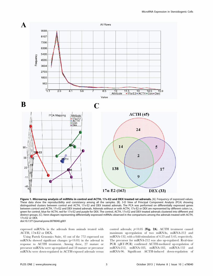
expressed miRNAs in the adrenals from animals treated with
ACTH, 17a-E2 or DEX.
Using Partek Genomics Suite, 45 out of the 772 expressed rat
miRNAs showed significant changes (p,0.05) in the adrenal in
response to ACTH treatment. Among these, 27 mature or
precursor miRNAs were up-regulated and 18 mature or precursor
miRNAs were down-regulated in ACTH-exposed adrenals versus
control adrenals; p,0.05 (Fig. 2A). ACTH treatment caused
maximum up-regulation of two miRNAs, miRNA-212 and
miRNA-132, with a fold-stimulation of 4.23 and 3.43, respectively.
The precursor for miRNA-212 was also up-regulated. Real-time
PCR (qRT-PCR) confirmed ACTH-mediated up-regulation of
miRNA-212, miRNA-183, miRNA-182, miRNA-132 and
miRNA-96. Significant ACTH-induced down-regulation of
Figure 1. Microarray analysis of miRNAs in control and ACTH, 17a-E2 and DEX treated rat adrenals. [A]. Frequency of expressed values.These data show the reproducibility and consistancy among all the samples. [B]. 3-D View of Principal Component Analysis (PCA) showingdistinguished clusters between control and ACTH, 17a-E2 and DEX treated adrenals. The PCA was performed on differentially expressed genesbetween control and ACTH, 17a-E2 and DEX treated adrenals. Adrenals without or with ACTH, 17a-E2 or DEX are represented by different colors i.e.,green for control, blue for ACTH, red for 17a-E2 and purple for DEX. The control, ACTH, 17a-E2 and DEX treated adrenals clustered into different anddistinct groups. [C]. Venn diagram representing differentially expressed miRNAs observed in the comparisons among the adrenals treated with ACTH,17a-E2 or DEX.doi:10.1371/journal.pone.0078040.g001
MicroRNA Expression in Steroidogenic Cells
PLOS ONE | www.plosone.org 5 October 2013 | Volume 8 | Issue 10 | e78040

miRNA-466b, miRNA-214, miRNA-503 and miRNA-27a was
also observed (Fig. 3). While this work was in progress, a recent
microarray study reported the expression profile of miRNAs in
mouse adrenals in response to acute treatment of animals with
ACTH and demonstrated no similarities with our observations
[41].
We also performed a microarray analysis to screen the
expression profiles of miRNAs in adrenals from rats chronically
treated with a hypocholesterolemic and possible ACTH secreta-
gogue, 17a-E2 [45–47]. The expression levels of 163 mature
miRNAs varied significantly (p,0.05) in response to 17a-E2
treatment (Fig. 2B, C), of which, 63 miRNAs exhibited changes
more-than 1.5 fold (p,0.05). The expression levels of miR-183
(4.61-fold), miR-96 (4.56-fold), and miR-182 (4.29-fold) were most
highly up-regulated, whereas miR-122 (9.79-fold), miR-503 (5.88-
fold), and miR-139-3p (1.94-fold) showed the greatest down-
regulation as a result of 17a-E2 treatment. qRT-PCR measure-
ments confirmed that the expression of miR-212, miRNA-183,
miRNA-182, miRNA-132, miRNA-370, miRNA-377 and
miRNA-96 was up-regulated and that of miRNA-122, miRNA-
200b, miRNA-466b, miRNA-138, miRNA-214, miRNA-503 and
miRNA-27a down-regulated in adrenals from 17a-E2 treated rats
(Fig. 3).
To identify a set of adrenal miRNAs that are potentially
regulated by a potent synthetic glucocorticoid agonist and an
inhibitor of corticosteroidogenesis, dexamethasone [45], three rats
were treated with a single dose of dexamethasone (100 mg) sc for a
24 h period. Again, changes in the adrenal miRNA profile on
dexamethasone treatment were assessed using microarray. Anal-
ysis of the miRNA array showed that expression of 33 miRNAs
significantly changed (p,0.05) following treatment with dexa-
methasone (Fig. 2D). The expression of miRNA-483, miRNA-
181a-1, miRNA-490 and miRNA and miRNA-181b-1 was up-
regulated in response to dexamethasone treatment. In contrast,
dexamethasone down-regulated the expression of several of the
miRNAs by more than 1.5 fold, i.e., miR-122 (8.2-fold), miR-466b
(2.31-fold), miR-200b (1.9-fold) miR-877 (1.61-fold), miR-296
(1.61-fold)and precursor of miR-504 (1.53-fold) (Fig. 2D). Ex-
pression of miRNA-27a (1.32-fold) was also down-regulated by
DEX. Using qRT-PCR, we confirmed the down-regulation of
miRNA-200b, miR-122, miR-19a, miRNA-466b, and miRNA-
27a expression (Fig. 3).
Multiple Hormonal Regulation of Adrenal miRNAsWe also examined whether the expression of any miRNAs is
altered by more than one hormone treatment, i.e., by ACTH/
17a-E2, ACTH/DEX, 17a-E2/DEX or ACTH/17a-E2/DEX.
The results are presented in Table 1. The level of expression of
miR-212 and miR-132 was up-regulated (.1.5-fold) by both
ACTH and 17a-E2 treatments. ACTH and DEX down-regulated
miR-466b more than 1.5-fold, but the effect of 17a-E2, although it
showed a similar trend, was not statistically significant (p = 0.084).
miR-296 and miR-122 were down-regulated (.1.5 fold) by both
17a-E2 and DEX. The levels of miR-27a and miR-551b were
Figure 2. MicroRNA (miRNA) expression profiles in adrenals from rats treated with ACTH, 17a-E2, DEX or saline (control). [A]. Theheat map represents the expression levels of 45 miRNAs in two conditions (control and ACTH). [B, C]. The heat map represents the expression levelsof 163 miRNAs in two conditions (control and 17a-E2). 74 miRNAs were up-regulated [B] and 89 miRNAs were down-regulated with 17a-E2 [C]. [D].The heat map represents the expression levels of 33 miRNAs in two conditions (control and DEX). Red, up-regulated genes; blue, down-regulatedgenes.doi:10.1371/journal.pone.0078040.g002
MicroRNA Expression in Steroidogenic Cells
PLOS ONE | www.plosone.org 6 October 2013 | Volume 8 | Issue 10 | e78040

down-regulated by all three hormones, ACTH, 17a-E2 and DEX.
Microarray data demonstrated that the levels of miR-183 and
miR-182 were up-regulated with 17a-E2 treatment, but not with
ACTH (p= 0.065) treatment; qRT-PCR measurements, however,
showed significant increases in their expression in response to
either ACTH or 17a-E2 treatment.
cAMP Induced Regulation of miRNA Expression inPrimary Rat Granulosa Cells and Mouse Leydig TumorMLTC-1 Cells
We next examined whether the expression of some of the
miRNAs that were found to be hormone-sensitive in the adrenal
were also regulated in granulosa and MLTC-1 cells treated with a
cAMP agonist, Bt2cAMP. More specifically, we assessed the
impact of Bt2cAMP treatment on the expression of miRNA-212,
miRNA-122, miRNA-27a, miRNA-466b, miRNA-200b, miRNA-
138, miRNA-214, miRNA-183, miRNA-182, miRNA-132,
miRNA-96 and miRNA-19a. qRT-PCR measurements indicated
that exposure of primary rat granulosa cells to Bt2cAMP for 24 h
inhibited the expression of miRNA-200b, miRNA-466b, miRNA-
27a, miRNA-214, and miRNA-138 and miRNA-19a while
enhancing the expression of miRNA-212, miRNA-183, miRNA-
182, and miRNA-132 (Fig. 4). Treatment of MLTC-1 cells with
Bt2cAMP for 6 h increased the expression of miRNA-212,
miRNA-183, miRNA-132, miRNA-182 and miRNA-96 and
inhibited the expression of miRNA-138 and miRNA-19a
(Fig. 4B).
Correlation of Expression Levels of Selected miRNAs withtheir Predicted Targeted Genes
Having identified miRNAs that are subject to hormonal
regulation, we next examined potential correlations between
selected hormone-sensitive miRNAs with their predicted target
genes. We first used microRNA.org and TargetScan 4.0 to predict
target genes for selected hormone-responsive miRNAs. Some
miRNAs down-regulate large numbers of target mRNAs through
interaction with 39 UTRs (Lim et al., 2005). The number of target
genes predicted by a single miRNA varied greatly, ranging from
several to hundreds. Using NCBI databases for functional
screening of the putative target genes, we further identified a
number of target genes directly involved in steroidogenesis
(Table 2). We also performed a reverse prediction strategy, based
on the sequence of the 39 UTRs of the gene of interest, to make a
prediction about the miRNAs which may target some critical
steroidogenic genes, such as CYP11A1, StAR, LDL-R and
NR5A1. CYP11A1, the gene encoding cholesterol side-chain
cleavage enzyme (P450scc), was predicted to be the target gene of
miRNA-134. StAR may be a target gene of miR-376b, miR-150,
miR-330 and miR-138. NR5A1 was predicted to be the target
gene of miR-342, while LDL-R was predicted to be the target gene
of miR-182 and miR-466b. MiR-183, miR-96 and miR-19a were
predicted to target the ABCA1 gene. ABCG1 may be a target gene
of miR-542.
In a follow-up study, we performed real-time PCR and Western
blot analysis to monitor expression of predicted target genes and
their protein products in response to hormone treatment of rat
adrenals and ovarian granulosa cells. Three genes, Mecp2, Ctbp1
and p250 GAP, have been recently identified as targets of miR-132
[36]. However, in our RT-PCR assay adrenal mRNA levels of
Mecp2, Ctbp1 and Rics were not impacted by ACTH, DEX or 17a-
E2 treatment. Likewise, expression of another predicted target
gene of miR-132, HDAC3, was also unchanged by ACTH, 17a-
E2 or DEX treatment (Fig. 5A).
As summarized in Table 2, several genes involved in lipid
metabolism and steroidogenesis were predicted to be the target
genes of different miRNAs. We examined the expression of some
of these predicted lipid/steroidogenic genes in the adrenals from
control and hormone treated rats. qRT-PCR data showed that
mRNA levels of SF-1, StAR, CYP11A1 and LDL-R were all up-
regulated in the rat adrenal gland in response to ACTH and 17a-
E2, but down-regulated with DEX treatment (Fig. 5A). Both
ACTH and 17a-E2 treatment of rats caused a significant
reduction in mRNA levels of LXRa, LXRß, and DAX-1.
Interestingly, mRNA levels of LXRa were up-regulated by DEX
treatment. Adrenal mRNA levels of ABCA1, ABCG1 and C/
EBPa were significantly reduced in 17a-E2 treated animals, while
ACTH treatment increased the mRNA expression of ABCA1.
Figure 3. Quantitative RT-PCR (qRT-PCR) validation of miRNA-212, miRNA-200b, miRNA-183, miRNA-122, miRNA-19a, miRNA-466b, miRNA-182, miRNA-132, miRNA-138, miRNA-370, miRNA-96, miRNA-503, miRNA-27a and miRNA-214 levels in control,ACTH-, 17a-E2 or DEX-treated adrenals in vivo. Expression of U6 was used for normalization. The experiments were performed independentlythree times. Data are presented as mean 6 standard error. *p,0.05; **p,0.01; ***p,0.001.doi:10.1371/journal.pone.0078040.g003
MicroRNA Expression in Steroidogenic Cells
PLOS ONE | www.plosone.org 7 October 2013 | Volume 8 | Issue 10 | e78040

Finally, the mRNA expression of SREBP-1c was significantly
attenuated in response to 17a-E2 or DEX treatment.
We also determined the mRNA levels of these genes in rat
granulosa cells treated with or without Bt2cAMP (Fig. 5B). In rat
granulosa cells, the mRNA levels of Rics, Ctbp1, HDAC3 and
MECP2 were not affected following treatment with Bt2cAMP. SF-
1, StAR, CYP11A1, LDL-R, LXRa, LXRß, ABCA1, ABCG1,
SREBP-1c, and C/EBPa mRNA levels were all up-regulated in
cAMP-treated rat granulosa cells, whereas mRNA levels of DAX-
1 transcription factor were significantly reduced. Using Western
blot, we also assessed the effects of hormonal treatment of rat
adrenal gland and Bt2cAMP treatment of rat granulosa cells on the
levels of StAR, SF-1, SREBP-1c, LDLR and HDAC3 proteins,
and the results are presented in Fig. 5C. Western blot analysis
showed no significant changes in the nuclear protein levels of
HDAC3 in ACTH, 17a-E2 or DAX treated adrenals. The
adrenal protein levels of StAR and SF-1 were increased
significantly following treatment of rats with ACTH or 17a-E2,
although DEX treatment showed no significant effect on the
protein levels of either of these two proteins. The protein levels of
lipogenic transcription factor, SREBP-1c, were decreased in the
17a-E2, as well as DEX, treated adrenals. In contrast and as
expected, a significant increase in LDL-R protein levels was noted
in the 17a-E2 treated adrenal samples. In ovarian granulosa cells,
protein levels of StAR, SF-1, SREBP-1c and LDL-R were all
increased following Bt2cAMP treatment. As before, HDAC3
levels, however, were not impacted by Bt2cAMP stimulation.
miRNA-132 and miRNA-214 Suppress SREBP-1c and LDLRby Targeting Specific Site(s) within the 39 UTR of SREBP-1c and LDLR, Respectively
The results presented above raised the possibility that some of
the hormone sensitive miRNAs might posttranscriptionally/
translationally alter the expression of certain proteins involved in
lipid metabolism. Here, we directly assessed the binding of
miRNA-138, miRNA-132 and miRNA-182/miRNA-214 to the
39UTR of StAR, SREBP-1c, and LDLR, respectively, and
regulation of their expression levels, by carrying out luciferase
reporter gene assays. Individual fragments of the 39 UTR region of
the StAR gene containing site I or site II binding site for miRNA-
138-5p, the 39-UTR of SREBP-1c containing a binding site for
miRNA-132-5p, the 39-UTR of LDLR containing a binding site
for miRNA-182-5p or the 39-UTR of LDLR containing site I, site
II, or site III binding site for miRNA-214-3p were inserted
downstream of the luciferase open reading frame of pMIR-
REPORT vector. CHO cells were co-transfected individually with
StAR 39-UTR (containing the putative site I or site II for miRNA-
138 binding) 6 pre-miRNA-138-5p (panel B), SREBP-1c 39-UTR
(containing the putative binding site for miRNA-132) 6 pre-
miRNA-132-3p, LDLR 39-UTR (containing the putative binding
site for miRNA-182), or LDLR 39-UTR (containing the putative
site I, site II or site III for miRNA-214 binding) 6 pre-miRNA-
214-3p for 36h, followed by determination of luciferase activities.
Overexpression of pre-miRNA-132 and pre-miRNA-214 signifi-
cantly decreased the luciferase activity of the 39UTR of the
SREBP-1c and LDLR reporter containing micRNA-132 and
miRNA-214 binding sites, respectively (Fig. 6). In contrast, no
inhibitory effect of pre-miRNA-138 on the StAR 39 UTR (with 2
putative binding sites) reporter construct and pre-miRNA-182 on
the LDLR 39UTR (with a single putative binding site) reporter
construct was detected.
Discussion
Steroid hormone synthesis occurs predominantly in the
steroidogenic cells of the adrenal gland, ovary and testis and is
under the control of trophic peptide hormones secreted from the
pituitary. The rate limiting step in steroidogenesis is the trophic
hormone2/cAMP-stimulated and StAR-mediated translocation
of cholesterol from the outer mitochondrial membrane to the
inner mitochondrial membrane where the side-chain cleavage
enzyme (P450scc; Cyp11A1) carries out the first committed step in
steroidogenesis, i.e., conversion of cholesterol to pregnenolone [1–
4]. This step is subject to both acute [3], [5–8] and chronic [2],
[3], [9–12] stimulation, and trophic hormones regulate this step
mainly at the level of gene transcription. Although limited
information is also available to suggest that posttranscriptional
and posttranslational events may be involved in the regulation of
steroidogenesis, relatively little information is available on the
Figure 4. Quantitative RT-PCR (qRT-PCR) analysis of miRNAs inmouse rat granulosa and MLTC-1 cells treated without or withBt2cAMP (2.5 mM) for 24 h or 6 h. [A] Granulosa cells: groups ofRNA samples were analyzed by qRT-PCR. The levels of expression ofmiRNA-212, miRNA-122, miRNA-138, miRNA-214, miRNA-183, miRNA-182, miRNA-132, miRNA-96, miRNA-466b, miRNA-200b, and miRNA-19aare shown. Expression of U6 was used for normalization. [B] MLTC-1cells: groups of RNA samples were analyzed by qRT-PCR. The levels ofexpression of miRNA-212, miRNA-122, miRNA-138, miRNA-214, miRNA-183, miRNA-182, miRNA-132, miRNA-96, miRNA-466b, miRNA-200b, andmiRNA-19a are shown. Expression of U6 was used for normalization.*p,0.05; **p,0.01; ***p,0.001.doi:10.1371/journal.pone.0078040.g004
MicroRNA Expression in Steroidogenic Cells
PLOS ONE | www.plosone.org 8 October 2013 | Volume 8 | Issue 10 | e78040

biological factors that possibly mediate these events. Emerging
evidence showing hormonal regulation of miRNAs in steroido-
genic cells [19], [36–42], coupled with the identification of a
diverse and large number of miRNAs [21–25], strongly suggest
that miRNAs may be involved in the posttranscriptional/
posttranslational regulation of steroidogenesis. In this study, we
first carried out a comprehensive analysis of miRNA profiling
using control and in vivo hormone treated rat adrenals to identify
miRNAs whose expression is altered in response to ACTH, 17a-
ethinyl estradiol (17a-E2) or dexamethosone (DEX) treatment.
Taking cues from the adrenal data, we also examined the effects of
Bt2cAMP (the second messenger of trophic hormone action)
stimulation of rat ovarian granulosa cells and mouse testicular
Leydig tumor cells, MLTC-1, on the expression of some of the
relevant miRNAs.
Chronic ACTH treatment in vivo significantly altered the levels
of many miRNAs in rat adrenal glands. In general, more miRNAs
were upregulated than downregulated in response to ACTH
treatment. Real-time PCR (qRT-PCR) measurements demon-
strated that ACTH treatment upregulated the expression of
miRNA-212, miRNA-183, miRNA-182, miRNA-132 and
miRNA-96, while down-regulating the expression of miRNA-
466b, miRNA-214, miRNA-503 and miRNA-27a. However, the
levels of expression of these miRNAs differed considerably when
Table 2. miRNAs and gene targets.
miRNAChromosomallocation (rat) Regulation Predicted Target Genes
miR-212/2132 10 AU, EU SOX5, FOXA1, FOXO3,CREB5, ABCG4, RICS, SREBP-1c, GPAT2, MECP2
miR-154 6 AU, EU DOCK1, MECP2, WNT5A
miR-183 4 AU, EU, DU FOXO1, ABCA1, NR3C1
miR-182 4 AU, EU FOXO3, IGF1R,, ABCD1, CREB1, SOX6, CEBPA, LDL-R, SIK1, PIK3R1, FOXO1
miR-207 5 AD CHREBP, LEPTIN
miR-218 14 AD SNF1LK2, PIK3R1, ABCG4, SOX5, MECP2
miR-450a X AD CREB1, IGF1
miR-96 4 EU SOX5, ABCD1, FOXO1, FOXQ1, VLDLR, IGF1R, ABCA1, FOXO4, SNF1LK, ABCA2,FOXO3, PIK3R1, MECP2
miR-494 6 EU, AU PTEN, ROCK1, IGF1R
miR-376b 6 EU, DD CTBP2, IGF1R, STAR
miR-377 6 EU NR6A1, SMAD4, WNT5A
miR-32 5 EU PTEN, GATA2, NR4A3, SNF1LK, ABCG4
miR-150 1 ED LXRB, STAR, ACBD3
miR-504 X ED, AD CYP11B1, HSD17B7, HSD17B8
miR-370 6 EU FOXO3
miR-19a 15 EU IGF2R, MECP2, PPARA, PIK3R3, FOXP2, PTEN, ABCA1, FOXP1, WNT1, SMAD4, SOX5,IGF1R
miR-138 8 AD, ED PPARD, PPARGC1A, SNAP25, STAR
miR-125b-2 11 ED, AD CYP24A1
miR-503 X AD, ED CYP26B1, SNF1LK, IGF1R, WNT3A, SOX5, PAPPA, WNT4
miR-122 18 ED, DD GATA4, FOXO3
miR-99b 1 ED IGF1R, CYP26B1
miR-466b 17 AD, DD IGF1R, LDLR, SREBP1
miR-483 1 DU IGF1, SMAD4
miR-200b 5 DD NR5A2, CREB5, SNAP25, GATA4, SNF1LK2, CYP1B1, SOX2, PPARA, SOX1, PTEN,MECP2, FOXO3
miR-881 X DD PDK2
miR-342 6 ED SOX6, NR5A1
miR-330 1 AD MECP2, STAR
miR-27a 19 AD, ED, DD SNAP25, PPARG, NR5A2, SMAD5, CREB1, ABCA1, CYP39A1, GATA2, PDK1, WNT3A,FOXO1, CYP1B1, PPARA, GSK3B, IGF1
miR-542 X ED CYP17A1, LXRB, HSD17B11, ABCG1, ABCA2, IGF2
miR-350 13 EU GATA3, PIK3R3, CYP26B1, SNAP25, DAX1, FOXA2, CTBP1, IGF1R, HOXA1, CREB5,SOX5, FOXO3
miR-134 6 EU CYP11A1
miR-214 13 AD, ED VLDLR, WNT3, PPARGC1a, FOXO4, LDLR, CREB1, SCARB1, UCP2
miR-1 18 AU, EU, DU IGF1, CREB5, SNAP25, NR4A2, SMAD4
AU, ACTH up-regulated; EU, 17a-E2 up-regulated; DU, DEX up-regulated; AD, ACTH down-regulated; ED, 17a-E2 down-regulated; DD, DEX down-regulated.doi:10.1371/journal.pone.0078040.t002
MicroRNA Expression in Steroidogenic Cells
PLOS ONE | www.plosone.org 9 October 2013 | Volume 8 | Issue 10 | e78040
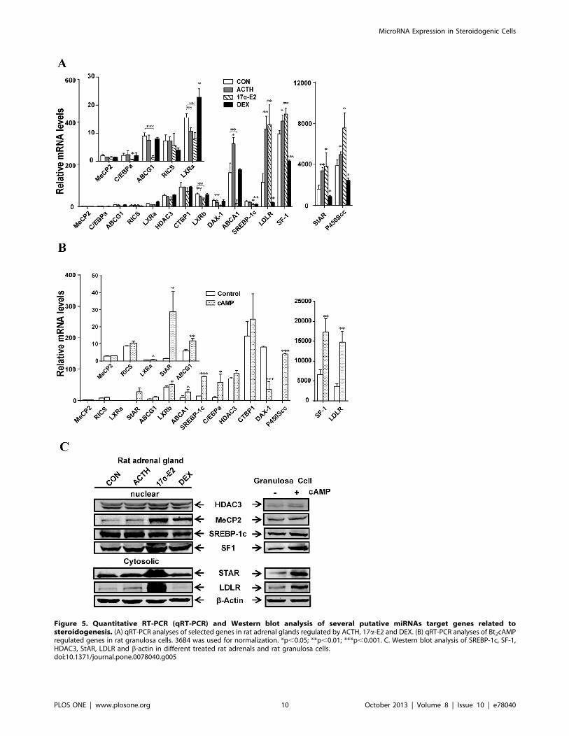
Figure 5. Quantitative RT-PCR (qRT-PCR) and Western blot analysis of several putative miRNAs target genes related tosteroidogenesis. (A) qRT-PCR analyses of selected genes in rat adrenal glands regulated by ACTH, 17a-E2 and DEX. (B) qRT-PCR analyses of Bt2cAMPregulated genes in rat granulosa cells. 36B4 was used for normalization. *p,0.05; **p,0.01; ***p,0.001. C. Western blot analysis of SREBP-1c, SF-1,HDAC3, StAR, LDLR and b-actin in different treated rat adrenals and rat granulosa cells.doi:10.1371/journal.pone.0078040.g005
MicroRNA Expression in Steroidogenic Cells
PLOS ONE | www.plosone.org 10 October 2013 | Volume 8 | Issue 10 | e78040

Figure 6. miRNA-132 and miRNA-214 binding sites in the 39 UTR of the mouse SREBP-1c and LDLR genes mediate thedownregulation of SREBP-1c and LDLR expression by miRNA-132 and miRNA-214, respectively. [A]. Seed sequences of the putativemiRNA-138-5p, miRNA-132-3p and miRNA-182-5p/miRNA-214-3p binding sites in the 39-UTR of mouse StAR, SREBP-1c and LDLR genes, respectively.For the reporter gene assay, the 39 UTR region of the StAR gene containing site I or site II binding site for miRNA-138-5p, the 39-UTR of SREBP-1ccontaining a binding site for miRNA-132-5p, the 39-UTR of LDLR containing a binding site for miRNA-182-5p or the 39-UTR of LDLR containing site I,site II, or site III binding site for miRNA-214-3p was inserted downstream of the luciferase open reading frame of pMIR-REPORT vector. CHO cells were
MicroRNA Expression in Steroidogenic Cells
PLOS ONE | www.plosone.org 11 October 2013 | Volume 8 | Issue 10 | e78040

measured by real-time-PCR as compared to their expression
values detected by microarray analysis. This result is most likely
due to the detection of both precursor and mature forms of
miRNAs by microarray, and only the mature form by PCR [54].
While our work was in progress, a microarray study reported the
expression profile of mouse adrenal miRNAs under basal
conditions (0 time) and in response to acute treatment of mice
(10, 30 or 60 min) with ACTH [41]. In that study, 16 miRNAs
were identified, whose levels of expression were maximally up-
regulated following 10 min treatment of mice with ACTH (range:
1.1180–1.8437), whereas expression of one miRNA, mmu-
mRNA-433, was down-regulated (–1.1465). Those miRNAs
differentially expressed on the microarrays with greatest fold
changes, miRNA-101a, miRNA-142-3p, miRNA-433 and
miRNA-96, were further analyzed. Both microarray and qRT-
PCR data measurements indicated that the expression of these
four miRNAs varied considerably with respect to ACTH
treatment and time after treatment [41]. Moreover, significant
differences were also noted between microarray and qRT-PCR
measurements. Interestingly, a comparison of our gene array list to
the list presented in this publication [41] indicates that none of the
transcripts overlap. The reasons for this observed disparity are not
clear, but may stem from many factors, including the use of two
different types of rodent adrenals (mouse vs rat) and two different
ACTH treatment regimens (chronic vs acute ACTH treatment).
In addition to ACTH, we also performed a microarray analysis
to screen the expression profiles of adrenal miRNAs from rats
chronically treated with 17a-E2, a hypocholesterolemic and
possible ACTH secretagogue [46–48]. 17a-E2 treatment, like
ACTH treatment, results in the induction of both adrenal LDL-R
(the current study) and SR-BI [46]. To our knowledge, this is a
first report describing the effects of 17a-E2 on the expression of
adrenal miRNAs. Significant differences in expression of 163
miRNAs were observed between the adrenals from 17a-E2-
treated rats and control rats, with 63 miRNAs showing a change
greater than 1.5-fold. The expression levels of miR-183, miR-96,
and miR-182 were most highly up-regulated, whereas miR-122,
miR-503, and miR-139-3p exhibited the greatest down-regulation
as a result of 17a-E2 treatment. Real-time quantitative PCR
measurements confirmed that the expression of miR-212, miRNA-
183, miRNA-182, miRNA-132, miRNA-370, miRNA-377 and
miRNA-96 was up-regulated and that of miRNA-122, miRNA-
200b, miRNA-466b, miRNA-138, miRNA-214, miRNA-503 and
miRNA-27a down-regulated in adrenals from 17a-E2 treated rats.
Again, as noted above for ACTH treatment, the expression levels
of miRNAs differed significantly between measurements made by
microarray analysis and qRT-PCR. Furthermore, a comparison of
ACTH data with that of 17a-E2 data demonstrated that only
,25% of the transcripts overlap. This suggests that 17a-E2-
induced hypocholesterolemia or direct estrogen effects on the
adrenal, but not increased ACTH secretion, is most likely
responsible for the observed alterations in the levels of specific
miRNAs in adrenals of 17a-E2-treated rats.
The hypothalamus-pituitary-adrenal (HPA) axis consists of a set
of direct influences and feedback responses between the hypo-
thalamus, the pituitary gland and the adrenal that control
reactions to stress and glucocorticoid secretion. Glucocorticoid
(cortisol in humans and corticosterone in rodents) secretion by the
adrenal cortex inhibits the functions of both the hypothalamus and
the pituitary gland by a negative feedback mechanism. This
reduces the secretion of CRH and vasopressin and directly reduces
the cleavage of pro-opiomelanocortin (POMC) into ACTH and ß-
endorphin. In our study, we examined the impact of a synthetic
glucocorticoid, dexamethasone (DEX)-mediated inhibition of the
HPA axis and ACTH secretion, on miRNA expression profiles in
the adrenals [53]. DEX treatment up-regulated the expression of
miRNA-483, miRNA-181a-1, miRNA-490 and miRNA-181b-1,
while it down-regulated the levels of miR-122, miR-466b, miR-
200b, miR-877, miR-296, miRNA-27a and precursor of miR-504.
Furthermore, such DEX alteration of adrenal miRNA levels
demonstrates that DEX suppression of endogenous ACTH
secretion modulates a set of adrenal miRNAs, with the exception
of miRNA-96, miRNA-466, and miRNA-27a, that are distinct
from those modulated by treatment with exogenous ACTH.
Interestingly, the expression of miRNA-96 is up-regulated in
response to ACTH treatment, but is down-regulated following
DEX treatment. Considering the current view that miRNAs act as
negative regulators of gene expression, their altered expression in
response to DEX may enhance and/or reduce the expression of
target steroidogenic genes, leading to possibly down-regulation of
adrenal steroid hormone synthesis and secretion.
Our data further demonstrate that expression levels of some
miRNAs are regulated by more than one hormone, i.e., by
ACTH/17a-E2, ACTH/DEX, 17a-E2/DEX or ACTH/17a-
E2/DEX; Table 1. The most striking similarity was observed
between ACTH and 17a-E2. Both ACTH and 17a-E2 up-
regulated the expression of miRNA-212, miRNA-132, miRNA-
154, miRNA-494, miRNA-872, miRNA-194, and miRNA-24-1,
but reduced the expression of miRNA-322, miRNA-20b, miRNA-
339, miRNA-27a, miRNA-551b, and miRNA-1224. We also
observed that miRNA-30a was up-regulated in adrenals treated
with ACTH, but down-regulated by 17a-E2 exposure. A
comparison of effects of ACTH and DEX shows that both
hormones increased the expression miRNA-181b, miRNA-672,
and miRNA-100, and significantly decreased the levels of miRNA-
92a, and miRNA-466b. In addition to ACTH/17a-E2 and
ACTH/DEX, we observed that a total of 11 miRNAs are
regulated by both 17a-E2 and DEX. Among these, three mRNAs
were up-regulated in response to in vivo treatment of adrenals with
17a-E2 or DEX, and the remaining eight miRNAs were down-
regulated in treated adrenals with either of the two hormones.
Finally the expression levels of miRNA-27a and miRNA-551b
were significantly reduced in adrenals of ACTH, 17a-E2 or DEX
treated animals. Together, these data raise the possibility that
some of these miRNAs (with sensitivity towards two or three
hormones) may be intimately involved in the complex regulation
of adrenal steroidogenesis.
We next evaluated the effects of Bt2cAMP stimulation of rat
ovarian granulosa cells and of mouse MLTC-1 Leydig tumor cells
on the expression of twelve miRNAs (miRNA-212, miRNA-122,
miRNA-183, miRNA-200b, miRNA-466b, miRNA-182, miRNA-
96, miRNA-27a, miRNA-132, miRNA-214, miRNA-138 and
miRNA-19a) whose adrenal expression was differentially altered in
response to treatment of rats with ACTH, 17a-E2 or DEX. qRT-
co-transfected individually with the StAR 39-UTR (containing putative site I or site II for miRNA-138 binding) 6 pre-miRNA-138-5p (panel B), theSREBP-1c 39-UTR (containing putative binding site for miRNA-132) 6 pre-miRNA-132-3p (panel C), the LDLR 39-UTR (containing putative binding sitefor miRNA-182) (panel D), or the LDLR 39-UTR (containing putative site I, site II or site III for miRNA-214 binding) 6 pre-miRNA-214-3p for 36 h (panelE). Reporter gene assays were performed using a dual-luciferase kit as described in Materials and Methods. The results are expressed as relativeluciferase activities (firefly luciferase/Renilla luciferase).doi:10.1371/journal.pone.0078040.g006
MicroRNA Expression in Steroidogenic Cells
PLOS ONE | www.plosone.org 12 October 2013 | Volume 8 | Issue 10 | e78040

PCR measurements indicated that in granulosa cells, miRNA-138
and miRNA-19a are expressed at very high levels as compared to
other miRNAs. Significant expression was also observed for
miRNA-27a, miRNA-132 and miRNA-214, whereas very low
expression was noted for all of the remaining (seven) miRNAs.
Bt2cAMP stimulation of granulosa cells caused down-regulation of
a majority of miRNAs, including miRNA-200b, miRNA-466b,
miRNA-27a, miRNA-214, miRNA-138 and miRNA-19a, but
expression levels of miRNA-212, miRNA-183, miRNA-182, and
miRNA-132 were significantly increased. The expression levels of
miRNA-122 and miRNA-96, however, were not affected by
cAMP stimulation. A few earlier studies have examined the
expression of miRNAs, although these studies were mainly focused
on identifying miRNAs in whole ovaries or follicular/luteal tissues
from various mammalian species, including humans [55], mice
[56–58], pigs [59], cattle [60–63] and sheep [64] using cloning-
based or next generation sequencing strategies [for review see 65–
67]. Some studies also identified and characterized miRNAs that
are expressed in specific ovarian compartments, including
follicular mouse [36], [68] and horse [69] granulosa cells, cow
cumulus-oocyte complexes [70], equine follicular fluid [69] and
bovine corpora lutea [71]. In addition, other studies reported
differences in miRNA expression between different ovarian or
follicular compartments. For example, miRNA-503, miRNA-224
and miRNA-383 are expressed almost exclusively in mouse
granulosa cells and oocytes [68], [72], whereas a large number
of miRNAs are differentially expressed in bovine ovarian cortex,
cumulus cells and corpus luteum [60]. Furthermore, a correlation
was recently reported between miRNA levels of horse follicular
fluid and granulosa cells [69]. Despite these various findings, very
little information is currently available about the hormonal
regulation of miRNAs in the ovary. One study reported a robust
induction of miRNA-21, miRNA-132 and miRNA-212 following
in vivo stimulation of mouse ovaries with LH/hCG [36]. More-
over, cultured mouse granulosa cells exhibited a robust induction
of miRNA-132 and miRNA-212 when challenged with 8BrcAMP
[36]. Another in vitro study reported up-regulation of 17 miRNAs
and down-regulation of 14 miRNAs following 12 h exposure of
mouse granulosa cells to FSH [73]. Our studies, while confirming
some of these findings, have identified several additional miRNAs
whose expression is up- or down-regulated in response to second
messenger (cAMP) treatment of rat granulosa cells.
We also examined the cAMP regulation of miRNA expression
in MLTC-1 cells, a model cell line of Leydig cells. Treatment of
MLTC-1 cells with Bt2cAMP for 6 h increased the expression of
miRNA-212, miRNA-183, miRNA-132, miRNA-182 and
miRNA-96, and inhibited the expression of miRNA-138 and
miRNA-19a. To our knowledge this is the first report showing
hormone-induced changes in the levels of the above mentioned
miRNAs in Leydig cells. Follow-up studies are in progress to more
critically examine their potential role in the regulation of
testosterone production by Leydig cells. Furthermore, as summa-
rized in Table 2, several genes involved in lipid metabolism and
steroidogenesis, whose expression levels are altered by hormones
in the adrenal, ovarian granulosa cells and testicular Leydig cell
line (MLTC-1), are predicted to be target genes of miRNAs.
Ongoing studies are also evaluating the actions of selected
hormone responsive miRNAs on potential target genes and
secondarily on steroidogenesis. In this context, we have evaluated
the effects of miRNA-138, miRNA-132 and miRNA-132/
miRNA-214 on the expression of StAR, SREBP-1c and LDLR,
respectively, by carrying out 39UTR luciferase assays. Overex-
pression of pre-miRNA-132 and pre-miRNA-214 significantly
decreased the luciferase activity of the 39UTR of the SREBP-1c
and LDLR reporter containing micRNA-132 and miRNA-214
binding sites, respectively. In contrast, no inhibitory effect of pre-
miRNA-138 on the StAR 39 UTR (with 2 putative binding sites)
reporter construct and pre-miRNA-182 on the LDLR 39UTR
(with a single putative binding site) reporter construct was
detected.
In conclusion, the current study provides the first comprehen-
sive analysis of hormonal regulation of miRNAs in steroidogenic
cells of the adrenal, ovary and testis. The results defined the
miRNA expression profiles in rat adrenals in response to treatment
with three different hormones (ACTH, 17a-E2 and DEX), and
identified several miRNAs that are subject to hormonal regulation
in ovarian granulosa cells and testicular Leydig cells. Understand-
ing their actions on potential target genes involved in lipid
metabolism should aid greatly in defining the post-transcriptional/
post-translational mechanisms by which specific miRNAs may
contribute to the regulation of steroidogenesis.
Author Contributions
Conceived and designed the experiments: ZH WJS FBK SA. Performed
the experiments: ZH YC. Analyzed the data: ZH XT LFL. Wrote the
paper: ZH SA. Edited the manuscript: ZH WJS FBK SA.
References
1. Payne AH, Hales DB (2004) Overview of steroidogenenic enzymes in the
pathway from cholesterol to active steroid hormones. Endocr Rev 25: 709–714.
2. LaVoie H, King SR (2009) Transcriptional regulation of steroidogenic genes:
STARD1, CYP11A1 and HSD3B. Exp Biol Med (Maywood) 234: 880–907.
3. Miller WL, Bose HS (2011) Early steps in steroidogenesis: intracellular
cholesterol trafficking. J Lipid Res 52: 2111–2135.
4. Hu J, Zhang Z, Shen WJ, Azhar S (2010) Cellular cholesterol delivery,
intracellular processing and utilization for biosynthesis of steroid hormones. Nutr
Metab (Lond) 7: 47.
5. Miller WL (2008) Steroidogenic enzymes. Endocr Dev 13: 1–18.
6. Pon LA, Hartigan JA, Orme-Johnson NR (1986) Acute ACTH regulation of
adrenal corticosteroid biosynthesis. Rapid accumulation of a phosphoprotein.
J Biol Chem 261: 13309–13316.
7. Pon LA, Orme-Johnson NR (1988) Acute stimulation of corpus luteum cells by
gonadotrophin or adenosine 39,59-monophosphate causes accumulation of
phosphoprotein concurrent with acceleration of steroid synthesis. Endocrinology
123: 1942–1948.
8. Epstein LF, Orme-Johnson NR (1991) Acute action of luteinizing hormone on
mouse Leydig cells: accumulation of mitochondrial phosphoproteins and
stimulation of testosterone synthesis. Mol Cell Endocrinol 81: 113–126.
9. Stocco DM, Clark BJ (1996) Regulation of the acute production of steroids in
steroidogenic cells. Endocr Rev 17: 221–244.
10. Miller WL (1988) Molecular biology of steroid hormone synthesis. Endocr Rev
9: 295–318.
11. Simpson ER, Waterman MR (1988) Regulation of the synthesis of steroidogenicenzymes in adrenal cortical cells by ACTH. Annu Rev Physiol 50: 427–440.
12. Payne AH, Youngblood GL, Sha L, Burgos-Trinidad M, Hammond SH (1992)
Hormonal regulation of steroidogenic enzymes gene expression in Leydig cells.J Steroid Biochem Mol Biol 43: 895–906.
13. Simpson E, Lauber M, Means G, Mahendroo M, Kilgore M, et al. (1992)
Regulation of expression of the genes encoding steroidogenic enzymes in theovary. J Steroid Biochem Mol Biol 41: 409–413.
14. Stocco DM (2001) StAR protein and the regulation of steroid hormonebiosynthesis. Annu Rev Physiol 63: 193–213.
15. Manna PR, Dyson MT, Stocco DM (2009) Regulation of the steroidogenic
acute regulatory protein gene expression: present and future perspective. MolHum Reprod 15: 321–333.
16. Rone MB, Fan J, Papadopoulos V (2009) Cholesterol transport in steroid
biosynthesis: Role of protein-protein interactions and implications in diseasestates. Biochim Biophys Acta 1791: 646–658.
17. Manna PR, Stocco DM (2011) The role of specific mitogen-activated protein
kinase signaling cascades in the regulation of steroidogenesis. J Signal Transduct2011: 821615.
18. Kraemer FB, Shen W-J (2002) Hormone-sensitive lipase: control of intracellular
tri-(di-)acylglycerol and cholesteryl ester hydrolysis. J Lipid Res 43: 1585–1594.
MicroRNA Expression in Steroidogenic Cells
PLOS ONE | www.plosone.org 13 October 2013 | Volume 8 | Issue 10 | e78040

19. Hu Z, Shen WJ, Kraemer FB, Azhar S (2012) MicroRNAs 125a and 455 repress
lipoprotein-supported steroidogenesis by targeting scavenger receptor class Btype I in steroidogenic cells. Mol Cell Biol 32: 5035–5045.
20. Hu Z, Hu J, Zhang Z, Shen WJ, Yun CC, et al. (2013) Regulation of expression
and function of scavenger receptor class B, type I (SR-BI) by Na+/H+ exchangeregulatory factors (NHERFs). J Biol Chem 288: 11416–11435.
21. Ambros VN (2004) The function of animal microRNAs. Nature 431: 350–355.
22. Bartlet DP (2004) MicroRNAs: genomics, biogenesis, mechanism, and function.
Cell 116: 281–207.
23. Kevin VN (2005) MicroRNA biogenesis: coordinated cropping, and dicing. NatRev Mol Cell Biol 6: 376–385.
24. Bartel DP (2009) MicroRNAs: target recognition and regulatory functions. Cell136: 215–233.
25. Fabian MR, Sonenberg N, Filipowicz W (2010) Regulation of mRNA
translation and stability by microRNAs. Annu Rev Biochem 79: 351–379.
26. Siomi H, Siomi MG (2010) Posttranscriptional regulation of microRNA
biogenesis in animals. Mol Cell 38: 323–332.
27. Finnegan EF, Pasquinelli AE (2013) MicroRNA biogenesis: regulating the
regulators. Crit Rev Biochem Mol Biol 48: 51–68.
28. Bushati N, Cohen SM (2007) microRNA functions. Annu Rev Cell Dev Biol 23:175–205.
29. Eulalio A, Huntzinger E, Izaurralde E (2008) Getting to the root of miRNA-mediated gene silencing. Cell 132: 9–14.
30. Filipowicz W, Bhattacharyya SN, Sonenberg N (2008) Mechanisms of post-
transcriptional regulation by microRNAs: are the answers in sight? Nat RevGenet 9: 102–114.
31. Ghildiya M, Zamore PD (2009) Small silencing RNAs: an expanding universe.Nat Rev Genet 10: 94–108.
32. Krishan K, Steptoe AL, Martin HC, Wani S, Nones K, et al. (2013) MicroRNA-182–5p targets a network of genes involved in DNA repair. RNA 19: 230–242.
33. Venkataraman S, Birks DK, Balakrishnan I, Alimova I, Harris PS, et al. (2013)
MicroRNA 218 acts as a tumor suppressor by targeting multiple cancerphenotype-associated genes in medulloblastoma. J Biol Chem 288: 1918–1928.
34. Gillen AE, Gosalia N, Leir SH, Harris A (2011) MicroRNA regulation ofexpression of the cystic fibrosis transmembrane conductance regulator gene.
Biochem J 438: 25–32.
35. Rottiers V, Naar AM (2012) MicroRNAs in metabolism and metabolicdisorders. Nat Rev Mol Cell Biol 13: 239–250.
36. Fiedler SD, Carletti MZ, Hong X, Christenson LK (2008) Hormonal regulationof microRNA expression in periovulatory mouse mural granulosa cells. Biol
Reprod 79: 1030–1037.
37. Sirotkin AV, Ovcharenko D, Grossmann R, Laukoa, Mlyncek M (2009)Identification of microRNAs controlling human ovarian cell steroidogenesis via
a genome-scale screen. J Cell Physiol 219: 415–420.
38. Carletti MZ, Fiedler SD, Christenson (2010) MicroRNA 21 blocks apoptosis in
mouse periovulatory granulose cells. Biol Reprod 83: 286–295.
39. Yao N, Yang B-Q, Liu Y, Tan X-Y, Lu C-L, et al. (2010) Follicle-stimulatinghormone regulation of microRNA expression on progesterone production in
cultured rat granulosa cells. Endocrine 38: 158–166.
40. Lin F, Li R, Pan ZX, Zhou B, Yu DB, et al. (2012) MiR-26b promotes granulosa
cell apoptosis by targeting ATM during follicular atresia in porcine ovary. PLoS
ONE 7(6): e38640. Doi:10.1371/journal.pone.0038640.
41. Riester A, Issler O, Spyroglou A, Rodrig SH, Chen A, et al. (2012) ACTH-
dependent regulation microRNA as endogenous modulators of glucocorticoidreceptor expression in the adrenal gland. Endocrinology 153: 212–222.
42. Zhang Q, Sun H, Jiang Y, Ding L, Wu S, et al. (2013) MicroRNA-181asuppresses mouse granulose cell proliferation by targeting activin receptor IIA.
PLoS ONE 8(3): e59667.doi:10.1371/journal.pone.0059667.
43. Reaven E, Tsai L, Azhar S (1996) Intracellular events in the ‘‘selective’’transport of lipoprotein-derived cholesteryl esters. J Biol Chem 271: 16208–
16217.
44. Lehoux J-G, Fleury A, Ducharme L (1998) The acute and chronic effects of
adrenocorticotropin on the levels of messenger ribonucleic acid and protein of
steroidogenic enzymes in rat adrenal in vivo. Endocrinology 139: 3913–3922.
45. Kovanen PT, Goldstein JL, Chappell DA, Brown MS (1980) Regulation of low
density lipoprotein receptors by adrenocorticotropin in the adrenal gland of miceand rats in vivo. J Biol Chem 255: 5591–5598.
46. Azhar S, Nomoto A, Reaven E (2002) Hormonal regulation of adrenal
microvillar channel formation. J Lipid Res 43: 861–871.
47. Kovanen PT, Brown MS, Goldstein JL (1979) Increased binding of low density
lipoprotein liver membranes from rats treated 17a-ethinyl estradiol. J Biol Chem254: 11367–11373.
48. Verschoor-Klootwyk AH, Verschoor L, Azhar S, Reaven GM (1982) Role of
exogenous cholesterol in regulation of adrenal steroidogenesis. J Biol Chem 257:7666–7671.
49. Livak Kj, Schittgen TD (2001) Analysis of relative gene expression data using
real-time quantitative PCR and the 22DDCt method. Methods 25: 402–408.50. Bartels CL, Tsongalis GJ (2009) MicroRNAs: Novel Biomarkers for Human
Cancer. Clin Chem 55: 623–31.51. Friedman RC, Farh KK, Burge CB, Bartel DP (2009) Most mammalian
mRNAs are conserved targets of microRNAs. Genome Res 19: 92–105.
52. Garcia DM, Baek D, Shin C, Bell GW, Grimson A, et al. (2011) Weak seed-pairing stability and high target-site abundance decrease the proficiency of lsy-6
and other microRNAs. Nat Struct Mol Biol 18: 1139–46.53. Keller-Wood ME, Dallman MF (1984) Corticosteroid inhibition of ACTH
secretion. Endocr Rev 5: 1–24.54. Pan Q, Luo X, Chegani N (2008) Differential expression of microRNAs in
myometrium and leiomyomas and regulation by ovarian steroids. J Cell Mol
Med 12: 227–240.55. Landgraf P, Rusu M, Sheridan R, Sewer A, Iovino N, et al. (2007) A
mammalian microRNA expression atlas based on small RNA librarysequencing. Cell 129: 1401–1414.
56. Rao S, Song R, Park C, Zheng H, Sanders KM, et al. (2007) Cloning and
expression profiling of small RNAs expressed in the mouse ovary. RNA 13:2366–2380.
57. Mishima T, Takizawa T, Luo S-S, Ishibashi O, Kawahigashi Y, et al. (2008)MiRNA (miRNA) cloning analysis reveals sex differences in miRNA profiles
between adult mouse testis and ovary. Reproduction 136; 811–822.58. Ahn HW, Morin RD, Zhao H, Harris RA, Coarfa C, et al. (2010) MicroRNA
transcriptome in the newborn mouse ovaries determined by massive parallel
sequencing. Mol Hum Reprod 16: 463–471.59. Li M, Liu Y, Wang T, Guan J, Luo Z, et al. (2011) Repertoire of porcine
microRNAs in adult ovary and testis by deep sequencing. Int J Biol Sci 7: 1045–1055.
60. Hossain MM, Ghanem M, Hoelker M, Rings F, Phatsara C, et al. (2009)
Identification and characterization of miRNAs expressed in the bovine ovary.BMC Genomics 10: 443.
61. Tripurani SK, Xiao C, Salem M, Yao J (2010) Cloning and analysis of fetalovary microRNAs in cattle. Anim Reprod Sci 120: 16–22.
62. Huang J, Ju Z, Li Q, Hou Q, Wang C, et al. (2011) Solexa sequencing of noveland differentially expressed mincroRNAs in testicular and ovarian tissues in
Holstein cattle. Int J Biol Sci 7: 1016–1026.
63. Miles JR, McDaneld TG, Wiedmann RT, Cushman RA, Echternkamp SE, etal. (2012) MicroRNA expression profile in bovine cumulus-oocyte complexes;
possible role of let 7 and miR-10a in the development of bovine oocytes. AnimalReprod Sci 130: 16–26.
64. McBride D, Carre W, Sontakke S, Hogg CO, Law AS, et al. (2012)
Identification of miRNAs associated with the follicular-luteal transition in theruminant ovary. Reproduction 144: 221–233.
65. Christenson LK (2010) MicroRNA control of ovarian function. Anim Reprod 7:129–133.
66. Donadeu FX, Schauer SN, Sontakke SD (2012) Involvement of miRNAs inovarian follicular and luteal development. J Endocrinol 215: 323–334.
67. Hossain MM, Sohel MMH, Schellander K, Tesfaye D (2012) Characterization
and importance of microRNAs in mammalian gonadal function. Cell Tissue Res349: 679–690.
68. Yao G, Yin M, Lian J, Tian H, Liu L, et al (2010) MicroRNA-224 is involved intransforming growth factor-beta-mediated mouse granulosa cell proliferation
and granulosa cell function by targeting Smad4. Mol Endocrinol 24: 540–551.
69. da Silveria JC, Veeramachaneni DNR, Winger QA, Carnevale EM, Bouma GJ(2012) Cell-secreted vesicles in equine ovarian follicular fluid contain miRNAs
and proteins: a possible new form of cell communication within the ovarianfollicle. Biol Reprod 86: 71, 1–10.
70. Tesfaye D, Worku D, Rings F, Phatsara C, Tholen E, et al. (2009) Identification
and expression profiling of microRNAs during bovine oocyte maturation usingheterologous approach. Mol Reprod Dev 76: 665–677.
71. Ma T, Jiang H, Gao Y, Zhao Y, Dai L, et al. (2011) Microarray analysis ofdifferentially expressed microRNAs in non-regressed and regressed bovine
corpus luteum tissue; microRNA-378 may suppress luteal cell apoptosis bytargeting the interferon gamma receptor 1 gene. J Appl Genet 52: 481–486.
72. Lei L, Jin S, Gonzalez G, Behringer RR, Woodruff TK (2010) The regulatory
role Dicer in folliculogenesis in mice. Mol Cell Endocrinol 315: 63–73.73. Yao N, Yang B-Q, Lin Y, Tan X-Y, Lu C-L, et al. (2010) Follicle-stimulating
hormone regulation of microRNA expression on progesterone production incultured rat granulose cells. Endocrine 38: 158–166.
MicroRNA Expression in Steroidogenic Cells
PLOS ONE | www.plosone.org 14 October 2013 | Volume 8 | Issue 10 | e78040