Embed Size (px)
Citation preview
Proc, Natl. Acad. Sci. USAVol. 84, pp. 388-392, January 1987Biochemistry
Homology probing: Identification of cDNA clones encodingmembers of the protein-serine kinase family
(oligonucleotide probes/HeLa celis/phosphorylase kinase/CDC2 and CDC28 genes)
STEVEN K. HANKSMolecular Biology Laboratory, Salk Institute for Biological Studies, P.O. Box 85800, San Diego, CA 92138
Communicated by Robert W. Holley, October 3, 1986 (received for review August 25, 1986)
ABSTRACT Mixed oligonucleotide probes were used toscreen a HeLa cDNA library for clones encoding amino acidcontiguities whose conservation is characteristic of the protein-serine kinase family. Eighty thousand clones were screened,from which 19 were identified as showing strong hybridizationto two distinct probes. Four clones were chosen for character-ization by partial DNA sequence analysis and 3 of these werefound to encode amino acid sequences typical of protein-serinekinases. One deduced amino acid sequence shares 72% identitywith rabbit skeletal muscle phosphorylase kinase V-subunit,while another is closely related to the yeast protein-serinekinases CDC2 in Schizosaccharomyces pombe and CDC28 inSaccharomyces cerevisiae. This screening approach shouldhave applications in the identification of clones encodingpreviously unknown or poorly characterized members of otherprotein families.
Protein phosphorylation events are of central importance inthe response of cells to various internal and external signals(reviewed in refs. 1 and 2). The number of unique proteinkinases and protein kinase activities that have been describednow exceeds 50. These can broadly be classified into twosubfamilies based on substrate specificity; protein-serinekinases phosphorylate serine and to a lesser extent threonineresidues, while protein-tyrosine kinases specifically modifytyrosine. Characterization commonly involves protein-puri-fication methodology coupled with a suitable in vitro assay bywhich substrate-specific phosphate additions can be moni-tored. In some instances, investigators have succeeded inpurifying a particular protein kinase (3-6) or protein kinase-derived peptide (7-10) to a degree and quantity sufficient foramino acid analysis. However, most protein kinase activitieshave not been highly purified and characterization is limitedto biochemical studies. Another route to protein kinaseidentification and characterization stems from the study ofoncogenic retroviruses. Many retroviral transforming pro-teins and their cellular counterparts have been shown topossess protein kinase activity (reviewed in refs. 11 and 12).Amino acid sequences for this group have been deduced fromDNA coding sequences, circumventing the often laboriousprotein purification procedures.The availability of amino acid sequence data for a number
of protein kinases leads to an alternative approach to theidentification and subsequent characterization of additionalmembers of this family of enzymes. Alignment of the se-quences for maximum homology reveals several shortstretches within the catalytic domain where amino acids arehighly conserved throughout the protein kinase family (5, 12,13). Furthermore, some residues within these stretchesappear to distinguish between subfamilies, being specific foreither protein-serine or protein-tyrosine kinases. DNA se-
quences encoding these short stretches of homology canserve as specific hybridization targets for synthetic oligonu-cleotide probes and, thereby, DNA clones encoding mem-bers of a particular subfamily can be identified from a largelibrary of sequences, I have tested the validity of thisapproach by screening a HeLa cDNA library with probesdesigned to recognize clones encoding protein-serinekinases.
METHODScDNA Library Construction. HeLa cells, obtained from the
American Type Culture Collection, were grown inDulbecco's modified Eagle's medium supplemented with10% calf serum. Total RNA was isolated from an exponen-tially growing cell population using the guanidinium thiocya-nate method (14) and poly(A)+ RNA was selected by tworounds of oligo(dT) chromatography (15). The poly(A)+ RNAwas used to construct a cDNA library as described byOkayama and Berg (16). The cDNA constructs were trans-formed into Escherichia coli strain DH-5 using the highefficiency method of Hanahan (17).
Library Screening with Oligonucleotide Probe Mixtures.Oligodeoxynucleotide mixtures were synthesized by thephosphoramidite method (18) on a Systec model 1450Asynthesizer. The cDNA library was initially screened bycolony hybridization (19) on nitrocellulose filters using oli-gonucleotide mixtures labeled with 32P by polynucleotidekinase (20). Hybridizations were carried out at 370C in 5 xSSPE/5x Denhardt's solution (0.74 M NaCl/50 mMNaH2PO4.H20/5 mM Na2EDTA'2H20/0.1% Ficoll/0.1%polyvinylpyrrolidone/0.1% bovine serum albumin) supple-mented with hydrolyzed yeast RNA (100 ,ug/ml) (21) and0.05% Na4P207*10H20 as additional blocking agents. Afterhybridization, filters were washed in 3 M (CH3)4NCl/50 mMTris HCl, pH 8.0/2 mM Na2EDTA'2H20/0.1% NaDodSO4according to Wood et al. (22). The wash temperature was setat 52°C to select for long contiguous matches. Autoradi-ography was for 16-24 hr at room temperature.For secondary screening, plasmids were first purified from
positive colonies by a small-scale alkaline lysis procedure(23). Plasmids were digested with Pst I, then DNA fragmentswere separated by agarose gel electrophoresis and trans-ferred to nitrocellulose (24). Hybridizations to oligonucleo-tide probes and washing solutions were as described for thecolony hybridization procedure. Sequential washings of in-creasing stringency were performed by increasing the washtemperature from 42°C to 47°C to 52°C, between which theblots were processed for autoradiography. To allowrehybridization of the filters, probes were removed bydenaturation (25).DNA Sequence Analysis. DNA sequence analysis was by
the dideoxy-chain termination method (26) following sub-cloning into the Pst I site of M13mpl8 (27). The orientationsof inserts in the single-stranded templates were determinedby dot hybridization (28) using 32P-labeled oligonucleotide
388
The publication costs of this article were defrayed in part by page chargepayment. This article must therefore be hereby marked "advertisement"in accordance with 18 U.S.C. §1734 solely to indicate this fact,
Proc. Natl. Acad. Sci. USA 84 (1987) 389
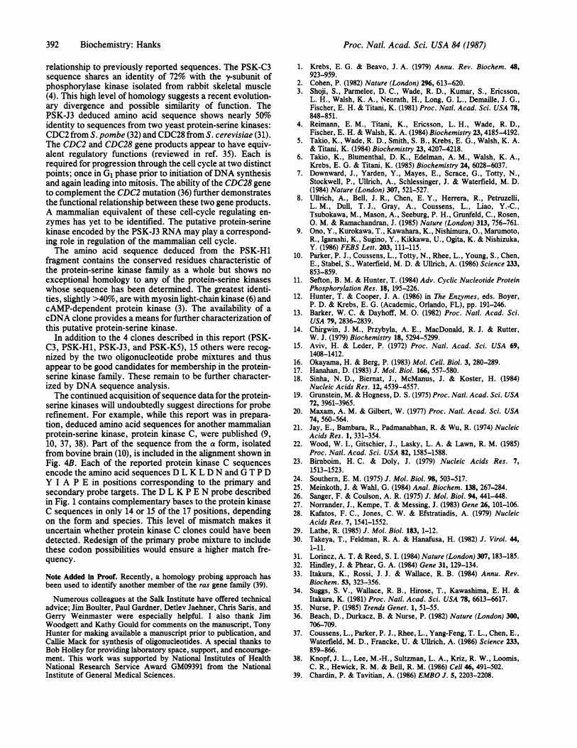
PRIMARY TARGET SECONDARY TARGET
cAPK (161)cGPK (478)
A PhK-Y (144)MLCK (177)consensus
D L IG I IN I VR V L
:Y R D L K P E N L L:Y R D L K P E N L INH R D L K P E N I LN L D L K P E N I LrD L K P E N I
I D QL D HL D DC V N
d
Nt -Asp-Lu-Lys-Pro-Glu-Asn- Ct
5I' -CA6-UUt-AA+-CCN-CAt-A6- a ons
B65' -GA6-CUa-AA+-CCN-GA+AA-- 3'
.. . . . (196)T W T L C GTP E Y L AP E I I L S K
.. . . . (614) T W T F C CT P E Y V A P E I I L N K
.. . . . . . (188) L R E V C C T P S Y L A P E I I E C S
.. . . (216) L K V N F G T P E F L S P E V V N Y Dc T P *y II P E I I
Co:tdus Nt -GIy-Thr-Pro-Glu-Tyr-Leu-Als-Pro-Clu- Ct
6'CUN
G+eUA6UUt- *CCN-GAA- 3'
StoIn* 65 -GGC-ACC-CC6-GAG-UA6-CU -CC-CCN-GA - 3'
3* CT2-GA2-TT-QGN-CT--TT 6' cD AProds*
T3' CCG-TGG-GGR-CTC-AT2-GA2-CGG-GGN-CT 65
A
FIG. 1. (A) Alignment of amino acid sequences from the catalytic domains of four mammalian protein-serine kinases reveals two separatehighly conserved segments. Sequences shown are as follows: cAPK, cAMP-dependent protein kinase from bovine cardiac muscle (3); cGPK,cGMP-dependent protein kinase from bovine lung (5); PhK-y, phosphorylase kinase y subunit from rabbit skeletal muscle (4); MLCK, myosinlight-chain kinase from rabbit skeletal muscle (6). The single-letter amino acid code is used. Only the conserved stretches and surroundingresidues are shown. Numbers in parentheses indicate amino acid position from amino termini. For MLCK, where the amino-terminal portionof the protein has not been determined, numbering initiates at the amino-terminal end of the reported sequence. Residues conserved in eachof the four sequences are shown in the consensus line as uppercase letters and conservation in three of the four sequences is indicated withlowercase letters. Consensus sequences chosen for probe targeting are underlined. (B) Oligonucleotide probe design. The primary probe is amixture of 64 17-mers complementary to RNA sequences encoding the conserved sequence: Asp-Leu-Lys-Pro-Glu-Asn (D L K P E N). Allpossible codons are represented except for the leucine position, where two of the six possible codons were selected. The two selected leucinecodons are used at a combined frequency of 0.68 in human protein-coding sequences (29). More extensive codon selection was used in designingthe secondary probe, a mixture of 96 26-mers complementary to RNA sequences encoding the consensus sequence Gly-Thr-Pro-Glu-Tyr-Leu-Ala-Pro-Glu (G T P E Y L A P E). The minimum pairing frequency between the most complementary oligonucleotide in the secondary mixtureand a target sequence is 0.73 (19 of 26 bases paired), while the predicted best-pairing frequency, based on codon usage (29), is 0.91.Oligodeoxynucleotide mixtures used as probes are enclosed in boxes.
probes and hybridization conditions as described above.Sequencing from templates with inserts in each orientationenabled sequence determination for the entire insert. Per-forming the sequencing reactions at 50'C aided in readingthrough oligo(dG) tails present in some of the inserts.
RESULTSDesign of oligonucleotide probes was based on amino acidsequence alignment (5, 12) of catalytic domains from fourmammalian protein-serine kinases (Fig. lA). Two highlyconserved segments were selected for oligonucleotide probetargeting. The primary target was chosen as nucleotidesencoding the amino acid sequence: Asp-Leu-Lys-Pro-Glu-Asn (single letter code, D L K P E N). This sequence,beginning at position 166 in cAMP-dependent protein kinase(3), is also contained in each of three other reported proteinkinase sequences: cGMP-dependent protein kinase (5),phosphorylase kinase y-subunit (4), and myosin light-chainkinase (6). In the protein-tyrosine kinases, the two sequencesAsp-Leu-Arg-Ala-Ala-Asn (D L R A A N) and Asp-Leu-Ala-Ala-Arg-Asn (D L A A R N) are most characteristic of thisconserved region (12), and this subfamily therefore will notbe recognized by probes directed toward the D L K P E Nhomology.The secondary target sequence encodes the consensus
motif Gly-Thr-Pro-Glu-Tyr-Leu-Ala-Pro-Glu (G T P E Y L AP E). This region of homology lies 29 residues COOH-terminal to the D L K P E N sequence in cAMP-dependentprotein kinase and shows a small level of divergence amongthe other protein-serine kinases (Fig. LA). Among the pro-tein-tyrosine kinases, this region has diverged considerably(12) and is typified by the sequence Gly-Ala-Lys-Phe-Ile-
Lys-Trp-Thr-Ala-Pro-Glu (G A K F P I K W T A P E) foundin the transforming protein of Rous sarcoma virus (30).To ensure high match frequencies, mixtures of oligonucle-
otides representing many of the codon possibilities were usedas probes (Fig. 1B). Codon selection was based on utilizationfrequencies in human protein-coding sequences (29). Speci-ficities of probes were tested by conducting a computer-assisted homology search against the National Institutes ofHealth GenBank library.*
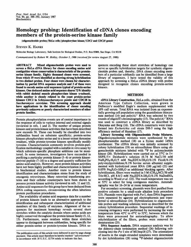
Initially, the D L K P E N probe was used to screen a HeLacell cDNA library by colony hybridization. Following ahigh-stringency wash, a positive signal was detected in 89 of=80,000 colonies screened. Plasmids from each positiveclone were purified to facilitate secondary screening. Eachplasmid was cleaved with Pst I and DNA fragments wereseparated by agarose gel electrophoresis (Fig. 2 Upper). Thefragments were then transferred to nitrocellulose filters andhybridized to the secondary G T P E Y L A P E probe (Fig.2 Middle). Of the 89 positive clones identified with the D LK P E N probe, 19 also showed significant hybridization tothe GT P E Y L A P E probe.To identify small fragments recognized by both probes, the
G T P E Y L A P E probe was removed by denaturation andthe blots were rehybridized with the D L K P E N probe (Fig.2 Lower). Several small Pst I fragments were detected thathybridized strongly to both. Four such fragments ranging insize from 300 to 450 base pairs (bp) were chosen forpreliminary DNA sequence analysis. These fragments rep-resented four individual clones: PSK-C3 (putative protein-seine kinase; filter C, colony 3), PSK-H1, PSK-J3, and
*National Institutes of Health (1985) Genetic Sequence Databank:GenBank (Research Systems Div., Bolt, Beranek, and Newman,Inc., 10 Moulton St., Cambridge, MA 02238), Tape Release 38.
Biochemistry: Hanks
Proc. Natl. Acad. Sci. USA 84 (1987)
A
40
40
"GTPEYLAPE"
46.
EDLKPEN" %
4 Ep
FIG. 2. Secondary screening of positive clones. Results for only22 of the 89 clones are shown. (Upper) Ethidium bromide-stainedplasmid DNA fragments after digestion with Pst I and separation byagarose gel electrophoresis. Two Pst I cutting sites are present in thevector, giving rise to the 1.3-kbp band present in all lanes and a largerfragment containing both vector and cDNA insert sequences. Ad-ditional bands arise from cutting sites within the insert. The centraland two end lanes contain molecular size standards (BethesdaResearch Laboratories' 1-kb ladder fragments shown are, from majorband at bottom, 0.51, 1.02, 1.64, 2.04, 3.05,4.07, 5.09, 6.11, 7.13, and8.14 kbp). (Middle) Pattern of hybridization to the G T P E Y L A PE probe mixture. Several fragments give strong hybridization signals(arrows). (Lower) The same blot rehybridized to the D L K P E Nprimary probe mixture. The 450-bp fragment indicated by the openarrow (Upper) shows very strong hybridization to both probes (largearrows). This fragment is part of an -3.2-kbp insert from the plasmiddesignated PSK-H1.
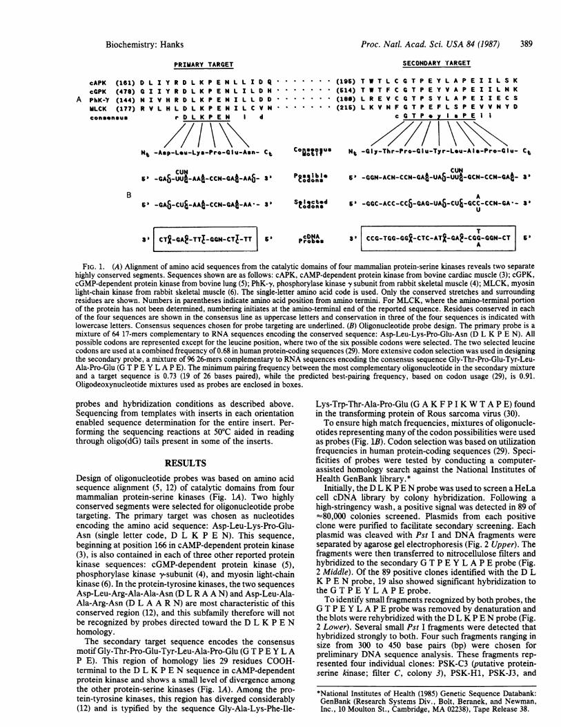
PSK-K5. The fragments were subcloned into M13 andsingle-stranded templates were prepared from severalplaques containing each PSK fragment. As a final screeningprocedure prior to sequencing, dot hybridizations to thetemplate preparations were performed to determine whether1oth D L K P E N and G T P E Y L A P E probes recognizethe same strand. For clone PSK-K5, hybridization was toopposite strands (not shown). Therefore, this clone was notfurther characterized.Each of the three remaining PSK hybridizing fragments
was sequenced and found to contain a single open readingframe encoding an amino acid sequence characteristic of theprotein-serine kinase family (Fig. 3). All three contain nu-
cleotides encoding the primary target sequence D L K P EN.For PSK-C3, this sequence contains one position of probemismatch, while all codons in PSK-H1 and PSK-J3 are
represented in the probe. PSK-C3 and PSK-H1 both alsoencode an amino acid sequence very closely related to thesecondary target consensus, G T P G Y L A P E in PSK-C3and G T P E Y I A P E in PSK-H1. The secondary targetsequences contained in PSK-C3 and PSK-H1 match se-
quences in the G T P E Y L A P E probe at 20 and 23 of the26 positions, respectively. The PSK-J3 secondary targetencodes the rather highly divergent sequence V T L W Y RA P E. A clear hybridization signal was produced despite thefact that 8 of the 26 nucleotide bases were mismatched withthe probe. Interestingly, this sequence is present in probableprotein-serine kinases encoded by genes that complementcell-cycle mutations in two species of yeast-CDC28 inSaccharomyces cerevisiae (31) and CDC2 in Schizosaccha-romyces pombe (32).The amino acid sequences encoded by the three Pst I
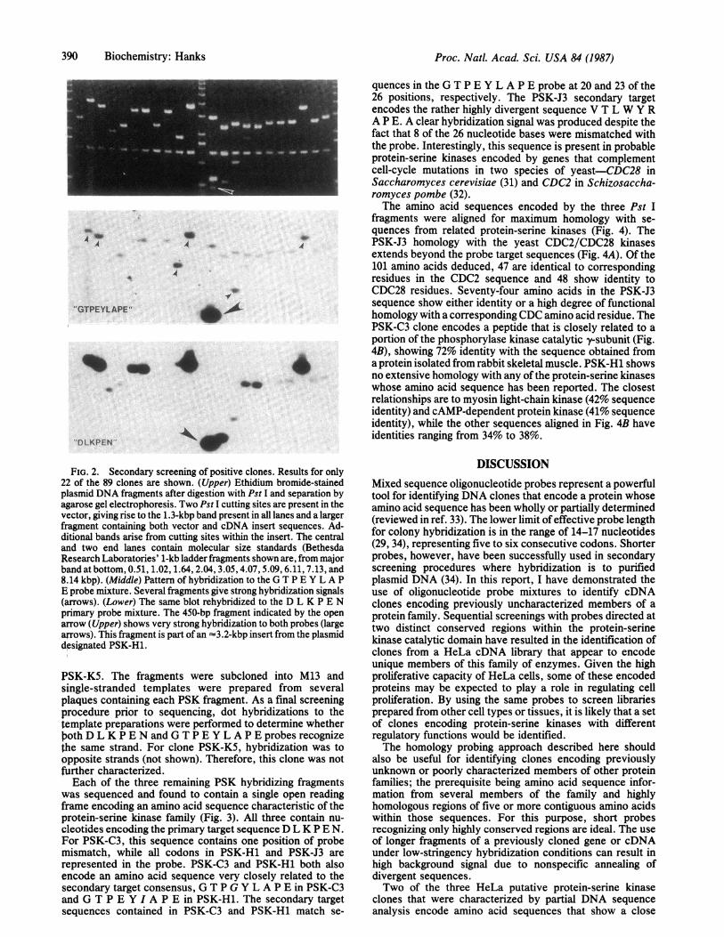
fragments were aligned for maximum homology with se-quences from related protein-serine kinases (Fig. 4). ThePSK-J3 homology with the yeast CDC2/CDC28 kinasesextends beyond the probe target sequences (Fig. 4A). Of the101 amino acids deduced, 47 are identical to correspondingresidues in the CDC2 sequence and 48 show identity toCDC28 residues. Seventy-four amino acids in the PSK-J3sequence show either identity or a high degree of functionalhomology with a corresponding CDC amino acid residue. ThePSK-C3 clone encodes a peptide that is closely related to aportion of the phosphorylase kinase catalytic y-subunit (Fig.4B), showing 72% identity with the sequence obtained froma protein isolated from rabbit skeletal muscle. PSK-H1 showsno extensive homology with any of the protein-serine kinaseswhose amino acid sequence has been reported. The closestrelationships are to myosin light-chain kinase (42% sequenceidentity) and cAMP-dependent protein kinase (41% sequenceidentity), while the other sequences aligned in Fig. 4B haveidentities ranging from 34% to 38%.
DISCUSSIONMixed sequence oligonucleotide probes represent a powerfultool for identifying DNA clones that encode a protein whoseamino acid sequence has been wholly or partially determined(reviewed in ref. 33). The lower limit of effective probe lengthfor colony hybridization is in the range of 14-17 nucleotides(29, 34), representing five to six consecutive codons. Shorterprobes, however, have been successfully used in secondaryscreening procedures where hybridization is to purifiedplasmid DNA (34). In this report, I have demonstrated theuse of oligonucleotide probe mixtures to identify cDNAclones encoding previously uncharacterized members of aprotein family. Sequential screenings with probes directed attwo distinct conserved regions within the protein-serinekinase catalytic domain have resulted in the identification ofclones from a HeLa cDNA library that appear to encodeunique members of this family of enzymes. Given the highproliferative capacity of HeLa cells, some of these encodedproteins may be expected to play a role in regulating cellproliferation. By using the same probes to screen librariesprepared from other cell types or tissues, it is likely that a setof clones encoding protein-serine kinases with differentregulatory functions would be identified.The homology probing approach described here should
also be useful for identifying clones encoding previouslyunknown or poorly characterized members of other proteinfamilies; the prerequisite being amino acid sequence infor-mation from several members of the family and highlyhomologous regions of five or more contiguous amino acidswithin those sequences. For this purpose, short probesrecognizing only highly conserved regions are ideal. The useof longer fragments of a previously cloned gene or cDNAunder low-stringency hybridization conditions can result inhigh background signal due to nonspecific annealing ofdivergent sequences.Two of the three HeLa putative protein-serine kinase
clones that were characterized by partial DNA sequenceanalysis encode amino acid sequences that show a close
390 Biochemistry: Hanks
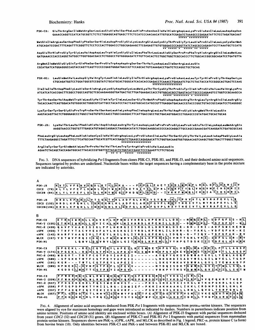
Biochemistry: Hanks Proc. Nati. Acad. Sci. USA 84 (1987) 391
PSK-C3: luThrArgS-rIl-ietArgSorL-uL-uGluAlaVeIS-rPh-L-uHisProAsnAsnIleVaIHisArgAspL*uLysPro~luAsnII-L-uLouAspAspAsnGAAACCAGGTCCATCATGCGGTCTCTCCTGGAGGCAGTGAGCTTTCTCCATCCCAACAACATTGTGCATCGAGATCT^AAGCCCGAGAATATTCTCCTAGATGACAAT
***** ***********
M*tGlnIlArgLeuSerAspPheGlyPheSerSerHisL-uAspProGlyGluLysLouArgGluLeuCysClyThrProGlyTyrLeuAlaProGluIleLeuLysCysSerM*tATGCAGATCCGACTTTCAGATTTCGGGTTCTCCTCCCACTTGGACCCTGGCGAGAAGCTTCGAGAGTTGTGTGGGACCCCAGGTTATCTAGCGCCAGAGATCCTTAAATGCTCCATG
** ***** . ***** ** *****
AspGluThrHisProGlyTyrGlyLeuValAspAspLeuTrpAlaCys~lyGluIleLeuPheThrLeuLeuAlaGlyS*rProProPh*TrpHisArgArglnlleLeuMetL*uGATGAAACCCACCCAGGCTATGGCTTGGTGGACGACCTCTGGGCCTGTGGGGAGATCTTGTTCACACTCCTGGCTGGCTCGCCACCCTTCTGGCACCGGCGGCAGATCCTGATGTTA
ArgietIIeMtGluGlyGInTyrGInPheS-rSerProGluTrpAspAspArgSerSerThrVaILysAspL*ulleS*rArgLeuLouGInCGCATGATCATGGAGGGCCAGTACCAGTTCAGTTCCCCCGAGTGGGATGACCGTTCCAGCACTGTCAAAGACCTGATCTCCAGGCTGCTGCAG
PSK-H1: L*uGInMetVaILeuAspGlyValArgTyrLeuHisAlaLeuGlyIleThrHisArgAspL*uLysProGluAsnL*uL*uTyrTyrHisProGlyThrAspS*rLysCTGCAGATGGTCCTGGATGGCGTCCGGTATCTGCATGCACTGGGCATCACACACCGAGACCTCAAACCTGAGAATCTGCTCTACTACCATCCGGGCACTGACTCCAAG
11..llI eThrAspPhGlyLeuAlaSerAlaArgLysLysGlyAspAspCysLeuMetLysThrThrCyGlyThrProGluTyrl*AlaProGluV ILeuValArgLysProATCATCATCACCGACTTCGGCCTGGCCAGTGCTCGCAAGAAGGGTGATGACTGCTTGATGAAGACCACCTGTGGCACGCCTGAGTACATTGCCCCAGAAGTCCTGGTCCGCAAGCCA
***.******* * ********
TyrThrAsnS*rValAspMetTrpAIaLeuGlyVaIIleAlaTyrIIeLeuLeuSerGlyThrMVtProPh.GluAspAspAsnArgThrArgLeuTyrArgGlnlleL-uArgGlyTACACCAACTCAGTGGACATGTGGGCGCTGGGCGTCATTGCCTACATCCTACTCAGTGGCACCATGCCGTTTGAGGATGACAACCGTACCCGGCTGTACCGCCAGATCCTCAGGGGC
LysTyrS*rTyrS-rGlyGluProTrpProS-rVaIS-rAsnL-uAleLysAspPh-Il-AspArgL-uLouThrValAspProGlyAloArgVetThrAlaL~uGlnAAGTACAGTTACTCTGGGGAGCCCTGGCCTAGTGTGTCCAACCTGGCCAAGGACTTCATTGACCGCCTGCTGACAGTGGACCCTGGAGCCCGTATGACTGCACTGCAG
PSK-J3: LysVaIThrL-uValPheGluHisValAspGlnAspLeuArgThrTyrLeuAspLysAlaProProProGlyL*uProAlaGluThrlleLysAspL-uM*tArglnAAGGTAACCCTGGTGTTTGAGCATGTAGACCAGGACCTAAGGACATATCTGGACAAGGCACCCCCACCAGGCTTGCCAGCCGAAACGATCAAGGATCTGATGCGCCAG
Ph*L*uArgGlyLeuAspPheLeuHisAlaAsnCysIlIVaIHisAr9AspLeuLysProGluAsnIleLeuVaIThrS*rGlyGlyThrVaILysL*uAIa~spPh*GlyL*uAlaTTTCTAAGAGGCCTAGATTTCCTTCATGCCAATTGCATCGTTCACCGAGATCTGAAGCCAGAGAACATTCTGGTGACAAGTGGTGGAACAGTCAAGCTGGCTGACTTTGGCCTGGCC
ArgII.TyrS*rTyrGlnMetAlaLeuThrProValValValThrLeuTrpTyrArgAlaProGluVaILeuL-uGInAGAATCTACAGCTACCAGATGGCACTTACACCCGTGGTTGTTACACTCTGGTACCGAGCTCCCGAAGTTCTTCTGCAG
* ** * * *e**** ********
FIG. 3. DNA sequences of hybridizing Pst I fragments from clones PSK-C3, PSK-H1, and PSK-J3, and their deduced amino acid sequences.Sequences targeted by probes are underlined. Nucleotide bases within the target sequences having a complementary base in the probe mixtureare indicated by asterisks.
APSK-J3 QVLVEVD[ 1 T[4YjL]APSK-~~~~J3 IF L|R T J K A - - - P [E P P A E T I DLs R IF L R Ci L D NLCDC2 (79) K L Y L V F E F L D M D L IK Y M RIR S E T G A 1D P R L Y Q LIV NHC V NICHHS|R| R I ICDC28 (84) K L Y L V F E F L D L K R Y C E C P K D Q DL I FIF V C K KI A H S H R I L
PSK-J3 H R D L K PE VT SC I Y S Y Q -ALTPV VVTLWYRAPEVL QCDC2 (132) H R D L K P Q N L ID E C N L K L A D F C L A R S F G V P L R Y T H E I V T L W Y R A P E V L ClCDC28(136) H R D L K P Q N L L I NKDDC N L K LCD F C L A R A F C V P L R A Y T H E I V T L W Y R A P E V L L C
BPSK-C3 S| RS A V S IF[ P NNI V HR D L K P E N I L L DD I- N Q R - S[D FC IF SS- HPhK-7 (126) RKI AI V I C A K LN I VH R D L K P EN I L L D D - - D N K T D F G F S C - QLPKC-a (439) Q A V F Y A A E I S I C L F F L H K R G I I Y R D L K L D N V H L D S E - C H - I K I - A D F C H C K E H U
CGPK (469) T T R F Y T A C V V E A F A Y L H S K C I I Y R D L K P E N L I L D H R -G - Y A K L - V D FG IFA K - K IcAPK (142) HA R F Y A A Q I V L T F E Y L H S L D L I Y R D L K P E N L L I D Q Q - C - Y I Q V -T D FC FA K -R V
VLCK (167) V D T V V F R Q I C[ I LIFHWI K R V L[ LD L K P EN I C V N T TC H L V K IFG AR- [ilYPSK-H1 L Q V V L V R Y L A L G I T RlD L K P E NlL L Y Y H P G T D S K I 5IT D F AAS A K
PSK-C3 D P[ E K L - R E L a T P C Y L A P E I L K D E T H P G Y G L V D L _ C GEI L FT L L A S PPhK-7 (174) D P C E K L - R E V C G T P SY L A P E I I ECS HN D N H P C Y. K E V S T V Y T L L A[ S PPKC-a (489) H D G V T - T R T FC C T P D Y I A P E I I - - - - - - A Y Q P Y G K S V D W W A Y G V L L Y E H L ACG Q PcGPK (568) - G F G K K T W T FC G T P E Y V A P E I I - - - - - - L N K C H D I S A D Y W S L C I L H Y E L L T C S PcAPK (191) K C R - - - T W T L C C T P E Y L A P E I I - - - - - - L S K C Y N K A V D W W A L C V L I Y E H A A a Y PVLCK (209) N P N E K - V N F|G T P E|IF L SP E V|V - - - - - - N Y D Q I S D KT[D H W S T[VlTLfSG|L SPSK-H1 K C D D C Ld T T C|G T P E|Y I AP E V|L - - - - - - V R K P Y T N S VD WA GVIJA I L L S CT
PSK-C3 lP F W H R|R Q I|L M L R M I M E Q|Q FS|S P E W D DIR1 S|T V K D L|I S L QPhK-7 (228)|P F W H R|K Q V|L H L R H I M S H Q S P E W D D|Y S D|T V K D L V IF V V Q P Q K R Y T A E EPKC-a (537) P F D G E D E D E L F Q S I M E HN V S Y - --P K S L S K E A V S I C K C L T K H P G K R L CC PcGPK (666) P F S G P D P H K T Y N I I L R G - I D H I E F - P K K I A K N AAN L I K K L C R D N P S E R L G N L KcAPK (237) P F F A D Q P I Q I Y E K I V S C K V R F - -- - P S H F S S D L K D L L R N L L Q V D L T K R F C N L KVLCK (267) F|L GM D DM E T L NHN V n Sr N W Y F D E E T F E A ID Er K DFV S N n I V K E Q[ FR H|S [A 1
PSK-H1UV FEIDDaLTVDPN R JR L Y R Q I TR TKJa w P S IU KA W Del T VDM. a R H0 LA 9J
FIG. 4. Alignment of amino acid sequences deduced from PSK Pst I fragments with sequences from proteiii-serine kinases. The sequenceswere aligned by eye for maximum homology and gaps were introduced as indicated by dashes. Numbers in parentheses indicate position fromamino termini. Positions of amino acid identity are enclosed within boxes. (A) Alignment of PSK-J3 fragment with partial sequences deducedfrom yeast CDC2 (32) and CDC28 (31) genes. (B) Alignment of PSK-C3 and PSK-H1 Pst I fragments with partial sequences from mammalianprotein-serine kinases. Sequences shown are PhK-y, cGPK, cAPK, and MLCK as described in Fig. 1 legend; PKC-a, protein kinase C (a form)from bovine brain (10). Only identities between PSK-C3 and PhK-y and between PSK-H1 and MLCK are boxed.
Proc. Natl. Acad. Sci. USA 84 (1987)
relationship to previously reported sequences. The PSK-C3sequence shares an identity of 72% with the v-subunit ofphosphorylase kinase isolated from rabbit skeletal muscle(4). This high level of homology suggests a recent evolution-ary divergence and possible similarity of function. ThePSK-J3 deduced amino acid sequence shows nearly 50%identity to sequences from two yeast protein-serine kinases:CDC2 from S. pombe (32) and CDC28 from S. cerevisiae (31).The CDC2 and CDC28 gene products appear to have equiv-alent regulatory functions (reviewed in ref. 35). Each isrequired for progression through the cell cycle at two distinctpoints; once in G1 phase prior to initiation of DNA synthesisand again leading into mitosis. The ability of the CDC28 geneto complement the CDC2 mutation (36) further demonstratesthe functional relationship between these two gene products.A mammalian equivalent of these cell-cycle regulating en-zymes has yet to be identified. The putative protein-serinekinase encoded by the PSK-J3 RNA may play a correspond-ing role in regulation of the mammalian cell cycle.The amino acid sequence deduced from the PSK-H1
fragment contains the conserved residues characteristic ofthe protein-serine kinase family as a whole but shows noexceptional homology to any of the protein-serine kinaseswhose sequence has been determined. The greatest identi-ties, slightly >40%, are with myosin light-chain kinase (6) andcAMP-dependent protein kinase (3). The availability of acDNA clone provides a means for further characterization ofthis putative protein-serine kinase.
In addition to the 4 clones described in this report (PSK-C3, PSK-H1, PSK-J3, and PSK-K5), 15 others were recog-nized by the two oligonucleotide probe mixtures and thusappear to be good candidates for membership in the protein-serine kinase family. These remain to be further character-ized by DNA sequence analysis.The continued acquisition of sequence data for the protein-
serine kinases will undoubtedly suggest directions for proberefinement. For example, while this report was in prepara-tion, deduced amino acid sequences for another mammalianprotein-serine kinase, protein kinase C, were published (9,10, 37, 38). Part of the sequence from the a form, isolatedfrom bovine brain (10), is included in the alignment shown inFig. 4B. Each of the reported protein kinase C sequencesencode the amino acid sequences D L K L D N and G T P DY I A P E in positions corresponding to the primary andsecondary probe targets. The D L K P E N probe describedin Fig. 1 contains complementary bases to the protein kinaseC sequences in only 14 or 15 of the 17 positions, dependingon the form and species. This level of mismatch makes ituncertain whether protein kinase C clones could have beendetected. Redesign of the primary probe mixture to includethese codon possibilities would ensure a higher match fre-quency.
Note Added in Proof. Recently, a homology probing approach hasbeen used to identify another member of the ras gene family (39).
Numerous colleagues at the Salk Institute have offered technicaladvice; Jim Boulter, Paul Gardner, Detlev Jaehner, Chris Saris, andGerry Weinmaster were especially helpful. I also thank JimWoodgett and Kathy Gould for comments on the manuscript, TonyHunter for making available a manuscript prior to publication, andCallie Mack for synthesis of oligonucleotides. A special thanks toBob Holley for providing laboratory space, support, and encourage-ment. This work was supported by National Institutes of HealthNational Research Service Award GM09391 from the NationalInstitute of General Medical Sciences.
1. Krebs, E. G. & Beavo, J. A. (1979) Annu. Rev. Biochem. 48,923-959.
2. Cohen, P. (1982) Nature (London) 296, 613-620.3. Shoji, S., Parmelee, D. C., Wade, R. D., Kumar, S., Ericsson,
L. H., Walsh, K. A., Neurath, H., Long, G. L., Demaille, J. G.,Fischer, E. H. & Titani, K. (1981) Proc. Natl. Acad. Sci. USA 78,848-851.
4. Reimann, E. M., Titani, K., Ericsson, L. H., Wade, R. D.,Fischer, E. H. & Walsh, K. A. (1984) Biochemistry 23, 4185-4192.
5. Takio, K., Wade, R. D., Smith, S. B., Krebs, E. G., Walsh, K. A.& Titani, K. (1984) Biochemistry 23, 4207-4218.
6. Takio, K., Blumenthal, D. K., Edelman, A. M., Walsh, K. A.,Krebs, E. G. & Titani, K. (1985) Biochemistry 24, 6028-6037.
7. Downward, J., Yarden, Y., Mayes, E., Scrace, G., Totty, N.,Stockwell, P., Ulirich, A., Schlessinger, J. & Waterfield, M. D.(1984) Nature (London) 307, 521-527.
8. Ullrich, A., Bell, J. R., Chen, E. Y., Herrera, R., Petruzelli,L. M., Dull, T. J., Gray, A., Coussens, L., Liao, Y.-C.,Tsubokawa, M., Mason, A., Seeburg, P. H., Grunfeld, C., Rosen,0. M. & Ramachandran, J. (1985) Nature (London) 313, 756-761.
9. Ono, Y., Kurokawa, T., Kawahara, K., Nishimura, O., Marumoto,R., Igarashi, K., Sugino, Y., Kikkawa, U., Ogita, K. & Nishizuka,Y. (1986) FEBS Lett. 203, 111-115.
10. Parker, P. J., Coussens, L., Totty, N., Rhee, L., Young, S., Chen,E., Stabel, S., Waterfield, M. D. & Ullrich, A. (1986) Science 233,853-859.
11. Sefton, B. M. & Hunter, T. (1984) Adv. Cyclic Nucleotide ProteinPhosphorylation Res. 18, 195-226.
12. Hunter, T. & Cooper, J. A. (1986) in The Enzymes, eds. Boyer,P. D. & Krebs, E. G. (Academic, Orlando, FL), pp. 191-246.
13. Barker, W. C. & Dayhoff, M. 0. (1982) Proc. Natl. Acad. Sci.USA 79, 2836-2839.
14. Chirgwin, J. M., Przybyla, A. E., MacDonald, R. J. & Rutter,W. J. (1979) Biochemistry 18, 5294-5299.
15. Aviv, H. & Leder, P. (1972) Proc. Natl. Acad. Sci. USA 69,1408-1412.
16. Okayama, H. & Berg, P. (1983) Mol. Cell. Biol. 3, 280-289.17. Hanahan, D. (1983) J. Mol. Biol. 166, 557-580.18. Sinha, N. D., Biernat, J., McManus, J. & Koster, H. (1984)
Nucleic Acids Res. 12, 4539-4557.19. Grunstein, M. & Hogness, D. S. (1975) Proc. Natl. Acad. Sci. USA
72, 3961-3965.20. Maxam, A. M. & Gilbert, W. (1977) Proc. Natl. Acad. Sci. USA
74, 560-564.21. Jay, E., Bambara, R., Padmanabhan, R. & Wu, R. (1974) Nucleic
Acids Res. 1, 331-354.22. Wood, W. I., Gitschier, J., Lasky, L. A. & Lawn, R. M. (1985)
Proc. Natl. Acad. Sci. USA 82, 1585-1588.23. Birnboim, H. C. & Doly, J. (1979) Nucleic Acids Res. 7,
1513-1523.24. Southern, E. M. (1975) J. Mol. Biol. 98, 503-517.25. Meinkoth, J. & Wahl, G. (1984) Anal. Biochem. 138, 267-284.26. Sanger, F. & Coulson, A. R. (1975) J. Mol. Biol. 94, 441-448.27. Norrander, J., Kempe, T. & Messing, J. (1983) Gene 26, 101-106.28. Kafatos, F. C., Jones, C. W. & Efstratiadis, A. (1979) Nucleic
Acids Res. 7, 1541-1552.29. Lathe, R. (1985) J. Mol. Biol. 183, 1-12.30. Takeya, T., Feldman, R. A. & Hanafusa, H. (1982) J. Virol. 44,
1-11.31. Lorincz, A. T. & Reed, S. I. (1984) Nature (London) 307, 183-185.32. Hindley, J. & Phear, G. A. (1984) Gene 31, 129-134.33. Itakura, K., Rossi, J. J. & Wallace, R. B. (1984) Annu. Rev.
Biochem. 53, 323-356.34. Suggs, S. V., Wallace, R. B., Hirose, T., Kawashima, E. H. &
Itakura, K. (1981) Proc. Natl. Acad. Sci. USA 78, 6613-6617.35. Nurse, P. (1985) Trends Genet. 1, 51-55.36. Beach, D., Durkacz, B. & Nurse, P. (1982) Nature (London) 300,
706-709.37. Coussens, L., Parker, P. J., Rhee, L., Yang-Feng, T. L., Chen, E.,
Waterfield, M. D., Francke, U. & Ullrich, A. (1986) Science 233,859-866.
38. Knopf, J. L., Lee, M.-H., Sultzman, L. A., Kriz, R. W., Loomis,C. R., Hewick, R. M. & Bell, R. M. (1986) Cell 46, 491-502.
39. Chardin, P. & Tavitian, A. (1986) EMBO J. 5, 2203-2208.
392 Biochemistry: Hanks