Embed Size (px)
Citation preview

04/2
0 |
Volu
me
146
| Th
annh
ause
n, G
erm
any,
Apr
il 15
, 202
0
e n g l i s h
2 0 2 004
The Golgi Apparatus – Cellular Detox Super Hero
Green Extraction with a New Solvent for Performant Biomimetic Skin Actives
Ethyl Macadamiate as a Botanical Silicone Alternative in Hair Conditioners
skin & hair care
Soft Focus Effects: Natural Particles for Optimum Skin Care
colour cosmetics
EU-harmonized Product Notification, Obligations and Deadlines
home care
Combination of Lactic Acid with 1,2-hexanediol – a New Possibility to Stabilise Rinse-off Formulations
stabilisation
personal care |
sofwjournal | 146 | 04/206
skin care
Introduction
The Golgi apparatus – the coordinator of autophagy and the cellular post office
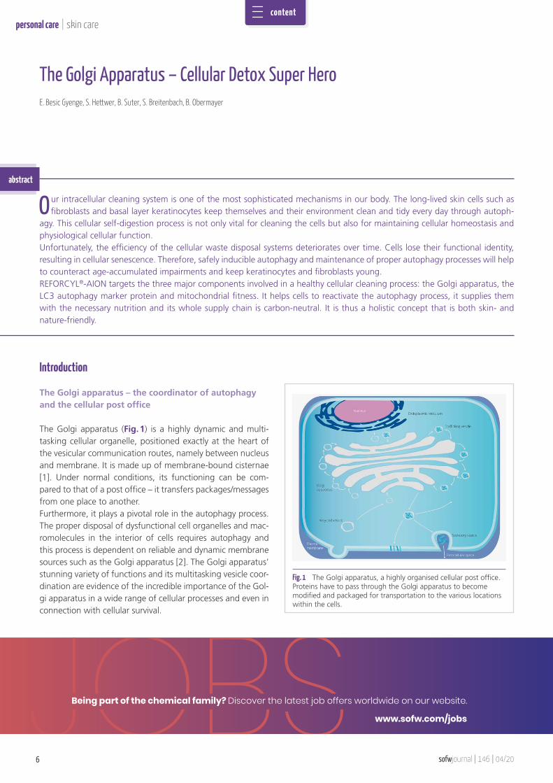
The Golgi apparatus (Fig. 1) is a highly dynamic and multi-tasking cellular organelle, positioned exactly at the heart of the vesicular communication routes, namely between nucleus and membrane. It is made up of membrane-bound cisternae [1]. Under normal conditions, its functioning can be com-pared to that of a post office – it transfers packages/messages from one place to another. Furthermore, it plays a pivotal role in the autophagy process. The proper disposal of dysfunctional cell organelles and mac-romolecules in the interior of cells requires autophagy and this process is dependent on reliable and dynamic membrane sources such as the Golgi apparatus [2]. The Golgi apparatus’ stunning variety of functions and its multitasking vesicle coor-dination are evidence of the incredible importance of the Gol-gi apparatus in a wide range of cellular processes and even in connection with cellular survival.
The Golgi Apparatus – Cellular Detox Super HeroE. Besic Gyenge, S. Hettwer, B. Suter, S. Breitenbach, B. Obermayer
Fig. 1 The Golgi apparatus, a highly organised cellular post office. Proteins have to pass through the Golgi apparatus to become modified and packaged for transportation to the various locations within the cells.
abstract
Our intracellular cleaning system is one of the most sophisticated mechanisms in our body. The long-lived skin cells such as fibroblasts and basal layer keratinocytes keep themselves and their environment clean and tidy every day through autoph-
agy. This cellular self-digestion process is not only vital for cleaning the cells but also for maintaining cellular homeostasis and physiological cellular function. Unfortunately, the efficiency of the cellular waste disposal systems deteriorates over time. Cells lose their functional identity, resulting in cellular senescence. Therefore, safely inducible autophagy and maintenance of proper autophagy processes will help to counteract age-accumulated impairments and keep keratinocytes and fibroblasts young. REFORCYL®-AION targets the three major components involved in a healthy cellular cleaning process: the Golgi apparatus, the LC3 autophagy marker protein and mitochondrial fitness. It helps cells to reactivate the autophagy process, it supplies them with the necessary nutrition and its whole supply chain is carbon-neutral. It is thus a holistic concept that is both skin- and nature-friendly.
Being part of the chemical family? Discover the latest job offers worldwide on our website.
www.sofw.com/jobs
content
personal care|
04/20 | 146 | sofwjournal 7
skin care
The mitochondrion – cellular and autophagy energy supplier
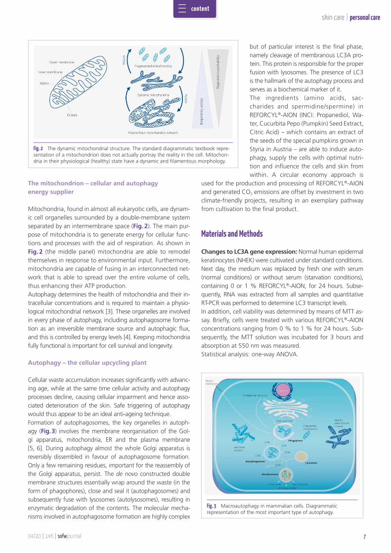
Mitochondria, found in almost all eukaryotic cells, are dynam-ic cell organelles surrounded by a double-membrane system separated by an intermembrane space (Fig. 2). The main pur-pose of mitochondria is to generate energy for cellular func-tions and processes with the aid of respiration. As shown in Fig. 2 (the middle panel) mitochondria are able to remodel themselves in response to environmental input. Furthermore, mitochondria are capable of fusing in an interconnected net-work that is able to spread over the entire volume of cells, thus enhancing their ATP production. Autophagy determines the health of mitochondria and their in-tracellular concentrations and is required to maintain a physio-logical mitochondrial network [3]. These organelles are involved in every phase of autophagy, including autophagosome forma-tion as an irreversible membrane source and autophagic flux, and this is controlled by energy levels [4]. Keeping mitochondria fully functional is important for cell survival and longevity.
Autophagy – the cellular upcycling plant
Cellular waste accumulation increases significantly with advanc-ing age, while at the same time cellular activity and autophagy processes decline, causing cellular impairment and hence asso-ciated deterioration of the skin. Safe triggering of autophagy would thus appear to be an ideal anti-ageing technique. Formation of autophagosomes, the key organelles in autoph-agy (Fig. 3) involves the membrane reorganisation of the Gol-gi apparatus, mitochondria, ER and the plasma membrane [5, 6]. During autophagy almost the whole Golgi apparatus is reversibly dissembled in favour of autophagosome formation. Only a few remaining residues, important for the reassembly of the Golgi apparatus, persist. The de novo constructed double membrane structures essentially wrap around the waste (in the form of phagophores), close and seal it (autophagosomes) and subsequently fuse with lysosomes (autolysosomes), resulting in enzymatic degradation of the contents. The molecular mecha-nisms involved in autophagosome formation are highly complex
but of particular interest is the final phase, namely cleavage of membranous LC3A pro-tein. This protein is responsible for the proper fusion with lysosomes. The presence of LC3 is the hallmark of the autophagy process and serves as a biochemical marker of it.The ingredients (amino acids, sac-charides and spermidine/spermine) in REFORCYL®-AION (INCI: Propanediol, Wa-ter, Cucurbita Pepo (Pumpkin) Seed Extract, Citric Acid) – which contains an extract of the seeds of the special pumpkins grown in Styria in Austria – are able to induce auto-phagy, supply the cells with optimal nutri-tion and influence the cells and skin from within. A circular economy approach is
used for the production and processing of REFORCYL®-AION and generated CO2 emissions are offset by investment in two climate-friendly projects, resulting in an exemplary pathway from cultivation to the final product.
Materials and Methods
Changes to LC3A gene expression: Normal human epidermal keratinocytes (NHEK) were cultivated under standard conditions. Next day, the medium was replaced by fresh one with serum (normal conditions) or without serum (starvation conditions), containing 0 or 1 % REFORCYL®-AION, for 24 hours. Subse-quently, RNA was extracted from all samples and quantitative RT-PCR was performed to determine LC3 transcript levels. In addition, cell viability was determined by means of MTT as-say. Briefly, cells were treated with various REFORCYL®-AION concentrations ranging from 0 % to 1 % for 24 hours. Sub-sequently, the MTT solution was incubated for 3 hours and absorption at 550 nm was measured.Statistical analysis: one-way ANOVA.
Fig. 2 The dynamic mitochondrial structure. The standard diagrammatic textbook repre-sentation of a mitochondrion does not actually portray the reality in the cell. Mitochon-dria in their physiological (healthy) state have a dynamic and filamentous morphology.
Fig. 3 Macroautophagy in mammalian cells. Diagrammatic representation of the most important type of autophagy.
content
personal care |
sofwjournal | 146 | 04/208
skin care
Since autophagy and cell death are intimately linked, a cell viability assay was performed. The results showed that REFORCYL®-AION did not induce apoptosis in NHEK cells but kept them alive at concentrations up to 1 % during 24 hours of exposure (data not shown).
Morphological manifestations of autophagy in healthy cells
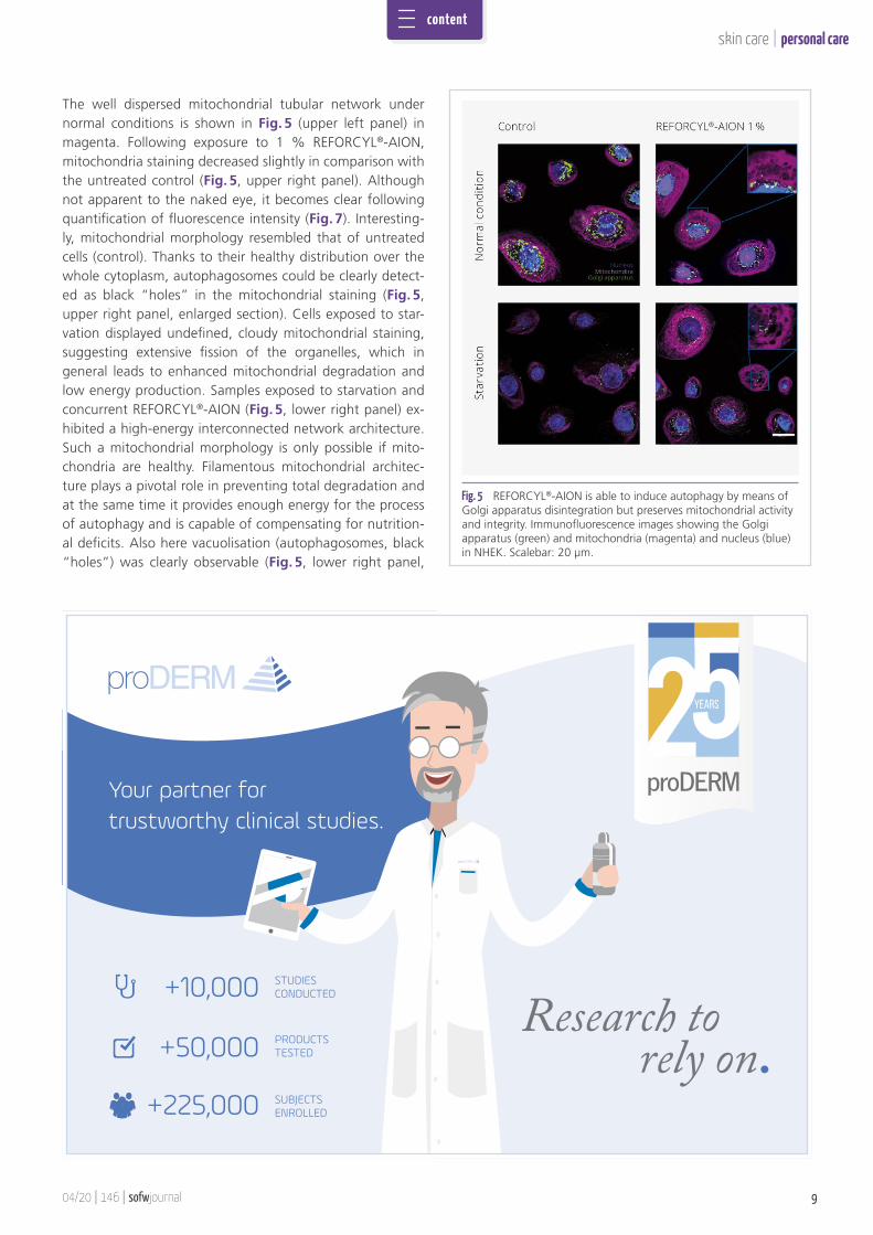
Under normal conditions, cells exhibited a prominent juxta-nuclear Golgi apparatus (stained in green, Fig. 5, upper left panel). With exposure to 1 % REFORCYL®-AION, there was an extensive decrease in Golgi apparatus staining, confirming progressive autophagy and increased autophagosome forma-tion (Fig. 5, upper right panel) as suggested by elevated LC3. This was also observed in the case of the samples ex-posed to starvation, or starvation and concurrent 1 % REFORCYL®-AION, as shown in the lower panels of Fig. 5. It is noteworthy that the remaining residues of the Golgi apparatus in the starvation control were scattered over the cytoplasm, resembling the type of distribution seen during cellular death [15], while the remaining residues in the REFORCYL®-AION samples remained at the juxtanuclear lo-cation, suggesting a highly organised disassembly process as seen in living and healthy cells.
Induction of autophagy in healthy cells and strength-ening of mitochondria under stressed conditions: NHEK were cultivated on a coverslip for 24 hours. Afterwards, the medium was replaced by fresh medium with serum (con-trol) or without serum (starvation) containing 0 or 1 % REFORCYL®-AION, for 24 hours. On completion of the incuba-tion period, samples were immunostained with MitoTracker™ Deep Red FM staining for 45 minutes (mitochondria) and An-ti-Giantin antibody (Golgi apparatus) overnight. Nuclei were stained with DAPI.
General improvement in skin conditions: The in vivo study was conducted in accordance with the World Medical Asso-ciation’s Declaration of Helsinki. A double blind, placebo-con-trolled, randomised study in 55 volunteers (female and male, equally distributed in number) was performed. Emulsion con-taining 0% or 3% REFORCYL®-AION was applied twice daily for 56 days to the skin of the face. To determine subjective impressions of efficacy, a five-point scale was employed (5: I strongly agree; 4: I agree; 3: I neither agree nor disagree; 2: I disagree; 1: I strongly disagree). Those volunteers who responded by selecting options 5 or 4 were considered to be satisfied with the results of treatment.Statistical analysis was performed using an unpaired Stu-dents’ t-test.
Results
Upregulation of LC3A gene expression
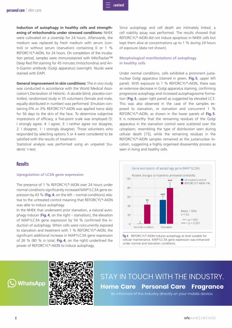
The presence of 1 % REFORCYL®-AION over 24 hours under normal conditions significantly increased MAP1LC3A gene ex-pression by 43 % (Fig. 4, on the left – normal conditions) rela-tive to the untreated control meaning that REFORCYL®-AION was able to induce autophagy. In the NHEK that underwent prior starvation, a natural auto-phagy inducer (Fig. 4, on the right – starvation), the elevation of MAP1LC3A gene expression by 54 % confirmed the in-duction of autophagy. When cells were concurrently exposed to starvation and treatment with 1 % REFORCYL®-AION, the significant additional increase in MAP1LC3A gene expression of 26 % (80 % in total; Fig. 4, on the right) underlined the power of REFORCYL®-AION to induce autophagy.
Fig. 4 REFORCYL®-AION induces autophagy at level suitable for cellular maintenance. MAP1LC3A gene expression was enhanced under normal and starvation conditions.
STAY IN TOUCH WITH THE INDUSTRY.Home Care Personal Care Fragrance
Be informed of the industry directly on your mobile device.
content
04/20 | 146 | sofwjournal 9
personal care|skin care
The well dispersed mitochondrial tubular network under normal conditions is shown in Fig. 5 (upper left panel) in magenta. Following exposure to 1 % REFORCYL®-AION, mitochondria staining decreased slightly in comparison with the untreated control (Fig. 5, upper right panel). Although not apparent to the naked eye, it becomes clear following quantification of fluorescence intensity (Fig. 7). Interesting-ly, mitochondrial morphology resembled that of untreated cells (control). Thanks to their healthy distribution over the whole cytoplasm, autophagosomes could be clearly detect-ed as black “holes” in the mitochondrial staining (Fig. 5, upper right panel, enlarged section). Cells exposed to star-vation displayed undefined, cloudy mitochondrial staining, suggesting extensive fission of the organelles, which in general leads to enhanced mitochondrial degradation and low energy production. Samples exposed to starvation and concurrent REFORCYL®-AION (Fig. 5, lower right panel) ex-hibited a high-energy interconnected network architecture. Such a mitochondrial morphology is only possible if mito-chondria are healthy. Filamentous mitochondrial architec-ture plays a pivotal role in preventing total degradation and at the same time it provides enough energy for the process of autophagy and is capable of compensating for nutrition-al deficits. Also here vacuolisation (autophagosomes, black “holes”) was clearly observable (Fig. 5, lower right panel,
Fig. 5 REFORCYL®-AION is able to induce autophagy by means of Golgi apparatus disintegration but preserves mitochondrial activity and integrity. Immunofluorescence images showing the Golgi apparatus (green) and mitochondria (magenta) and nucleus (blue) in NHEK. Scalebar: 20 μm.
+10,000
+50,000
+225,000
STUDIES CONDUCTED
PRODUCTS TESTED
SUBJECTS ENROLLED
Your partner for trustworthy clinical studies.
content
personal care |
sofwjournal | 146 | 04/2010
skin care
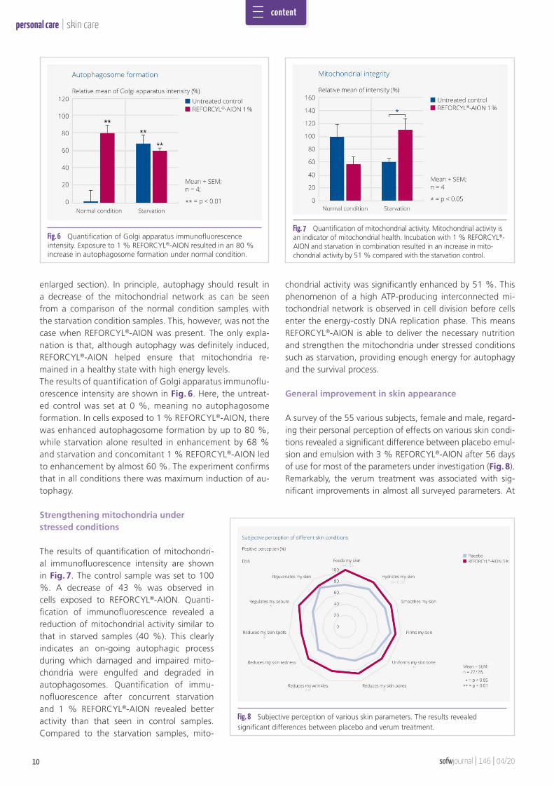
enlarged section). In principle, autophagy should result in a decrease of the mitochondrial network as can be seen from a comparison of the normal condition samples with the starvation condition samples. This, however, was not the case when REFORCYL®-AION was present. The only expla-nation is that, although autophagy was definitely induced, REFORCYL®-AION helped ensure that mitochondria re-mained in a healthy state with high energy levels.The results of quantification of Golgi apparatus immunoflu-orescence intensity are shown in Fig. 6. Here, the untreat-ed control was set at 0 %, meaning no autophagosome formation. In cells exposed to 1 % REFORCYL®-AION, there was enhanced autophagosome formation by up to 80 %, while starvation alone resulted in enhancement by 68 % and starvation and concomitant 1 % REFORCYL®-AION led to enhancement by almost 60 %. The experiment confirms that in all conditions there was maximum induction of au-tophagy. Strengthening mitochondria under stressed conditions
The results of quantification of mitochondri-al immunofluorescence intensity are shown in Fig. 7. The control sample was set to 100 %. A decrease of 43 % was observed in cells exposed to REFORCYL®-AION. Quanti-fication of immunofluorescence revealed a reduction of mitochondrial activity similar to that in starved samples (40 %). This clearly indicates an on-going autophagic process during which damaged and impaired mito-chondria were engulfed and degraded in autophagosomes. Quantification of immu-nofluorescence after concurrent starvation and 1 % REFORCYL®-AION revealed better activity than that seen in control samples. Compared to the starvation samples, mito-
chondrial activity was significantly enhanced by 51 %. This phenomenon of a high ATP-producing interconnected mi-tochondrial network is observed in cell division before cells enter the energy-costly DNA replication phase. This means REFORCYL®-AION is able to deliver the necessary nutrition and strengthen the mitochondria under stressed conditions such as starvation, providing enough energy for autophagy and the survival process.
General improvement in skin appearance
A survey of the 55 various subjects, female and male, regard-ing their personal perception of effects on various skin condi-tions revealed a significant difference between placebo emul-sion and emulsion with 3 % REFORCYL®-AION after 56 days of use for most of the parameters under investigation (Fig. 8). Remarkably, the verum treatment was associated with sig-nificant improvements in almost all surveyed parameters. At
Fig. 8 Subjective perception of various skin parameters. The results revealed significant differences between placebo and verum treatment.
Fig. 7 Quantification of mitochondrial activity. Mitochondrial activity is an indicator of mitochondrial health. Incubation with 1 % REFORCYL®- AION and starvation in combination resulted in an increase in mito-chondrial activity by 51 % compared with the starvation control.
Fig. 6 Quantification of Golgi apparatus immunofluorescence intensity. Exposure to 1 % REFORCYL®-AION resulted in an 80 % increase in autophagosome formation under normal condition.
content
04/20 | 146 | sofwjournal 11
personal care|skin care
least 80 % of the volunteers using REFORCYL®-AION emul-sion were satisfied with the results of treatment. Noteworthy is the fact that 100 % of the verum users described their skin as being better fed. Around 90 % of the subjects evaluated skin hydration, smoothness, firmness and sebum regulation as positive while 86 % in general were satisfied with results in terms of skin uniformity, skin redness, pore reduction and skin rejuvenation.
Conclusion and Discussion
In view of the overall results, it can be concluded that REFORCYL®-AION is able to induce autophagy under normal conditions, employing 80 % of the Golgi apparatus’ cister-nae and resulting in the degrading of impaired mitochondria. Interestingly, when autophagy is induced by starvation, an emergency reserve of rapidly available amino acids, proteins and carbohydrates is released, transferring mitochondria into a high-energy fused network that serves as a defence against apoptosis and at the same time provides enough energy for an effective autophagy process.REFORCYL®-AION combines cutting-edge molecular findings with the principles of carbon-neutrality and it represents a holistic upcycling concept beneficial for the skin and our en-vironment.
References
[1] Farquhar, M.G. and G.E. Palade, The Golgi apparatus (complex)-(1954-1981)-from artifact to center stage. J Cell Biol, 1981. 91(3 Pt 2): p. 77s-103s.
[2] Geng, J. and D.J. Klionsky, The Golgi as a potential membrane source for au-tophagy. Autophagy, 2010. 6(7): p. 950-1.
[3] Youle, R.J. and A.M. van der Bliek, Mitochondrial fission, fusion, and stress. Science, 2012. 337(6098): p. 1062-5.
[4] Rambold, A.S. and J. Lippincott-Schwartz, Mechanisms of mitochondria and autophagy crosstalk. Cell Cycle, 2011. 10(23): p. 4032-8.
[5] Mizushima, N. and M. Komatsu, Autophagy: renovation of cells and tissues. Cell, 2011. 147(4): p. 728-41.
[6] Wang, J., et al., Autophagosome formation: Where the secretory and autoph-agy pathways meet. Autophagy, 2017. 13(5): p. 973-974.
contact
Emina Besic GyengeStefan HettwerBrigit SuterSandra BreitenbachBarbara ObermayerRAHN AGDörflistrasse 1208050 Zurich | Switzerland
REFORCYL®-AION Garb’Ageing Clean-Up
· Activates autophagy· Maintains mitochondrial fitness· Detoxifies and repairs aged skin
NEW
listed
COSMOS
comp
liant
CHINA
comp
liant
NAGOYA