Embed Size (px)
Citation preview

HL Topic 11.2: Movement
Chapter 49.5

Locomotion

11.2 Movement: Understandings:
1. Bones and exoskeletons provide anchorage for muscles and act
as levers.
2. Synovial joints allow certain movements, but not others (e.g. elbow
synovial joint allows flexion and extension but not pronation and
supination).
3. Movement of the body requires muscles to work in antagonistic
pairs.
4. Skeletal muscle fibres are multinucleate and contain specialized
endoplasmic reticulum.
5. Muscle fibres contain many myofibrils.
6. Each myofibril is made up of contractile sarcomeres.
7. The contraction of the skeletal muscle is achieved by the sliding of
actin and myosin filaments.
8. ATP hydrolysis and cross bridge formation are necessary for the
filaments to slide.
9. Calcium ions and the proteins tropomyosin and troponin control
muscle contractions.
11.2 Applications and skills: A1. Antagonistic pairs of muscles in an insect leg.
S1. Annotation of a diagram of the human elbow to show
function (cartilage, synovial fluid, joint capsule, named
bones and antagonistic muscles).
S2. Drawing labelled diagrams of the structure of a
sarcomere (Z lines, actin filaments, myosin filaments with
heads, and the resultant light and dark bands).
S3. Analysis of electron micrographs to find the state of
contraction of muscle fibres. (measurement of the length of
sarcomeres will require calibration of the eyepiece scale of
the microscope.
Movement: Internal use only 11.2.1 State the roles of bones, ligaments, muscles, tendons and nerves
in human movement.
11.2.2 Label a diagram of the human elbow joint, including cartilage, synovial fluid, joint capsule, named bones and antagonistic muscles (biceps and triceps).
11.2.3 Outline the functions of the above-named structures of the human elbow joint (11.2.2).
11.2.4 Compare the movements of the hip joint and the knee joint.
11.2.5 Describe the structure of striated muscle fibres, including the myofibrils with light and dark bands, mitochondria, the sarcoplasmic reticulum, nuclei and the sarcolemma.
11.2.6 Draw and label a diagram to show the structure of a sarcomere, including Z lines, actin filaments, myosin filaments with heads, and the resultant light and dark bands.
11.2.7 Explain how skeletal muscle contracts, including the release of calcium ions from the sarcoplasmic reticulum, the formation of cross-bridges, the sliding of actin and myosin filaments, and the use ATP to break cross-bridges and re-set myosin heads.
11.2.8 Analyse electron micrographs to find the state of contraction of muscle fibres. (Muscle fibres can be fully relaxed, slightly contracted, moderately contracted and fully contracted.)
11.2.9 Be able to measure the length of sarcomeres (will require calibration of the eyepiece scale of the microscope.)

Movement
Movement is the hallmark of animals. In order to catch food, an animal must either move through its environment or move the surrounding water or air past itself.
A large portion of their time and energy is spent actively searching for food and escaping from danger and looking for mates.
Locomotion: active travel from place to place

Modes of locomotion
Diverse modes. Animals swim, crawl, walk,
run, or hop and fly.
Requirements: energy must be expended to
overcome friction and gravity.

Cost of Transportation

Summary: Cost of Transport
A. Running animals generally consume more energy per meter traveled than similarly sized animals specialized for swimming because running demands overcoming gravity.
B. Swimming is the most efficient mode of transport.
C. Flying animals use more energy for the same amount of time, however.
D. A larger animal travels more efficiently than a smaller species specialized for the same mode of transport. (E.g. a horse consumes less energy per kg of body weight than a cat running the same distance.)

Requirements for movement
1. Muscles arranged as antagonistic pairs.
(e.g. Flexors and extensors).
2. A skeleton that provides support and
attachment points for muscles. (e.g.
endoskeleton made of cartilage/bone;
exoskeleton made of chitin/calcium
carbonate; hydrostatic skeleton).
3. Nervous enervation to stimulate and
coordinate muscle contraction.

1. Swimming
Overcoming gravity is less of a problem than
for species that move on land or through
air.
However, water is a much denser medium
than air.
Friction (resistance) is a major problem for
aquatic animals.

Adaptations for swimming
• Sleek, torpedolike shape is common for fast swimmers.
• Glands in skin of a bony fish secrete a mucus that gives the animal its characteristic sliminess, an adaptation that reduces drag during swimming.
• Bony fish have swim bladders, an air sac that helps control the buoyancy of the fish. Bony fish can remain almost motionless.

Swimming

Example: bony fish swimming
• Bony fish swim by moving their body and tail from side to side.
• They generate a force that pushes the fish forwards by beating the tail from side to side.
• There are bony projections from the vertebra that are used for muscle attachment.
• The muscle on the left side causes the tail to move to the left while the muscle on the right side causes the tail to move to the right. (Antagonistic muscle bundles).
• Bony fish are maneuverable swimmers, their flexible fins are better for steering and propulsion.

Flying
• Overcoming gravity is a major problem. The wings must develop enough lift to overcome the downward force of gravity.
• Key: shape of the wings which act as airfoils, structures whose shape creates lift by altering air currents.
• The leading edge is thicker than the trailing edge and its upper surface is somewhat convex and its undersurface is concave or flattened.
• Air passing over the wing travels faster than air passing under the wing. Low pressure on top, higher pressure exerted on bottom: lift. Birds generate this lift by flapping wings up and down.

Flying
Adaptations for bird flight
• Bones are strong but light.
• Internal structure is honeycombed.
• Another adaptation is to reduce weight:
absence of some organs. Females have
only one ovary (e.g.) Modern birds are
toothless.
• Keen eyesight.

Bird Flight
• Providing power for flight, birds flap their wings
by contractions of large pectoral (breast)
muscles anchored to a keel on the sternum
(breastbone). Pectoralis major pulls the wing
down, pectoralis minor pulls the wings up. The
tendon of the pectoralis minor is attached to the
upper surface of the shoulder bone.
• Flapping rate varies with species. Shape and
arrangement of feathers form wing into airfoil.

3. Crawling: earthworm • Peristaltic locomotion of an earthworm.
• Earthworms have a hydrostatic skeleton (fluid held under pressure in a closed body compartment).
• They control their form and movement by using muscles to change the shape of the fluid-filled compartments. Peristalsis is a type of locomotion produced by rhythmic waves of muscle contractions passing from head to tail.
• Earthworms have 2 sets of muscles- one elongates the body (circular) while the other shortens (longitudinal) it. It also has bristles (chaetae) that will hold the substrate.

Earthworm: Peristalsis

Walking e.g. arthropod
• Arthropods are segmented, have a hard exoskeleton (also called a cuticle) made of chitin and jointed appendages.
• Cuticle is thick over some parts of body but paper thin and flexible in joints.
• Muscles are attached to knobs and plates of the cuticle that extend into the interior of the body.
• Exoskeletons are shed with growth. Crabs (an arthropod) walk by flexing (bending) and extending (straightening) the segments of their legs.
• Extensors: muscles cause extension while flexor muscle causes flexion.
• Extension: Increasing the angle of articulation. Flexion: Decreasing the angle of articulation.

Insect leg

Human and Grasshopper
extensors and flexors


Humans with endoskeleton
• Endoskeleton consists of bones either
fused (think of skull bones) or connected
at joints by ligaments that allow freedom of
movement (think elbow/hip/shoulder).
Muscles are connected to bones by
tendons.

Human Skeleton: Axial vs.
Appendicular Skeleton

Human Joints

Roles of Bones, Muscles, Nerves,
Ligaments and Tendons
1. Bones: Provide an anchorage for muscles, aid in movement by giving muscles something firm to work against, they act as levers, changing the size or direction of forces generated by muscle.
2. Muscles: Muscles move skeletal parts by contracting. The ability to move parts of the body in opposite directions requires that muscles be attached to the skeleton in antagonistic pairs, each muscle working against the other.

Nerves, Tendons, Ligaments
3. Nerves: stimulate muscles to contract. They stimulate each of the different muscles used in locomotion to contract at the correct time so the movement is coordinated.
4. Tendons: attach muscles to bone.
5. Ligaments: connect bone to bone, restricting movement at joints and helping to prevent dislocation.

The Human Elbow Joint
1. Bones: Upper arm bone is the humerus
which provides a firm anchorage for the
muscles. Acts a lever.
Lower arm bones are the ulna which
transmits forces from the triceps through
the forearm and the radius which transmits
forces from the biceps through the forearm.
(If you get confused between 2 bones,
radius is bone closest to your thumb; ulna
is your “funny bone”. Both act as levers.

2. Muscles: Biceps, which is a flexor
muscle used to bend the arm at the
elbow and the Triceps, which is an
extensor muscle used to straighten the
arm.
3. Connective tissues: a) tendons which
attach muscle to bone and b) ligaments
which connect bone to bone, are tough
cords of tissue and prevent dislocation.

4. Synovial Cavity
• Feature of freely movable joints (synovial joints). The synovial cavity separates the articulating bones.
• Another characteristic of such joints is the presence of articular cartilage. Articular cartilage covers the surfaces of the articulating bones but does not bind the bones together.
• The cartilage is a layer of smooth and tough tissue that covers the ends of the bones where they meet to reduce friction.

The elbow

More Synovial Cavity • A sleevelike articular capsule surrounds and
encloses the synovial cavity and unites the
articulating bones.
• The capsule is both flexible and strong, also
resisting dislocation. The outer fibers of this
capsule extend and make up the ligaments.
• The inner layer of this capsule is formed by a
synovial membrane. It secretes synovial
fluid which fills the synovial cavity,
lubricates the joint, and provides
nourishment for the articular cartilage.


Knee vs. Hip Joint: A) Knee
• Knee (like the elbow) is also a hinge joint which
means that movement is primarily in a single
plane. (Bones: upper femur, lower tibia and
fibula)
• Movement is usually flexion and extension.
• Flexion decreases the angle between
articulating bones. Extension increases the
angle between articulating bones, often to
restore the leg to its anatomical position after it
has been flexed.

Knee vs. Hip: B) Hip
• The hip joint between the ball-like surface of the femur and the cuplike depression of the pelvic bone, allows movement in 3 planes:
1. Flexion and extension or protraction (movement forward on a plane parallel to the ground) /retraction (movement of a protracted part of the body backward on a plane parallel to the ground).
2. Abduction (movement of bone away from the midline) and adduction (movement toward the midline)
3. Rotation (bone moves in a single plane around its longitudinal axis).

Vertebrate Skeletal Muscle AKA striated muscle because of the repeating pattern of light and dark bands seen under the microscope.
Characterized by a hierarchy of smaller and smaller parallel units.
Muscle consists of a bundle of long muscle fibers running the length of the muscle.
Each fiber is a single cell with many nuclei which reflects its formation by the fusion of many embryonic cells.

Skeletal Muscle
Sarcolemma: membrane of
muscle fiber

Skeletal muscle continued
C. Each fiber is itself a bundle of smaller myofibrils arranged longitudinally.
D. The myofibrils are composed of two kinds of myofilaments. The thin filaments consist of two strands of actin and one strand of a regulatory protein, tropomyosin, coiled around one another, while thick filaments are staggered arrays of myosin molecules.

Muscle Contraction

• The repeating unit of the myofibril is called a sarcomere, the basic contractile unit of the muscle.
• The borders of the sarcomere are called the Z-lines.
• The thin filaments are attached to the Z lines and project toward the middle of the sarcomere.
• The thick filaments are centered in the sarcomere.
• Around each myofibril is a special type of endoplasmic reticulum called the Sarcoplasmic reticulum
• There are also mitochondria between the myofibrils

T-tubules and Sarcoplasmic
Reticulum
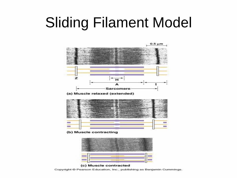
Sliding Filament Model

• At rest: the thick and thin filaments do not
overlap by much.
• During contraction: the length of each
sarcomere is shortened (distance from
one Z line to the next becomes shorter).
• These changes can be explained by the
sliding filament model of muscle
contraction.

Sarcomere: Unit of skeletal
muscle

Sarcomere. How large? In order to measure the length of
a sarcomere with a light
microscope:
1. Measure the distance in mm
from the start of 1 dark band
to the start of a dark band 10
bands away.
2. Divide by 10.
3. Convert mm to um by X
1000.
4. Find actual length by dividing
this length by magnification
of the micrograph, 200X.

• Sliding Filament Model: Based on the interaction of the structural protein molecules that make up thin and thick filaments.
• Myosin consists of a long, fibrous “tail region” with a globular “head” region sticking off to one side (I think of it as a golf club).
• The tail is where the individual myosin molecules cohere to form the thick filament. The myosin head is the center of bioenergetic reactions that power muscle contractions.

Skeletal Muscle Contraction

Control of muscle contraction:
Troponin,tropomyosin and Ca++ • 1. Skeletal muscle only contracts when
stimulated by a motor neuron. When the muscle is at rest the myosin binding sites on the actin molecules are blocked by the regulatory protein, tropomyosin.
• Another set of regulatory proteins, the troponin complex, controls the position of tropomyosin on the thin filament. For a muscle cell to contract, the myosin binding sites on the actin must be uncovered.
• 2. This occurs when calcium ions bind to troponin, altering the interaction between troponin and tropomyosin. The calcium binding rearranges the tropomyosin-troponin complex, exposing the myosin binding sites on actin.
• Calcium concentration in the cytosol of the muscle cells is regulated by the sarcoplasmic reticulum. The membrane of the SR actively transports calcium from the cytosol into the interior of the reticulum, which is thus an intracellular storehouse for calcium.

• 3. The stimulus leading to the contraction of a skeletal muscle cells is an action potential in a motor neuron that makes a synaptic connection with the muscle cell.
• a) synaptic terminal of the motor neuron releases neurotransmitter (acetylcholine) at the neuromuscular junction.
• b) the postsynaptic muscle cell is depolarized triggering an action potential in the muscle cell.
• c) the action potential is the signal for contraction. The action potential spreads deep into the interior of the muscle cell along infoldings of the plasma membrane called T (transverse) tubules.

• d) when T tubules contact the sarcoplasmic reticulum,
the action potential changes the permeability of the
sarcoplasmic reticulum, causing it to release calcium
ions.
• e) calcium ions bind to troponin allowing the muscle to
contract .

Myosin – Actin Interaction

4. Cross-bridge formation.
Using ATP • a) The head can bind ATP and hydrolyze
it into ADP and inorganic phosphate (we say that the head has ATPase activity).
• b) Some of the energy released by cleaving ATP is transferred to the myosin, which changes shape to a high-energy configuration. Myosin head is “cocked”.
• c) This energized myosin can bind to a specific site on actin, forming a cross bridge.

• 4. When this happens, the stored energy is released and the myosin head relaxes to its low-energy configuration.
• 5. This relaxation changes the angle of attachment of the myosin head to the tail. So as the myosin bends inward on itself, it exerts tension on the thin actin filament to which it is bound, pulling the thin filament toward the center of the sarcomere.
• 6. When a new molecule of ATP binds to the myosin head, the cross-bridge is broken. In a repeating cycle, the free head can then cleave the new ATP to revert to the high energy configuration and attach to a new binding site on another actin molecule farther along the thin filament.

Relaxation of muscle
5. Muscle contractions stop when the
sarcoplasmic reticulum pumps the calcium
back out of the cytosol into SR and the
tropomyosin-troponin complex again blocks
the myosin binding sites as the
concentration of calcium falls.
Acetylcholinesterase breaks down
acetylcholine in the synapse.