Embed Size (px)
Citation preview
بسم اهللا الرمحن الرحيم
History Taking Introductory Course
Prepared by
Hanan Ibrahim Mansour
Edited by
Abdallah Beano Ala’a Azzouqa
Abdallah Mansour Anas Al
Rema Al-Jondi Doua’a Sallam
Abeer Yassin Fadwa Al
Eman Sadaqa Basma Al
Suha Abu-Khalaf Shatha Dmour
Sahar Almustafa Mohammad Zmaili
بسم اهللا الرمحن الرحيم
History Taking & OSCE Examinationourse for Fourth Year Medical Students
Ala’a Azzouqa
Mansour Anas Al-Bawaliz
Jondi Doua’a Sallam
Abeer Yassin Fadwa Al-Qadi
Basma Al-Nashash
Shatha Dmour
Mohammad Zmaili
Published by
OSCE Examination for Fourth Year Medical Students
Published by

2 | P a g e

3 | P a g e
Preface OSCE is the abbreviation of “Objective Structured Clinical Examination”. This examination tests your clinical skills in gathering medical information from patients by history taking and physical examination. It is structured around a differential diagnosis of the presenting complaint; which is the systematic method of diagnosing a disorder (e.g., headache) that lacks unique symptoms or signs. OSCE is made up of few stations (6 for the introductory course); in each you spend few minutes (5mins for the introductory course) to gather the medical information required. Questions are easy to answer if you had studied them and come in short statements. The exam is done on models (your colleagues) not true patients. You are obligated to comment on each step as you proceed along the examination. An examiner will be present in the room, but he’s not supposed to talk neither instruct you through those 5 minutes … so don’t try to ask for his help, he’s is present to assign you a mark only. 5 minutes may seem short time, actually they are not! … 5 mins will be enough Insha’a Allah to answer the provided question if you used them wisely. Don’t panic, Don’t hesitate making ‘mmmm’ or ‘ahhhhh’, and be polite. Self-confidence and clear voice are essential to master this exam. Preparing for OSCE requires daily practice. Don’t leave its study for the last week of the semester. Try to practice one new physical examination skill each day, because practice is the key to master OSCE. Never be satisfied with your skills, try always to improve and fine tune them. This dossier is intentionally made for daily-life clinical practice, to make your understanding of the provided topics more comprehensive. I’ve intension to make an exam night review version, asking Allah to give me good well and time to manage that. The dossier is the gather of practice and learning from residents and colleges plus reading OSCE books and websites, but in the core is based on Macleaod’s Clinical Examination Book 12th edition. I highly recommend that you build a good relationship with Macleaod’s book; I think of it as the heart of clinical books. For any suggestions to improve the contents or the design, please contact me on; http://www.facebook.com/hananmnsr or email me on; [email protected] Your suggestions are welcomed may Allah bless you

4 | P a g e
Acknowledgement
I am grateful to all those medical students who helped or contributed once in the past for making OSCE dossier; for ours being a continuation of theirs. I would like to thank MD. Mai Al-toos, my introductory course instructor, for her tolerance, encouragement, and dedication for teaching. I greatly appreciate the work of the editors named, that was precise and of value to develop the content of this dossier. Finally, with a heart full of gratitude, I would like to thank our medical committee, named Saleh Roman, Anas Al-bawaliz, Abdallah Mansour, Belal Abu-hazem and Bara’a Al-sharqawi, for their giving and support through those hard years of medical school.
I am and all my work is the fruit of my parents, for whom I’m grateful to the end of my life, and for whom I dedicate this work.
بسم اهللا الرحمن الرحیم
( فمن یعمل مثقال ذرة خیرا یره ۞ ومن یعمل مثقال ذرة شرا یره ) سورة الزلزلة – اآلیتان 7 و 8
... اسھذا العمل وانفع بھ النفي في میزان حسناتنا، وبارك اللھم یلة برحمتكقاللھم تقبل أعمالنا خالصة لوجھك الكریم، واجعلھا ث آمین

5 | P a g e
Contents
Section 1: History
Gathering information ……………………………………………………………….. 8
Section 2: Physical Examination
Settings of the Physical Examination and General Rules …………...…….......... 18
The General Examination …………………………………………………………… 21
Examination of the Thyroid Gland Function …………………………………….… 32
Cardiovascular System Examination ………...………………………………….….. 36
Respiratory System Examination ……………………………………………….….. 46
Gastrointestinal & Renal Examination …………………………………………..… 54
Musculoskeletal System Examination …………………………………………..…. 61
Nervous System Examination………………………………………………….…… 80
Section 3: Appendix
Abbreviations ………………………………….……………………………..….…… 90
Previous years OSCE stations ………………………………………………………. 91
History application form …………………………………………………….….…… 92

6 | P a g e

7 | P a g e
Section
1
History

8 | P a g e
Gathering Information ||____ Patient profile:
Patient’s name, age, sex, marital status, address and job
Place of admission (floor, ICU, CCU, burn unit…etc)
Source & time of referral (OPD, ER, other hospitals…etc)
Source of history (patient himself, relative, healthcare worker…etc)
History taken by who, time & date of history taking
||____ Presenting/ chief complaint:
The major problem in the pt’s own words with its duration. Ex. cough of 3 days prior to admission, knee pain of 3 years duration … etc
Do NOT use medical terminology. Ex. use shortness of breath instead of dyspnea, vomitting of blood or dark vomitus instead of hematemesis, abnormal shaking movements instead of tremor… etc
Clarify what the patient means by any term he/ she uses; avoid jargons! Ex. if the patient says he has a "funny feeling in his head", clarify by asking "What do you exactly mean by "Funny feeling in your head"?? ** Medical Jargon is to use medical terms while talking with patients, they may consider it as a rude behavior.
The chief complaint must be precise and concise
||____ History of presenting illness:
It’s an analysis of the presenting complaint
Involves active listening & reflection:
Let the patient tell his own story, summarize what you have understood and ask for further clarification.

9 | P a g e
For any presenting complaint (typically pain but can be applied to ANY complaint with some modifications) describe ‘SOCRATES’:
1. Site; localized or generalized.
2. Onset; gradual, sudden, abrupt. Mention associated circumstances (ex. Playing football, watching TV, getting off bed…etc). Always begin with asking "What were you doing when the problem started?” the answer will reveal many details about the onset and the accompanying circumstances.
3. Character; sharp, dull, burning, tingling, boring, stabbing, crushing, tugging…etc It’s preferred to use the pt’s own description rather than offering suggestions.
4. Radiation ؟؟أو یبقى مكانھ ؟؟ بیسمع لمكان آخر في جسدك/ بینتقل/ بیضرب األلم
5. Associated symptoms usually ask about nausea, vomiting, fever, chills (قشعریرة)/ rigors ) ارتجاج الجسم من البرد( , weight loss, headache, sweating, cough or any other related symptoms and explain them briefly
6. Timing; duration, course, and pattern. Either episodic (give duration & frequency) or continuous (describe change in severity along a specified time coarse like a day or a week)
7. Exacerbating and relieving factors; is it exacerbated by movement, light, sound …etc is it relieved by rest, darkened room, pain killers, … etc
8. Severity; using a scale of 10 where (0) no pain and (10) maximum pain similar to toothache or pain of labor. An alternative way to assess the severity is to ask if the pain woke the patient from sleep (very severe), did he come to the hospital driving and if he is capable of doing activities of daily living (not very severe), you can comment "the pain was severe enough to wake the patient from sleep"...etc
# ask following SOCRATES, but write in history as follows; character, then site and radiation, then onset and timing, then severity, then exacerbating and relieving factors, finally associated symptoms with brief description for each… this way is more logically synchronous for listener to follow up with you #
10 | P a g e
Previous history of similar complaint. If present define change from current one.
Include review of the system of the presenting illness i.e. if the pt has cough, you must mention review of respiratory system.
Include other parts of the history if relevant ex) smoking status, family Hx, travel Hx,…etc
Write down investigations which were done for the presenting illness, and medications given before admission – as in the ER
Explain what happened to the pt from the time he sought medical advice to the current time
At the end of the conversation, summarize the main points mentioned by the patient back to him, giving him the chance to correct or add anything [active listening & reflection!!]
Effects on lifestyle ‘FIFE’ Feelings related to the illness Ideas on what is happening to him Functioning in the terms of impact on daily life Expectations of the illness and you the doctor; modern medicine may be unable to cure the problem, and the important issue is what you can do to help a pt to function [[it’s preferred to avoid this section of history (FIFE) at fourth-year level, because this will probably provoke patient’s feelings you are not trained to deal with them]]
Example (of SOCRATES modification); whenever the patient says that he has feeling of hotness you have to ask him; 1- duration 2- documented or not and if documented ask about the degree and the route by which it was measured ( orally, rectally ...) 3- onset (gradual or sudden and the rate of development 4- continuous or intermittent or if it has a specific pattern 5- associated symptoms (chills, rigor, sweating, night sweating...) 6- exacerbating and relieving factors 7- timing and diurnal variations
||____ Systematic enquiry (review of systems):
1. General:
>well being whether good or poor
> appetite whether good or poor
> weight change; whether recorded by
measuring weight or perceived by change
in size of clothing
> energy whether good or poor
> sleep whether good or poor
> mood whether good or poor

11 | P a g e
2. Cardiovascular system:
> Chest pain; ask SOCRATES
> breathlessness;
(a) on lying flat (orthopnea); ask for
number of pillows, and what happens if he
lay flat.
كأنك عم مخنوق و لى ظھرك تشعر نفسكأول ما تنام ع
!تغرق؟
(b) At night (PND); ask at which time
wakes him up at night
بتصحى باللیل بتحس حالك مخنوق و بدك ھوا؟؟
(c) on minimal exertion; ask about type of
exercise; walking to car or bathroom, dish
washing…etc or determine distance in
meters, stairs etc.
> Palpitation; ask pt to tap out with his
fingers for rate and rhythm, onset &
termination (abrupt/ gradual),
precipitating factors (e.g. coffee, exercise,
emotional stress), frequency and duration
of episodes, whether they’re exacerbated
or relieved by exercise
.خفقان بقلبك؟؟ أوصفلي اییاه/ بتحس برفة
> Pain in legs on walking (claudication),
ask about distance that provoke pain, and
if pain relieved on rest, unilateral or
bilateral, and location
>ankle swelling; ask if persistent or comes
and goes
3. Respiratory system
> SOB (exercise tolerance)
> cough; ask about its sound, dry/
productive, timing (daytime/ nocturnal),
associated features, exacerbating &
relieving factors
> wheeze; on inspiration or expiration,
persistent or not
> sputum production; ask about its type
(serous/ mucous), amount, color
(purulent/rusty), timing, smell, solid
material.
> hemoptysis; ask about its volume and
nature, duration and frequency
> chest pain due to inspiration or coughing
4. Gastrointestinal system:
> Mouth;
oral ulcers (painful/ painless, recurrent/
not), dental hygiene or recent dental
procedure
> difficulty swallowing (dysphagia);
determine if for solids or liquids, and
occurring at which level (ask the pt to
point!)
> Painful swallowing (odynophagia)
Make sure that the patient means either
odynophagia or dysphagia as they may
mix between them! Ask: Is it pain that
stop you from swallowing or you just can't
swallow??
> Nausea & vomiting;
describe vomitus in terms of color,
amount, content (intact or digested food),
projectile or not
> hematemesis
> indigestion (عسر ھضم)
> heartburn المعدة؟؟ رأس حرقة ب
> Abdominal pain; ask SOCRATES
> change in bowel habits; increased or
decreased frequency of passing stool.
Mention normal stool habit.
> Change in color of stool; pale, dark, tarry
black, fresh blood
> change in stool consistency.

5. urinary system
>pain passing urine (dysuria);
state if at beginning or end or throughout
urination
> urgency & frequency (at night; nocturia)
> hematuria
> incontinence (stress/ urge)
> libido; state if impaired
> multiple sexual partners (unprotected
intercourse)
6. Genital system:
[always begin with apologizing about the following questions but emphasize the importance of
asking such question in reaching the specific diagnosis , usually patients won't mind answering
these questions if you had a good approach.]
>for men ‘if appropriate!!’
Prostatic symptoms; hesitancy, poor stream or flow (Is the urine stream weak that it spoils your
clothes?), terminal dribbling, urethral discharge, erectile difficulties.
> For women
last menstrual period (consider pregnancy), timing & regularity of period, abnormal bleeding,
vaginal discharge, contraception, if appropriate ask about pain during intercourse
(dyspareunion)
7. Endocrine system:
> Heat or cold intolerance
> Change in sweating
> excessive thirst (polydipsia)
8. Musculoskeletal system:
> Joint pain
> stiffness
> joint swelling
> restricted mobility (limited range of
motion in a particular joint)
> falls (explain why the fall occurred, did it
cause any medical problems/disabilities
etc.)
9. Nervous system:
> Headache
> dizziness; vertigo (الدنیا بتلف فیك؟) or light-
headedness ) شعرت نفسك غیر متزن(
> faints (loss of consciousness) اغماء
> fits (seizures, abnormal contractions)
تشنجات
> altered sensation; like tingling, pins and
needles …etc
> weakness
> visual disturbances; like double vision,
loss of color vision …etc
> hearing problems; like tinnitus … etc
> memory & concentration change
10. Others for bleeding diathesis;
> bleeding or bruising
> skin rash

13 | P a g e
|| _____ Past Hx:
Medical: chronic illnesses (DEATH; Diabetes, Epilepsy, Asthma, Thyroid, Hypertension) and obstructive sleep apnea, or others as anemia and dyslipidemia …etc previous hospital admissions; when, where, why and length of stay history of blood transfusion; when, where, why, frequency and complications
Surgical: when, where, why, complications and anesthesia type and complications
Obstetrical (for females): Last menstrual period, age of menarche and menopause, number of pregnancies & complications, type of delivery & complications, any abortion & if a defined cause were given and family planning method.
|| _____ Drug Hx:
Ask about prescribed drugs, OTC and alternative remedies (herbs …etc)
For each medication know name (generic/ scientific), dose, dosage regimen, duration, indication and if any side effects were encountered upon use
Assess patient compliance; by asking the pt to describe how and when they take their medications, their names, and indication of usage. **give them permission to admit that they don’t take all their remedies by saying ‘that must be difficult to remember’
Ask about allergy, and clarify what the pt exactly means by it; is it simple rash or anaphylactic shock?
|| _____ Family Hx:
Start questioning by ‘are there any illnesses that run in your family’
Document illness or age of death in first degree relatives (parents, sibling, children)
If there is a suspicion of an inherited disease go back for three generations & obtain details of racial origin, consanguinity or adoption
Inquire if any family member suffers similar complaints as the pt.
Draw a pedigree chart

14 | P a g e
|| _____ Social Hx:
Understand your patient’s personal constraints and support. Patients may ask why these questions
are being asked, so you have to explain how every question is related to the patient's case/
complain.
Lifestyle: exercise, diet, marriage, homing (owned/ rented, living at which floor & if living with others), hobbies, pets
Occupational Hx: current & previous occupation, exposure to hazards, unemployment (reason & duration), attitude to job
Travel Hx: when, where, observations after return, type of accommodation, activities undertaken
Sexual Hx (Only when relevant): casual relationship; regular sexual partner (male/ female), irregular (how many in the past year, whether male/ female)
Tobacco & hubble-bubble (water pipe): ever smoked; how long, what type, how much (use pack years = (no. of cigarettes smoked per day* years ) / 20)… classify as current smoker vs. x-smoker ask non-smoker about exposure to smoke at work or home (passive smoking)
Alcohol: amount and type of drink, daily/ weekly pattern of drinking, usual place of drinking, alone or accompanied, purpose, amount of money spent on alcohol, attitude to alcohol (CAGE; cut down, annoyed, guilty, eye opener) state whether drinks or doesn’t drink alcohol [don’t say not alcoholic] calculate units drunk per week; 1 unit = 25 ml of 40 % alcohol = 10 ml of ethanol X % = X units of alcohol / L
Hx of vaccination ask if fully underwent national vaccination program at school. For travelers, ask about boosters taken before travel. For health care workers, ask about hepatitis B vaccine and influenza vaccine.
Religion
Drug abuse
Insurance system *** very important to ask

15 | P a g e
History Taking Technique (for medicine and surgery rounds)
1. Prerequisite: Introduce yourself, ask for permission, and ensure conversation is private.
2. Take Patient profile.
3. Ask about chief complaint and its duration.
4. Define complaint and establish the cause using a mind-built list of differential diagnosis.
5. Inquire about the presence of previous similar complaints, if present compare to the current one.
6. Establish risk factors.
[[Any disease can be inquired about using the aforementioned steps. So, in your medicine and surgery rounds that will come try to build your studying of history taking using those step, you will find it very helpful and easier to remember enshallah ]]
At the end of the dossier you will find a History taking form, my suggestion is to use it for a month so as to memorize it then to leave it and start taking history by your own

16 | P a g e
17 | P a g e
Section
2
Physical
Examination

18 | P a g e
The Settings of the Physical Examination
and General Rules
||____ Settings at each station:
Greeting ( بركاتھ و اهللا رحمة و علیكم السالم )
Introduce yourself ( .... سنة طب ـة\طالب ،........ اسمي )
Address patient by name, ‘also if possible by date of birth and file number’
Ask for permission and be specific ( .... صدرك، بطنك، افحص ممكن )
Wash your hands with water and soap or ask for sterillium
Examination room should be; 1. Private
Ask for chaperone if to examine female patients, tactfully ask all relatives to leave the place. Parents should always be present when you examine children.
2. Warm 3. Well lit (prefer sunlight) Comment by saying (There is adequate privacy, warmth and illumination)
Exposure; o Seek permission before exposure and be specific. o Expose the specified area only and cover the rest of the pt’s body with a blanket to
ensure the pt doesn’t become cold!! o Re-cover the pt’s body before commenting on the examination
Position of the pt
Position of the examiner
Thank pt when you finish examination ( ( خیرا اهللا جزاك. ..جزیال شكرا
19 | P a g e
||____ Don’t speak in English with your pt, unless he can understand english
||____ How to assess consciousness and orientation (in time, place, and person) [this is a simplified examination, as you go further in your neurology study Inshallah you would be
given detailed examination of consciousness and orientation under cognition tests]
Conscious >> Spontaneous eye opening
Oriented in time >> ؟في أي وقت نحن اآلن من الیوم
Oriented in place >> أین أنت اآلن ؟
Oriented in person >> ؟ھل الشخص الذي یجلس بجوارك قریبك أو من أنا؟
||____ Rules of inspection:
Don’t touch the pt
Look from all angles to all sites (i.e. rotate your head around the pt’s body)
||____ Rules of palpation:
Ask for permission
Ask for any site of pain and leave its examination to the end; if you provoke additional
pain, pt will refuse completion of examination.
Warm your hands very well by rubbing them to each other.
Maintain eye-to-eye contact throughout palpation **Students often miss this point!
||____ Rules of percussion:
Make sure to percuss using the correct maneuver
I.e. use the tip (not pad) of right middle finger to tap the dorsal surface of left middle
phalanx of middle finger, swinging your hand at the wrist joint with elbow held still
Assess symmetry: whenever you percuss a point, percuss the point opposite to it on the
other side of the midline i.e. in a zigzag pattern.
||____ Rules of auscultation:
Warm head of stethoscope before putting it on the pt’s body
Assess symmetry: whenever you auscultate a point, auscultate the point opposite to it on
the other side of the midline i.e. in a zigzag pattern.

20 | P a g e
||____ Examination of System Vs Organ Vs examination for a disease:
Examination of a system;
when you are asked to examine a specific system (as for example gastrointestinal
system), first fulfill the settings, then do first impression and go through all parts of the
specified system to be examined
Examination of an organ;
here you examine a specified organ (as for example the abdomen), first fulfill the
settings, then go directly to examine the specified organ without going through first
impression, nor vitals, nor hands nor face
# in the settings of an OSCE examination, you may be even asked to do part of an organ
examination only (as for example to auscultate the abdomen)
Examination for a disease;
if you were asked to examine a patient for a specified disease (as for example jaundice),
you need to look for signs of this disease all over the patient’s body taking in
consideration any system affected by the disease, plus looking for clues of possible
causative conditions (as in this example signs of chronic liver disease, liver failure,
hemolytic anemia and obstructive causes)
||____ Whenever you are asked to examine a paired part in the body, compare it to the other side
Ex. If you were asked to examine a limb in one side, compare it to the other side limb. If you were asked to examine right eye, compare to the left eye If you were asked to examine right breast, compare to the left breast … etc [I.e. for symmetry vs. asymmetry]
||____ Make it a story, and build your own MINDMAP to memorize the examination sequence
and details. Don’t try to memories the examination in a similar way as a robot does, not making
connections between points. Be smart by doing categorization and nested lists.

21 | P a g e
The general examination
||___ First impressions
[When u see a pt, u first look at his face, smile at him and shake his hand, then look to general body
demeanor, clothing and skin complexion… pull a chair to sit on beside the pt and try to take a deep
breath smelling him for any recognizable odor] كن لماحا
Describe the pt body position when you first saw him
Ex) the pt was sitting in a chair, lying flat on his bed, sitting at the edge of his bed,
standing, leaning forward…etc
Connections
Ex) connected to IV fluid line, has a cannula in his right hand, connected to a Foley’s
catheter, on an oxygen mask …etc
Handshake;
describe whether the hands upon handshaking were hot/cold, dry/sweaty, large and
fleshy, delayed relaxation of grip, deformed hands/ fingers…
Facial expression and general demeanor
mention whether the pt maintain eye-to-eye contact or not, anxious, apathetic, startled,
agitated, lugubrious, comfortable, breathless, in pain, cachectic, obese…etc
Clothing
Ex) dirty, baggy, tattoos, MedicAlert bracelet, necklace highlights…etc
Unusual skin complexion
Ex) cyanosed, pale, yellowish, orangish, whitish albino…etc
Body odors
Ex) mousy, fishy, sweet, fetid, tobacco, alcohol, marijuana, halitosis…etc
Spot diagnoses of a specific disease
[[This skill –first impression- will improve by time, so be patient and DO PRACTICE!]]

22 | P a g e
||___ Assess this pt VITAL SIGNS
1. Pulse
For 1 min, from radial artery by 3 fingers against radius bone
Don’t focus too much on the watch while counting; so as not to miss count
Record in bpm (beat per minutes)
2. Respiratory rate (RR)
Take for 1 min; record as breaths/min (1 breath = inspiration + expiration)
Ask pt to put his palm hand above his chest; for ease of examination
Act as if you’re taking the pulse, so that the pt doesn’t get anxious affecting the rate
of breathing
3. Blood pressure (BP)
From brachial artery; bladder of the sphygmomanometer cuff over brachial artery,
pump of the sphygmomanometer on radial side (thumb side)
Measure blood pressure bilateral, in supine and standing positions
If you can’t take standing take it sitting with legs hanging out of examination couch
Record in mmHg, which arm were used, in which body position (supine/ standing)
4. Temperature (Temp)
Use a thermometer; oral, axillary, or rectal
For oral; put under tongue, ask pt to close on it by lips, leave for at least 3 min
Record in degree Celsius (oC)
5. Body mass index (BMI)
Weigh pt and take his height
Record in kg/m2

23 | P a g e
||___ Examine this pt’s HANDS
[[Examine hands bilateral (right and left) unless specified in an exam situation]]
1. Settings Greet your patient, introduce yourself then ask for permission to examine his hands
Wash your hands. Ensure adequate privacy, warmth and illumination of the room.
Tactfully expose patient’s hands up to the elbow, remove any jewelry.
Put a cushion under the pt’s hands and then stand on the right side of the pt.
2. inspection Look for dorsal then palmer aspects of the hand then between fingers
Comment on: 1. swelling 2. scar 3. polydactylism 4. arachnodactyly 5. flexed hand deformity 6. Dupuytren’s contracture 7. Single palmer crease 8. Palmer creases
pigmentation 9. Tobacco (tar) staining 10. venipuncture marks 11. fungal spores 12. Palmar erythema
At the dorsum, look for: 1. Swelling 2. Scars 3. Polydactylism 4. Flexed hand deformity 5. Arachnodactyly 6. Tobacco (tar) staining
At the palm, look for: 1. Swelling 2. Scar 3. Flexor surface of the wrist and forearm for
venipuncture marks 4. Single palmer crease 5. Palmer creases pigmentation 6. Dupuytren’s contracture 7. Palmar erythema
Ask pt to abduct fingers and look in between for
fungal spores
Skin normally hair is only present on the dorsum of the
hand and the proximal phalanx
Comment on: 1. Hair 2. Callosities
Nails
examination for finger clubbing requires doing 3
maneuvers
1. look from lateral side of the finger for loss of
normal angle between nail and nail bed, and
increased nail curvature
2. fluctuation of nail bed
3. nails of opposite hands against each other, for
clubbing there is loss of space
Comment on: 1. cyanosis 2. koilonychia 3. leukonychia 4. Beaus’ lines 5. splinter hemorrhages 6. onycholysis 7. dilated capillaries in the
proximal nail fold 8. Finger clubbing;

24 | P a g e
Soft tissues *Wasting of both thenar and hypothenar eminencies present as flat palm. *Muscle wasting of intrinsic muscles of the hand present as dorsal guttering.
Comment on: Muscle wasting of thenar, hypothenar muscles, and intrinsic muscles of the hand
3. palpation General rules of palpation: Ask for permission to put your hands on the pt. Ask for any area of pain & examine it last Warm your hands up & maintain eye-to-eye contact throughout examination
Comment on: 1) Tenderness, Masses, skin
texture 2) temperature 3) tendons 4) joints
Assess temperature
Using the dorsum of your hands; as your palm is mostly sweaty and hot so will not give you a good perception of examined hand temperature. Always compare both hands and different levels of the same hand
Tendons Flexor and extensor tendons (as mentioned in MSS examination of the hand)
Joints MCPJs, PIPs, DIPs (as mentioned in MSS examination of the hand)
4. maneuvers Tremor or any abnormal movements
Fine tremor; arms extended, hands extended and pronated (you may put a sheet of paper above the dorsum of hand observe for shaking of the paper) you may not use the paper if the hands are obviously shaking
Flapping (coarse/ asterixis); hands extended and pronated at wrist, ask pt to dorsiflex hands and maintain, look for abnormal movements # you can also look for asterixis by asking the pt to squeeze your index and middle fingers and maintain this for 30-60 s. pt with a flapping tremor can’t maintain this posture
Others; like resting tremor (pill rolling of Parkinson)…etc
Comment on: 1) Fine tremor 2) Flapping tremor 3) Resting tremor

25 | P a g e
||___ Examine this pt’s TONGUE
1. Settings Greet your patient, introduce yourself then ask for permission to examine his
tongue.
Wash your hands. Ensure adequate privacy, warmth and illumination of the
room.
Tactfully ask patient to open his mouth for you to see his tongue, use a pen-light
torch for better view, and then stand opposite to the patient from front.
2. Inspection Ask the pt to open his mouth, look to his
tongue while inside, then ask the pt to put
out his tongue, move it right and left, and to
touch his palate with the tip of his tongue.
Tongue fasciculation is examined while the
tongue is inside the mouth
Comment on: 1) Tremor 2) Fasciculation 3) Macroglossia 4) Tongue furring 5) Geographic tongue 6) White patches 7) Glossitis 8) Central cyanosis
3. Sensory
examination
Facial and glossopharyngeal nerves are
responsible for taste sensation of the tongue,
examine by allowing pt to taste different
types of food. It’s of importance that pt can
sense difference of food taste rather to
exactly name it.
Trigeminal nerve is responsible for touch
sensation of anterior two-thirds of the
tongue; ask the pt to open his mouth, close
his eyes, and to make a sound each time he
feels a thing touching his tongue. Do it twice
at each tongue halves (once left, another
right)
Comment on: Tongue sensation of touch and taste
4. Motor
examination
Hypoglossal nerve examination; as
mentioned in CN XII examination in nervous
system.
Comment as in motor exam of CN XII

26 | P a g e
||___ Examine the LYMPH NODES
1. Settings Greet your patient, introduce yourself then ask for permission to examine the
specified lymph node area
wash your hands. Ensure adequate privacy, warmth and illumination of the
room.
Tactfully expose the area of interest and its drainage territory
Position of pt and examiner varies from LN to another
2. Inspection Look for visible lymphadenopathy Comment; no/visible enlargement at the site of the lymph nodes of examination
3. Palpation General rules of palpation: Ask for permission to put your hands on the pt. ask for any area of pain & examine it last warm your hands up & maintain eye-to-eye contact throughout palpation
Comment;
Enlarged LN; located at….., it’s ….. cm in diameter, hard/ soft/ rubbery/ matted in consistency, non/ tender, not fixed/ fixed to underlying tissue and/ or skin
If no enlargement comment; no palpable LN and no tenderness
Lymph node:
Palpate one side at a time using the pad of your finger ,compare with the nodes on the contralateral side
Assess; Site, size, consistency, tenderness
Determine whether the nodes are fixed to;
Surrounding and deep structures; by moving it along X and Y axis if overlying a muscle (ex sternomastoid) put the muscle into action and assess mobility thereafter, if still palpable then it’s not fixed to the muscle.
Skin; by pinching a skin fold over it.
Drainage territory Comment; no/a swelling/ wound/ inflamed area has been detected along drainage territory

27 | P a g e
Cervical LN:
Pt sitting, using 3 fingers in a rotational movement
Examiner position and LN group examined;
From behind;
- submental, submandibular, pre-auricular; both sides can be palpated simultaneously.
- tonsillar, deep cervical nodes in the anterior triangle, supraclavicular, and scalene; palpate
each side at a time.
# Scalene by placing index between the sternomastoid muscle and clavicle, ask pt to tilt head
to the same side and press firmly downwards toward the first rib.
From front;
posterior auricular, occipital, deep cervical of posterior triangle; both sides can be palpated
simultaneously.
Axillary LN:
With the patient seated, position yourself in front of him or to his side, and abduct his arm
and support it with your ipsilateral hand. Palpate with the finger tips of your contralateral
hand. E.g. if the right was to be examined, your right hand will support the patient’s arm and
you’ll palpate with your left.
Apical, medial, anterior, posterior and then lateral groups;
Compress the LN of the anterior and posterior groups against the anterior and posterior
axillary fold, respectively.
Epitrochlear LN:
Pt sitting, elbow partially flexed, wrist supported with contralateral hand, use thumb of
ipsilateral hand to palpate medial aspect of the elbow
Inguinal LN:
Pt lying supine
Palpate over the horizontal chain (just below and parallel to inguinal ligament) and vertical
chain (along the saphenous vein)
Popliteal LN
Pt lying supine
Palpate under the knee in the popliteal fossa

28 | P a g e
||___ Examine this LUMP (or this SWELLING)
1. Settings
Greet your patient, introduce yourself then ask for permission to examine his lump
wash your hands. Ensure adequate privacy, warmth and illumination of the room.
Exposure and position depends on the lump location
2. Inspection [4S];
Describe Site, Shape, Size and Skin overlying the lump
3. Palpation [TT CS FAP light];
General rules of palpation: Ask for permission to put your hands on the pt.
ask for any area of pain & examine it last
warm your hands up & maintain eye-to-eye contact throughout examination
1. Tenderness
2. Temperature; using the dorsum of your hand
3. Consistency; soft/hard
4. Surface and edge; whether surface is smooth/nodular. And whether the edge is; well
delineated/ ill defined, regular/irregular, sharp/rounded
5. Fluctuation; in both axis.
Remember to fix the lump in place with your middle finger and thumb, while doing the
maneuver with your index.
6. Attachment; to overlying skin or underlying tissues
7. Pulsation, thrills or bruits
8. Transillumination; using a torch
4. Examine draining lymph nodes
[[This method is of MD. Sameer jab3ete, I find it more logically synchronous than the one in Macleod’s. You
can follow either]]

29 | P a g e
||___ Measure this pt WEIGHT AND HEIGHT
1. Settings
Greet your patient, introduce yourself then ask for permission to measure his weight
and height
wash your hands. Ensure adequate privacy, warmth and illumination of the room.
measure the pt weight in his in-door cloths barefooted
2. Inspection
1. abnormal stature or body proportions
2. Abnormal fat distribution. Ex) truncal obesity, back hump…etc
3. Evidence of malnutrition or specific vitamin deficiencies
3. Measure pt height in cm
4. Measure pt weight in kg
5. calculate and Record BMI in Kg/m2
6. measure waist circumference;
1. Pt standing
2. Measure at a point equidistant between the costal margin and the iliac crest
3. Record maximum diameter over any abdominal fat not under it.
7. Calculate waist: hip ratio
Quick info:
We measure waist
circumference and hip
ratio as part of definition
for ‘‘Metabolic Disease’’.

30 | P a g e
||___ Assess this pt HYDRATION STATUS
1. Settings Greet your patient, introduce yourself then ask for permission to assess his hydration status wash your hands. Ensure adequate privacy, warmth and illumination of the room. Tactfully expose the area of interest. Stand on the right side of the pt
2. First
impression
Check for :
Consciousness
Orientation
Sunken eyes
Comment on general appearance of the pt with noting ; 1. Consciousness 2. Orientation 3. Presence of sunken eyes
3. Vital signs Pulse in radial artery of both hands while supine then in one hand while standing. >>in dehydration it is elevated (by 30) unless the dehydration is severe then it’s decreased
BP is measured while supine and while standing to check for postural hypotension which indicates vascular volume depletion. >>In dehydration blood pressure is decreased (by 20 systolic or 10 diastolic).
Respiratory rate is increased in dehydration.
Temp >> not measured
BMI not calculated but weight loss is useful in determining the amount of fluid lost if usual weight is known (1 L fluid loss = 1 kg weight loss)
Comment on :
1. Pulse & postural tachycardia
2. BP & Postural hypotension
3. RR 4. Weight loss if usual
weight known
4. Hands We palpate for temperature and determine if they
were dry/ sweaty.
Comment if hot/cool and
dry/sweaty.
5. Face Inspect for sunken eyes
Inside the mouth for dry mucous membranes or dry
tongue (but not very reliable since commonly caused
by breathing through the mouth)
Comment on:
1. Appearance of the eyes
2. Presence/ absence of dry
mucous membranes in
the mouth.

31 | P a g e
6. Neck Check skin turgor by gently pinching a fold of skin at the manubrium of sternum and holding it for a few seconds then letting it go. If normal it will return to its original state promptly, while in dehydration it’s going to take longer time.
!! This area is free of subcutaneous tissue, hence being used.
Comment if there is loss of
skin turgor or not.
JVP examination
>> JVP is low in dehydration.
Comment on JVP
Examine axilla for sweating and dryness. Comment whether the axilla
is dry or sweaty.
7. edema Examine for sacral edema (in bedridden patients)
and ankle edema (in mobilized patients); by applying
firm pressure with your thumb for 15 seconds or
more. The pressure you apply should be enough to
turn your pink nail-bed white.
Comment if there is sacral or
ankle edema

32 | P a g e
Examination of the thyroid gland function
1. settings Greet your patient, introduce yourself then ask for permission to examine his neck
wash your hands. Ensure adequate privacy, warmth and illumination of the room.
Expose neck and upper chest.
Position the patient sitting upright on a chair and ask for a glass of water
Stand on the Rt side of pt for general assessment then move behind him (posterior
approach) to examine thyroid gland, but ensure to maintain eye contact as possible
2. first
impression
Assess consciousness and orientation; coma (acute
hypothyroidism), loss of consciousness (severe
hypothyroidism)
Describe appearance and general demeanor
(hyperactive/restless or slow, fat/thin, appropriateness
of clothing to weather).
Describe facial expression (apathy/startled) & abnormal
ticks.
Hand shake (temp and sweat; cold &dry in
hypothyroidism, sweaty &warm in hyperthyroidism.)
Ask the patient to speak and cough
Comment on:
1. Consciousness and orientation
2. Facial expression and general demeanor
3. Activity. 4. Hand temperature and
sweat. 5. Hoarseness of the voice 6. Bovine cough
3. Vitals Pulse; tachycardia and atrial fibrillation
(hyperthyroidism), bradycardia and first degree heart
block (hypothyroidism).
Test for collapsing pulse (for Graves).
Measure blood pressure, wide pulse pressure
(hyperthyroidism)
BMI; obese (hypothyroidism), cachexia
(hyperthyroidism)
Comment on :
1. Pulse Collapsing pulse.
2. RR 3. BP 4. Temperature 5. BMI
4. Hands Inspect both dorsal and palmar aspects.
Test for fine tremors.
Test for clubbing (thyroid acropachy).
Palpate for temp and sweating.
Comment on :
On palmer aspect;
1. Muscle wasting which is evident in thenars due to carpal tunnel syndrome in hypothyroidism
2. Temp and sweat.
33 | P a g e
On dorsal aspect; 3. Hand hair 4. Onycholysis. 5. Onychotillomania (hyper) 6. tremor 7. Digital acropachy
(clubbing, digital soft tissue swelling, subperiosteal new bone formation)
5. Face Inspect for:
Texture of skin ( dry-coarse in hypothyroidism and greasy-sweaty in hyperthyroidism)
Hair loss (occurs in both) or eyebrow thinning (outer 1/3 in hypothyroidism).
Assess for eyes manifestations:
1. Proptosis by looking at the seated pt from behind and above.
2. Exophthalmos: inferior limbus appears larger than the superior limbus in the eye, measured using exophthalmometer.
3. Lid retraction: superior limbus is visible. 4. Lid lag (ophthalmoplegia) by asking pt to follow
your finger as you move it quickly in a downward fashion from a point above the eye
5. Chemosis which is redness of the eye globe. 6. Periorbital edema 7. Corneal damage (ulcers) [1-7 signs of hyperthyroidism]
8. Bilateral ptosis (severe hypo) 9. Periorbital myxedema 10. Large tongue [8-10 signs of hypothyroidism]
Comment on :
1. Texture of skin. 2. Hair loss & eye brows 3. Proptosis 4. Exophthalmos 5. Lid retraction. 6. Lid lag (ophthalmoplegia). 7. Chemosis 8. Periorbital edema 9. Corneal damage 10. Ptosis
11. Periorbital myxedema. 12. macroglossia

34 | P a g e
6. Neck
<<Examine
thyroid
gland>>
Inspection [neck hyperextended]
Ask pt to swallow water and then look from lateral side of the neck for any moving bulging mass
With the pt’s jaw slightly open and ask him to protrude his tongue to note for thyroglossal duct cyst which moves on tongue protrusion (thyroid and goiters don’t move with tongue protrusion.)
Use flash light to look at the back of the mouth for lingual goiter.
Do Pemberton’s maneuver; ask the pt to abduct both his hands above his head to check for retrosternal extension of the thyroid goiter. if present, it would compress the SVC causing engorgement of the neck veins, a plethoric face, elevated JVP and sometimes even fainting (positive Pemberton’ sign)
Comment on :
1. Wounds/ Scars (thyroidectomy scars are cosmetic and difficult to detect)
2. Swellings, nodules or cysts.
3. Redness 4. Any mass that moves with
swallowing or protrusion of the tongue.
5. Presence of lingual goiter. 6. Positive or negative
Pemberton’ sign.
Palpation [neck slightly flexed]:
General rules of palpation: Ask for permission ask for any area of pain & examine it last warm your hands up & maintain eye-to-eye contact throughout examination
Rest your hands gently on each side of the lower aspect of the neck between the larynx/trachea and sternomastoid. You should be standing behind the pt. don’t use both hands in palpation; use one to push towards the contralateral side and the other to palpate in circular fashion over the thyroid lobe.
While palpating, ask the pt to swallow and comment if any movements occur with swallowing.
Palpate suprasternal notch; if there is retrosternal goiter the suprasternal notch will not be palpable.
Palpate cervical lymph nodes.
Comment on :
1. If thyroid is palpable or not (palpable in 25% of males); if palpable comment on : 1. Size and surface
(symmetry, smooth or not , nodularity)
2. Consistency (hard or soft)
3. Tenderness. 4. Mobility (Movement
with swallowing) 2. Hotness of overlying skin 3. Thrills 4. Non/ Palpable
suprasternal notch 5. Cervical
Lymphadenopathy

35 | P a g e
Percuss directly on the manubrium of sternum.
Normally resonant note, dull
if goiter reached upper
mediastinum.
Auscultate with diaphragm for bruits over both thyroid
lobes which is heard in hyperthyroidism.
Comment if bruits are heard.
7. limbs Upper limbs for proximal myopathy; To examine, ask
patient to abduct arms to 90 degrees and completely
flex his elbows. Ask him to maintain this stature while
you apply a downward force against his resistance.
(I like to call it chicken position )
Lower limbs for;
1. pretibial myxedema (non-thyrotoxic graves’
disease); which is associated with skin changes
of abnormal color, obvious creases and
ulceration
2. non-pitting edema (hypothyroidism)
3. proximal myopathy; gower sign
Examine tendon reflexes which are exaggerated in
hyperthyroidism. In hypothyroidism delayed relaxation
is observed.
Comment on;
1. proximal myopathy
2. pretibial myxedema
3. non-pitting edema
4. tendon reflexes

36 | P a g e
Cardiovascular system examination
1. settings Greet your patient, introduce yourself then ask for permission to examine him. Wash your hands. Ensure adequate privacy, warmth and illumination of the room. Exposure for anterior chest to the umbilicus Position the patient in semi recombinant position (45o to the horizontal plane). Stand at the right side of the pt
2. First
impression
Assess;
-Consciousness and orientation (CAO×3)
-abnormal facial colors
-distress or anxiety (restless pt)
- hoarseness of voice; suspect ascending
aortic aneurysm
-Horner’s syndrome occurs with carotid
aneurysm
Comment on:
1. Consciousness 2. Orientation 3. Cyanosis 4. Jaundice 5. Pallor 6. Distress or anxiety 7. Horsiness of voice 8. Horner’s syndrome
3. Vitals Take temperature, respiratory rate and
calculate BMI.
[leave pulse and BP examination to be
examined with arterial pulses]
Comment on;
1. RR
2. Temp
3. BMI
4. Hands -Inspect then palpate for temperature. -test all 3 maneuvers for clubbing -test for fine tremors by asking pt to hold hands outstretched. -test for flapping tremor; that occurs in the setting of heart failure due to CO2 retention.
Comment on:
1. Nails; Clubbing, Splinter hemorrhage, Peripheral cyanosis
2. Tar stain 3. Skin & tendon xanthomata 4. Osler's nodes (painful) & Janeway
lesions (painless) 5. Skin temperature 6. Fine Tremors 7. Flapping tremor

37 | P a g e
5. Pulses Radial artery: [Found at the flexor surface of wrist, lateral to the tendon of flexor carpi radialis] 1. Count radial pulse for a whole minute on each arm
using the pads of your 3 middle fingers of the same hand as the one examined.
2. Comment on rate in beats per min (bpm), rhythm (regular/ irregular), vessel wall (elastic, gently striking my fingers…etc)
3. Check for radio-radial asymmetry by palpating both radials simultaneously
4. Test for radio-femoral delay by palpating radial and femoral arteries simultaneously
5. Test for collapsing pulse by feeling pulse with the palm opposite to your metacarpal heads and slowly raising his arm passively up above his head; ask pt if he has pain in his shoulder. #aka Watson's water hammer pulse is the medical sign which describes a pulse that is bounding and forceful, rapidly increasing and subsequently collapsing, as if it were the hitting of a water hammer that was causing the pulse. It indicates aortic regurgitation
6. Test for pulse deficit by auscultation of apex beat, calculate rate and subtract from radial pulse. # Large deference indicates atrial fibrillation
Comment on:
1. Rate 2. Rhythm 3. Compressibility 4. Radio-radial asymmetry 5. Radio-femoral delay 6. Collapsing pulse 7. Pulse deficit. 8. For volume and
character you’ve to examine large arteries like brachial or carotid.
Brachial artery :
[Found in the cubital fossa, medial to the biceps tendon] 1. While the patient’s elbow is partially flexed, palpate the
brachial artery with your thumb and cup the rest of your fingers around the elbow. Use your right hand to examine the right brachial, and vice versa. The pulse must be counted for 1 minute and on the other arm as well.
2. Check for brachio-brachial delay. 3. Pulsus alternans is a beat-to-beat variation in pulse
volume, with regular rhythm. Occurs with left ventricular systolic impairment
Comment on :
1. Rate 2. Rhythm 3. Compressibility 4. Volume, Pulsus
alternans 5. Character 6. brachio-brachial delay 7. Blood pressure 8. Postural hypotension 9. Pulsus paradoxus.
38 | P a g e
4. Measure Blood Pressure; >>Ask pt if he smoked, drank coffee or soda, or went up
stairs just before examination; all those may artificially
raise BP… remember that BP changes momentary
Rest Pt for 5 min.
if one arm is known to record a higher pressure use
it, otherwise use either arms
Center of the bladder should be over the brachial
artery, use a proper size cuff (that surrounds 2/3 of
arm circumflex)
The arm should be rested comfortable, elbow
supported at the level of the heart and free of tight
clothing
Take bilateral readings, in both the supine and
standing positions (sitting with legs hanging down if
the patient couldn’t stand)
Palpate the brachial pulse as you inflate the cuff.
The pressure at which the pulse becomes
impalpable is a rough estimate of systolic pressure
Inflate cuff 30 mmHg above this value
Listen to brachial artery with the diaphragm;
decrease pressure of cuff by 2-3 mmHg/s
>>The systolic pressure is the value at which you start
hearing a taping sound
>>Diastolic pressure is the pressure at which the sounds completely disappear. In elderly a muffled sounds persists, in which the point of muffling is the best guide to diastolic pressure # Postural hypotension is a drop of >20 mmHg systolic or >10 mmHg diastolic on standing, the pt must have been standing longer than 2 mins for it to be pathological
5. Measure BP in one arm while pt holds deep inspiration to test for pulsus paradoxus, a decrease in SBP > 15 mmHg is pathological

39 | P a g e
Carotid artery:
[Felt between the larynx and the anterior border of sternomastoid below the angle of the mandible] 1. Measure carotid pulse on both sides using your
contralateral thumb of the side examined. # Never assess both carotids simultaneously; as it may cause those with carotid sinus hypersensitivity to faint.
2. Listen for carotid bruits using bell while pt holds his breath over the carotid bifurcation at level of superior border of the thyroid cartilage.
Comment :
1. Rate 2. Rhythm 3. Compressibility 4. Volume 5. Character 6. Bruits.
Examine lower limb pulses if asked to examine pulses alone
Femoral artery:
[Felt below the mid-inguinal point, half way between the
anterior superior iliac spine and symphysis pubis; lateral to
the femoral vein and medial to the femoral nerve. Use your
middle and index fingers]
1. Check for radio-femoral delay
2. Listen for bruit
Popliteal artery:
[Deep in the popliteal fossa, thumbs pushing against tibial
tuberosity, finger tips pushing deep behind knee]
Posterior tibial artery:
[2 cm below and posterior to medial malleolus, against
calcaneus ]
Dorsalis pedis artery:
[Lateral to tendon of extensor hallucis longus, against
navicular bone]
Comment if palpable or not.

40 | P a g e
6. Face General; look for pallor of the face, malar flush
Eyes; look for
1. Signs of hyperlipidemia;
corneal arcus, xanthelasma.
2. Sign of anemia; conjunctival pallor
3. Signs of infective endocarditis; petechial
hemorrhages in the conjunctiva, Roth spots (using
ophthalmoscope - just mention)
4. Diabetic and hypertensive retinopathy (using
ophthalmoscope - just mention)
5. Sign of jaundice; yellowish discoloration of sclera
Mouth; look for central cyanosis (lips and tongue), dental
caries, angular stomatitis, glossitis
Comment on:
1. pallor 2. Malar flush 3. Corneal arcus 4. Periorbital xanthelasma 5. Conjunctival pallor 6. Petechial hemorrhages
in the conjunctiva 7. yellowish discoloration
of sclera 8. retinopathy & Roth spots 9. Central cyanosis 10. Dental caries. 11. Angular stomatitis 12. glossitis
7. Neck inspection Comment on:
1. Visible masses 2. Visible veins 3. Scars
Do thyroid examination including only inspection for extra-
ocular manifestations (proptosis, lid lag…) only since other
aspects of the examination are covered in the general
examination of the hands, face…then palpate.
Comment on thyroid
examination.

41 | P a g e
JVP examination:
Pt semi-recumbent (45) put a pillow below his head to
relax neck muscles, ask pt to slightly turn his head to the Lt
side while you stand to his right.
Inspection (using a torch and look from a lateral angle):
Visible pulses.
Identify external jugular vein, to avoid it. Use internal jugular vein to calculate JVP
Ask pt to hold his breath at deep inspiration, which decreases the JVP #Kussmaul’s sign: if the venous wave increased with inspiration instead of decreasing.
JVP decreases if the patient sits up (it may even disappear), and increases if he lies down.
Palpation:
Try to palpate the venous pulse. venous pulse is impalpable
With neck obliteration, venous pulse disappears
Ask if anything hurts the pt in the abdominal area prior to abdomino-jugular reflux test; apply pressure on the abdomen over the liver for 10s.
Measure:
Place a ruler vertically at the sternal angle. Place any straight object horizontally to the ground at the highest point of the jugular venous wave. Record this height and add 5cm to it to obtain the length of JVP in cm. It’s normally less than 9cm total.
Auscultation
For Venous hum using diaphragm. They are heard throughout the cardiac cycle and may be confused with heart murmurs, but the hum is abolished with neck obliteration. The phenomenon is completely harmless
Comment:
By inspection 2 pulses were
visible an outward single
peaked arterial pulse and an
inward double waved
venous pulse. The venous
pulse decreases on
inspiration and disappears
on sitting upright
By palpation the venous
pulse is not palpable,
disappears with neck
obliteration and increases
with abdomino-jugular
reflex.
Measured as (normally
<9cm)
No venous hum.

42 | P a g e
8. Precordium Inspection
Start from foot of the bed then move to the Rt side
of the pt.
Make sure to lean forward looking at the axilla of
left side of the pt
# use the light of your torch to look for visible
pulsations.
Comment at foot of bed: 1. Deformities; pectus
excvatum, pectus carinatum
2. Body Hair distribution Comment at Rt side of the pt: 3. Scars or bulging masses 4. Visible pulsations 5. Dilated veins
Palpation:
General rules of palpation: Ask for permission ask for any area of pain & examine it last warm your hands up & maintain eye-to-eye contact throughout exam
1. assess for tenderness 2. Locate the apex beat. At first use your entire
palm infra-mammary then try to locate it using 2 fingers, if not palpable then tilt pt to the left side. Try to localize it counting the ribs from second
costal cartilage at level of sternal angle. >>Abnormalities of apical beat: 1. Generalized diffused; LV dilatation 2. Tapping apical beat; mitral stenosis 3. Double apical impulse; HOCM (hypertrophic
obstructive cardiomyopathies) 4. Apical heaves; LV hypertrophy >>Location could be displace in ventricular dilatation or dextrocardia
3. Palpate for left and right ventricular heaves, using your palm, at the apex of the heart and left sternal margin, respectively. The patient must hold his breath at expiration after a deep inspiration.
4. Palpate for thrills using the pulps of your fingers
at apex and both sides of sternum; Aortic stenosis (thrills on apex, lower sternum and neck) VSD (thrills on Rt and Lt sides of sternum).
Comment :
1. tenderness 2. Apex beat
normally is gently tapping (with no heave, not diffused, no double apical impulse, no palpable S1 “not tapping”) localized at the Lt 5th intercostal space on mid-clavicular line
3. Presence of heaves 4. Presence of thrills.

43 | P a g e
Auscultation:
1. use the diaphragm then the bell in 4 areas : 1. At the apex 2. Lower left parasternal area (Lt 4th
intercostal) 3. Left upper parasternal area ( 2nd or 3rd
intercostals) 4. Rt upper parasternal area. [[palpate the carotid while auscultation to time any murmurs]] # The bell can identify low-pitched sounds; characteristically S3, S4, murmur of mitral stenosis, and carotid bruits.
2. Listen at the aortic area for S2 splitting while the patient holds his breath on inspiration and for reverse splitting while he holds on expiration.
3. Listen for murmurs radiation using the bell over the carotids with pt holding his breath.
4. To accentuate the mid-diastolic and presystolic murmurs of mitral stenosis, roll the patient to his left and listen over the left mid-axillary line along the level of apex beat
5. Test for aortic regurgitation murmur by asking the pt to sit up, lean forward and hold his breath in full expiration. Listen using diaphragm over upper Lt costal edge.(3rd Lt intercostal)
6. Auscultate the bases of the lungs posteriorly while the pt is sitting up for respiratory crackles.
7. Test for sacral edema while the pt is sitting upright as well by pressing over sacral area using the tips of your fingers or the side of your hand.
Comment on:
1. (quiet,normal,loud )S1 2. (quiet,normal,loud)S2 3. Splitting of S2
“could or couldn’t hear, as its always present ”
4. Added sounds 5. Murmurs; describe
location , timing, duration, radiation, character and pitch
6. Crackles at bases of lung 7. Sacral edema.

44 | P a g e
9.Lower limb
[[Note
exposure of
both limbs up
to the
umbilicus, for
cultural
concerns up to
the mid-
thigh]]
Inspection;
Look to all leg aspects (anterior, posterior and
lateral, you’ve to kneel down for better view of the
leg. Make sure to check heels and in between toes)
>> ulcers:
Arterial; distal points
Venous; gaiter areas
neurotrophic; pressure points
>> Buerger’s test: While the pt is supine, ask about back or hip pain then from the foot of the bed raise the pt’s leg to
45 gradually and hold for 2-3 minutes. Note if it becomes pale at a lower angle then ask pt to sit up and hang his leg out of the bed. The test aims to assess Buerger’s angle, or the angle
at which the leg becomes pale. In a normal patient,
the leg must not become pale even if angles as high
as 90 are reached. In an ischemic leg, pallor and
possibly venous guttering become evident at the
lower angle of 20-30 (known as Buerger’s critical
angle). Reactive hyperemia is also notable on
dependency.
Comment on:
Signs of chronic LL ischemia:
1. Color usually white, with peripheral cyanosis
2. Skin texture (shiny and dry or not)
3. Hair loss (specially distal) 4. Hypertrophic nails 5. Decreased muscle bulk 6. Any ulcers in toes and
heels. 7. Buerger’s test
Signs of chronic venous
insufficiency:
8. Visible veins 9. Scars 10. Obvious swelling 11. Redness or
hyperpigmentation 12. Ulcers in gaiter’s areas 13. Thickened, eczematous
skin
Palpation:
Check for temperature bilaterally using the dorsum of your hand in 3 locations
[Don’t forget calf muscle]
Superficial palpation and squeeze muscle bulk on posterior aspect of the thigh; for tenderness, palpable masses, coarse skin, and tenseness calf muscle.
Test for capillary refill by compressing a toe nail, color should be restored maximally in 2 secs
Do neurological examination for power and sense.
Pulses; 1. Popliteal pulse. 2. Post. Tibial pulse. 3. Dorsalis pedis pulse.
Check for edema of the leg by pressing using your thumb at the shin of the tibia. Check ankle
Comment on:
1. Temperature difference 2. Superficial; tenderness,
palpable masses, coarse skin, tense calf muscle.
3. Duration of capillary refill 4. Power and sensation of
lower limb 5. Pulses if palpable or not. 6. edema of leg and ankle 7. Circumference measured
and if swelling is present. 8. Trendelenburg test

45 | P a g e
edema by pressing above medial malleolus for 15s with your thumb, you have to determine level of the edema >> if edema was found, measure JVP
Measure circumference of both legs at the same point (10 cm below tibial tuberosity in the line from tibial tuberosity to medial malleolous) note which leg is dominant because enlargement of the dominant leg of up to 2 cm is normal while enlargement of the non-dominant one is always abnormal and indicates swelling.
Trendelenburg test;
Ask pt to sit on the edge of the examination couch
Elevate leg as far as comfortable to the pt [to milk the blood in it!]
with the pt’s leg still elevated, press with your thumb or a tourniquet over the saphenofemoral junction (2-3 cm below and 2-3 cm lateral to pubic tubercle)
ask pt to stand while you maintain pressure over the saphenofemoral junction
If saphenofemoral junction regurgitation is present, the pt’s varicose veins will not fill until your digital pressure is removed. If they do fill before you release the pressure, venous regurgitation through the perforators is indicated.
Auscultation:
Over femoral artery for bruit
Comment on:
Femoral bruit
10.Abdomen Look for scars and visible pulsations
Palpate for palpable aorta above umbilicus,
hepatomegaly and splenomegaly.
Test for ascites with transmitted thrills and shifting
dullness
Listen for aortic bruits above umbilicus and renal
bruit 2cm above and lateral to umbilicus.
(for abdominal examination, the pt must be lying
supine and you sitting to his Rt)
Comment on :
1. Scars 2. visible pulsations 3. If Aorta palpable, with bruit 4. If liver is Palpable or not and
the liver span. 5. If spleen is palpable or not. 6. Presence of shifting dullness
or transmitted thrills.
7. Renal bruit

46 | P a g e
Respiratory System Examination
1. Settings Greet your patient, introduce yourself then ask for permission to examine him. Wash your hands. Ensure adequate privacy, warmth and illumination of the room. Exposure for anterior chest to the umbilicus, for the posterior chest expose the back to the iliac crest. Position the patient
In semi recombinant position (45o to the horizontal), hands apart >> anterior chest examination Sitting upright, scapula apart (arms crossed) >> posterior chest examination
Stand at Rt side of the pt; examination of chest starts at the foot of the bed before you move to the Rt side of the pt.
2. First
impression
Consciousness; in CO2 retention pt is
drowsy while if hypoxic he would be
agitated.
Pt may not be able to lay flat if he has
orthopnea, so you need to mention that.
Ask pt to cough, and then take a deep
breath with opened mouth; to comment
on stridor.
Look for signs of Horner’s syndrome
(ptosis, miosis, anhydrosis)
Comment on :
Look >> 1. Consciousness & Orientation 2. Position of pt 3. Connected to ventilator or not. 4. In distress/ breathlessness/ in-
pain 5. Use of accessory muscles for
breathing 6. Cyanosis 7. Horner’s syndrome
Smell >> 8. Breath smell (smoke/ alcohol)
Listen >> 9. Depth or any abnormal
breathing patterns or audible sounds with breathing (wheeze/ stridor/ hoarseness).
3. Vitals Measure:
temperature, respiratory rate, BP and
pulse on one hand.
Examine for pulsus paradoxus by
measuring systolic BP in deep inspiration
and normal breathing
Comment on :
1. Pulse 2. BP
& Pulsus paradoxus. 3. Temp. 4. RR 5. BMI

47 | P a g e
4. Hands First inspect then test for :
Test for fine tremors by asking pt to out stretch his hands.
Test for flapping tremors by asking pt to outstretch his hands and hyperextend the wrist and maintain this position for a few seconds.
Palpate for temp.
Clubbing all 3 maneuvers.
Palpate for tenderness at the wrists, indicating hypertrophic pulmonary osteoarthropathy.
Comment on :
On palmer aspect: 1. Temp. 2. Muscle wasting
on dorsal aspect:
3. Nail abnormalities 4. Yellow nail syndrome 5. Cyanosis 6. Clubbing 7. wrist tenderness 8. Tar stain. 9. Plethoric hand 10. Tremors (fine/ coarse)
5. Face
Look inside the mouth for cyanosis and at
the eye for conjunctival pallor.
Comment on :
1. Central cyanosis 2. Dental hygiene 3. Conjunctival pallor.
6. Neck Inspection Comment on :
1. Dilated veins 2. Masses 3. Scars.
Palpate for lymph nodes ;
Cervical, scalene
Comment if palpable or not and if
palpable comment on size, consistency,
tenderness, and fixation.
Examine for JVP elevation (page 43) Comment on JVP examination
7. Anterior
and lateral
chest.
Areas to test, on each side, for TVF, percussion and auscultation:
1. Apex of the lung (fingers pointing posteriorly) in supraclavicular area.
2. 3 intercostal spaces starting from the 2nd, 4th, (5th or 6th) 3 cm away from
midline.
3. And 3 intercostal areas on the lateral chest in the axilla
>> Whenever you examine any of the above locations, examine the point opposite to
it on the other side before moving on, so as to compare findings.
>>Position the pt in a semi-recombinant position, hands by side

48 | P a g e
Inspection:
Start from foot of the bed, while standing
there ask the pt to take a deep breath
through his mouth, note chest expansion
and pattern of breathing
From Rt side of the pt make sure to check
axillary area by asking the patient to
slightly abduct his arms for you to have a
look on his axilla.
Ask the pt to cough and then take a deep
breath to clarify any audible sounds of
breathing (stridor)
Comment on the nipples if being in-lined,
or one is deviated from the other, or the
presence of accessory nipple along milk
line.
Comment from the foot of BED on :
1. Breathing. Abdomino-thoracic/ thoraco-
abdominal, tachypnic/ Kussmaul/
Cheyne-Stokes
2. Expansion; comment if the chest moves with respiration or not only (don’t mention symmetry at this step)
3. Deformities; you can say something similar to “The chest is symmetrical elliptical in cross section,no pectus excavatum, no pectus carinatum, no kyphosis, no scoliosis, no barrel chest”
From right side of the pt comment on:
1. Accessory muscle use. 2. Scars 3. Visible veins 4. Visible masses 5. Skin marks or spots 6. Hair distribution 7. Nipples 8. Audible sounds of breathing
(stridor, wheeze, hoarseness).
Palpation: General rules of palpation: Ask for permission ask for any area of pain & examine it last warm your hands up & maintain eye-to-eye contact throughout examination 1. Superficial palpation:
Move your hand across the chest wall with minimal pressure. It’s important not to raise your hands from the chest wall and to cover the lateral chest in axillary areas as well as lung apex.
Comments:
1. Tenderness, subcutaneous/ superficial nodules and subcutaneous emphysema.
2. Mediastinal deviation; >Trachea location, cricosternal distance and tracheal tug. Ex. “Trachea centrally located
cricosternal distance 4 cm or 3
fingers and no tracheal tug.”
>Location of apex beat. Ex. “Apex beat in left 5th ICS in

49 | P a g e
comment on; tenderness, subcutaneous/ superficial nodules, subcutaneous emphysema [emphysema feels like a bubble sheet]
2. Mediastinal deviation:
Upper; by locating trachea.
Warn pt that you’re going to touch his neck, place your middle finger on trachea and the index and ring fingers on the heads of clavicles around. It’s normally central, or slightly deviated to the right
Palpate with your middle 3 fingers vertically the trachea to the first prominence which is the cricoids cartilage to assess cricosternal distance.
While finger pulps span the cricosternal distance test for tracheal tug by asking the pt to take a deep breath through the mouth and note the descent of the trachea
Lower; by locating apex beat.
3. Chest expansion; grab the lower part of the thorax with both of your hands so that your thumbs almost meet centrally and your fingers extend to encircle the sides of the thorax. Ask the patient to take a deep breath through the mouth and then note the distance between your thumbs. If your hands are small gently pinch a skin fold between your fingers and ask the pt to take a deep breath through the mouth and assess distance generated by chest expansion between the thumbs. It’s important not to compress the chest actively using our hands.
MCL.”
3. Chest expansion symmetry and distance.Ex. “Symmetrical bilateral chest expansion with thumbs 5 cm apart.”
4. TVF comment if any differences noted between sides. “Ex. Symmetrical bilateral TVF all
over the chest.”

50 | P a g e
4. TVF; gently rest your hand on one of the locations stated above and ask the patient to say 44 in Arabic (or 99 in English). Repeat on the other side and for the other locations mentioned. Each side should have 9 points examined in total. Start on the normal side if any one
side was known to be abnormal.
Percussion:
Tap in all locations mentioned above,
you’re allowed 2 taps. Again, whenever
you percuss one point, percuss the point
opposite to it on the other side before
moving on. One additional location to
percuss is the clavicle, on which you
percuss directly.
In percussion the fingers are placed
horizontally on the chest in-line with the
intercostals spaces, except at the lung
apices, where they are oriented vertically
so that the tips of the fingers are directed
posteriorly rather than horizontally.
Then percuss for the upper edge of the
liver in midclavicular line (3 cm away from
midline) starting from Rt 2nd intercostal
while pt holds expiration after deep
inspiration through mouth. A dull note
indicates that you are percussing over the
liver.
Comment: if any difference in percussion
note was noted
If normal, comment similarly to the
following: “symmetrical bilateral
resonant percussion note all over the
chest.”
And comment on the location of the
liver dullness;
normally “upper edge of liver dullness in
Rt fifth intercostals, at MCL.”
Auscultation
we always use the diaphragm, each
location compared to other side in a zigzag
pattern, avoid listening at the midline,
stay 3 cm away:
Comment on :
1. Breathing sounds (remember there is vesicular breathing and bronchial breathing) Normally “symmetrical bilateral
vesicular breathing sound, good air
entry bilaterally.”

51 | P a g e
1. Breathing: instruct the pt to take a deep breath through the mouth each time we place the stethoscope on the chest.
2. Vocal resonance: instruct the pt to say 44 in Arabic each time you place the stethoscope on the chest.
3. Aegophony: instruct the pt to say “E” in English (check his pronunciation before you start this test) each time you place the stethoscope on the chest.
4. Whispering pectoriloquy : instruct the pt to whisper (1,2,3) to himself each time you place the stethoscope on his chest. Before beginning this test, listen to the patient whisper without wearing your stethoscope to make sure he’s using an adequate volume of voice.
## Comment on presence of wheezes and crackles and pleural friction rubs. normally “NO wheeze, NO crackles, NO pleural friction rubs, No clicks.”
2. Vocal resonance Normally “symmetrical bilateral vocal resonance”
3. In Aegophony the E is heard A in areas with pathology normally “No aegophony”
4. Whispering pectoriloquy is normally not heard except in pathological areas normally “No whispering pectoriloquy.”
8. Posterior
and lateral
chest.
For examination of the posterior chest we assess TVF, percuss and auscultate in the
following locations:
1. Lung apices
2. 2 locations medial to scapula; above level of the spine and opposite medial
border
2 locations below the scapula
(note that the more downward we go the more laterally we try to go)
3. 3 locations in the axillary area for examination of the lateral chest.
>> Whenever you examine any of the above locations, examine the point opposite to
it on the other side before moving on, so as to compare your findings
>>Position the pt sitting upright with hands crossed anteriorly to pull away the
scapulas from the midline.

52 | P a g e
Inspection:
From Rt side of the pt while standing
there ask the pt to take a deep breath
through the mouth and make sure to
check axillary area by asking the patient
to move his arms away from his sides.
Ask the pt to cough and take a deep
breath from mouth to test for any
audible sounds of breathing (stridor)
From foot of BED Comment on :
1. Expansion; Symmetry of Movement with respiration.
2. Deformities Ex of what you can say in normal cases
“Symmetrical elliptical in cross section,
no deformities, and chest moves with
respiration.”
From right side of the pt comment on:
3. Scars 4. Hair distribution 5. Visible veins 6. Visible masses 7. Skin marks or spots 8. Audible sounds of breathing.
Palpation is the same as anterior chest
but don’t perform mediastinal shifts
Palpate for tenderness over the spine
Comments:
1. Tenderness, subcutaneous/ superficial nodules and subcutaneous emphysema.
2. Chest expansion symmetry and distance. Ex. “Symmetrical bilateral chest
expansion with thumbs 5 cm apart.”
3. TVF comment if any differences were noted between sides. Ex. “Symmetrical bilateral TVF all over
the chest.”

53 | P a g e
Percuss each location.
Remember, whenever you percuss one
point, percuss the point opposite to it
on the opposite side.
Instead of liver dullness check
diaphragmatic excursion. While the
patient holds at inspiration, percuss
downwards starting from the lower
border of the scapula until a dull note is
heard. After asking the patient’s
permission, draw a line at the point of
dullness. Now with the patient holding
at expiration, percuss downwards
starting from the mark until a dull note
is heard again. Draw another line at this
point, and measure the distance
between the 2 lines. Repeat for the
other side.
Comment if any difference in percussion
was noted
normally “symmetrical bilateral resonant
percussion note all over the chest.”
comment on distance of diaphragmatic
excursion on each side
normally 5-8 cm (reduced in paralysis of
the diaphragm)
Auscultation is the same maneuvers as
anterior chest but in the locations
mentioned for posterior chest exam
Comment on :
1. Breathing sounds
normally “symmetrical bilateral
vesicular breathing sound, good air
entry bilaterally. NO wheeze, NO
crackles, NO pleural friction rubs, No
click.”
2. Vocal resonance
normally “symmetrical bilateral vocal
resonance”
3. Aegophony
normally “NO aegophony”
4. whispering pectoriloquy
normally “NO whispering pectoriloquy”

54 | P a g e
Gastrointestinal & Renal Systems Examination
1. Settings Greet your patient, introduce yourself then ask for permission to examine him.
Wash your hands. Ensure adequate privacy, warmth and illumination of the room.
exposure ideally is from the nipples to the knees but for cultural constrains exposure
from the xyphoid process to the symphysis pubis.
Position: lying in flat position with head rested on one pillow (15-20 above horizontal)
To relax the abdominal muscles so as to ease examination, ask pt to put hands
by side, flex hip joint 45, and flex knee joint 90.
Stand at the Rt side of the bed; for abdominal examination start while standing at the
foot of the bed then SITTING (**students often forget this!) at the Rt side of the pt.
2. First
impression
From the Rt side of the pt Comment on :
1. Consciousness 2. Orientation 3. Position. 4. In pain or not 5. Pallor 6. Jaundice 7. Any specific odor or smells
(fetor hepaticas) 8. Obvious muscle wasting
3. Vitals Measure BP bilateral, in supine and standing
positions
Measure waist circumference
Comment on :
1. Pulse 2. RR 3. BP 4. Temp. 5. BMI, distribution of weight
(truncal/ generalized), waist circumference
4. Hands leukonychia, koilonychias, palmar erythema
(chronic liver disease, normal in preg.)
finger clubbing (liver cirrhosis, IBS,
malabsorption, celiac disease)
asterixis (flapping tremor: liver failure, also
in cardiac, respiratory & renal failure)
Dupuytren's contracture (alcoholism, liver
disease).
Comment :
On palmer aspect; 1. Temperature 2. Muscle wasting 3. Palmar erythema 4. Dupuytren’s contracture
on dorsal aspect; 5. Koilonychia, Leukonychia, or
any other nail abnormalities. 6. Loss of lunula

55 | P a g e
7. Clubbing
8. Arms; Look for needle marks, tattoo, and Axillary hair
9. Flapping tremors
5. Face Look in the eyes, ask pt to look up and down while you retract the eyelids to have a better view of the conjunctiva. Look inside mouth. Inspect for Parotid swelling
Comment:
In the eye; 1. Conjunctival pallor 2. yellowish sclera 3. Kayser-Fleischer rings
In the ears; 4. tophi
In the mouth; 5. Caries & loss of teeth 6. cyanosis 7. Glossitis 8. Angular stomatitis 9. Mouth ulcers 10. Ulcers on the tongue.
Salivary glands; 11. Parotid swelling.
6. neck Examine the thyroid gland. Comment on the thyroid gland.
7. Lymph
nodes
Palpate for;
o supraclavicular lymph nodes (Virchow is the
left one if palpable then positive Troisier’s
sign)
o axillary lymph nodes while sitting up
o Inguinal lymph nodes while supine.
Comment if any lymph nodes were
palpable and describe palpable ones.
8. Anterior
chest
Expose chest then cover it after inspection.
Spider nevi (> 5 in distribution of SVC chronic
liver disease)
Comment on :
1. Spider nevi 2. Gynaecomastia in males 3. Breast atrophy in females 4. Body hair distribution &
paucity of axillary hair

56 | P a g e
9. Abdomen Inspection
(1-4) from foot of the bed
(5-12) from the Rt side of the pt
(13) ask the pt to cough (14) Test for divarication of rectus abdominis muscle by asking the pt to raise his head and try to sit up, without actually sitting up all the way through. You should be supporting his shoulder.
Comment on the foot of the bed:
1. symmetry 2. movement with respiration 3. shape of abdomen (scaphoid
/ distended/ flat/ protuberant/ localized bulge/ flank fullness)
4. Site & shape of umbilicus.
Ex. “By inspection from the foot
of the bed, the abdomen is
symmetrical, flat and
thoracoabdominal movement
with respiration. Umbilicus is
inverted, centrally located
midway between xiphisternum
and symphysis pubis.”
Comment on right side of the pt:
5. Scars & stomas 6. Hair distribution 7. Lesions or scratch marks 8. Skin coloration & Bruises 9. Visible masses 10. Striae 11. Visible pulsations or
peristalsis 12. Visible dilated veins or caput
medusa 13. Visible cough impulse and
intact hernial orifices [describe reaction of pt to cough. i.e. lie still in bed or rolled on bed]
14. Divarication of recti.

57 | P a g e
Palpation (ask for a chair, u must be sitting)
General rules of palpation:
Ask for permission
ask for any area of pain & examine it last
warm your hands up & maintain eye-to-eye
contact throughout examination
1. Superficial palpation by passing your hand on all 9 regions of the abdomen with very minimal pressure.
2. Deep palpation using palms and fingers with semicircular fashion on all 9 regions. If abdomen is tense due to guarding or peritonitis, ask the pt to flex his knees bilaterally or distract him by asking him to count from 100 down to 0.
3. Special organ palpation Liver: Start from RIF. Press using the side of your index
with fingers directed diagonally toward the
umbilicus then ask the pt to take deep breath
through his mouth. While the pt is in expiration
move your hand upwards in the same position
for no more than 1 inch and instruct the pt to
take another deep breath through the mouth
every time you move your hand all the way up
to the costal margin, sense liver edge with your
index & middle fingers
Now for percussion of the liver start from 2nd
intercostal space in midclavicular line while the
pt holds expiration for upper edge of liver
dullness, mark it. Then percuss from RIF in MCL
up to liver dullness or costal edge. Make marks
on the body after taking permission then
measure liver span, normally 6-12 cm
Comment on :
Superficial palpation:
1. Superficial masses 2. Superficial tenderness 3. muscle tone of abdominal
wall (rigid or not) ex) “soft lax abdomen with no superficial tenderness or masses”
Deep palpation:
1. Deep tenderness 2. Deep masses
Special organ palpation:
1. Liver edge if palpable or not and the liver span and murphy’s sign.
2. Spleen if palpable or not and don’t comment on its percussion unless abnormal.
3. Comment if kidneys palpable or not. And if costal angle tenderness was found. ## Never use the terms splenomegaly or hepatomegaly
4. Sacral edema 5. Pulsatile abdominal aorta

58 | P a g e
Gall bladder:
Do murphy’s sign;
using your left hand, lay index on the pt’s right
costal margin, press with your thumb on the gall
bladder fundus (opposite 9th rib) then ask the pt
to take a deep breath using his mouth.
A positive test is indicated by a sudden
cessation of breathing.
Spleen: Do as in liver palpation but start from RIF then
move diagonally towards the spleen in the left
subcostal area and try to press your hand below
subcostal margin.
Then ask the pt to flex his knees and roll to his
Rt side. Place your left hand on the patient’s
back on the area opposite to the left
hypochondrium and gently apply pressure
towards the patient’s anterior. Now palpate the
spleen with your right hand.
percuss while the pt is in that position starting
from the umbilicus all the way to the lateral
axillary area between ribs 9,10,11.
Kidneys: Place your Rt hand superiorly in the flank area
lateral to lateral rectus muscle and your left in
the loin. Ask the pt to take a deep breath
through the mouth.
First palpate bimanually then test for
ballottement. Repeat for the other kidney using
same positions for your hands.
Check for costal angle tenderness by asking the
pt to sit up and gently palpate the
costovertebral angle for tenderness. If not
tender, gently percuss the angle with a closed
fist and ask if were painful.

59 | P a g e
Asses sacral edema
Aortic pulsation
Press down deeply in the midline above the
umbilicus; it might be palpable in most
individuals. A pulsatile mass greater than 3 cm
suggests AAA (abdominal aortic aneurism)
Percussion:
General percussion over all 9 regions. Percuss the midline from Xiphisternum to suprapubic area for bladder dullness.
[exclude pregnancy in females]
Ascites tests: 1. Shifting dullness:
percuss in midline for most tympanic area (usually supraumbilical) then percuss laterally to the left side until the furthest area of dullness is reached, maintain your finger at that position, roll pt to his Rt side, wait 10 s, then percuss the same site again. Positive test if dullness changed to tympanic again.
2. Transmitted thrills: place the pt’s hand vertically in the midline of the abdomen. Rest your hand on one side of the patient’s abdomen and gently tap on the other side using your other hand. Feeling a thrill after tapping indicates massive (tense) ascitis.
[normal peritoneal fluid volume is 50-100 ml, positive for shifting dullness if > 1.5-2 L, positive for transmitted thrills if > 5-6 L]
Comment on :
1. Percussion note (normally tympanic)
2. suprapubic dullness (don’t say bladder dullness)
3. Negative or positive shifting dullness or transmitted thrills.

60 | P a g e
Auscultate 7 areas all using the diaphragm:
1. For bowel sound on the right side of the umbilicus or RIF over ileocecal valve and count for one minute. You cannot conclude an absence of bowel sounds unless you listen for 3 min.
2. For aortic bruits in the midline in epigastric area.
3. For renal bruit 2 cm above and 2cm lateral to the umbilicus on both sides.
4. For liver friction rubs and liver bruits on the Rt lower costal area.
5. For spleen friction rubs on the Lt Lower costal area.
6. If dilated veins were found listen over them for venous hum.
Comment on:
1. (Active, hypoactive, hyperactive) bowel sounds and their rate. Describe them as gurgling which is normal, or tinkling which is pathological.
2. Presence of aortic bruits. 3. Presence of renal bruits 4. Presence of liver friction rubs
and bruits. 5. Presence of spleen friction
rubs. 6. Venous hum.
succession splash ;
First tell the pt what you are going to do, ask
him about the last time he had a meal or a drink
(normally absent 4 hours after the last meal).
Shake the pt’s pelvis while your ear is near
epigastric area to listen for a splash.
Comment if succession splash is
present or not.
10. finalizing As part of the abdominal examination you should say that you want to:
1. palpate the hernial orifices at rest and when the pt coughs
2. feel the femoral pulses
3. examine the Genitalia
4. Examine the Back.
5. Do Digital rectal examination (DRE).
6. Examine lower limbs for; clubbing, edema, hair loss, pyoderma gangrenosum
and erythema nodosum.
As part of GI system examination you should say that you want to perform urine
analysis to the patient

61 | P a g e
Musculoskeletal system
[[Even if were asked to examine one side of the body, do maneuvers only for the one to be examined
and comment on the need to repeat the examination on the other side for comparison]], this is
according to the rule of two; COMPARE ONE LIMB WITH THE OPPOSITE SIDE.
For any abnormality specify the site (right or left, medial or lateral, dorsal or palmar), the specific joint
involved, and look for multiplicity and symmetry.
Examination of the hand & wrist:
Settings Greet your patient, introduce yourself then ask for permission to examine him.
Wash your hands. Ensure adequate privacy, warmth and illumination of the room.
Position the Pt seated upright facing you with both arms exposed, hands rested on a pillow.
Ask for any site of pain so as to avoid touching or moving it.
Look [[Look to the palmar then dorsal aspect of the hand, ask pt to abduct fingers to look in-between, ask pt to make a fist looking to hill-valley-hill-valley aspect, ask to flex fingers pointing toward wrist]] Look for color changes like erythema (redness) suggesting acute inflammation, bruising, scars, and palmar erythema Obvious swelling of;
MCPJ, ask the pt to make a fist and comment on loss of interdigital indentation on the dorsum of the hand i.e. loss of hill-valley-hill-valley appearance.
IPJs with a spindling appearance Deformity;
Arachnodactyly (long and thin fingers), ulner deviation at MCPJ, duputren’s contracture (fixed flexion), displacement at the wrist joint.
rotational deformity by asking the pt to flex fingers together pointing to scaphoid tubercle
Ask pt to show you lateral side of his fingers; look for mallet, boutonniere, swan neck deformities, and Z-thumb.
Others;
muscle wasting; thenar, hypothenar, small muscles of the hand
nail changes specially psoriasis, onycholysis and vasculitis at the nail folds
Comment on: 1. color changes 2. swelling at MCPJ
and IPJ 3. deformities 4. muscle wasting 5. nail changes

62 | P a g e
Feel 1. Strike dorsum of the pt with your dorsum to assess
temperature. compare bilaterally , focus on joint areas & on
swellings if they exist
2. Muscle bulk of thenar and hypothenar eminences
3. Feel for consistency of any detected swelling
4. Squeeze with your thumb and index each IPJ separately to
detect sponginess
5. Squeeze with your thumb and index across all MCPJ together to
detect sponginess, ask pt to move his fingers while you
maintain this position to detect for crepitation
6. Palpate the flexor tendon sheath for each finger separately
with your thumb and index for swelling or tenderness.
**If you find swelling asses triggering by asking the pt to fully
extend a previously flexed finger
Comment on: 1. temperature 2. muscle bulk 3. consistency of
swelling (hard/ soft) 4. sponginess,
crepitation or tenderness at joints
5. Flexor sheath tenderness or swelling, and triggering**
Move
[active then passive if pt was unable to move actively] 1. At fingers: ask pt to make a fist then extend his fingers fully
2. At wrist: flexion, extension, adduction and abduction
Extension by Prayer’s sign; palms together, and extend
wrist fully, with elbow of both hands at same level
Flexion by Reverse prayer’s sign; dorsum opposing each
other, flex wrist fully, elbow of both hands at same level.
Adduction by ulner deviation and abduction by radial
deviation
3. To test grip, ask pt to squeeze your index and middle fingers.
4. Passively move all joints (wrist, MCPJs, IPs) separately, with
fixation of other joints to detect triggering.
Comment on: 1. Range of motion of
hand joints and wrist joint
2. Grip strength 3. Triggering or locking

63 | P a g e
Special
tests
Thumb movements: Around IPJ:
Flexion and extension Around MCPJ:
At the level of the palm> flexion; to the ulnar side> extension; to the radial side
Vertical on the level of the palm> adduction; downwards touching the hand side> abduction; upwards towards the ceiling
Opposition; by touching the tip of little finger with the tip of the thumb making a circle
Muscles and tendons: 1. Flexor digitorum profundus;
move DIPs
2. Flexor digitorum superficialis;
and then ask pt to flex one finger at a time
3. Extensor digitorum; ask pt to extend fingers with the wrist in
the neutral position with applying resistance
at the wrist
4. Flexor and extensor pollicis lougus; ask pt to flex and extend his
thumb while you hold his MCPJ
5. Ask pt to extend thumb at MCPJ
resistance
Flexion and extension
palm; to the ulnar side
to the radial side
Vertical on the level of the palm; downwards touching the hand side upwards towards the ceiling
Opposition; by touching the tip of little finger with the tip of e thumb making a circle
Comment on thumb
range of motion around
MCPJ and IPJ
Flexor digitorum profundus; hold the PIPs still, then ask pt to
Flexor digitorum superficialis; hold other fingers fully extended
ask pt to flex one finger at a time
Extensor digitorum; ask pt to extend fingers with the wrist in
the neutral position with applying resistance. Support pt hand
Flexor and extensor pollicis lougus; ask pt to flex and extend his
hold his MCPJ
Ask pt to extend thumb at MCPJ like a hitch-hiker against
Comment:
Intact (name muscle)
tendon
Comment on thumb
range of motion around
MCPJ and IPJ
Comment:
Intact (name muscle)
tendon

64 | P a g e
Nerves (motor and sensory function) 1. Median nerve:
Abduction of the thumb against resistance
Opposition of the thumb, try to open the formed circle with
your index (OK sign)
test for carpal tunnel syndrome (CTS);
a. tinel’s sign; by percussion with tip of your index over
median nerve close to wrist. ask pt for any shooting
tingling sensation elicited upon percussion
b. phalen’s sign; ask pt to do reverse prayer’s sign, ask
whether he felt any pain elicited
c. thenar muscle wasting
Test for sensation over the lateral 3 and half fingers on
palmer aspect with the nails on dorsal aspect, plus
sensation over thenar muscles.
2. Ulner nerve:
Palmar Interossei; put a sheet of paper with the fingers fully
extended between index and middle fingers, ask pt to hold
still as you try to pull the sheet
Dorsal interossei; ask pt to abduct index and little fingers
against resistance
Adductor pollicis; ask pt to grip a card btw palm and
adducted thumb; ask him to hold the card in this position
while u try to pull it. If he held it with thumb flexion this is
called (forment’s sign)
Test sensations over palmer aspect of medial one and a half
finger, medial third of palm and dorsum, and over
hypothenar eminence.
3. Radial nerve:
Ask pt to flex elbow to 90 and pronate the wrist
Ask pt to extend fingers, thumb then wrist against
resistance
Test sensation over lateral 2/3 of the dorsum
Comment:
Intact (name of nerve)
nerve motor and
sensory functions

65 | P a g e
Examination of the Elbow:
setting Greet your patient, introduce yourself then ask for permission to examine him.
Wash your hands. Ensure adequate privacy, warmth and illumination of the room.
Pt seated upright facing the examiner with both arms exposed.
Ask for pain at the elbow, so as to avoid touching or moving it.
Look While elbow fully extended inspect the joint from anterior and posterior aspects.
Look from anterior with full extension for carrying angle; normally valgus angle of 11o-13o
Look from side for fixed flexion deformity
comment on :
1. Redness 2. Swellings 3. Scars 4. bruises 5. Rheumatoid nodules or gouty
tophi or psoriatic plaques 6. Muscle atrophy 7. Deformity 8. Carrying angle
Feel Feel bony prominences at the elbow: 1. Medial epicondyle (origin of flexors) 2. Lateral epicondyle (origin of extensors) 3. Olecranon
Check for symmetry of the dorsal triangle formed by the bony prominences of the joint.
Feel for temperature bilateral
Comment on :
1. Tenderness over bony prominences
2. Symmetry of bony triangle, say; bony prominences aligned in full extension
3. Nodules or Swellings; describe soft, firm, or spongy.
4. temperature
Move
[active then move passively if active is impaired]
Demonstrate for the pt :
Flexion :touch the shoulder
Extension: straighten the arm
For supination and pronation ask the pt to hold his elbow in 90 degrees flexion adjacent to the trunk and then to supinate and pronate.
Comment on :
1. Extension and Flexion active range of movement (normally 0-145o)
2. Supination active range of movement (normally 0-90o)
3. Pronation active range of movement (normally 0-85o)
Special
tests
Two tests both while elbow is 90o flexed adjacent
to the trunk.
1. Tennis elbow (lateral epicondyle, arm pronated, ask to extend wrist against resistance)
2. Golfer’s elbow (medial epicondyle , arm supinated, ask to flex wrist against resistance)
Test for elbow crepitation while passive flexion.
Comment :
1. Negative or positive tennis
elbow and golfer’s elbow tests.
2. Presence or absence of the
crepitus.
66 | P a g e
Examination of the Shoulder:
settings Greet your patient, introduce yourself then ask for permission to examine him.
Wash your hands. Ensure adequate privacy, warmth and illumination of the room.
Pt seated upright facing the examiner with both arms exposed down to waist
Ask for pain at the shoulder, so as to avoid touching or moving it.
Look Inspect from :
Front
Behind
Axilla
Stand above the seated patient and look down on the shoulder
>>Swelling and redness are not very reliable since the joint is deep and those may not be obvious even if there is inflammation. Test for winging of the scapula by asking the pt to stand at a little more than an arm-distance, then to push the wall using his hands while you watch him from behind
Comment on:
1. Symmetry and contour of the shoulders for deformity
2. Position of scapula (elevated, depressed or winged or no abnormality)
3. Swelling, redness. 4. Scars, bruises 5. Muscle wasting 6. Winging of the scapula
Feel Palpate along a path that starts from :
Sternoclavicular joint
To Clavicle
To Acromioclavicular joint
To Scapular spine
Then anterior below the lateral end of the clavicle
palpate :
Cricoids; this might be uncomfortable for the pt so warn him.
Then palpate for :
Glenohumeral head
Bicipital tendon in bicipital groove on anterior upper aspect of the arm, do internal rotation at shoulder 15o = turn the groove from anteriolateral to anterior position.
Supraspinatus tendon by extending the shoulder and palpate anterior
Muscle for tenderness. >>Note that temp and swellings are not palpated for
since the shoulder joint is deep.
Comment on:
1. Tenderness
2. Swelling
3. masses

67 | P a g e
Move
(active if impaired do passive)
First for rough assessment, stand behind the pt and ask
him to :
Put hands behind head (abduction, flexion, external rotation)… رقبتك وراء تكتف
Put hands behind back (adduction, extension, internal rotation)… ظھرك وراء تكتف
Demonstrate for the pt, while putting your hand on the
patient’s shoulder to feel for crepitus:
Flexion
Extension
Abduction
Elevation, hold the lower pole of scapula between your thumb & index
For external and internal rotation either arm 90 flexed at elbow and adjacent to trunk or 90 flexion at elbow and 90 abduction at shoulder, in mid prone position
Circumduction.
Comment on:
1. range of motion around the
shoulder joint
NL;
Flexion (180)
extension (60-70)
abduction (180)
adduction (60-70)
internal rotation (120)
external rotation (60-70)
and symmetrical
2. crepitation
Special
tests
For bicipital tendonitis 2 tests:
1. Ask the pt to flex the fully extended elbow with a supinated hand against resistance. Feel the tendon simultaneously at elbow and the shoulder.
2. The patient's elbow is flexed 90 while their forearm
is fully pronated. The examiner holds their arm at the
wrist. Ask the patient to supinate his arm against
resistance.
Comment:
1. Intact (muscle name) functions
2. Tenderness over a muscle
tendon
3. Pain on movement
Teres minor and infraspinatus:
The pt is asked to hold his hand in 30o flexion against his
anterior trunk side, with his palm facing inward pointing
to the trunk. Ask the patient to try moving his arm away
from the body against resistance (external rotation!).

68 | P a g e
Subscapularis (lift-off test):
The patient is asked to hold his hand behind his back at
waist level with 90o flexion at elbow and the palm facing
out. Ask the patient to move his arm away from his body
without applying resistance (internal rotation!).
Supraspinatus:
1. Ask the pt to abduct his arm from 0o against
resistance.
2. Empty bottle test: is performed while the patient
elevates the arm to 90 o at the shoulder and the arm
should be in the scapular plane (scapular plane is a
plane between the sagittal plane and the frontal
plane). The examiner then exerts downward pressure
at the patient's elbow or wrist against the patient's
resistance.
This maneuver isolates the supraspinatus tendon.
Pain indicates a positive test, and weakness may
indicate a tear.
3. Painful arc test (impingement): passively abduct the
arm fully, and then ask the patient to lower it down
from that position slowly.
If there is tendonitis then pain occurs at 60o-120o of
passive abduction, if there is a tear of tendons then
arm will fall down during active adduction.
For deltoid:
Ask the pt to abduct his arm from 30o against resistance.

69 | P a g e
Examination of the Cervical Spine:
Settings Greet your patient, introduce yourself then ask for permission to examine him.
Wash your hands. Ensure adequate privacy, warmth and illumination of the room.
Pt seated upright on a chair facing the examiner with neck and upper thorax exposed.
Ask for any site of pain at the neck to avoid.
Look Face the patient, to observe any abnormal posture of the head or neck. Then turn around the patient to look for;
1. deformities (loss/ increased lordosis, torticollis, lateral flexion)
2. skin over the spine; colour, scars, rash, and hair patches
3. swelling over the back of the neck 4. muscle wasting in the neck
Comment on: 1. Head & Neck posture 2. Neck deformity 3. Skin colour, scars, rash, hair
patches 4. Swelling 5. Muscle wasting
Feel Palpate; 1. Midline spinous processes from occiput to T1 2. Paraspinal soft tissue 3. Supraclavicular fossa; for cervical rib 4. Anterior neck structures (thyroid gland) 5. Cervical lymph nodes
Comment on: 1. Tenderness 2. Thyroid gland 3. Cervical lymph nodes
enlargement
Move
[active, then passive if active is restricted] 1. Flexion; ask pt to put his chin on his chest (NL 80o) 2. Extension; ask pt to look at the ceiling as far back as
possible (NL 50o) 3. Lateral flexion; ask the pt to put his ear on to his
shoulder on right then on left sides (NL 45o) 4. Rotation; ask the pt to look over his right then left
shoulder (NL 80o) Move the neck passively, if range of active motion is reduced. Observe for sudden/ gradual resistance at the end of motion by stiffness or pain. Ask the pt if he felt any abnormal sensations shooting down his arms while you are moving his neck.
Comment on: 1. Range of active motion 2. Resistance by pain or stiffness
at the end of passive motion 3. Shooting paraesthesia or pain
on neck movements
Special
tests
Neurological assessment of the upper and lower limbs

70 | P a g e
Examination of the Thoracic Spine:
Settings Greet your patient, introduce yourself then ask for permission to examine him.
Wash your hands. Ensure adequate privacy, warmth and illumination of the room.
Pt standing upright facing the examiner with his neck, chest, and back exposed.
Ask for any site of pain at the back to avoid.
Look Inspect the patient posture from front, side and back while the patient is standing; comment if any abnormal posture or deformity was detected. inspect the skin overlying the thoracic spines; in terms of colour, scars, hairy patch, swelling, mass (lipoma may overlay congenital abnormality) Look for muscle wasting of the back.
Comment on; 1. Abnormal posture 2. Deformity 3. Skin; colour, scars, hairy patch,
swelling, mass 4. Muscle wasting
Feel Palpate spine of the thoracic vertebrae noting any prominent spinous process Palpate paraspinal soft tissue for tenderness
Comment on; 1. Alignment of spinous processes 2. Increased prominence of one or
more vertebrae 3. Paraspinal tenderness
Move
While the patient is sitting at the edge of a couch, ask him to cross his arms and then twist around both ways, right then left, maintaining forward gaze to test for rotation at the level of the thoracic vertebrae.
Comment on restricted movement by pain or stiffness

71 | P a g e
Examination of the Lumbar Spine:
Settings Greet your patient, introduce yourself then ask for permission to examine him.
Wash your hands. Ensure adequate privacy, warmth and illumination of the room.
Pt standing upright facing the examiner with his neck, chest, and back exposed.
Ask for any site of pain at the back to avoid.
Look Inspect from front, side, and back for abnormal body posture or deformity. look for skin colour, scars, hairy patches, mass, muscle wasting
Comment on; 1. Abnormal posture 2. Deformity 3. Skin; colour, scars, hairy patch,
swelling, mass 4. Muscle wasting
Feel Palpate midline spinous processes noting any abnormal alignment of the spine. Palpate paraspinal soft tissue for any focal area of tenderness. After warning the pt, gently percuss the lumbar spine with a closed fist. Ask the pt if he felt any shooting pain or paraesthesia that radiated down the legs upon percussion.
Comment on: 1. Alignment of spinous processes 2. Tenderness 3. Shooting leg pain or paraesthesia
upon percussion on lumbar spine.
Move
[active movement]
Flexion: ask the pt to touch his toes with the tip of his fingers, while keeping the legs straight .(المس األرض وركبتك مستقیمة)The lumbar spine must turn kyphotic
Extension: ask the pt to lean back as far possible (خلي جسمك مستقیم واحني ظھرك للخلف قدر استطاعتك) NL 10o-20o
Lateral flexion: ask the pt to slide his hand down his leg while keeping the leg straight (مشي ایدك على جانب جسمك بدون ما ترفعھا)
Rotation: ask the pt to twist around both ways
Comment on range of motion
Special
tests
Schober’s test:
Request a permission from the pt to draw 3 lines on his back.
Identify dimples of venus (at level of posterior superior iliac spine), at its level make a midline mark
Mark a line 5 cm below and another 10 cm above the aforementioned line
Ask the pt to bend forward as far as he can, measure the distance between the lowest and upper lines (NL > 20 cm, reduced in Ankylosing Spondylitis)
Comment on restriction of forward
flexion of the lumbar spine.
72 | P a g e
Root Compression Tests: 1. Straight leg raise test: [test L4, L5, S1 nerves root]
With the pt lying supine, passively flex the hip keeping the knee straight.
Stop if any limitation of motion by stiffness or pain occurred.
Measure the angle formed between the leg & the horizon. (NL 80o-90o) >> Bragard’s test: while the pt’s leg is left up, lower it down slightly then dorsiflex the foot to confirm nerve root tension. >> root tension shall be relieved by flexion at the knee
2. Tibial nerve stretch test: [test L4-5, S1-3]
Flex the hip to 90o, and extend the knee.
Press over hamstring tendons once and over popliteal fossa another. Note if pain occurred >> Positive test is pain when pressing across popliteal fossa. >>Pressure over center of popliteal fossa bears on the posterior tibial nerve which is bowstringing across it, causing pain locally or radiating to the back.
3. Femoral nerve stretch test: [test L2-4]
With the pt prone, flex the knee backwards. >> positive test is pain felt in the thigh >> If pain didn’t occur, you might extend the hip as well.
4. Flip test (اختبار الكذب):
Ask the pt to sit on the end of the couch with the hips and knees flexed to 90o
Examine the knee reflex
Extend the knee, as if to examine the ankle reflex.
The pt with a prolapsed disc will not bear the
tension on the nerve and will lie back to relieve it.
Comment on:
1. Straight leg raise test angle
2. Bragard’s test
3. Relieve by knee flexion
Comment Positive/negative tibial
nerve stretch test
Comment Positive/negative Femoral
nerve stretch test
Comment Positive/negative flip test

73 | P a g e
Examination of the Hip joint:
settings Greet your patient, introduce yourself then ask for permission to examine him.
Wash your hands. Ensure adequate privacy, warmth and illumination of the room.
Pt exposed from toes up to iliac crests.
Ask for any site of pain at the hip to avoid.
look Assess the gait by asking the pt to walk a straight
path head and forth;
normally smooth, symmetrical, ergonomically
economical, with each leg 50% out of phase with
the other (fancy ha! )
while the pt is standing inspect from:
o Front: stance straight or not, deformity, pelvic
tilt, shoulder levels with ground, pelvic muscle
wasting.
o Sides: lumbar lordosis. (increased or
decreased)
o Behind: spinal deformity and gluteal atrophy
o Around the hips: scars, sinuses, dressings, or
any other skin changes.
[[Compare limbs bilaterally!!]]
Comment on:
1. gait 2. Stance 3. Shoulders parallel to the
ground and symmetrically placed over pelvis or not
4. Pelvic tilt present or not 5. Deformity 6. Muscle wasting 7. Presence of stoop or increase
in lumbar lordosis 8. Spine straight or there is
deformity (ex. scoliosis) 9. Gluteal atrophy 10. Scars, sinuses or any skin
changes.
Feel Palpate greater trochanter, lesser trochanter, and
ischial tuberosity.
Note any tenderness suggesting bursitis or muscle
strains.
Comment on tenderness
Move Flexion:
While the pt is supine facing up. Move the leg of the
pt with your Rt hand to flex the hip and the knee.
place Lt hand below the pt back on lumbar spine; to
detect masking of movement with lumbar spine
flexion
Comment on range of passive hip
flexion
[NL 0-120o]
Adduction and abduction:
Stabilize the pelvis with your Lt hand placed on the
opposite iliac crest, then move the leg with your Rt
hand laterally and then medially crossing to opposite
leg.
Comment on range of passive hip
abduction [NL 45o] and adduction [NL
25o]

74 | P a g e
>> You can asses both limbs together from foot of the
bed, by adducting and abducting at the level of the
hip bilaterally.
External and internal rotation:
Roll the extended leg over the couch
OR with the knee flexed at 90 move foot medially for
external rotation and laterally for internal rotation.
Comment on range of passive hip
external and internal rotation [NL 45o
each]
Extension:
While the pt is facing downwards (prone), stabilize
the pelvis with your Lt hand, then assess hip
extension bilaterally.
Comment on range of passive hip
extension [NL 0- 20o]
Special
tests
Shortening:
While the pt is supine facing upwards and legs
maximally stretched measure each limb from
umbilicus to medial malleolus (apparent length) and
from anterior superior iliac spine ASIS to medial
malleolus (true length)
>> confirm leg length discrepancy by Block test:
ask the pt to stand with bare foots on the ground,
raise the shorter leg using a series of blocks until the
pelvis is leveled. Assess by palpating both ASIS.
Comment if there is true or apparent
length discrepancy or shortening.
Thomas test:
With your left hand placed below the pt‘s back,
passively flex both legs (hips & knees) with your Rt
hand as far as possible. Then ask the pt to extend the
tested hip actively while you hold non-tested hip
flexed.
Incomplete extension of the tested leg indicated fixed
flexion deformity.
[make sure the pt didn’t undergone hip replacement
before performing this test!!!]
comment on presence or absence of
fixed flexion deformities in the hip
joint.
Trendelenberg’s sign.
Ask the pt to stand on one leg for 30 s, pt trunk will
lean to the affected side and contralateral pelvis will
drop.
ثانیة 30لمدة الوراء الى ویرجعھا األرض عن قدمھ یرفع
Comment if present or absent
Trendelenberg’s sign.
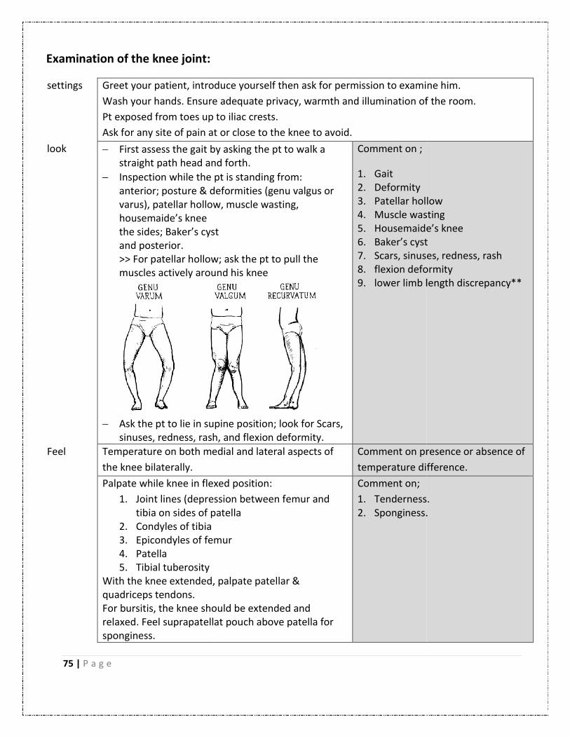
75 | P a g e
Examination of the knee joint:
settings Greet your patient, introduce yourself then ask for permission to examine him.
Wash your hands. Ensure
Pt exposed from toes up to iliac crest
Ask for any site of pain at or close to the knee
look First assess the gait by asking the pt to walkstraight path head and forth
Inspection while the pt is standing anterior; posture & deformities (genu valgus or varus), patellar hollowhousemaide’s knee the sides; Baker’s cystand posterior. >> For patellar hollow; askmuscles actively around his knee
Ask the pt to lie in supine positionsinuses, redness, rash, and flexion deformity.
Feel Temperature on both medial and lat
the knee bilaterally.
Palpate while knee in flexed
1. Joint lines (depression between femutibia on sides of patella
2. Condyles of tibia 3. Epicondyles of femur4. Patella 5. Tibial tuberosity
With the knee extended,quadriceps tendons. For bursitis, the knee should be extendedrelaxed. Feel suprapatellat pouchsponginess.
Greet your patient, introduce yourself then ask for permission to examine him.
Ensure adequate privacy, warmth and illumination of the room
up to iliac crests.
Ask for any site of pain at or close to the knee to avoid.
First assess the gait by asking the pt to walk a straight path head and forth.
while the pt is standing from: posture & deformities (genu valgus or
patellar hollow, muscle wasting,
Baker’s cyst
For patellar hollow; ask the pt to pull the muscles actively around his knee
in supine position; look for Scars,
sinuses, redness, rash, and flexion deformity.
Comment on ;
1. Gait 2. Deformity 3. Patellar hollow4. Muscle wasting5. Housemaide’s knee6. Baker’s cyst7. Scars, sinuses, redness, rash 8. flexion deformity9. lower limb length discrepancy
on both medial and lateral aspects of Comment on presence or absence of
temperature difference.
flexed position:
Joint lines (depression between femur and tibia on sides of patella Condyles of tibia Epicondyles of femur
, palpate patellar &
knee should be extended and suprapatellat pouch above patella for
Comment on;
1. Tenderness2. Sponginess.
Greet your patient, introduce yourself then ask for permission to examine him.
of the room.
Patellar hollow Muscle wasting Housemaide’s knee Baker’s cyst Scars, sinuses, redness, rash flexion deformity lower limb length discrepancy**
Comment on presence or absence of
difference.
Tenderness. Sponginess.

76 | P a g e
For effusion first inspect for the obliteration of the
parapatellar grooves then do;
Mild effusion >> Ripple test milk above patella 5 cm then milk medial side = accumulation of fluid on lateral side of the knee. and then push laterally and view a bulge medially.
Moderate effusion >>Patellar tap ## absent in massive effusion
Massive effusion>> Fluctuation hold patella in between your hands, press upwards once and wait, press downwards once and wait.
Comment;
1. Obliteration of parapatellar grooves.
2. Positive or negative ripple test, fluctuation and patellar tap.
Move Actively :
Ask the pt to flex his knees as far as he can and then to extend it fully, while you place your palm over his knee joint to feel any crepitation.
Ask the pt to rise his lower limb at the hip keeping the knee extended to check for extension and flexion deformity. If the pt was unable to rise his limb unless with a flexed knee, passively extend the leg; If can be extended passively then it’s an extensor lag, if not then it’s a flexion deformity which is due to a mechanical obstruction.
Comment on:
Range of active movement of
extension and flexion [NL 140o,
symmetrical] and presence of
crepitation.
And comment on presence or
absence of extensor lag or flexion
deformity.
Passively ;
Passively flex the knee
For extension raise both legs together and observe laterally for hyperextension (genu recurvatum > 10o).
Comment on ; Range of Passive movement and presence of hyperextension of the knee.

77 | P a g e
Special
tests
Tests of knee stability:
1. Collateral ligaments stability:
apply first while leg fully extended 0o then at 30o
flexion of the knee (to omit effect of cruciate
ligaments and capsule), put pt’s knee between your
elbow and side, feel with your thumbs the joint lines
o Medial collateral ligament>> valgus stress o Lateral collateral ligament>> varus stress >> major opening of the joint lines indicate a tear
2. Cruciate ligaments stability:
Put legs of the pt flexed together then inspect from the
side for posterior sag sign. warn the pt that you are going
to sit on his foot, perform:
Anterior drawer test >> for anterior cruciate lig # false positive if posterior sag is present
Posterior drawer test >> for posterior cruciate lig
Comment if negative or positive
valgus and varus stress tests.
Comment from inspection on
presence of posterior sag sign and
then comment positive or negative
anterior and posterior drawer tests.
Mcmurray tests for meniscal tear:
1. Flex the knee to its full extent.
2. Hold from pt sole of foot.
Medial meniscus >> external rotation at the hip
and varus stress at the knee
Lateral >> internal rotation at the hip and valgus
stress at the knee
3. Extend the knee smoothly. Hear for a click or
clunk accompanied by discomfort.
Comment positive or negative
medial and lateral Mcmurray tests
Patellar apprehension test:
With the pt’s knee fully extended, push the patella
laterally with your thumb, and then flex the knee
slowly. If the pt actively resists flexion, this suggest
previous patellar dislaocation or insatability
Comment if positive or negative
Patellar apprehension test.
Squat test:
Ask the pt to squat, keeping the feet and heels flat on
the ground. if pt can’t perform this test; this indicates
incomplete knee flexion on the affected side which is
caused by a tear of the posterior horn of the menisci
Comment if pt was able to perform
squat test.
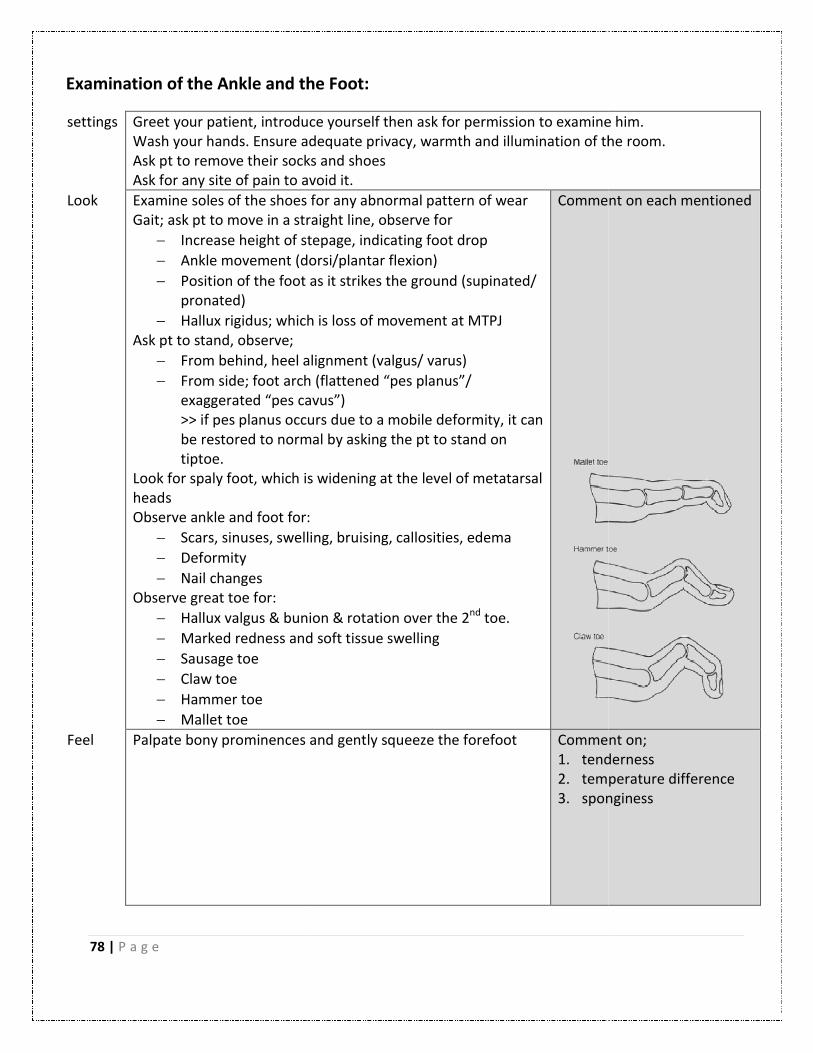
78 | P a g e
Examination of the Ankle and the Foot:
settings Greet your patient, introduce yourself then ask for permission to examine him.Wash your hands. Ensure adequate privacy, warmth and illuminationAsk pt to remove their socks Ask for any site of pain to avoid it.
Look Examine soles of the shoes for any abnormal pattern of wearGait; ask pt to move in a straight line, observe for
Increase height of step
Ankle movement (dorsi/plantar flexion)
Position of the foot as it strikes the ground (supinated/ pronated)
Hallux rigidus; which is loss of movement at MTPJAsk pt to stand, observe;
From behind, heel alignment (valgus/ varus)
From side; foot arch (flattenedexaggerated “pes cavus”>> if pes planus occurs due to a mobile deformity, it can be restored to normal by asking the pt to stand on tiptoe.
Look for spaly foot, which is widening at the level of metatarsal heads Observe ankle and foot for:
Scars, sinuses, swelling,
Deformity
Nail changes Observe great toe for:
Hallux valgus & bunion & rotation over the 2
Marked redness and soft tissue swelling
Sausage toe
Claw toe
Hammer toe
Mallet toe
Feel Palpate bony prominences and
Examination of the Ankle and the Foot:
Greet your patient, introduce yourself then ask for permission to examine him.adequate privacy, warmth and illumination of the room
Ask pt to remove their socks and shoes Ask for any site of pain to avoid it.
Examine soles of the shoes for any abnormal pattern of wear Gait; ask pt to move in a straight line, observe for
Increase height of stepage, indicating foot drop
Ankle movement (dorsi/plantar flexion)
Position of the foot as it strikes the ground (supinated/
Hallux rigidus; which is loss of movement at MTPJ
From behind, heel alignment (valgus/ varus)
From side; foot arch (flattened “pes planus”/ “pes cavus”)
>> if pes planus occurs due to a mobile deformity, it can be restored to normal by asking the pt to stand on
Look for spaly foot, which is widening at the level of metatarsal
Observe ankle and foot for:
Scars, sinuses, swelling, bruising, callosities, edema
& bunion & rotation over the 2nd toe.
Marked redness and soft tissue swelling
Comment on each mentioned
Palpate bony prominences and gently squeeze the forefoot Comment on;1. tenderness2. temperature3. sponginess
Greet your patient, introduce yourself then ask for permission to examine him. of the room.
Comment on each mentioned
Comment on; tenderness temperature difference sponginess

79 | P a g e
Move
1. Ask the pt to flex his foot up, then down, then inside, then outside.
2. Move the pt‘s foot passively to asses range of motion; at ankle; dorsiflexion [NL 15o] & planter flexion [NL 45o] at subtalar joint (while foot is dorsiflexed); inversion [NL 20o] & eversion [NL 10o]
>> If range of passive dorsiflexion is restricted, asses the contribution of gastrocnemius by applying dorsiflexion of the ankle once while knee is flexed, another when the knee is extended. If the movement is harder while knee is extended, this suggests gastrocnemius contracture.
Comment on range of motion
Special
tests
Achilles tendon: Ask the pt to kneel with both knees on a chair, and then palpate gastrocnemius and Achilles tendon
Comment on:
1. Tenderness
2. Soft tissue swelling
3. Gap in the Achilles tendon
Thomson’s (simmond’s) test: Ask the pt to kneel with both knees on a chair, and then squeeze the calf just distal to the level of maximum circumference. if the Achilles tendon is intact, planter flexion of the foot will occur
Comment if planter flexion
occurred or not.

80 | P a g e
Examine conscious level of the patient (
Nervous system
of the patient (glaswa comma scale):
Eye opening:four-eye guyVerbal response:Jackson 5motor response:6 wheeled vehicle © Eman Alkhateb
Eye opening: eye guy
Verbal response: 5
motor response: 6 wheeled vehicle
Alkhateb MD.

81 | P a g e
Examination of Stance and Gate:
Settings Greet your patient and introduce yourself to him.
Explain the examination and ask for his consent to carry it out.
Wash your hands. Ensure adequate privacy, warmth and illumination of the room.
Ask if the pt is currently experiencing any pain.
Stance Ask the pt to stand upright with feet close together,
eyes open, shoes off, and hands by side.
Comment on:
1. Swaying with opened
eyes.
2. swaying with closed eyes
(sensory ataxia) = positive
Romberg’s test
Observe for abnormal posture from front and side.
Perform Romberg’s test;
ask the pt to stand with closed eyes and observe for
body swaying (truncal ataxia).
Be ready to support the pt in case he lost his balance.
# Positive Romberg’s test indicates posterior column
disease.
Gait Ask the pt to move a measured straight 10 meters path
back and forth
Comment on:
1. time needed to complete
the walk
2. stride length
3. arm swing
4. steadiness during turning
5. limping
6. ability to perform tandem
gait
Perform Tandem gait;
ask the pt to walk the same distance heel to toe in a
straight line.
# Ataxia on a narrow-based gait suggests a cerebellar or
vestibular lesion.
## Forms of gait abnormalities:
hemiplegic gait >> semi-circular foot motion.
Bilateral UMNL >> scissor-like gait.
Cerebellar dysfunction >> broad-based, unsteadiness.
Parkinson gait >> delayed initiation, short steps, loss of arm swing, festinating gait.
Proximal myopathy >> waddling gait.
Huntington’s gait >> bizarre gait “dancing-like”.

82 | P a g e
Examination of the Cerebellum (Co-ordination):
Settings Greet your patient and introduce yourself to him.
Explain the examination and ask for his consent to carry it out.
Wash your hands. Ensure adequate privacy, warmth and illumination of the room.
Ask if the pt is currently experiencing any pain.
General Listen to the pt speech when he welcomes you back. Comment if the pt has
dysarthria, staccato or
slurred speech
Inspection Look for resting tremor and intention tremor
# resting tremor is a sign of Parkinson’s, while intention tremor is a
sign of cerebellar disease.
Comment if resting
tremors present or not
Tests
from up
to down
Test for Nystagmus:
with both eyes opened, ask the pt to hold head still, and
follow the tip of your finger. Draw an H shape in front of him.
observe for Nystagmus on lateral gaze specially
Comment if horizontal
Nystagmus present or not
Examine the pt speech by asking him to explain to you how he
get down to the hospital or say his long name.
Note any slurring in speech, slowness in rhythm or dysarthria.
Comment on dysarthria,
staccato or slurred speech
Finger-nose test:
Place your index finger at about half a meter from the pt’s
face. Ask him to touch the tip of his nose then the tip of your
finger with the tip of his index. Once he is able to do this, ask
him to do it as fast as he can. Ask him to repeat the test with
his other hand.
Dysmetria (past-pointing) and intention tremor at the end of
the movement are cerebellar signs.
Comment on dysmetria
and intention tremor
Rapid alternating movement:
Ask the pt to clap with alternating the dorsal then palmar
surfaces of one hand. Once he is able to do this, ask him to do
it as fast as he can.
Ask him to repeat the test with his other hand.
Comment on
Dysdiadochokinesis

83 | P a g e
Rebound phenomenon:
ask the pt to stretch his arms out in front and maintain this
position, then push the pt’s wrist quickly downwards and
observe the returning movement if became pendular.
Comment on positive
rebound phenomenon
Heel-to-shin test:
lie the pt on a couch. Ask him to run the heel of one leg down
the shin of the other, and then to bring the heel back up to
the knee and to start again. Ask him to repeat the test with
his other leg.
Comment on dysmetria
and intention tremor in
the lower limbs
Stance & gait:
Ask the pt to stand upright with feet close together, eyes
open, shoes off, and hands by side. Observe for body swaying
from front and side >>> truncal ataxia is a cerebellar sign.
ask the pt to perform tandem gait by walking a straight path
heel to toe >> inability to perform tandem gait is a cerebellar
sign
Comment on truncal ataxia
and ability to perform
tandem gait
Finishing Examine muscle tone in the upper limbs and lower limbs,
comparing both sides.
Comment if normal or
reduced muscle tone
Examine deep tendon reflexes in the upper and lower limbs Comment if normal or
exaggerated reflexes. And
if pendular
## If you were asked to list cerebellar signs, simply remember the acronym D2ANISH2:
Dysdiadochokinesis and Dysmetria
Ataxia
Nystagmus
Intention tremor
Slurred/ staccato speech
Hypotonia and hyporeflexia

84 | P a g e
Examination of the Cranial Nerves:
Settings Greet your patient and introduce yourself to him.
Explain the examination and ask for his consent to carry it out.
Wash your hands. Ensure adequate privacy, warmth and illumination of the room.
Ask if the pt is currently experiencing any pain.
I (olfactory
nerve)
1. Check nasal passages patency; by asking the pt directly to report any feeling of
nasal blockage, then close one nostril and ask pt to breathe in while you listen to
sound of breathing, and then repeat for the other side.
2. Look for septal deviation
3. Ask the pt to close his eyes, then present to him commonly available odors and
ask pt to identify it.
** test each nasal passage alone
>anosmia (Absence of the sense of smell): in head injury and basal ganglia
disease
> parosamia (disorder in the sense of smell): in head trauma, past sinusitis, S/E
of drugs
>olfactory hallucination: in Alzheimer’s and focal epilepsy
II (Optic nerve) 1. Look for pupils symmetry, regularity, and size (NL 3-5 mm in light, 4-9 mm in
dark)
2. Examine visual acuity by snellen/logMar chart
3. Examine colour vision by ishihara series
4. Fundoscopy to examine posterior retina by fundoscope
5. Examine visual field;
sit directly facing the pt at an eye level about 1 meter away (confrontation), ask
pt to fix his eyes on your nose. Examine:
a- homonymous defect with both eyes open at 2, 4, 8, 10 o’clock
b- sensory inattention with both eyes open
c- peripheral visual fields for each eye at a time at 2, 4, 8, 10 o’clock
d- central visual field for each eye at a time, using red hatpin.
Ask pt to identify its colour, then test if can see the same at four quadrants of
vision.
e- Size of blind spot: located 15° to temporal side of point of visual fixation.
6. Pupillary examination; light reflex and accommodation reflex

85 | P a g e
III (oculomotor
nerve),
IV (Trochlear
nerve),
VI (Abducens
nerve)
Inspect for:
1. Head & eyelid position:
head tilt, strabismusm, proptosis (exophthalmos) from above and
behind, ptosis, lid-lag by asking pt to follow your finger from 45o above
horizon
2. pupils symmetry
3. periorbital edema, lacrimal apparatus, eye lid margin, conjunctiva, sclera,
cornea with dye
Test movements of extra-ocular muscles;
sit in confrontation, move your finger in H motion.
Look for nystagmus, ask pt to report double vision
comment: smooth pursuit, no divergence of gaze, no nystagmus
Do cover test (for strabismusm if present)
Do Pupillary examination; light reflex and accommodation reflex
V (trigeminal
nerve)
Motor:
1- Inspect for muscle wasting
2- Ask pt to clench his teeth, then feel for muscle bulk of masseter and
temporalis
3- Ask pt to open his jaw while you apply upward resistance
Sensory:
Ask the pt to close his eyes, test sensations at dermatomes of 3 branches
(ophthalmic, maxillary, mandibular) bilaterally of the following sensory
modalities;
1- light touch using cotton wool
2- pain using neurotip
examine touch sensation of anterior 2/3 of tongue using orange stick
Reflexes:
1- Corneal reflex.( Afferent: CN 5 ,,, Efferent : CN 7)
2- Jaw reflex ( Afferent : CN 5 ,, Efferent :CN 5 )

86 | P a g e
VII (facial
nerve)
Inspect for:
Facial asymmetry, difference in blinking, involuntary movements,
presence of nasolabial folds, deviation of the angle of the mouth
Motor:
1- Ask pt to look up, inspect for forehead wrinkles
2- Ask pt to close his eyes tightly against resistance, examine power
3- Ask pt to blow out his cheeks against resistance, examine power
4- Ask pt to whistle (or purse his lips)
5- Ask pt to smile widely (show me your teeth smile :D ), examine deviation of
the mouth
Test taste sensation of anterior 2/3 of the tongue by present commonly
available food.
Schirmer's tear test
Corneal reflex
# Hyperacusis (increased auditory volume in an affected ear) may be produced by
damage to the VII cranial nerve as it innervates the stapedius muscle.
VIII (Vestibulo
-cochlear
nerve)
Hearing part (cochlear branch):
1- Whispered voice test from 15cm away, ask pt to repeat what he hears while
closing other ear (mask hearing in contralateral. side by pushing tragus)
2- Weber’s test: NL symmetrical at midline
3- Rinne’s test: NL air conduction better than bone conduction ‘positive test’
Vestibular branch:
1- Dix-Hallpike maneuver
2- Vestibular reflex that includes caloric reflex and oculocephalic reflex
IX (glosso
-pharyngeal),
X (vagus nerve)
1- Examine pt speech
comment on dysarthria & dysphonia
note the quality and sound of the patient's voice. Is it hoarse or nasal?
2- Ask pt to cough; comment if bovine
3- Ask pt to say ‘aah’ while you look at the back of his mouth using the light of your
torch for deviation of uvula of contraction of pharyngeal muscles
# ( in the unilateral weakness it will deviate to the normal side )
4- Examine gag reflex by water swallow test in a conscious pt
5- Ask pt to puff out to examine for nasal escape

87 | P a g e
XI (accessory
nerve)
Inspect for:
Muscle atrophy, involuntary movements, symmetry of shoulder contour
Palpate muscle bulk
Examine power
1- Ask pt to shrug his shoulders while you apply downward pressure to
examine trapezius
2- Ask pt to turn his head to one side against resistance to examine
sternomastoid
(this examine contralateral side)
XII (hypoglossal
nerve)
Inspect for:
wasting of the tongue, tremor, fasciculation, involuntary movements
Ask pt to protrude his tongue. Look for deviation of tip of the mouth
Note for deviations of the tongue from midline, a complete lack of ability to
protrude the tongue or any involuntary movement.
# Tongue will deviate towards the side of a peripheral lesion, and to the
opposite side of a central lesion.
Ask pt to move his tongue from side to side
Examine power; press tongue against cheek with resistance
Examine lingual speech by asking pt to say ‘lah lah lah’
Examine swallowing by water swallow test
For examination of Motor System, Sensory System, Speech, And Signs of Meningeal
Irritation please refer to Macleaod’s book.


89 | P a g e
Section
3
Appendix

90 | P a g e
Abbreviations
Pt = patient ARDS = acute respiratory distress syndrome
Hx = history COPD = chronic obstructive pulmonary disease
Px = physical examination ASA= acetyl salicylic acid (aspirin)
Dx = diagnosis CXR = chest x-ray
DDx = deferential diagnosis AXR = abdominal x-ray
y/o = year old CBC = complete blood count (lab test)
ICU = intensive care unit c/o = complains of
CCU = cardiac care unit EUA = examination under anesthesia
OPD = out-patient department FNA = fine needle aspiration (lab test)
ER = emergency room NGT = nasogastric tube
PTP = prior to presentation NPO = nothing per os
PTA = prior to admission POD = post operative day
CC = chief complaint RTC = return to clinic
HPI = history of presenting illness RTA = road traffic accident
ROS = review of system RTA = renal tubular acidosis
Wt = weight S /P = status post (Ex. s/p hernia repair)
PND = paroxysmal nocturnal dyspnea Lap choly = laparoscopic cholecystectomy
SOB = shortness of breath Op = operation
LMP = last menstrual period DM = diabetes mellitus
OCP = oral contraceptive CKD = chronic kidney disease
NKDA = no known drug allergy lab results symbol with normal ranges ;
CAOX3 = conscious and oriented in time, place
and person
LOC = loss/level of consciousness
GAEB = good air entry bilateral
Bx = biopsy
Rx = treatment regimen
MTX = methotrixate
CTX = chemotherapy
RTX = radiotherapy
ttt = treatment
= diagnosed with, or trimester in pregnancy
ABG = arterial blood gases or air bone gap in ENT
e = electrolytes
AAA = abdominal aortic aneurysm
Na (135 -148)
K (3.5 -5)
BUN (15-46)
Cr (0.6 – 1.4)
RBS random blood sugar
(70-120)
WBC (4-10)
Hemoglobin (M 16±2, F 14±2)
Hematocrit
(36-53)
Platelets (140-440)

91 | P a g e
Past years OSCE stations
Summer 2013
‘this is the first time when introductory exam get history taking stations!’
Stations of the first day group:
1. take a history from a patient complaining of cough and ask him the questions that
help you to differentiate asthma from COPD
2. take a history from patient with MI
3. take a history from patient with jaundice (points mentioned in figure 8.25 page 196
of Macleaod’s should be asked)
4. examine the patient’s knee doing only the passive and active movements
5. do inspection and palpation of the abdomen
6. do motor examination of the lower limb
Stations of the second day group:
1. examination of cerebellum
2. inspection and percussion of the abdomen
3. physical examination for knee effusion
4. history of chest pain asking the questions that help you differentiate stable from
unstable angina
5. (Oral station) the instructor asked about the signs and symptoms of iron deficiency,
low WBCs count and low platelets count.
6. take a history from patient complaining of cough and ask him the questions that help
you differentiate asthma from COPD
Summer 2012
1. examine stability of the right knee
2. do inspection of the abdomen
3. do percussion and auscultation of the anterior chest
4. do palpation and auscultation of the precordium
5. examine the patient radial pulse
6. examine facial nerve
92 | P a g e
Single
Married
Divorced
Widow
Living in _________________________________________,
Works as ________________________________________ .
History application form
II___ patient profile:
Mr. / Mrs. / Miss ____________________________________________________, _______ years old, Lady
Gentleman
The pt was admitted to ______________ via ____________ , on _______________________________
at ______________________ History was taken from _____________________ by ________________
______________________________________ on _______________________ at _______________.
II___ chief complaint:
__________________________________________________________________________________
__________________________________________________________________________________
II___ HPI:
__________________________________________________________________________________
__________________________________________________________________________________
__________________________________________________________________________________
__________________________________________________________________________________
__________________________________________________________________________________
__________________________________________________________________________________
__________________________________________________________________________________
__________________________________________________________________________________
__________________________________________________________________________________
__________________________________________________________________________________
__________________________________________________________________________________
__________________________________________________________________________________
__________________________________________________________________________________
__________________________________________________________________________________
__________________________________________________________________________________
__________________________________________________________________________________
__________________________________________________________________________________
__________________________________________________________________________________
__________________________________________________________________________________
__________________________________________________________________________________

93 | P a g e
__________________________________________________________________________________
__________________________________________________________________________________
__________________________________________________________________________________
__________________________________________________________________________________
__________________________________________________________________________________
__________________________________________________________________________________
__________________________________________________________________________________
__________________________________________________________________________________
__________________________________________________________________________________
__________________________________________________________________________________
__________________________________________________________________________________
__________________________________________________________________________________
__________________________________________________________________________________
__________________________________________________________________________________
__________________________________________________________________________________
__________________________________________________________________________________
__________________________________________________________________________________
__________________________________________________________________________________
__________________________________________________________________________________
__________________________________________________________________________________
__________________________________________________________________________________
__________________________________________________________________________________
__________________________________________________________________________________
__________________________________________________________________________________
__________________________________________________________________________________
__________________________________________________________________________________
__________________________________________________________________________________
__________________________________________________________________________________
__________________________________________________________________________________
__________________________________________________________________________________
__________________________________________________________________________________
__________________________________________________________________________________
__________________________________________________________________________________
__________________________________________________________________________________
__________________________________________________________________________________
__________________________________________________________________________________
__________________________________________________________________________________
__________________________________________________________________________________

94 | P a g e
II___ ROS:
General:
Well being;_______________________
Appetite;_________________________
Energy;__________________________ Cardiovascular system:
Chest pain
Breathlessness; o on lying flat, relived by ____ pillows o at night around _____ o’clock o on minimal exertion like ___________________
Respiratory system:
SOB
Cough; dry/productive, sound (_______), timing (________), associated with ____ _______, exacerbated by _____________ relieved by _________________________
Wheeze on ________________________ Gastrointestinal system:
Oral ulcers; if painful
Dental hygiene; _____________________
Dysphagia; to solids or liquids, at ____________________________level.
Odynophagia
Nausea
Vomiting; the vomitus is ________________ ___________________________________
Hematemesis
urinary system:
pain passing urine (dysuria);____________
frequency; if at night (nocturia)
hematuria
Genital system: for men:
hesitancy
poor stream
terminal dribbling
urethral discharge
erectile difficulties
Sleep;____________________________
Mood;____________________________
Wt change;________________________
Palpitation;__________________________ ___________________________________
Claudication; at ______________ distance
Ankle swelling
Sputum; that is _______________ (timing), in ________ color, in__________ amount, with _________ smell, ______ contain solid materials.
Hemoptysis; _________________________
Chest pain on inspiration or coughing
Indigestion
Heartburn
abdominal pain
Change in bowel habits. Normally pass feces __________ times a day, this has increased / decreased to __________ a day.
change in color of stool;_________________
incontinence (stress/ urge)
libido; normal or changed
unprotected intercourse (multiple sexual partners)
For women:
LMP ________________________________
Period is _____________________________
Heavy bleeding; state if contain clots
vaginal discharge
OCP
dyspareunia

95 | P a g e
Endocrine system:
Heat or cold intolerance
change in sweating
Musculoskeletal system:
Joint pain
Stiffness
joint swelling
Nervous system:
Headache
Dizziness
Faints
fits
altered sensation Others:
bleeding or bruising
II___ Past Hx:
Medical: chronic illnesses; ( D / E / A / T / H ), OSA, __________________________________________________ previous hospital admissions (when, where, why, length of stay); ______________________________________________________________________________________ ______________________________________________________________________________________ ______________________________________________________________________________________ history of blood transfusion (when, where, why, how many, complications); ______________________________________________________________________________________
Surgical (when, where, why, complications, anesthesia) : ______________________________________________________________________________________________________________________________________________________________________________________________________________________________________________________________________________________________________________________________________________________________________________________________________________________________________________
Obstetrical: LMP _____________________________, age of menarche __________ and menopause __________, number of pregnancies _______ & complications __________________________________________, type of delivery ____________________________ & complications___________________________, abortion __________________________________& cause _________________________________, family planning method ___________________________________.
Weakness
visual disturbances
hearing problems
memory & concentration changes
Skin rash
restricted mobility
falls
excessive thirst

96 | P a g e
II ___ Drug Hx:
________________________________________________
________________________________________________
________________________________________________
________________________________________________
________________________________________________
________________________________________________
________________________________________________
________________________________________________
________________________________________________
________________________________________________
________________________________________________
________________________________________________
Allergy; _________________________________________
II ___ Family Hx (draw a pedigree chart):
II ___ Social Hx:
Lifestyle: exercise_____________, diet_______________, marriage____________, homing_____________ ________________________________________, hobbies____________, pets ________________ .
Occupational Hx: previous occupation _______________________, unemployment (reason_____________________ _____________________duration___________), attitude to job ___________________________ .
Travel Hx: __________________________________________________________________________________
Sexual Hx:
regular sexual partner, M/ F
irregular (how many per year ________, M/ F)
Tobacco & water pipe:
smoker; ________________ pack years
X-smoker; stopped from _______ years
passive smoking at work or home
Alcohol: ___________________________________________________________________________________________________________________________ Hx of vaccination _________________________________________________________________________________
Religion _________________________________
Drug abuse _________________________________________________________________________________
Insurance system
________________________________________