Embed Size (px)
Citation preview
Vet RecoRD | 1
PaPer
History, clinical findings and outcome of horses with radiographical signs of equine odontoclastic tooth resorption and hypercementosisVahideh Rahmani ,1 Lotta Häyrinen,1 Ilona Kareinen,2 Mirja Ruohoniemi1
AbstractThe progression of equine odontoclastic tooth resorption and hypercementosis (EOTRH) has not been completely evaluated, and currently, the only effective treatment is extraction of severely affected teeth. We aim to describe how the disease relates to the history and clinical findings and to report on the outcome in individual horses. This case series comprises data collected from 20 horses (age 14–29 years old) with radiographic findings of EOTRH in their incisor and/or canine teeth. Most horses affected with EOTRH in this study were admitted for dental problems, but some for other complaints such as colic. Of the 288 teeth evaluated radiographically, 224 teeth were abnormal. Radiographic findings were most frequently located in the apical aspect and reserve crown of the teeth, and lesions were also commonly found in clinically normal teeth. Histopathology of extracted teeth showed inflammation in the periodontal ligament and revealed that resorption often extended to the dentine. Some owners were unwilling to allow extraction of their horses’ severely affected teeth, even though this treatment has been shown to increase the wellbeing of the horse. As EORTH is a life-long condition, the progression of the disease has to be continuously monitored and the treatments adjusted accordingly.
IntroductionEquine odontoclastic tooth resorption and hypercementosis (EOTRH) is a recently well-documented but probably underdiagnosed painful and always progressive disorder of unknown aetiology, affecting the teeth of older horses.1 2 The condition primarily involves the incisor and canine teeth; however, one case report describes lesions in the cheek teeth of two horses.3 Recent literature has proposed that breed and sex may be risk factors associated with EOTRH.4 5 EOTRH has been reported to occur most commonly in Thoroughbreds and Warmbloods,2 5 6 but the condition has also been diagnosed in several other breeds.3 5 7 8 Several reports suggest that geldings are
most commonly affected.5 9 Older age is a risk factor for EOTRH, but moderate-to-severe radiographic changes have been identified in middle-aged horses (11–13 years).4 8
EOTRH is characterised by internal and external resorption of dental structures and is often associated with excessive production of irregular cementum on the surface of the tooth.2 6 9 The following three EOTRH patterns have been described: predominant tooth resorption, predominant hypercementosis and combined resorption and hypercementosis.5 It appears that the disorder first affects the corner incisors (triadan 03 s) and then progresses to the middle and central incisors (triadan 02 s and then 01 s).10
Diagnosis of EOTRH is based on clinical findings and dental radiographs, and histopathology can help to differentiate the disease from other proliferative dental diseases.9 11 The reported clinical signs of EOTRH vary from no signs to subtle signs of pain, difficulties in biting or mastication, decreased appetite, dysphagia, weight loss and behavioural changes.9 10 However, EOTRH has commonly been reported to be asymptomatic at the time of diagnosis despite advanced dental lesions.10 12 As the disease progresses, it is characterised by gingivitis, gingival oedema and recession,
10.1136/vetrec-2018-105253
Veterinary Record (2019) doi:10.1136/ vetrec-2018-105253
1Department of Equine and Small Animal Medicine, Faculty of Veterinary Medicine, University of Helsinki, Helsinki, Finland2Department of Veterinary Biosciences, Faculty of Veterinary Medicine, University of Helsinki, Helsinki, Finland
E-mail for correspondence: Dr Vahideh Rahmani; vahideh. rahmani@ helsinki. fi
Provenance and peer review Not commissioned; externally peer reviewed.
Received November 3, 2018Revised July 12, 2019Accepted July 29, 2019
on October 13, 2019 at U
niversity of Helsinki. P
rotected by copyright.http://veterinaryrecord.bm
j.com/
Veterinary R
ecord: first published as 10.1136/vr.105253 on 10 October 2019. D
ownloaded from
| Vet RecoRD2
associated periodontal disease, fistula formation, tooth mobility and displacement, tooth fracture and/or tooth loss.2 10 13 Radiography is a practical and useful method for diagnosis of EOTRH, also helping to detect dental resorption and/or hypercementosis even when no obvious lesions are present on clinical examination.1 11 Typical radiographic findings are bulbous enlargement of the intra-alveolar crown root, resorptive lesions of the reserve crown, apex and/or surrounding bone, widening of the periodontal ligament space and tooth fractures.9 Histopathological examination has revealed infiltration of inflammatory cells in the resorptive lesions with odontoclasts surrounding the margins.2 Odontoclastic resorption precedes and probably precipitates hypercementosis as a reparative reaction by cells of the periodontal ligament secondary to the resorptive lesions.2
Although no single aetiology has been established for EOTRH, periodontal inflammation has been suspected to trigger the initial tooth resorption.5 14 Biomechanical stresses and strains on the periodontal ligaments, followed by a secondary infection with micro-organisms such as Treponema and Tannerella species have been suggested to contribute to the aetiology.2 6 Hypercementosis is considered a reparative rather than the primary pathological process.1
Currently, exodontia is the only treatment for the condition in clinically affected horses. Surgical extraction of severely affected and painful teeth has been shown to be an effective treatment and may improve the quality of life in the long term.9 12 Other treatments, such as systemic antibiotics, anti-inflammatories and oral flushes, have been used to reduce periodontal disease, but at best they offer only transient relief of symptoms.13 15 16
Here, we present a case series describing clinical, radiographic and histopathological findings of EOTRH in incisor and canine teeth in a patient population from a University Equine Hospital in Helsinki, Finland. The aim was to describe how the disease relates to the history and clinical findings of the affected horses and to report on the outcome of individual horses, as current knowledge of the course of the disease contains many gaps. We also discuss challenges in client communication regarding the treatment of EOTRH, a painful and progressive condition impeding the quality of life of affected horses, with a highly invasive treatment (exodontia) as the only effective option available.
Materials and methodsFor this retrospective study, we reviewed the records of horses radiographed at the Equine Hospital, Faculty of Veterinary Medicine, University of Helsinki, between January 2010 and March 2018. All cases having radiographs of the skull or teeth, taken for any clinical reason, were first collected and reviewed, and those taken of the incisor and canine teeth (n=83) were
evaluated in detail by the first or second author. Of these, the cases that had any radiographic signs suggestive of EOTRH were included in the study (n=20). All uncertain cases were reviewed by at least two authors before the decision on inclusion was made.
At a minimum, the following two diagnostic quality radiographs were available for all horses except one: 1) an occlusal view of the maxillary incisors and the canine teeth and 2) an occlusal view of the mandibular incisors and the canine teeth. The exception was a horse radiographed for a traumatic fracture with only one occlusal view of the mandible. All teeth in the radiographs from individual horses were evaluated for resorption, hypercementosis and other lesions by three authors (VR, LH and MR) using the previously described criteria.9 10 17 All cases were discussed by the authors until consensus regarding the findings in each tooth had been reached.
We reviewed the histories and clinical records of the horses included in the study including age, breed and sex. Clinical examination findings of the mouth and teeth were evaluated if recorded. In addition, the horses’ main complaint(s) and outcome were recorded.
Thirteen extracted teeth from three horses (horses 12, 13 and 14) were examined grossly and histopathologically. The teeth were processed using previously reported methods.2 Briefly, extracted teeth were fixed with 10 percent neutral buffered formalin and transversely sectioned into 10 mm sections using a diamond saw. The sectioned samples were then decalcified in a buffer containing 3 per cent neutral buffered formalin and 26 per cent formic acid (two to four weeks), embedded in paraffin, cut into 3–4 mm thick sections, and stained using H&E. The histological sections were examined via light microscope and classified as described previously.5
The modified triadan system18 for numbering dentition is used throughout the results.
ResultsHorsesTwenty horses (17 geldings and 3 mares) met the inclusion criteria for this study. The horses were aged between 14 and 29 years (mean 20.9 years, median 21.5 years), and included several different breeds (table 1). The first clinical case was diagnosed in 2013 and was confirmed histopathologically. Subsequently, two horses were radiographed and diagnosed in 2014, two in 2015, five in 2016, five in 2017 and five until the end of March 2018.
The case details, the owner’s complaint and history of the 20 horses are summarised in table 1. Only five horses came in for a routine dental examination, and seven were admitted to the hospital because of signs relating to eating problems or weight loss. Additionally, four horses were admitted to the hospital primarily because of colic (two of them associated with sand
on October 13, 2019 at U
niversity of Helsinki. P
rotected by copyright.http://veterinaryrecord.bm
j.com/
Veterinary R
ecord: first published as 10.1136/vr.105253 on 10 October 2019. D
ownloaded from

Vet RecoRD | 3
Table 1 Case details, owner’s complaints and history of 20 horses with equine odontoclastic tooth resorption and hypercementosis
Horse number
Age at initial presentation(years) Sex Breed
Complaint
HistoryPossible teeth-related symptoms Colic
Routine dental check Other
1 15 G Warmblood Dropping food − − − Colic <1 year ago2 19 G Pony Poor appetite − − − Colic >1 year ago, sand
enteropathy3 16 G Pony Poor appetite, thin, resists
the bit− − − Sand enteropathy
4 19 G Finnhorse − + − − −5 29 M Shetland pony Weight loss − − − −6 21 G Icelandic horse − − + − Sand enteropathy7 27 M Warmblood Weight loss − − − PPID8 23 G Finnhorse − − − Canine tooth fracture PPID9 24 G Shetland pony Halitosis + − − −
10 20 G Standardbred − − − Mandibular fracture Colic >1 year ago11 21 G Warmblood − − + − Colic >1 year ago12 18 M Warmblood − − + − −13 23 G Finnhorse Poor appetite + − − PPID14 24 G Pony Draining abscess, halitosis − − − −15 22 G Warmblood Weight loss − − Oesophageal
obstruction−
16 14 G Warmblood - − − Referred for radiology only
−
17 17 G North Swedish horse - − − Referred for radiology only
−
18 22 G Icelandic horse Keeps the tongue between the incisors
− + − −
19 23 G Warmblood Tooth problems (extracted cheek teeth, suspected EOTRH)
+ − − −
20 22 G Pony Changes noted in incisor area − + − −
-, no;+, yes; EOTRH, equine odontoclastic tooth resorption and hypercementosis; G, gelding; M, mare;PPID, pituitary pars intermedia dysfunction.
Figure 1 Intraoral radiograph of the mandible of horse 19 showing hypercementosis and resorptive lesions in teeth 302, 303, 304, 401, 402 and 403. Hypercementosis predominates in the teeth on the right side, whereas resorption is more evident on the left side. L, left.
accumulation in the colon); three of these also had signs related to dental disease. One horse was referred to the hospital because of a mandibular fracture and one for a fractured canine tooth. Two horses were referred by their treating veterinarian only for radiography (table 1).
Out of the 20 horses referred to the hospital, three horses had a history of pituitary pars intermedia dysfunction (PPID), four horses had a history of colic,
and three horses had a history of sand enteropathy (table 1).
Type and extent of EOTRH lesionsAll 20 horses had radiographic evidence of both tooth resorption and hypercementosis in their teeth. A representative image is shown in figure 1. Of the 288 teeth (225 incisors, 63 canines) evaluated radiographically, 224 teeth were abnormal and 64 teeth were considered normal. Five teeth were missing from the normal dentition. Of the 224 radiographically abnormal teeth, 92 teeth had resorption only, 98 teeth had concurrent resorption and hypercementosis and 23 teeth had hypercementosis without any radiographic evidence of resorption. Additionally, five teeth had a pathological fracture in association with resorptive lesions. Well-circumscribed, rounded and radiopaque fragments were seen in the apical region of six teeth.
The periodontal ligament was irregularly widened in 48 teeth, and the alveolar bone surrounding 46 teeth showed resorption.
The triadan 03 teeth were most commonly affected, followed by the triadan 02 teeth. The resorptive lesions within the teeth were most frequently located in both the apical aspect and the reserve crown (91/190 affected teeth), followed by the apical aspect (59/190 affected teeth) and the reserve crown (31/190 affected teeth).
on October 13, 2019 at U
niversity of Helsinki. P
rotected by copyright.http://veterinaryrecord.bm
j.com/
Veterinary R
ecord: first published as 10.1136/vr.105253 on 10 October 2019. D
ownloaded from
| Vet RecoRD4
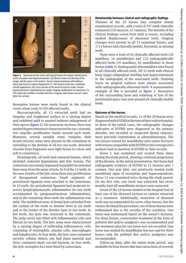
Figure 2 Removed incisor tooth with typical lesions for equine odontoclastic tooth resorption and hypercementosis. (a) Clinical crown is at the top of the image, and the apex at the bottom. Severe hypercementosis with bulbous apical and reserve crown enlargement (*). The clinical crown has a relatively normal appearance. (b) Cross-section at the level of reserve crown. Severe hypercementosis characterised by rough, irregular proliferation of cementum (*). This tooth also exhibits a locally extensive, irregular, dark brown necrotic space (lytic foci, arrow).
Resorptive lesions were rarely found in the clinical crown alone (only 9/190 affected teeth).
Macroscopically, all 13 extracted teeth had an irregular and roughened surface to a varying degree and exhibited mild to marked bulbous enlargement of their apices (figure 2). On transverse sections, there was marked hypercementosis characterised by tan-coloured, tree ring-like proliferative bands around each tooth. Moreover, several variably sized, irregular, dark brown necrotic areas were present in the cementum or extending to the dentine in all but one tooth. Attached alveolar bone fragments were light brown in colour and soft in consistency.
Histologically, all teeth had cemental lesions, which included cemental hyperplasia and lytic lesions. The cementum was mainly organised in parallel incremental lines away from the areas of lysis. In 9 of the 13 teeth, in the near vicinity of the lytic areas there was proliferation of disorganised cementum. Small segments of periodontal ligament were attached to the cementum. In 10 teeth, the periodontal ligament had moderate-to-severe lymphoplasmacytic inflammation (in one tooth accompanied by pyogranulomatous inflammation), and in the remaining three teeth the inflammation was mild. The multifocal areas of dental lysis extended from the surface of the tooth to dentine level in six teeth and to the border of the dentine layer in two teeth. In five teeth, the lysis was restricted to the cementum. The pulp cavity was filled with inflammatory cells and fibrosis in two teeth. The lytic areas were characterised by a varying degree of infiltrating inflammatory cells consisting of neutrophils, plasma cells, macrophages and lymphocytes. In four teeth, the lytic area contained necrotic cellular debris; one had plant material and three contained small coccoid bacteria. In four teeth, the lytic resorptive foci were lined by osteoclasts.
Relationship between clinical and radiographic findingsThirteen of the 20 horses had complete dental examination records, with a total of 197 teeth clinically evaluated (150 incisors, 47 canines). The severity of the clinical findings varied from mild to severe, including marked displacement of abnormal teeth. Gingival changes were present in all 13 examined horses, and 7/13 horses had clinically mobile, fractured, or missing teeth.
There were a total of 44 clinically affected teeth (28 maxillary, 16 mandibular) and 152 radiographically affected teeth (70 maxillary, 82 mandibular) in these horses (table 2). Radiographic abnormalities were found in all clinically affected teeth. All 33 teeth with clinical bony (juga) subgingival swelling had hypercementosis in the radiographs of the associated teeth. Draining tracts on gingival surfaces were always associated with radiographically abnormal teeth. A representative example of this is provided in figure 3. Resorption of the surrounding bone and widening or loss of the periodontal space was seen around all clinically mobile teeth.
Outcome of the horsesBased on the medical records, 15 of the 20 horses were diagnosed with EOTRH at the time of their visit to hospital. In three of the initial cases, the radiographic changes indicative of EOTRH were diagnosed as the primary disorder, but recorded as suspected dental tumours, more precisely cementomas or odontoblastomas. The two horses treated for mandibular and canine fractures with lesions compatible with EOTRH in this retrospective analysis had no mention of EOTRH in their records.
Horse 1 was examined and treated three times during the study period, showing continual progression of the disease. At the initial presentation, this horse had radiographic evidence of EOTRH in 11 incisors and 3 canines. One year later, one previously normal tooth manifested signs of resorption and hypercementosis. Horse 12 was examined twice during the study period. On the first visit, one tooth was extracted, but seven months later all mandibular incisors were extracted.
Seven of the 18 horses treated at the hospital had at least one of the diseased incisors or canines extracted as a treatment. Additionally, extraction of diseased teeth was recommended for seven other horses, but the owners declined the procedure; two of these horses were euthanased due to the severity of the condition. One horse was euthanased based on the owner’s decision. For three horses, conservative treatment in the form of pelleted diet and/or analgesics was recommended and the treatment plan for one horse was not recorded. One horse was treated for mandibular fracture and for three horses only the pelleted diet and/or analgesics was recommended.
Follow-up data, after the initial study period, was available for four horses that had extractions of severely
on October 13, 2019 at U
niversity of Helsinki. P
rotected by copyright.http://veterinaryrecord.bm
j.com/
Veterinary R
ecord: first published as 10.1136/vr.105253 on 10 October 2019. D
ownloaded from

Vet RecoRD | 5
Table 2 Clinical findings, affected teeth and total number of clinically and radiographically affected teeth in 13 of the 20 horses with EOTRH
Horse number
Clinical findingsNumber of clinically affected teeth
Number of radiologically affected teeth
Gingivitis±gingival recession
Bony subgingival swelling
Draining tracts
Abnormal dentition
Maxilla Mandible Maxilla MandibleMobile Fractured Lost
1 + 102 104 101104
303 204 4 1 7 7
2 + 203 − 402 − − 1 1 5 63 − 101–103
201–203− − − − 6 − 7 7
6 + − 303 − − 403 − 1 4 79 + − − − − 203
403− − 5 7
11 + − 103 203
− − − 2 − 4 8
12 + 103203
− − − − 2 − 5 3
13 + 103203304403–404
304 − 204 − 3 3 6 7
14 + 101–102201–203301–303401–402
303 402
303 − 103403
5 5 7 7
15 + 303304404
203 403
− − 103 1 4 4 6
18 + 303 − − − − − − 4 619* + 103
203 403 − − − 2 1 7 6
20 + 103203
− − − − 2 − 5 5
*The horse had seven maxillary incisor teeth.Where horses were examined more than once during the study period, the findings of the first visit only are included.-, not present; +, present; EOTRH, equine odontoclastic tooth resorption and hypercementosis.
Figure 3 (a) Representative photograph of horse 13 showing a small draining tract (arrow) on the gingival surface of tooth 304. Mild bony swelling of the gingiva is also evident. (b) Intraoral radiograph of the mandibular incisor and canine teeth of horse 13 demonstrating hypercementosis and resorptive lesions of varying degree in teeth 301, 302, 303, 403 and 404 (the lesions appear mild relative to those presented in figure 1). L, left.
affected teeth. Three horses had been described as ‘happier, less withdrawn’, or as having improved mastication and now being able to eat hay and food pellets without difficulties. One horse had been euthanased.
DiscussionSince 2013, the number of horses suffering from EOTRH has increased steadily in the patient population of the University Equine Hospital, due to increased awareness of the disease and advancements in equine dental knowledge. The previously reported three different
manifestations of EOTRH,5 that is, predominant tooth resorption, predominant hypercementosis and combined resorption and hypercementosis, were all represented in this case series. The histopathological findings in incisor and canine teeth, and the periodontal ligament were similar to those reported earlier,2 5 confirming the diagnosis of EOTRH. All extracted teeth exhibited hypercementosis and lytic resorptive foci histopathologically, with inflammation of the periodontal ligament also a common feature. As dentine is considered a physically sensitive living tissue19 and the lesions reported here extended to the level of the dentine, this is a further indication of the painful nature of EOTRH.
It has already been established that EOTRH is a disease that primarily affects the teeth of older horses. The horses in most of the previous reports have been older than 15 years when first presented,2 5 9 which is in agreement with our observations. However, one recent study noted that age is not a significant risk factor,4 and Smedley and others5 describe pathological manifestations of the disease in a 10-year-old horse. This is supported by radiographic evidence of resorption in even younger horses, although the frequency of all types of tooth resorption does increase significantly with age.1 8 Most of the horses in the study had severe
on October 13, 2019 at U
niversity of Helsinki. P
rotected by copyright.http://veterinaryrecord.bm
j.com/
Veterinary R
ecord: first published as 10.1136/vr.105253 on 10 October 2019. D
ownloaded from
| Vet RecoRD6
lesions that appear to have progressed over a prolonged period before the diagnosis was made, which accounts for the bias towards aged horses. The wide range of breeds and predomination of geldings in the study is in line with previous reports on EOTRH.1 5 6 8 9 The two horses in the study that had more than one clinical and radiographic examination serve as examples of the progression of this disease. The progression of EOTRH over time has been reported elsewhere,9 and the study shows that the changes can worsen markedly within a matter of months.
The resorptive lesions were most frequently located in both the apical and reserve crown portion, consistent with previous reports,1 2 and this makes the clinical diagnosis more challenging. All teeth with bony subgingival swellings identified at clinical examination showed hypercementosis on radiography as well. When hypercementosis is present in the late stages of the disease, the periodontium can be grossly distorted.1 Only 23 teeth (10.26 per cent) in the study had hypercementosis without radiographic signs of resorption. In some cases, excessive radiopacity, due to a severely thickened cementum, may have obscured underlying regions of resorption. In the present study, radiographs revealed fragmentation of the roots of six incisor teeth. A recent case report7 showed similar fragmentation in the root area with odontoclastic resorption in all mandibular incisors and both third maxillary incisors. The cause of this fragmentation was unknown.
In line with previous reports, data in the study also support the notion that clinical signs may be subtle even in the presence of marked radiographic changes.9 While the clinical crown of the incisor may appear healthy, there may be advanced lesions in its apical and reserve crown. Gingivitis, gingival recession, bony subgingival swelling and draining tracts should direct suspicion towards EOTRH in older horses. Even small draining tracts on gingival surfaces were associated with radiographically abnormal teeth and resorption of the surrounding bone. The fact that most horses had mobile, fractured or lost teeth in the rostral mouth is an indication of the advanced stage of the disease at the time of presentation. Where older horses present with fractured teeth originally thought to be of traumatic origin (as seen here), the presence of EOTRH should be considered as a predisposing factor.
Since the horses in this study may also have had treatment elsewhere, their clinical histories may be incomplete. Three of the horses had a history of PPID. Horses with endocrine conditions, such as PPID, have been reported to be more susceptible to developing EOTRH than healthy horses.4 Recent research indicates that small animals with Cushing’s disease have a higher prevalence of ligament laxity, presumably because cortisol weakens the periodontal ligaments, causing increased risk of periodontal disease.4 Many
of the horses had colic on admission to hospital or a history of colic, and five horses were found to have sand enteropathy on admission or in their history. Good dental health is pivotal for proper functioning of the equine digestive system,20 and ensuring oral comfort and effective masticatory ability is of great importance in the ageing horse.13 16 Dental abnormalities can predispose horses to colic. In a recent study, more than half of the horses with a history of colic had dental abnormalities, whereas only 30 per cent of non-colicy horses had dental problems.21 If mastication is impaired because of dental pain, food may not be digested properly, which could lead to gastrointestinal problems. Pain in the rostral aspect of the mouth can also disrupt the selection of the food by a horse, which in turn could increase the possibility of ingesting geo-sediment.
To date, dental extraction remains the only treatment that has been effective for relieving pain caused by EOTRH.9 12 15 The clinical crown and surrounding soft tissues may not necessarily look severely diseased until the condition is very advanced, making it hard for the owner to appreciate that lesions only visible radiographically could cause pain. Although systematic pain assessment in horses has been developed during the past decade,22 relatively little is known about dental pain. Not all horses show chronic pain clearly, and it may be difficult to notice the discomfort that this gradually progressing condition causes to an individual horse. This may partly explain why some horses presented with marked displacement of their incisors, although this was not their primary complaint on admission. People involved in the daily care of the horse tend to underestimate impairments in the horse’s expressions of wellbeing.23 Eleven horses in this study had clear signs of dental pain according to the owners. However, the owners’ perception of pain in half of the cases in this study was not considered ‘severe enough’ to warrant exodontia where recommended. Additionally, the progressive nature of EOTRH raises the potential prospect of future exodontia and the cosmesis associated with ‘en bloc’ extractions15 and subsequent tongue protrusion may deter owners from undertaking treatment. Given that horses suffering from EOTRH are typically older, are no longer working and might have other geriatric problems, it is easy to appreciate why owners may be reluctant to spend money. Additionally, from the cosmetic viewpoint, Foster15 points out that if the incisors are removed en bloc, the owner should be aware that the horse may extend its tongue out of the mouth.
The treating veterinarian may also experience uncertainty of when to proceed to an invasive procedure. At present, a clinician must rely on somewhat scarce evidence for how to deal with this disease. Extraction of the diseased tooth is the only applicable treatment, but specifying the threshold for a tooth to be ‘diseased enough’ to require extraction to relieve the pain is
on October 13, 2019 at U
niversity of Helsinki. P
rotected by copyright.http://veterinaryrecord.bm
j.com/
Veterinary R
ecord: first published as 10.1136/vr.105253 on 10 October 2019. D
ownloaded from
Vet RecoRD | 7
problematic. This study shows that clinical appearance clearly underestimates the severity of the condition relative to radiographic findings. Radiographs of the incisors of older horses should be recommended when even mild clinical signs indicative of EOTRH are noted. Radiographs might also underestimate the involvement of the sensitive dentine, as evidenced here in the histological examination of the teeth. A clinician should consider all of the above matters when assessing the quality of life of an individual horse and emphasise to the owner that chronic pain should not go untreated. In their review article, Parker and Yeates24 stated that a structured assessment of the horse’s quality of life may prove useful for difficult situations such as chronic, progressive disease in geriatric patients and may help in making decisions together with the owner. While evidence clearly indicates that radical teeth extractions can markedly improve the quality of life of a horse,7 17 further research on the long-term effects of extractions is warranted.
ConclusionIn conclusion, the clinical, radiographic and histopathological features of EORTH in this study were similar to those reported previously. The authors also confirmed that dental radiography is necessary for diagnosis, as even mild clinical signs may have advanced radiographic changes. The condition is painful and underdiagnosed, and even drastic changes in the incisors and canine teeth may go unnoticed by the owner. Thus, veterinarians should consider the existence of EOTRH in older horses regardless of the initial complaint. Unfortunately, extraction of the affected teeth is currently the only effective treatment option, which may be considered radical by both the owner and the clinician, especially in early cases. Further studies are warranted to identify options for early treatment and prevention of the disease.
Funding The authors have not declared a specific grant for this research from any funding agency in the public, commercial or not-for-profit sectors.
Competing interests None declared.
Ethics approval This study was approved by the Ethics Committee of Viikki Campus Research, University of Helsinki (licence no. 13/2017).
Data availability statement All data relevant to the study are included in the article.
Open access This is an open access article distributed in accordance with the Creative Commons Attribution Non Commercial (CC BY-NC 4.0) license, which
permits others to distribute, remix, adapt, build upon this work non-commercially, and license their derivative works on different terms, provided the original work is properly cited, an indication of whether changes were made, and the use is non-commercial. See: http:// creativecommons. org/ licenses/ by- nc/ 4. 0/.
© British Veterinary Association 2019. Re-use permitted under CC BY-NC. No commercial re-use. Published by BMJ.
ORCID iDVahideh Rahmani http:// orcid. org/ 0000- 0002- 8406- 5615
References 1 Henry TJ, Puchalski SM, Arzi B, et al. Radiographic evaluation in clinical practice of the
types and stage of incisor tooth resorption and hypercementosis in horses. Equine Vet J 2017;49:486–92.
2 Staszyk C, Bienert A, Kreutzer R, et al. Equine odontoclastic tooth resorption and hypercementosis. Vet J 2008;178:372–9.
3 Moore NT, Schroeder W, Staszyk C. Equine odontoclastic tooth resorption and hypercementosis affecting all cheek teeth in two horses: clinical and histopathological findings. Equine Vet Educ 2016;28:123–30.
4 Pearson AM, Mansfield G, Conaway M. Associated risk factors of equine odontoclastic tooth resorption and hypercementosis. In: Proceedings of the 59th annual convention of the American association of equine practitioners. Nashville, Tennessee, USA, 2013: 59. 65–70.
5 Smedley RC, Earley ET, Galloway SS, et al. Equine odontoclastic tooth resorption and hypercementosis: histopathologic features. Vet Pathol 2015;52:903–9.
6 Sykora S, Pieber K, Simhofer H, et al. Isolation of Treponema and Tannerella spp. from equine odontoclastic tooth resorption and hypercementosis related periodontal disease. Equine Vet J 2014;46:358–63.
7 Arnbjerg J. Dental resorption in an old pony: case report. Dent Oral Craniofac Res 2016;2:276–9.
8 Rehrl S, Schröder W, Müller C, et al. Radiological prevalence of equine odontoclastic tooth resorption and hypercementosis. Equine Vet J 2018;50:481–7.
9 Lorello O, Foster DL, Levine DG, et al. Clinical treatment and prognosis of equine odontoclastic tooth resorption and hypercementosis. Equine Vet J 2016;48:188–94.
10 Earley E, Rawlinson JT. A new understanding of oral and dental disorders of the equine incisor and canine teeth. Veterinary Clinics of North America: Equine Practice 2013;29:273–300.
11 Hole SL, Staszyk C. Equine odontoclastic tooth resorption and hypercementosis. Equine Vet Educ 2018;30:386–91.
12 Rawlinson JT, Earley E. Advances in the treatment of diseased equine incisor and canine teeth. Vet Clin North Am Equine Pract 2013;29:411–40.
13 du Toit N, Rucker BA. The gold standard of dental care: the geriatric horse. Vet Clin North Am Equine Pract 2013;29:521–7.
14 Kreutzer R, Wohlsein P, Staszyk C, et al. Dental benign cementomas in three horses. Vet Pathol 2007;44:533–6.
15 Foster DL. The gold standard of dental care for the adult performance horse. Vet Clin North Am Equine Pract 2013;29:505–19.
16 Nicholls VM, Townsend N. Dental disease in aged horses and its management. Vet Clin North Am Equine Pract 2016;32:215–27.
17 Lee S. Equine odontoclastic tooth resorption and hypercementosis. Aust Vet J 2010;88:N23–4.
18 Floyd MR. The modified Triadan system: nomenclature for veterinary dentistry. J Vet Dent 1991;8:18–19.
19 Dacre IT. Equine Dental Pathology. In: Baker GJ, Easley J, eds. Equine dentistry. 2th edn. Oxford: W.B: Saunders, 2005: 91–109.
20 Ralston SL, Foster DL, Divers T, et al. Effect of dental correction on feed digestibility in horses. Equine Vet J 2001;33:390–3.
21 Olusa TAO, Akinrinmade JF. Do dental abnormalities predispose horse to colic? J Vet Med Anim Health 2014;6:192–7.
22 de Grauw JC, van Loon JPAM. Systematic pain assessment in horses. The Veterinary Journal 2016;209:14–22.
23 Lesimple C, Hausberger M. How accurate are we at assessing others' well-being? the example of welfare assessment in horses. Front Psychol 2014;5:21.
24 Parker RA, Yeates JW. Assessment of quality of life in equine patients. Equine Vet J 2012;44:244–9.
on October 13, 2019 at U
niversity of Helsinki. P
rotected by copyright.http://veterinaryrecord.bm
j.com/
Veterinary R
ecord: first published as 10.1136/vr.105253 on 10 October 2019. D
ownloaded from


![Outcome of Allogeneic Adult Stem Cell Therapy in Dogs ...downloads.hindawi.com/journals/sci/2018/7309201.pdftreating tendon and ligament injuries in horses [17]. In this study, we](https://img.pdfslide.us/doc/110x75/5f02e6417e708231d4069062/outcome-of-allogeneic-adult-stem-cell-therapy-in-dogs-treating-tendon-and-ligament.jpg)