Embed Size (px)
Citation preview

1
Large Animal Cardiology Created by V’22 cardio group revised from Dr. Suzanne Cunningham
History and Exam History and Observation
- Information that may indicate a cardiovascular problem o Reduced performance: more often will be attributed to musculoskeletal or
respiratory issue o Cough o Dyspnea o Stunted growth o Lethargy o Collapse
- Observe from a distance prior to physical exam. Note the following - o Animal’s disposition o Alert, dull, nervous, or quiet o Dyspnea present? o Any peripheral edema? o Jugular pulsation or distension?
§ Some degree of pulsation = normal in large animals § If animal lowers head to graze à head is lower than right atrium à
distension of entire jugular occurs • This is NOT pathologic
§ To determine pathologic distension: observe jugular vein when animal’s head is in normal position
• Pathologic = jugular vein pulsating beyond lower 1/3 of the neck à jugular distension. Consider pericardial or cardiac disease
Cardiovascular Exam
- Mucous membrane color and capillary refill time (CRT) o Normal = moist, pink, CRT < 2 seconds
- Peripheral pulse o Facial artery: rostroventral border of the mandible o Transverse facial artery: caudal to the lateral canthus of the eye, ventral to the
zygomatic arch o Brachial artery: high on the medial aspect of the forelimb (also used for pulse
detection) - Cardiac apex beat palpate thorax wall with flat of hand. Useful in detecting thrills
o Thrill: a palpable vibration caused by turbulent blood flow. Present in murmurs graded V-VI/VI
- Auscultation of all regions of heart on left and right side of thorax

2
Cardiac Auscultation
- Entire precordium should be auscultated during exam - Point of maximum intensity (PMI) of valves
o Mitral valve: left 5th ICS, dorsal to olecranon (cardiac apex) o Aortic valve: left 3rd ICS, below point of shoulder and medial to triceps muscle
mass (heart base) o Pulmonic valve: slightly cranioventral to aortic valve at left heart base o Tricuspid valve: right 3rd – 4th ICS medial to triceps just above olecranon
Valve locations for auscultation (P = pulmonic, A = aortic, M = Mitral, T = Tricuspid)
- Heart rate and rhythm assessment o Horse: 44 BPM (23-48 range) o Cow: 30 BPM (60-80 range) o Sheep: 75 BPM (60-120 range) o Goat: 90 BPM (70-120 range) o Swine: 68 BPM (58-86 range)
- It is normal to hear 2, 3, or 4 heart sounds in horse & cow! o S1 = closure of mitral and tricuspid valve
§ Systole occurs between S1 & S2 o S2 = closure of aortic and pulmonic valve o S3 = vibrations resulting from termination of rapid ventricular filling in early
diastole o S4 = late diastole, during atrial contraction
Diagnostic Procedures Electrocardiography (ECG)
- Use in large animals is limited to determination of heart rate and rhythm

3
o CANNOT get accurate information about chamber enlargement or mean electrical axis because of the “category B” distribution pattern of Purkinje fibers in ventricles of horse/cow – depolarization happens in single “burst” of activation, so multiple lead tracings are not accurate
- Can use single lead system (base-apex lead) o Right forelimb (white) electrode à along jugular groove on right side of neck o Ground (green) electrode à right base of neck o Left limb (black and red) electrodes à over left precordium behind olecranon
(left apex) Echocardiography
- Non-invasive diagnostic technique allowing for real-time evaluation of blood flow and movement of walls/valves of heart
- Can measure wall thickness and luminal dimensions - Best non-invasive diagnostic test for congenital and acquired heart disease - When is an echo indicated?
o New/loud murmur o Arrhythmia detected o To rule out CHF o Impaired athletic performance after musculoskeletal and respiratory disease are
excluded o Fever of unknown origin
Radiography
- Most helpful in foals: lateral AND dorsoventral views possible - Angiography to identify congenital lesions - Provides limited information from large adult horses
o Can only obtain lateral views o Cardiac enlargement must be significant to detect o +/- information about pulmonary infiltrates to aid in diagnosis of CHF
Bloodwork
- CBC: helpful to look for signs of infectious disease o Endocarditis o Bacterial pericarditis
- Serum chemistry: electrolytes, liver and kidney values, etc. - Arterial blood gas: information about oxygenation and intracardiac shunts - Serum cardiac Troponin I (cTnI): evaluate myocardial damage
Blood Pressure
- Dinamap indirect BP measurement on coccygeal artery (OR metatarsal in foals) - Direct BP measurement = gold standard
o Requires arterial catheterization

4
o Useful for monitoring patient on cardioactive drug (i.e. Hydralazine – arterial vasodilator)
Cardiac Catheterization
- Not common in large animals - Useful for critical care patients or diagnostic challenges - Can demonstrate…
o Cardiac output o Pulmonary hypertension o Pressure profiles o Oxygen contents of different cardiac chambers
- Pacemakers sometimes needed in donkeys & horses with symptomatic high grade AV block
Cardiac Murmurs
- Clinical causes include o Valvular disease
§ Stenosis § Insufficiencies
o Intra- or extra-cardiac shunts § Septal defects § PDA § Sinus of Valsalva rupture
o Conditions à increased cardiac output o Conditions à increased velocity of blood flow
§ Anemia § Fever § High adrenergic tone
- Diastole = longest phase of cardiac cycle, therefore, longer murmurs are more likely to be diastolic
o Diastolic murmurs = MUCH more common in horses than small animals § Aortic insufficiency = common finding in older horses
Murmur Description Grade I: Soft murmur, heard while listening in a quiet room/stall after listening for a prolonged period of time Grade II: Soft murmur that can be heard almost immediately Grade III: Low – moderate intensity murmur Grade IV: Moderately intense murmur without a palpable thrill Grade V: Loud murmur with the presence of a palpable precordial thrill

5
Grade VI: Loud murmur, palpable thrill. The murmur is audible with the stethoscope removed from the thorax
Congenital Murmurs
- Clinical signs of congenital defects = dependent upon severity of hemodynamic derangement
o Asymptomatic, reduced exercise tolerance à cyanosis and death - VSD = most common defect seen in foal & calf - Valvular defects = next in frequency
1. Ventricular Septal Defect (VSD)
a. Most common cardiac defect in horse/cattle b. Harsh, holosystolic murmur overriding S2; PMI = right side, +/- additional
murmur over pulmonic region (= relative pulmonic stenosis) c. Cardiac cath – elevated RV pressures and PO2 d. Echo – may visualize defect; agitated saline injection may show bubbles in
BOTH R & L ventricles (bubble study) 2. Patent Ductus Arteriosus (PDA)
a. Continuous, machinery murmur; PMI = L base of heart, murmur radiates to manubrium of sternum & R cardiac base
b. Uncommon to auscult continuous murmur in foal (systolic murmur = more common). If a murmur does NOT disappear by day 4 of life = pathologic/abnormal murmur
3. Tetralogy of Fallot a. Systolic murmur transmitted widely over R & L thoracic wall b. Radiographs – RVE c. Cardiac cath – elevated RV pressure, lower pulmonary arterial pressure d. Stunting of growth, cyanosis, dyspnea
4. Misc. defects

6
a. Persistent truncus arteriosus b. Atrial septal defects c. Atrioventricular septal defects d. Valvular dysplasia or stenosis e. Other complex congenital lesions
Acquired Murmurs (valvular disease)
- Most valvular lesions do not halt athletic horses, however, it’s possible performance may decrease over time
o Exception: ruptured chordae tendinae à profound effect on performance - Survival prognosis = good
1. Functional (physiologic) murmur
a. Benign, typically high frequency, low intensity, grade I-II/VI at rest (intensity can increase > III/VI if horse is excited or w/ colic)
b. Usually crescendo-decrescendo; PMI = pulmonic/aortic valve (heart base) c. Common in young/athletic horses d. Functional murmurs disappear at rest
2. Aortic Stenosis (AS) a. Crescendo-decrescendo (ejection quality) systolic murmur; PMI = Ao valve region b. Most outflow tract murmur in horse = Ao valve > pulmonic valve c. MOST likely of clinical significance if…
i. Grade III/VI or louder ii. Heard on both sides of chest
d. Acquired AS is more common than congenital (subaortic) AS in horses 3. Mitral insufficiency/regurgitation (MR)
a. Holosystolic, plateau murmur; PMI = mitral area, may radiate towards aortic valve
b. Second most common acquired valvular disease in horses, cattle, pigs c. Severe MR can à LCHF w/ coughing, exercise intolerance, dyspnea d. Mild MR may not be associated w/ clinical signs e. Acutely ruptured chordae tendinae à CV collapse w/ fulminant pulmonary
edema 4. Ruptured chordae tendinae
a. Results in marked prolapse of MV leaflets w/ severe regurgitation b. Widely radiating pansystolic murmur c. Increased LA pressure à more pronounced/prolonged S3 d. +/- Acute dyspnea, frothy nasal discharge due to pulmonary edema
5. Tricuspid Insufficiency a. Harsh, holosystolic plateau murmur; PMI = right AV valve b. Most common acquired murmur in cattle, pigs, sheep (secondary to
endocarditis)

7
c. TR occasionally produces murmur in horse (common in standardbred racehorses)
d. +/- Prominent jugular pulsations e. May be associated w/ high altitude disease in cattle or other causes of PHT
6. Aortic Insufficiency a. Long diastolic decrescendo murmur; PMI = left hemothorax. Musical sounding b. The most common acquired valvular defect in aged horses c. Severe AI may à bounding, “waterhammer” arterial pulses due to diastolic
runoff d. Echo: guarded – poor prognosis if…
i. Fractional shortening < 30% ii. LA > 16 cm diameter
iii. Pulmonary artery > aorta 7. Pulmonic insufficiency and stenosis: rare in large animals
Cardiac Arrhythmias NORMAL Equine ECG
- Heart rate = 30 – 45 BPM at rest - P Wave: bifid in lead II (can be single/polyphasic, too). Maximum amplitude ~ 0.5 mV.
Duration 0.08 – 0.2 seconds - PR Interval: duration 0.22 – 0.56 seconds - QRS Complex: lower amplitude and more variation than in the dog. Not enough
definitive diagnostic criteria to diagnose ventricular enlargement patterns - T Wave: usually positive (leads III & aVF) or biphasic (lead II, aVR, aVF). Changes in T
waves in normal horses with exercise or excitement o T wave changes & ST segment changes: hypoxia, shock, septicemia, toxemia
Normal VARIATIONS in Equine ECG - Sinus Arrhythmia and Wandering Pacemaker: occur in <30% normal horses at rest.
Associated with high vagal tone, disappear with exercise or atropine. Not associated with respiration (like in dog)
- Low grade AV block: 1st degree AV block (PR interval > 0.40 sec) & Mobitz Type I (Wenckebach) 2nd degree AV block occurs in <20% of normal resting horses
o Are considered normal and due to high vagal tone if there are NO associated clinical signs and the rhythm returns to sinus rhythm with exercise, excitement, or atropine
o High-grade AV block or AV block that persists with exercise = pathologic. Dropping of every other beat/ 2+ beats in succession is NOT normal
- Sinus arrest and Sinoatrial Block: occasional pauses or breaks in normal sinus rhythm (NSR). Pauses usually disappear with exercise or atropine and do NOT require therapy
o Prolonged sinus arrest can à syncope, sometimes necessitating pacemaker placement

8
ABNORMAL Rhythms - Atrial Fibrillation: common in horses, develops in two different populations of horses:
(1) young horses of racing age without evidence of other cardiovascular disease, and (2) older horses with loud murmurs and significant cardiac pathology found upon necropsy
o Idiopathic atrial fibrillation: young horses, racing age. Present with sudden (an
unexplained) loss of stamina § Slow and irregular heart rate – may be confused with 2nd degree AV block
or sinus arrest on auscultation § Absent – soft murmur, usually without signs of heart failure early on § Good prognosis for return to NSR with treatment
o Atrial fibrillation with concurrent heart disease: older horses, usually with loud murmurs of MR or TR
§ Often evidence of heart failure § Heart rate tends to be fast (70 – 100 BPM) § May present for exercise intolerance, dyspnea, edema, weakness, or
collapse o Diagnosis: based on physical exam, detection of an irregular heartbeat & pulse.
Confirmed by ECG. Evaluate for presence of underlying heart disease via echo o Treatment
§ When heart disease is absent – mild: conversion of atrial fibrillation to NSR via Quinidine
• Decreases automaticity, slows conduction, prolongs effective refractory period of atrial cells
• Negative inotrope with anti-vagal effects (like atropine) • Well absorbed via PO administration through a nasogastric tube • New techniques of transvenous cardioversion of atrial fibrillation
in horses are beginning to replace oral cardioversion with Quinidine
• Possible SE: tachycardia, depression, urticaria, edema of nasal mucosa, anorexia, colic, diarrhea, laminitis, seizures, ventricular arrhythmias
• ONLY give Quinidine if animal has no systemic illness or CHF present

9
§ When heart disease is present: initiate treatment to relieve signs of CHF and control the ventricular response rate to the fibrillating atria
• Administer Furosemide IV or IM (0.5 – 1.0 mg/kg) to control edema
• Maintenance Digoxin BID o Prognosis: fair – good when onset is recent and loud murmurs/signs of CHF are
absent § 75 – 85% of horses with atrial fibrillation will convert to NSR § 20 – 30% of converted cases may revert back to atrial fibrillation § Prognosis is dependent upon underlying heart disease § The longer a horse has been in atrial fibrillation, the greater chance of
recurrence (even after cardioversion) - Atrial and ventricular arrhythmias
o If consistent or frequent – signs of heart disease (i.e. myocarditis). Can also be associated with…
§ Septicemia § Toxemia § Acute GI disease § Viral or bacterial diseases specific to horses
o Disease associations in horses § Atrial tachycardia develops with myocardial disease § Ventricular tachycardia develops with septicemia, toxemia, GI
disturbances (non-cardiac causes) - Atrial Tachycardias
o Usually managed by digitalization supplemented with Quinidine in refractory cases
o Lidocaine: causes convulsions in horses so ONLY used at very low doses (0.25-0.5 mg/kg slow IV) if necessary, for ventricular tachycardia (VT)
§ Ensure serum potassium levels are normal prior to administration
10
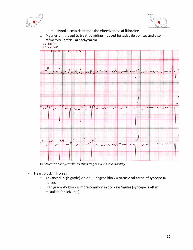
§ Hypokalemia decreases the effectiveness of lidocaine o Magnesium is used to treat quinidine induced torsades de pointes and also
refractory ventricular tachycardia
Ventricular tachycardia to third degree AVB in a donkey
- Heart block in Horses o Advanced (high grade) 2nd or 3rd degree block = occasional cause of syncope in
horses o High grade AV block is more common in donkeys/mules (syncope is often
mistaken for seizures)
11
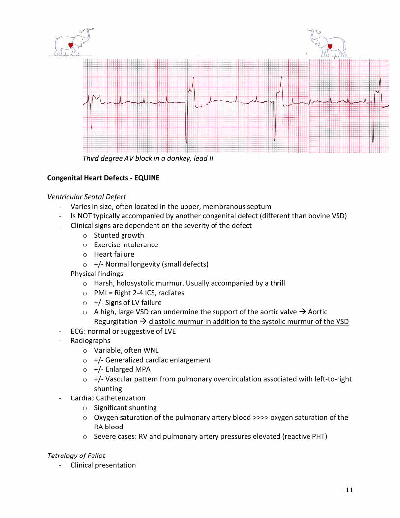
Third degree AV block in a donkey, lead II
Congenital Heart Defects - EQUINE Ventricular Septal Defect
- Varies in size, often located in the upper, membranous septum - Is NOT typically accompanied by another congenital defect (different than bovine VSD) - Clinical signs are dependent on the severity of the defect
o Stunted growth o Exercise intolerance o Heart failure o +/- Normal longevity (small defects)
- Physical findings o Harsh, holosystolic murmur. Usually accompanied by a thrill o PMI = Right 2-4 ICS, radiates o +/- Signs of LV failure o A high, large VSD can undermine the support of the aortic valve à Aortic
Regurgitation à diastolic murmur in addition to the systolic murmur of the VSD - ECG: normal or suggestive of LVE - Radiographs
o Variable, often WNL o +/- Generalized cardiac enlargement o +/- Enlarged MPA o +/- Vascular pattern from pulmonary overcirculation associated with left-to-right
shunting - Cardiac Catheterization
o Significant shunting o Oxygen saturation of the pulmonary artery blood >>>> oxygen saturation of the
RA blood o Severe cases: RV and pulmonary artery pressures elevated (reactive PHT)
Tetralogy of Fallot
- Clinical presentation

12
o Stunting of growth o Cyanosis o Dyspnea o Exercise intolerance
- Physical findings o Systolic murmur radiating widely over cardiac area
- ECG: RVE - Radiographs: RVE - Cardiac catheterization
o Elevated RV pressure o Normal – low pulmonary artery pressure o Low aortic blood oxygenation
Patent Ductus Arteriosus
- Foals: grade II-III/VI continuous murmur, PMI = 3rd – 4th left ICS at level of point of shoulder
o Heard until 4 - 5 days post birth Other defects
- Complex cardiac defects reported in Arabian foals - Atrial septal defect - Endocardial cushion defects
Congenital Heart Defects – BOVINE Ventricular Septal Defect
- Often coexists with another heart defect (i.e. truncus arteriosus) – unlike horses - Two-dimensional and Doppler echo = diagnostic - Cardiac cath is rarely performed
Other defects
- Ectopia cordis: the heart is located most often in the neck - PDA: usually diagnosed based on characteristic murmur
Acquired Cardiac Diseases Aortic Valve Insufficiency in the Horse
- Pathology o Diffusely thickened aortic valve OR nodules/bands affecting valve o Nodules = most common, loose fibrous connective tissue, fibroblast abundant
- Clinical Signs o Diastolic decrescendo murmur, PMI = L heart base o +/- Bounding pulses

13
o CHF & cardiomegaly in older animals (after peak working years) - Diagnosis
o Based on physical exam o ECG suggestive of L heart enlargement o Echo to document aortic insufficiency o Presence of fever/leukocytosis/peripheral embolization à consider bacterial
endocarditis - Treatment
o Most affected horses are NOT symptomatic, are old and not being worked o No adequate therapy o If CHF - Digitalis and diuretics
- Aortic valve more commonly affected than mitral valve Mitral Valve Insufficiency in the Horse
- Pathology o Localized or diffuse thickening, nodular thickening OR combination of lesions o Diffuse fibrous thickening = most common
- Clinical Signs o Most affected horses are asymptomatic o Middle – older age: can be severe enough to à exercise intolerance +/- CHF o Grade III+/VI plateau-shaped holosystolic murmur, PMI = mitral area, radiating to
right side - Diagnosis
o Based on physical exam o Echo to support: L heart volume overload in absence of AI o Normal Thoroughbreds: small amount of MR/TR present, especially after training o Mitral valve prolapse can occur with MR (less severe than degenerative valvular
disease) - Prognosis
o Good: mitral insufficiency rarely à CHF in horse o If progression to CHF occurs = poor prognosis
§ Poor oral absorption of diuretics & ACE-inhibitors Ruptured Chordae Tendinae in the Horse
- Chordae tendinae: anchor valve cusps to papillary muscles - Rupture à grossly incompetent valve - Large volume of blood regurgitated into LA during systole à decreased CO, increased
LVEDV & LVEDP à pulmonary congestion & fulminant life-threatening edema = LCHF - +/- Sudden death - Causes of ruptured MV chordae
o Blunt trauma o Severe physical exertion o Underlying primary MVD

14
- Clinical signs o Acute dyspnea with white foamy discharge from nostrils o Loud pansystolic murmur, PMI = MV, usually with palpable thrill o Decreased CO à signs of low output heart failure or CV collapse
- Diagnosis o Echo to visualize severe MR and a flail MV leaflet o Radiographs may show pulmonary infiltrates from LCHF
- Prognosis o Grave: most animals will be euthanized for fulminant CHF
Aorto-Cardiac Fistula (Ruptured Sinus of Valsalva) in the Horse
- Acquired cardiac fistula - Middle-aged, breeding stallions - Occurs suddenly à rapid death or severe distress - Clinical signs
o Acute collapse o Respiratory distress o Severe exercise intolerance o Continuous (PDA-like) murmur, PMI = R 4th ICS
- Diagnosis o Rupture should be suspected in breeding stallions with characteristic continuous
murmur and appropriate clinical signs o Echo to confirm diagnosis: visualize fistula from R aortic sinus with continuous
leftà right shunting from LVOT à RV or RA o ECG may show unifocal ventricular tachycardia
- Treatment o Supportive therapy – complete rest o Furosemide o Ventricular antiarrhythmics or digoxin
- Prognosis o Usually grave: survival time = 24 hr. – 4 yr. (if able to stabilize) o Sudden death possible
Bacterial Endocarditis in Ruminants
- Degenerative valvular disease = uncommon in ruminants - In ruminants: bacterial endocarditis appears as vegetative lesions, mostly on the right
side of circulation – Tricuspid and Pulmonic valves - Pathogenic Organisms (vary w/ species)
o Horse: Streptococcus, Actinobacillus, Pasturella o Cattle & Goats: Arcanobacterium pyogenes, Streptococci o Swine: Erysipelothrix rhysiopathiae, Streptococci o Lambs: Enterococci
- Clinical Signs

15
o Recurrent fever o Anorexia o Weight loss o Poor milk production o Shifting leg lameness o Tachycardia o Tachypnea o History of traumatic reticuloperitonitis or pneumonia o 1/6 cases admitted with primary complaint of heart disease
- Physical Examination: nonspecific and non-localizing o Systolic murmur may be present over TV and PV o Diastolic murmur of PI may be present o Jugular distension & jugular pulses together with distension of mammary veins
may be observed à development of ventral edema, ascites, cachexia - Diagnosis
o History, physical signs (murmur, fever), positive blood cultures o Echo visualization of vegetative lesions o Leukocytosis o Neutrophilia o Lymphopenia o Hyperglobulinemia o Anemia due to chronic infection
- Treatment o High dose antibiotics 4-6 wk. (penicillin or ampicillin) unless c/s indicate
otherwise - Prognosis
o Varies with stage of disease § Early (no heart failure): fair § Heart failure present: guarded – poor
Myocardial Disease in the Horse
- Primary myocardial disease = cardiomyopathy, not well documented in horses - Myocardial failure in the horse = most commonly from ingestion of monensin - Myocarditis (w/o CHF) = most frequently recognized myocardial disease in the horse - Myocarditis
o Inflammatory or degenerative myocardial lesions found in 2-15% necropsied horses
o Bacterial, viral, or parasitic causes (Streptococcus equi, influenza, purpura hemorrhagica, equine infectious anemia, strongyle larvae)
§ Strongyle larvae damage aortic root à microemboli à arteriosclerosis & myocardial necrosis
o Other causes: endocarditis, thoracic/abdominal abscessation, guttural pouch infections, colic, toxemias

16
o EHV-1 à myocarditis = well documented in aborted equine fetuses o Clinical signs = often absent
§ Decreased performance § Dyspnea on exertion § Tachycardia disproportionate to fever § Arrhythmias § Murmurs § Loud gallop sounds § Fainting/collapse § Sudden death: reported in horses recently recovered from
strangles/influenza § Development of CHF
o Treatment: directed at primary illness (cause of myocarditis) § Stall rest for 4 – 6 mo. § Antiarrhythmic therapy if serious arrhythmia is detected § Digitalis & diuretics if CHF present
- Monensin toxicosis o Horses = more sensitive to monensin than cattle, develop cardiac signs if ingest
treated cattle feed o Clinical signs
§ Acutely affected • Abdominal pain • Diarrhea • Acute circulatory failure • Death
§ Delayed syndrome: 3 – 6 mo. post-ingestion § Necropsy
• Myocardial degeneration • Pericardial effusion • Evidence of heart failure
o Treatment: supportive, myocardial damage is irreversible - White snakeroot intoxication
o Ingestion à severe myocardial degeneration, hemorrhages, infarcts o Arrhythmias & sudden death = frequent sequelae
Myocardial Disease in Cattle
- Cardiomyopathy is rare in cattle o Bovine DCM reported in some breeds
- Myocardial disease in cattle is typically secondary to… o Systemic infection
§ Pneumonia § Traumatic reticulopericarditis

17
§ Neonatal infection § Mastitis
o Nutritional deficiency § Vitamin E § Selenium
o Neoplastic infiltration § Lymphosarcoma
o Ingestion of toxins § Monensin § Gossypol § White snakeroot § Cassia occidentalis
- Clinical syndromes will vary (sudden death – chronic CHF) Traumatic Pericarditis in Cattle
- Most common cause of CHF in cattle - Perforation or migration of a foreign body (i.e. wire) into the pericardial sac. Disease
course varies from few d. – few wk./mo. - Pathology
o Significantly thickened pericardial sac o Adhesions between pericardium & diaphragm and pericardium & epicardium o +/- Visualize foreign body o Variable amounts of pus, fibrin, gas
- Clinical signs: signs reflect presence of infection due to reticuloperitonitis with evidence of cardiac tamponade
o Depression, anorexia, weight loss o Diarrhea, constipation, rumen atony o Decreased milk production o Arched back, abducted elbows, pain o Fever, tachycardia, tachypnea o Venous distension of the jugular and mammary veins o Ventral and cervical edema, ascites later in disease course
- Physical findings o Early in disease: pericardial friction rub may be present o Later in disease
§ Pericardial effusion develops à muffled heart sounds § Gas and fluid accumulate à splashing, tinkling, gurgling sounds
synchronous with heartbeat - Diagnosis
o Rule outs § Heart failure from congenital defects § Infective endocarditis § Altitude sickness (PHT)
18
§ Lymphosarcoma § Other myocardial diseases
o To confirm diagnosis § Echo § Pericardiocentesis at 4th – 5th ICS on Left side (idiopathic effusions can
occur and will often resolve) - Treatment
o Drain pericardial sac when heart failure is present § Pericardiocentesis § Instillation of antibiotics, debriding enzymes, placement of pericardial
drain/surgical exposure and drainage - Prognosis = poor
Pericardial Disease in the Horse
- An “epidemic” of pericarditis and mare reproductive loss in Spring-Summer of 2001 in Kentucky (previously very rare). Actinobacillus sp. were the principal isolates
o Cause of outbreak: “Septic penetrating setal emboli (SPSE)” from infestation of eastern tent caterpillars
- Clinical signs o Fever, anorexia, lethargy o Muffled heart sounds +/- pericardial friction rub o Signs of RCHF
§ Jugular distension § Jugular pulses § Ventral edema § Ascites § Pleural effusion § Tachycardia
- Diagnosis o Rule out other causes of RCHF o Rule out pleural effusion due to pleuritis o Echo or successful pericardiocentesis to confirm diagnosis o Can measure RA pressure before and after pericardiocentesis o +/- Low voltage QRS complexes or electrical alternans on ECG (not as helpful in
diagnosing as in small animals) - Treatment
o Signs of systemic congestion: drain and lavage pericardial sac o Systemic antibiotics, sometimes even if negative culture o If large number of eosinophils OR if infectious etiology is excluded à consider
corticosteroid therapy Neoplasia
- Lymphosarcoma = most common neoplasia of the heart in cattle

19
o Consider as a differential diagnosis for most cardiac diseases in cattle o Less frequent in horse – if it occurs, may include heart