Embed Size (px)
Citation preview
1
George D. Soltes, M.D.Assistant Professor
Interventional RadiologyUniversity of Washington
History: 38 year old female with left lower extremity swelling and pain over 3 years with recent worsening
Bilateral Femoral Venograms

2
A. Central propagation of femoral-popliteal thrombus
B. An anatomic variantC. Compression of the left common
iliac vein by the right common iliac artery
D. AtherosclerosisE. Iliac arteriovenous fistula
A. Angioplasty aloneB. Balloon-mounted stent
placementC. Covered stent (stent graft)
placementD. Self-expanding stent
placementE. All of the above

3
AKA: Iliac vein compression syndrome, Cockett syndrome
Etiology: Compression of the left common iliac vein by the overlying right common iliac artery venous intimal injury webs (“spurs”), stenosis, thrombosis
Most common in females 2nd-4th decadeTypical symptoms: Left leg swelling and
pain without other risk factors for venous disease
Imaging:• Doppler US: will detect iliac DVT, but won’t
visualize iliac vein compression and webs; may infer by abnormal waveform w/ resp maneuvers
• CT/MRI: Compression of left iliac vein between right iliac
artery and spine R/O other causes of extrinsic compression (LN, mass,
aneurysm, etc.) May be a normal anatomic finding (2/3 of
asymptomatic patients have at least 25% left iliac vain compression)

4
Right common iliac arteryRight common iliac artery
Left common iliac veinLeft common iliac vein
Compression of left common iliac vein between right common iliac artery and spine
Imaging:• Venography: Left common iliac vein
stenosis, usually smooth and short, but may be long and irregular

5
Imaging:• Venography: Left common iliac vein
stenosis, usually smooth and short, but may be long and irregular
May progress to occlusion
Hemodynamic significance indicated by: Regional collateral vein
filling Pressure gradient > 3-4
mmHg
Occlusion of left common iliac vein
Collateral veins
Treatment:• Surgical: Fem-fem or CFV-IVC bypass• Placement of self-expanding stents (e.g.,
Wallstents); usually 14 – 16 mm diameter• “Kissing stents” if stent will encroach on IVC• Angioplasty alone insufficient due to extrinsic
compression• Balloon-expanded stents avoided as may get
crushed • +/- Thrombolysis if acute clot present• +/- Anticoagulation following stent placement

6
Pre Treatment Post “Kissing” Iliac Wallstents
IR Treatment Outcomes:• Successful recanalization in > 90%• 50 – 80% 1-year primary patency• Major complications in 7%: access site
bleeding/hematoma, stent malposition, stent migration

7
History: 38 year-old male with left iliofemoral DVT and GI bleeding on anticoagulation
IVC filter requested
IVC gram
A
B
C
D
E

8
A. Circumaortic left renal veinB. Duplicated IVCC. Azygos continuation of IVCD. Artifact from overlying bowelE. IVC thrombus
Selective venogram of circumaortic left renal vein

9
Filter placed with apex at level of lowest (circumaortic) renal vein
Improper filter placement above retroaortic vein may allow clot to pass through renal sinus to the lung

10
Preaortic vein Retroaortic vein
CT Appearance
Left IVC• 0.2%-0.5%• L IVC usually joins L renal vein
Duplicated IVC• O.2%-3%• L IVC usually crosses over via L renal vein• Need bilat IVC filters or suprarenal filter
Azygos continuation of IVC• 0.6%• No intrahepatic IVC; hepatic veins drain to RA

11
Retroaortic left renal vein• 2%• Usually below level of R renal vein
Circumaortic left renal vein• 1.5%-8.7%• Most common IVC anomaly• Usually 1-2 cm below anterior L renal vein
78 year old female with bilateral lower extremity deep venous thrombosis and pulmonary embolism.
Recent intracranial hemorrhage and craniotomy.
IVC gramDiameter of infrarenal IVC: 30.5 cm

12
A. Placement of permanent Bird’s Nest IVC filter.
B. Full anticoagulationC. Placement of retrievable
IVC filterD. Placement of bliateral
common iliac vein retrievable filters
E. A and D
IVC transverse diameter > 28 mm1% to 2% of populationOptions for filter placement:
• Bird’s Nest Filter (only filter for diam > 35mm)• Standard filters (permanent or retrievable) in
each common iliac vein

13
Infrarenal Bird’s Nest IVC filter placed
Alternative: Bilateral common iliac vein filters

14
Widely accepted:• Contraindication to
anticoagulation• Complication of
anticoagulation• Failure of anticoagulation
Moderately accepted:• Massive PE• Free-floating iliofemoral or
IVC thrombus (“Widowmaker”)
• Severe cardiopulmonary dz• Poor anticoagulation
candidate (e.g., falls, compliance)
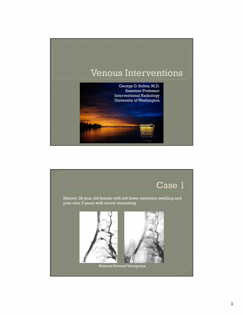
• Recurrent PE with filter
15
Controversial:• Prophylaxis: Severe trauma Immobilized patients Preoperative with multiple
risk factors for venous thromboembolism Advanced malignancy
• Pre LE thrombolysis• Pregnancy with proximal
DVT
Young patientProphylaxis (major
trauma, ICU, pre-op)
During LE thrombolysis
During pregnancy

16
49 year old male with elevated liver enzymes, ascites and abdominal pain
A. Alcoholic cirrhosisB. Hepatocellular
carcinomaC. Primary biliary
cirrhosisD. Sclerosing cholangitisE. Budd-Chiari syndrome

17
A. Hepatic vein angioplastyB. Hepatic vein stentC. TIPSD. Liver transplantE. All of the above
Hepatic vein outflow obstruction Level of obstruction:
• Hepatic venules Hepatic venoocclusive disease Etiol: chemotherapy, irradiation for stem cell/BMT
• Major hepatic veins Classic Budd-Chiari Etiol: thrombophilic states (polycythemia vera, OCPs,
malignancy), neoplastic invasion from HCC• Hepatic vein confluence/IVC/RA Etiol: membranous IVC obstruction, bland thrombus, tumor,
extrinsic compression, RA tumor, constrictive pericarditis)

18
• Depends on chronicity• Classic triad: hepatomegaly, abdominal pain,
ascites• Splenomegaly, jaundice, variceal bleeding• May be asymptomatic
CT/MRI:• Ascites, hepatomegaly,
splenomegaly• Mottled/patchy
enhancement of liver• Atrophy of right lobe
with sparing/hypertrophy of caudate lobe
• Slit like/obliterated hepatic veins
• Hepatic vein to hepatic vein collaterals
• Hepatic vein or IVC webs

19
US:• Absent or
dampened hepatic vein flow with loss of transmitted atrial waves
Absent flow in middle hepatic vein
Venography:• Classic:
“Spiderweb” appearance of small collateral veins
• Narrowing/webs/occlusions of hepatic veins or IVC

20
IR:• Thrombolysis if acute thrombus• Angioplasty and/or stenting of hepatic or IVC
stenoses• TIPS
Surgical:• Mesocaval/mesoatrial shunt• Liver transplantation
Web-like hepatic vein stenosisGradient: 18 mmHg
Post stent placementGradient: 2 mmHg
Symptoms resolved

21
28 year old male house painter with acute onset of heaviness, pain, and swelling of right arm.
Doppler R axillary and subclavian thrombosis Venography performed
RUE Venogram
A. Surgical decompressionB. VenoplastyC. ThrombolysisD. Stent placementE. Anticoagulation alone

22
A. ArteriesB. VeinsC. NervesD. All about equal
Caused by anatomical abnormalities involving the thoracic outlet• cervical rib• congenital bands• hypertrophied scalene
muscles• Bony exostoses• Anomalous muscle or
ligament anatomy http://my.clevelandclinic.org

23
Subgroups:• Neurologic Most common (95%) Compression of brachial
plexus
• Venous 3-4% Compression of
subclavian vein
• Arterial 1-2% Compression of
suclavian artery
AKA: Paget-Schroetter syndrome, effort thrombosis Minority (5-20%) of UE venous thrombosis M:F 2:1 2nd through 5th decade; mean 30’s Typically young & active 60-80% recent vigorous UE exercise R > L; Bilat in 6%

24
Subclavian vein runs within anterior part of the thoracic outlet• subclavius muscle and
costoclavicularligament anteromedially
• anterior scalene muscle posterolaterally
Extrinsic compressionIntimal injuryStenosisThrombosis

25
Typically young person with sudden onset
Often in setting of repeated activities involving arm elevation/exertion
UE swelling, heaviness, pain, fatigue, cyanosis
Enlarged subcutaneous veins of arm, shoulder, chest wall
Much less likely than LE DVT to present with symptomatic PE (9% vs 29%)
Semin Intervent Radiol. 2012 Mar; 29(1): 44–51.
Ultrasound:• Color doppler will show
axillo-subclavianthrombus
• Duplex US flat tracing without normal respvariation and reflected atrial activity
• Hard to directly visualize more central veins
• Duplex has 80%-100% sensitivity and specificity
Thrombosed subclavian vein
http://www.ultrasoundcases.info
Normal
Central occlusion

26
Venography:• Occlusion of
subclavian vein at costoclavicularjunction
• Distal extension of thrombus
• Collaterals• Arm abduction for
subtle/early cases
J Vasc Surg 2010; 51(6):153
Important to treat early!Catheter-directed thrombolysisSurgical decompression (1st rib
resection)No pre-operative stent placement
• May fracture from external compressionPost-operative venoplasty/stent if
residual stenosisPost-treatment anticoagulation (3-6 mos)Follow by clinical symptoms and US

27
Initial venogram
Post thrombolysis
• Surgical decompression subsequently performed• Symptoms resolved
38 year old female with sickle cell anemia
Attempted central venous catheter placement

28
A. Catheter directed thrombolysis
B. Systemic thrombolysis
C. Balloon angioplastyD. Stent placementE. Surgical bypass
A. Central venous catheters
B. Bronchogenic carcinoma
C. MediastinitisD. Pacemaker wiresE. Lymphoma

29
Malignant (90%)• Bronchogenic
carcinoma (#1 cause)
• Lymphoma, mets, mesothelioma, germ-cell, thymoma
Right hilar mass occluding SVC
Chest wall collaterals
group.bmj.com
Benign• Central venous
catheters/pacers (75% of benign)
• Granulomatous mediastinitis (TB, histoplasmosis, sarcoidosis)
• Aortic aneurysms• Radiation
Left innominate/SVC occlusionfrom central venous catheter

30
Edema of face, neck and upper extremities
Enlarged cutaneous collaterals
Headache, dizziness, syncope
DysphagiaChest painRespiratory distressDeath
passpaces.com
CT:• Most frequently used• Site and extent of
occlusion• Presence of thrombus• Presence of collaterals• Primary cause of
occlusion
group.bmj.com

31
MRI:• Similar advantages to
CT• MRV may help
delineate venous obstruction
Occlusions of innominate veins
group.bmj.com
Venography:• “Gold standard”• Cannot identify cause
of obstruction• Used to determine
extent of obstruction and guide treatment
Upper extent of obstruction
Lower extent of obstruction

32
Chemo, XRT, steroids• Bronchogenic carcinoma: Chemo and XRT provide
relief in 60-80% of patients 20% recurrence May take 2-4 weeks to have
effect
• Chemo is 1st line treatment for lymphoma
SVC Stenting:• 1st line treatment for
bronchogenic ca and mets
• Rapid relief of symptoms while underlying etiology is treated

33
Usually more insidious courseNo role for steroidsSurgical bypass—high morbidityThrombolysis if secondary to thrombusStenting
• Now considered primary treatment • May require repeat intervention
No randomized controlled study comparing SVC stenting to chemo or XRT
Stents 87%-100% effective in relieving symptoms at initial presentation
Re-obstruction rate as high as 40% in long-term follow-up
Stent patency restored in majority by secondary interventions

34
2% mortality:• Hemorrhage, cardiac events, resp failure, PE
4% major complications:• Stent migration, bleeding, infection, thrombotic
events, SVC rupture, pericardial tamponade, arrhythmias
3.2% minor complications: Puncture site hematoma, chest pain
38 year old female with sickle cell anemia
Attempted central venous catheter placement
Head and neck edema

35
Sheaths placed from jugular and femoral
Guidewire advanced across occlusion from below
Snare placed from above

36
Guidewire snared and pulled through jugular sheath “body floss”
Venoplasty of occlusion

37
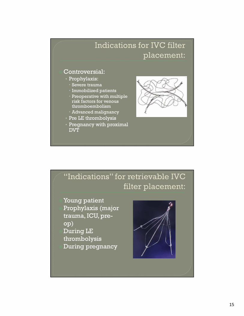
Stent placed
Post stent venogram
38
Almost immediate resolution of symptoms
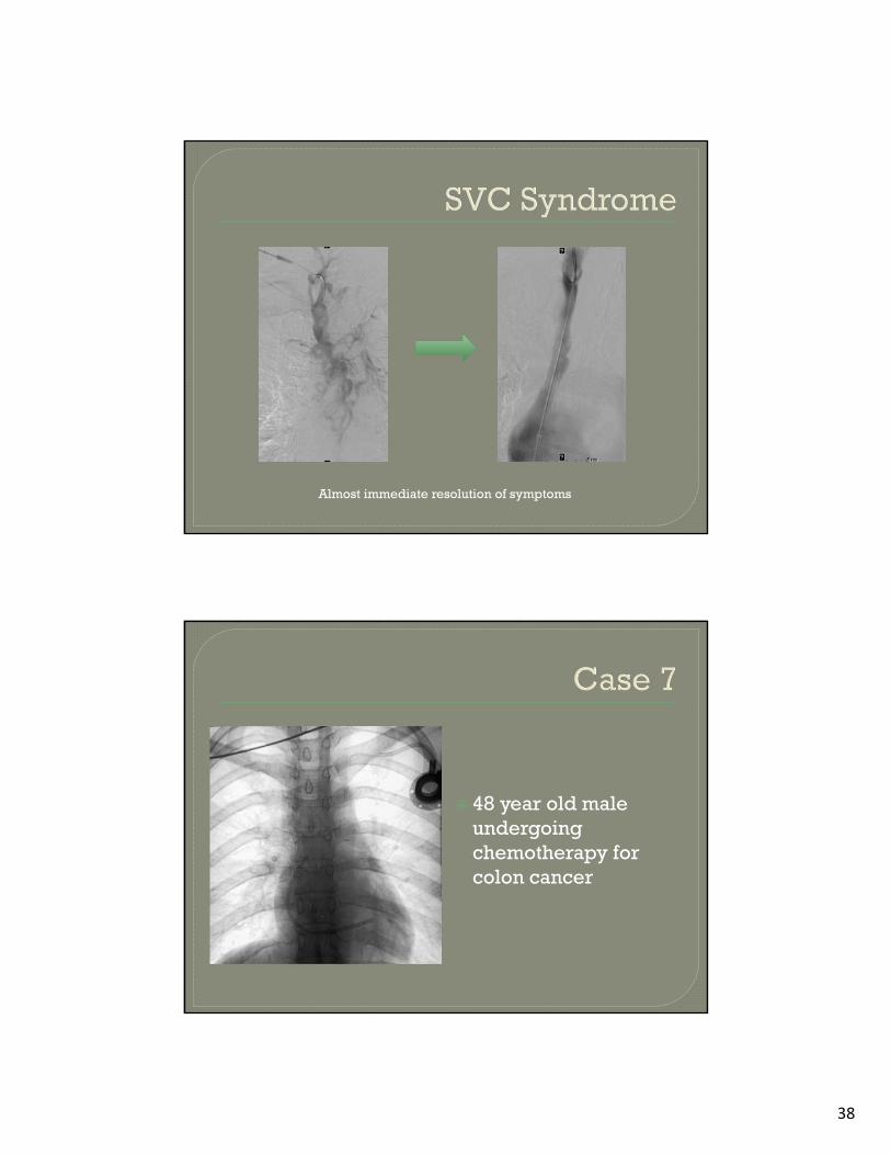
48 year old male undergoing chemotherapy for colon cancer

39
A. More lateral subclavianvein puncture site
B. More medial subclavianvein puncture site
C. Internal jugular vein access
D. Use of a polyurethane catheter
E. Use of a silicone catheter
Excessively medial entry into subclavian vein Catheter extravascular in costoclavicular space Catheter compressed between clavicle and first rib
nursingcenter.com

40
Jugular vein access If subclavian access, lateral vein entry
pointRecognize “pinch-off sign” on CXR
Grade 0• Normal appearance
Grade 1• Catheter deviation
Grade 2• Catheter narrowing
Grade 3• Transection with
embolization

41
Ruptured subclavianchest port with catheter fragment in RA/RV
Fragment snared…

42
…pulled into IVC…
…and out groin
43
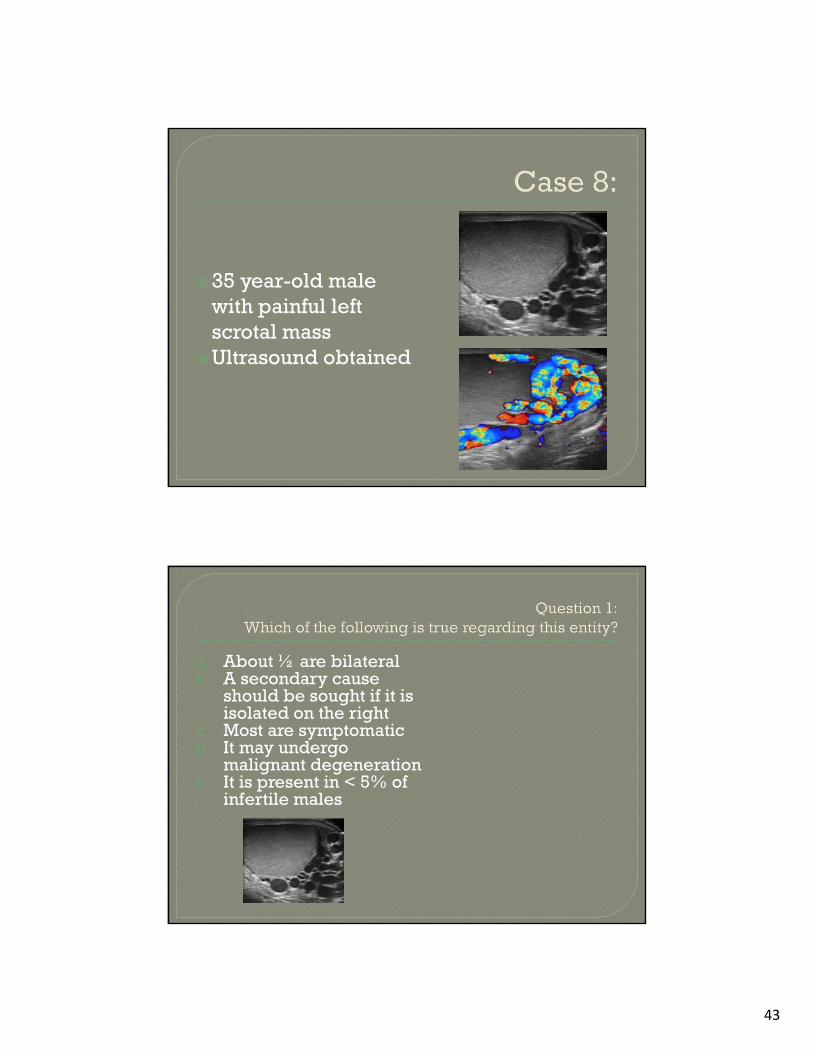
35 year-old male with painful left scrotal mass
Ultrasound obtained
A. About ½ are bilateralB. A secondary cause
should be sought if it is isolated on the right
C. Most are symptomaticD. It may undergo
malignant degenerationE. It is present in < 5% of
infertile males

44
A. Vein rupture is a rare but catastrophic complication
B. Particulate embolicssuch as PVA particles are preferred
C. Coils should be placed only at the level of the inguinal ligament
D. Recurrence rates are equivalent to surgery
Dilation of pampiniform plexus in the scrotum
Etiology: • Incompetent gonadal vein valve allowing reflux
of blood• Secondary varicocele Abdominal or pelvic mass impeding drainage of
pampiniform plexus Look for neoplasm if sudden onset in older man or
isolated right sided varicocele
45
5 to 17% of malesL:R 10:1Bilateral 10% Isolated to right: 1-2%
80% asymptomaticPain Infertility
• 40% of males evaluated for infertility have varicocele
Testicular atrophyScrotal swelling

46
Physical exam:• Palpable scrotal
abnormality Increases in size with
Valsalva and upright position
Preferred imaging modality
Findings:• Dilated pampiniform
plexus (> 2mm)• Doppler/color flow
reflux with Valsalva
ultrasoundcases.info

47
Indications for treatment:• Infertility• Groin pain • Adolescent varicocele• Recurrence post surgery
ConservativeSurgical
• Spermatic vein ligation Interventional
• Spermatic vein embolization

48
Testicular Hypothermia Device (THD)• AKA Refrigerated Underwear
Urology 1984;23
Ligation of internal spermatic vein
Open or laparoscopic
OutpatientApproximately 2
week recovery Recurrence rate:
• 10-20%• Usually caused by
collateral flow around ligature

49
Diagnostic venogram:• Right internal
spermatic vein usually drains into IVC and left into left renal vein
• Document valvularincompetence, collaterals
Left internal spermatic venogram
Diagnostic venogram:• Right internal
spermatic vein usually drains into IVC and left into left renal vein
• Document valvularincompetence, collaterals
Left internal spermatic venogram

50
Embolic agent:• Coils • May add liquid sclerosing agent Sotradecol Cyanoacrylate (glue)
Begin occlusion at superior pubic ramus and continue to vein origin
Coils placed along entire length of vein to prevent collateralization
51
With solitary left varicocele, only need to perform right-sided embolization if:• Treating infertility and• Right internal spermatic vein incompetent
Multiple spermatic veins may be present and need to be embolized
If use liquid agents, care to avoid reflux into scrotum and thrombosis of pampiniform plexus
Similar results to surgeryTechnical success rate > 90%Recurrence rate 10% Improved sperm counts in 80%Failures due to:
• Inability to canalize internal spermatic vein• Missed additional veins• Collateralization • Recanalization of occluded segments
Good outcomes after failed ligation

52
Minor flank pain or scrotal pain
Low grade feverVein rupture
(usually self-limited)
Migration of coil to lung
Thrombosis of pampiniform plexus
Vein rupture
THANKS!