Embed Size (px)
Citation preview
Cell Stem Cell
Perspective
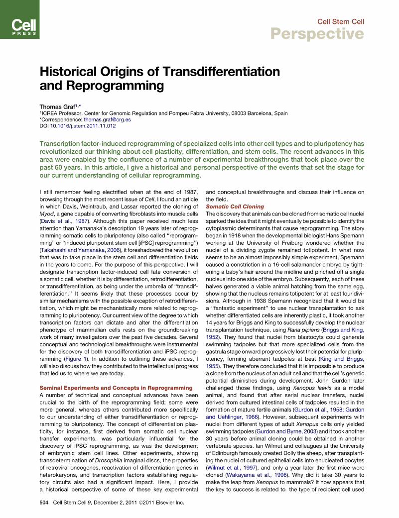
Historical Origins of Transdifferentiationand Reprogramming
Thomas Graf1,*1ICREA Professor, Center for Genomic Regulation and Pompeu Fabra University, 08003 Barcelona, Spain*Correspondence: [email protected] 10.1016/j.stem.2011.11.012
Transcription factor-induced reprogramming of specialized cells into other cell types and to pluripotency hasrevolutionized our thinking about cell plasticity, differentiation, and stem cells. The recent advances in thisarea were enabled by the confluence of a number of experimental breakthroughs that took place over thepast 60 years. In this article, I give a historical and personal perspective of the events that set the stage forour current understanding of cellular reprogramming.
I still remember feeling electrified when at the end of 1987,
browsing through the most recent issue of Cell, I found an article
in which Davis, Weintraub, and Lassar reported the cloning of
Myod, a gene capable of converting fibroblasts into muscle cells
(Davis et al., 1987). Although this paper received much less
attention than Yamanaka’s description 19 years later of reprog-
ramming somatic cells to pluripotency (also called ‘‘reprogram-
ming’’ or ‘‘induced pluripotent stem cell [iPSC] reprogramming’’)
(Takahashi and Yamanaka, 2006), it foreshadowed the revolution
that was to take place in the stem cell and differentiation fields
in the years to come. For the purpose of this perspective, I will
designate transcription factor-induced cell fate conversion of
a somatic cell, whether it is by differentiation, retrodifferentiation,
or transdifferentiation, as being under the umbrella of ‘‘transdif-
ferentiation.’’ It seems likely that these processes occur by
similar mechanisms with the possible exception of retrodifferen-
tiation, which might be mechanistically more related to reprog-
ramming to pluripotency. Our current view of the degree towhich
transcription factors can dictate and alter the differentiation
phenotype of mammalian cells rests on the groundbreaking
work of many investigators over the past five decades. Several
conceptual and technological breakthroughs were instrumental
for the discovery of both transdifferentiation and iPSC reprog-
ramming (Figure 1). In addition to outlining these advances, I
will also discuss how they contributed to the intellectual progress
that led us to where we are today.
Seminal Experiments and Concepts in ReprogrammingA number of technical and conceptual advances have been
crucial to the birth of the reprogramming field; some were
more general, whereas others contributed more specifically
to our understanding of either transdifferentiation or reprog-
ramming to pluripotency. The concept of differentiation plas-
ticity, for instance, first derived from somatic cell nuclear
transfer experiments, was particularly influential for the
discovery of iPSC reprogramming, as was the development
of embryonic stem cell lines. Other experiments, showing
transdetermination of Drosophila imaginal discs, the properties
of retroviral oncogenes, reactivation of differentiation genes in
heterokaryons, and transcription factors establishing regula-
tory circuits also had a significant impact. Here, I provide
a historical perspective of some of these key experimental
504 Cell Stem Cell 9, December 2, 2011 ª2011 Elsevier Inc.
and conceptual breakthroughs and discuss their influence on
the field.
Somatic Cell Cloning
Thediscovery that animals canbecloned fromsomatic cell nuclei
sparked the idea that itmight eventually bepossible to identify the
cytoplasmic determinants that cause reprogramming. The story
began in 1918 when the developmental biologist Hans Spemann
working at the University of Freiburg wondered whether the
nuclei of a dividing zygote remained totipotent. In what now
seems to be an almost impossibly simple experiment, Spemann
caused a constriction in a 16-cell salamander embryo by tight-
ening a baby’s hair around the midline and pinched off a single
nucleus into one side of the embryo. Subsequently, each of these
halves generated a viable animal hatching from the same egg,
showing that the nucleus remains totipotent for at least four divi-
sions. Although in 1938 Spemann recognized that it would be
a ‘‘fantastic experiment’’ to use nuclear transplantation to ask
whether differentiated cells are inherently plastic, it took another
14 years for Briggs and King to successfully develop the nuclear
transplantation technique, using Rana pipiens (Briggs and King,
1952). They found that nuclei from blastocyts could generate
swimming tadpoles but that more specialized cells from the
gastrula stage onward progressively lost their potential for plurip-
otency, forming aberrant tadpoles at best (King and Briggs,
1955). They therefore concluded that it is impossible to produce
a clone from the nucleus of an adult cell and that the cell’s genetic
potential diminishes during development. John Gurdon later
challenged those findings, using Xenopus laevis as a model
animal, and found that after serial nuclear transfers, nuclei
derived from cultured intestinal cells of tadpoles resulted in the
formation of mature fertile animals (Gurdon et al., 1958; Gurdon
and Uehlinger, 1966). However, subsequent experiments with
nuclei from different types of adult Xenopus cells only yielded
swimming tadpoles (Gurdon andByrne, 2003) and it took another
30 years before animal cloning could be obtained in another
vertebrate species. Ian Wilmut and colleagues at the University
of Edinburgh famously created Dolly the sheep, after transplant-
ing the nuclei of cultured epithelial cells into enucleated oocytes
(Wilmut et al., 1997), and only a year later the first mice were
cloned (Wakayama et al., 1998). Why did it take 30 years to
make the leap from Xenopus to mammals? It now appears that
the key to success is related to the type of recipient cell used
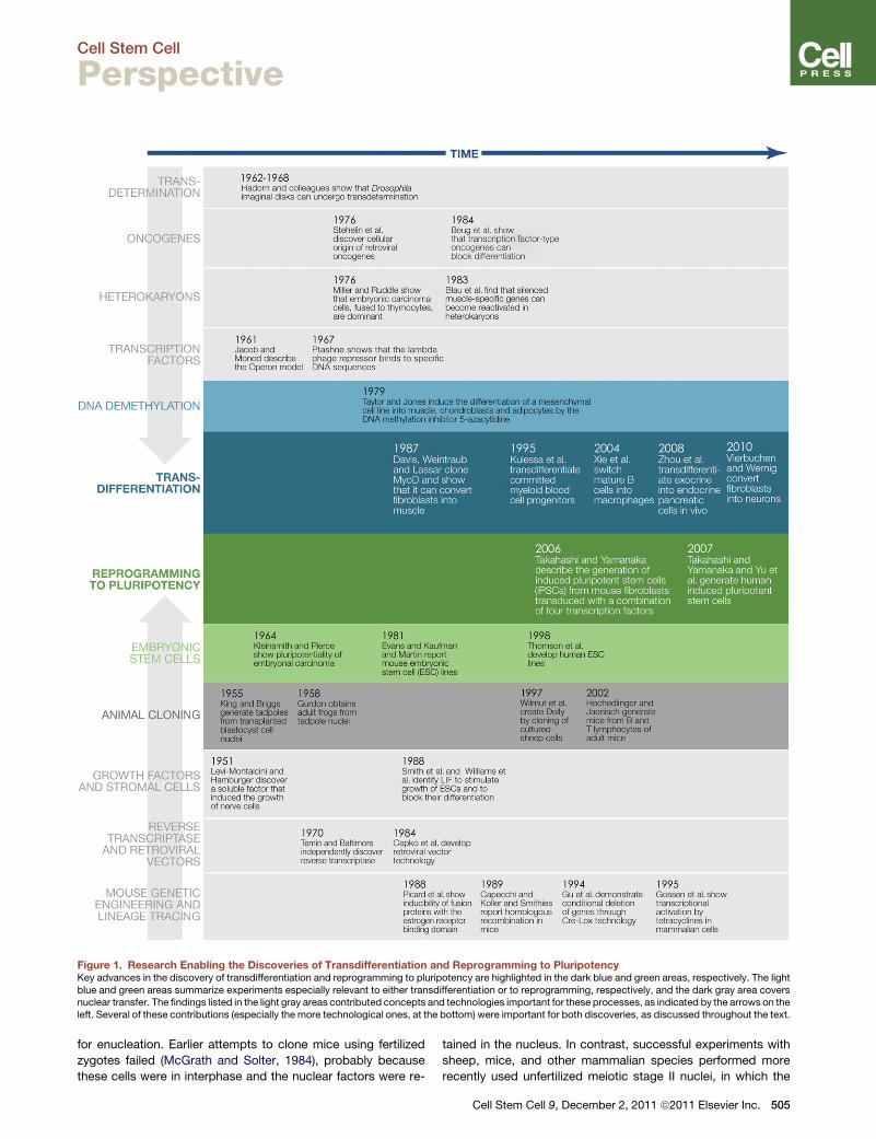
Figure 1. Research Enabling the Discoveries of Transdifferentiation and Reprogramming to PluripotencyKey advances in the discovery of transdifferentiation and reprogramming to pluripotency are highlighted in the dark blue and green areas, respectively. The lightblue and green areas summarize experiments especially relevant to either transdifferentiation or to reprogramming, respectively, and the dark gray area coversnuclear transfer. The findings listed in the light gray areas contributed concepts and technologies important for these processes, as indicated by the arrows on theleft. Several of these contributions (especially the more technological ones, at the bottom) were important for both discoveries, as discussed throughout the text.
Cell Stem Cell
Perspective
for enucleation. Earlier attempts to clone mice using fertilized
zygotes failed (McGrath and Solter, 1984), probably because
these cells were in interphase and the nuclear factors were re-
tained in the nucleus. In contrast, successful experiments with
sheep, mice, and other mammalian species performed more
recently used unfertilized meiotic stage II nuclei, in which the
Cell Stem Cell 9, December 2, 2011 ª2011 Elsevier Inc. 505
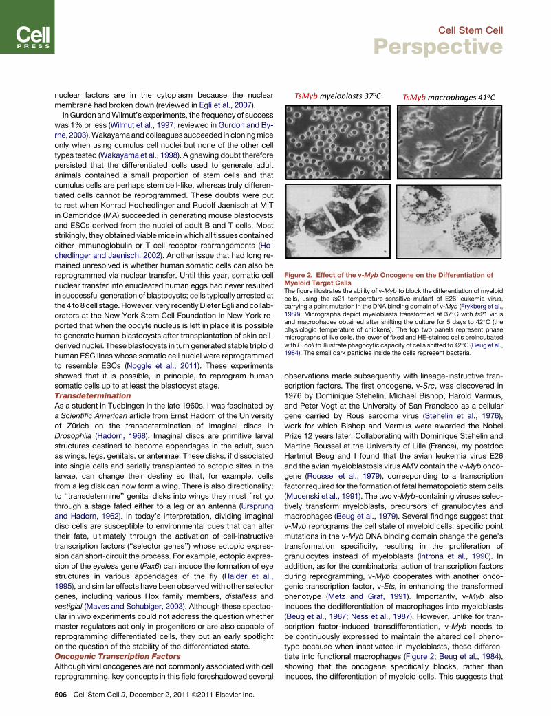
Figure 2. Effect of the v-Myb Oncogene on the Differentiation ofMyeloid Target CellsThe figure illustrates the ability of v-Myb to block the differentiation of myeloidcells, using the ts21 temperature-sensitive mutant of E26 leukemia virus,carrying a point mutation in the DNA binding domain of v-Myb (Frykberg et al.,1988). Micrographs depict myeloblasts transformed at 37�C with ts21 virusand macrophages obtained after shifting the culture for 5 days to 42�C (thephysiologic temperature of chickens). The top two panels represent phasemicrographs of live cells, the lower of fixed and HE-stained cells preincubatedwith E. coli to illustrate phagocytic capacity of cells shifted to 42�C (Beug et al.,1984). The small dark particles inside the cells represent bacteria.
Cell Stem Cell
Perspective
nuclear factors are in the cytoplasm because the nuclear
membrane had broken down (reviewed in Egli et al., 2007).
InGurdon andWilmut’s experiments, the frequency of success
was 1% or less (Wilmut et al., 1997; reviewed in Gurdon and By-
rne, 2003).Wakayamaandcolleagues succeeded in cloningmice
only when using cumulus cell nuclei but none of the other cell
types tested (Wakayama et al., 1998). A gnawing doubt therefore
persisted that the differentiated cells used to generate adult
animals contained a small proportion of stem cells and that
cumulus cells are perhaps stem cell-like, whereas truly differen-
tiated cells cannot be reprogrammed. These doubts were put
to rest when Konrad Hochedlinger and Rudolf Jaenisch at MIT
in Cambridge (MA) succeeded in generating mouse blastocysts
and ESCs derived from the nuclei of adult B and T cells. Most
strikingly, they obtained viablemice inwhich all tissues contained
either immunoglobulin or T cell receptor rearrangements (Ho-
chedlinger and Jaenisch, 2002). Another issue that had long re-
mained unresolved is whether human somatic cells can also be
reprogrammed via nuclear transfer. Until this year, somatic cell
nuclear transfer into enucleated human eggs had never resulted
in successful generation of blastocysts; cells typically arrested at
the 4 to 8cell stage.However, very recentlyDieter Egli andcollab-
orators at the New York Stem Cell Foundation in New York re-
ported that when the oocyte nucleus is left in place it is possible
to generate human blastocysts after transplantation of skin cell-
derived nuclei. These blastocysts in turn generated stable triploid
human ESC lines whose somatic cell nuclei were reprogrammed
to resemble ESCs (Noggle et al., 2011). These experiments
showed that it is possible, in principle, to reprogram human
somatic cells up to at least the blastocyst stage.
Transdetermination
As a student in Tuebingen in the late 1960s, I was fascinated by
a Scientific American article from Ernst Hadorn of the University
of Zurich on the transdetermination of imaginal discs in
Drosophila (Hadorn, 1968). Imaginal discs are primitive larval
structures destined to become appendages in the adult, such
as wings, legs, genitals, or antennae. These disks, if dissociated
into single cells and serially transplanted to ectopic sites in the
larvae, can change their destiny so that, for example, cells
from a leg disk can now form a wing. There is also directionality;
to ‘‘transdetermine’’ genital disks into wings they must first go
through a stage fated either to a leg or an antenna (Ursprung
and Hadorn, 1962). In today’s interpretation, dividing imaginal
disc cells are susceptible to environmental cues that can alter
their fate, ultimately through the activation of cell-instructive
transcription factors (‘‘selector genes’’) whose ectopic expres-
sion can short-circuit the process. For example, ectopic expres-
sion of the eyeless gene (Pax6) can induce the formation of eye
structures in various appendages of the fly (Halder et al.,
1995), and similar effects have been observedwith other selector
genes, including various Hox family members, distalless and
vestigial (Maves and Schubiger, 2003). Although these spectac-
ular in vivo experiments could not address the question whether
master regulators act only in progenitors or are also capable of
reprogramming differentiated cells, they put an early spotlight
on the question of the stability of the differentiated state.
Oncogenic Transcription Factors
Although viral oncogenes are not commonly associated with cell
reprogramming, key concepts in this field foreshadowed several
506 Cell Stem Cell 9, December 2, 2011 ª2011 Elsevier Inc.
observations made subsequently with lineage-instructive tran-
scription factors. The first oncogene, v-Src, was discovered in
1976 by Dominique Stehelin, Michael Bishop, Harold Varmus,
and Peter Vogt at the University of San Francisco as a cellular
gene carried by Rous sarcoma virus (Stehelin et al., 1976),
work for which Bishop and Varmus were awarded the Nobel
Prize 12 years later. Collaborating with Dominique Stehelin and
Martine Roussel at the University of Lille (France), my postdoc
Hartmut Beug and I found that the avian leukemia virus E26
and the avianmyeloblastosis virus AMV contain the v-Myb onco-
gene (Roussel et al., 1979), corresponding to a transcription
factor required for the formation of fetal hematopoietic stem cells
(Mucenski et al., 1991). The two v-Myb-containing viruses selec-
tively transform myeloblasts, precursors of granulocytes and
macrophages (Beug et al., 1979). Several findings suggest that
v-Myb reprograms the cell state of myeloid cells: specific point
mutations in the v-Myb DNA binding domain change the gene’s
transformation specificity, resulting in the proliferation of
granulocytes instead of myeloblasts (Introna et al., 1990). In
addition, as for the combinatorial action of transcription factors
during reprogramming, v-Myb cooperates with another onco-
genic transcription factor, v-Ets, in enhancing the transformed
phenotype (Metz and Graf, 1991). Importantly, v-Myb also
induces the dedifferentiation of macrophages into myeloblasts
(Beug et al., 1987; Ness et al., 1987). However, unlike for tran-
scription factor-induced transdifferentiation, v-Myb needs to
be continuously expressed to maintain the altered cell pheno-
type because when inactivated in myeloblasts, these differen-
tiate into functional macrophages (Figure 2; Beug et al., 1984),
showing that the oncogene specifically blocks, rather than
induces, the differentiation of myeloid cells. This suggests that
Cell Stem Cell
Perspective
v-Myb perturbs lineage-specific regulatory networks, linking cell
proliferation and differentiation. The observed v-Myb transgene
dependence of cell transformation seems to be a common
feature of oncogenes, because in many cancers, inactivation
of the causative oncogene, such as cyclin D1 or Myc, induces
tumor inhibition (Arber et al., 1997; Jain et al., 2002). However,
relief from such ‘‘oncogene addiction’’ may not necessarily be
due to the terminal differentiation of the transformed cells,
because shrinking of tumors is typically associated with cell
death.
Heterokaryons
The analysis of heterokaryons between embryonic stem cells
and various types of somatic cells had a strong impact on the
discovery of iPSC reprogramming. Early experiments in which
R.A. Miller and Frank Ruddle from Yale University (New Haven,
CO) fused embryonal carcinoma cells with thymus cells and
injected them into mice revealed the formation of teratocarci-
nomas containing a range of differentiated tissues (Miller and
Ruddle, 1976) and suggested that pluripotency is dominant.
Tada and colleagues reached similar conclusions after fusing
T cells with ESCs (Tada et al., 2001). The observation of ESC
dominance in heterokaryons raised the possibility that ESCs,
like oocytes, contain trans-acting factors capable of reprogram-
ming somatic cell nuclei (for reviews see Egli et al., 2007; Piccolo
et al., 2011; Yamanaka and Blau, 2010).
A separate line of research showed that genes repressed in
differentiated cells can be reactivated by experimental manipu-
lations. Henry Harris at Oxford University showed that in
hybrids of HeLa cells and chicken red blood cells (which are
nucleated), the red cell nuclei swelled and started synthesizing
RNA (Harris and Watkins, 1965). However, these early experi-
ments failed to report reactivation of differentiation genes,
concentrating instead on cell proliferation, dominance of tumor-
igenesis, and exchange of nuclear membrane components.
Then, almost two decades later, Helen Blau, a young faculty
member at Stanford University with a background in genetic
counseling, fused human amniocytes and mouse muscle cells
in an attempt to develop a prenatal diagnostic test for tissue-
specific diseases. To her astonishment, she found that in
heterokaryons (which maintained separate nuclei), several
human muscle-specific genes became reactivated within
24 hr after fusion, under conditions where there was no DNA
replication (Blau et al., 1983). Subsequently she extended these
findings to human keratinocytes and hepatocytes, which like-
wise reactivated muscle genes after fusion with mouse muscle
and also found that the relative dosage of ‘‘factors,’’ resulting
from the skewed nuclear ratio of the fused cells, determined
the direction of differentiation, i.e., whether nuclear genes
were silenced or activated (Blau et al., 1985). ‘‘I was very
excited, as these discoveries brought mammalian differentia-
tion and gene regulation under the broad umbrella of the prin-
ciples adduced in prokaryotes by the great French scientists
Jacob and Monod, and by Ptashne in the U.S.’’ (H. Blau,
personal communication). In an influential essay, Blau went
on to propose that differentiation requires continued regulation,
by both positive and negative regulators (Blau and Baltimore,
1991). At the time this suggestion seemed bold, but transdiffer-
entiation and iPSC reprogramming experiments have since fully
supported the idea.
Transcription Factors and Regulatory Switches
Studies of regulatory circuits in prokaryotes, yeast, Drosophila,
and sea urchins also left a permanent imprint on thinking within
the reprogramming field. The influential work of Francois Jacob
and Jacques Monod at the Institute Pasteur in Paris examining
how Escherichia coli digests lactose eventually culminated in
the Operon model. This model gave a plausible explanation for
gene regulation through a circuit containing a cis element (the
operator) and a trans-acting factor acting as a repressor when
lactose is absent (Jacob and Monod, 1961), a concept for which
Jacob and Monod won the Nobel Prize only 4 years later.
Another regulatory circuit, which resembles those later identified
for eukaryotes, was discovered in bacteriophage lambda. This
phage can exist in either a lytic state or a lysogenic (dormant)
state that is controlled by a repressor that binds with high affinity
to specific DNA sequences and is responsive to signaling by
external factors, thus permitting adaptation to changing environ-
mental conditions. This simple system thus represents a bi-
stable switch that evokes the picture of a transcription factor-
driven binary decision during lineage commitment of mammalian
cells (Ptashne, 1967, 2011). The similarities to the phage run
even deeper: much as GATA-1 antagonizes PU.1 during myeloid
to erythroid lineage reprogramming (Graf and Enver, 2009), the
same molecule that activates the genetic program for lysogeny
represses the genetic program of the lytic cycle and stabilizes
the switch by maintaining its own expression, thus perpetuating
a defined gene expression state. It is very gratifying and inspiring
to understand a switching mechanism in great molecular detail,
and so I always keep a copy of Mark Ptashne’s book A Genetic
Switch within reach of my desk. Although vertebrates are vastly
more complex, as work by Eric Davidson on sea urchins has
impressively documented (Davidson, 2010), the underlying
principles remain basically the same.
Embryonic Stem Cell Lines
An early advance was the demonstration that single cells from
a teratoma cell line are pluripotent, capable of generating tera-
tomas containing cells from all three major germ layers (Klein-
smith and Pierce, 1964). The development of ESCs provided
both a conceptual breakthrough and an indispensable tool for
the discovery of iPSC reprogramming. The development of
ESCs as the main pillar on which reprogramming to pluripotency
is based has been reviewed extensively and the reader is referred
to a recent historical review about the subject (Evans, 2011).
Nevertheless, the two most important breakthroughs should be
mentioned here. The first was the establishment of mouse ESC
lines by Martin Evans and Matthew Kaufman at the University of
Cambridge (UK) and by Gail Martin at the University of San Fran-
cisco (Evans and Kaufman, 1981; Martin, 1981). Martin Evans
shared the Nobel Prize with Capecchi and Smithies in 2007 for
this discovery. The second breakthrough was the establishment,
17 years later, of human ESCs by James Thomson, at the Univer-
sity ofWisconsin (Madison,WI) (Thomsonet al., 1998). ShinyaYa-
manakamentioned thatThomson’sdiscoverywasoneofhismain
motivations toattempt reprogrammingfibroblasts intopluripotent
cells (http://www.youtube.com/watch?v=AD1sZU1yk-Y).
By transferring mouse ESCs into a blastocyst that is then im-
planted in vivo, they can contribute to the germline and therefore
after breeding can generate animals entirely derived from the
ESCs. This technique has become the gold standard for the
Cell Stem Cell 9, December 2, 2011 ª2011 Elsevier Inc. 507
Cell Stem Cell
Perspective
pluripotency, or even totipotency, of iPSCs, especially when
using the technology of tetraploidmorula aggregation developed
by Andras Nagy and Janet Rossant from the Samuel Lunenfeld
Research Institute, Mount Sinai Hospital (Toronto, ON, Canada)
(Nagy et al., 1990). The fact that ESCs can also be induced to
differentiate in vitro into a large number of different cell types
enabled the identification of transcription factors that are essen-
tial for the maintenance of their phenotype (Nichols et al., 1998),
such as Oct4, which is expressed in preimplantation mouse
embryos (Scholer et al., 1990). ESCs also represent a key tech-
nological advance that provided important cellular parameters,
such as growth conditions and markers, which could be used
as a blueprint for the cells to be generated by reprogramming.
Reprogramming TechnologiesAside from the conceptual advances discussed so far and the
development over the past five decades of basic tools for cellular
and molecular biology, such as tissue-culture methods with
defined media, restriction enzymes, molecular cloning, and
DNA sequencing, a number of technologies, discussed below,
were more specifically required for the discoveries of transdiffer-
entiation and reprogramming to pluripotency.
Growth Factors and Stromal Cells
The first fibroblast cultures grown in defined medium needed to
be supplemented with bovine serum (which we now know
contain a number of growth factors such as EGF, PDGF, and
FGF), and it soon became evident that many specialized cell
types require additional tissue-specific growth factors. The first
growth factor identified, nerve growth factor, was described by
Rita Levi-Montalcini and Viktor Hamburger at Washington
University (St. Louis, MO), who discovered that conditioned
medium from a tumor cell line triggered the outgrowth of chick
neurons in culture (Levi-Montalcini and Hamburger, 1951).
Levi-Montalcini shared the Nobel Prize in 1986 with Stanley
Cohen, who purified the factor. We have since learned that
specialized cells require specific environments (niches) to
develop in the body. These niches consist of both soluble factors
and direct cell-cell interactions that can activate specific recep-
tors, thus triggering signaling pathways. The signaling pathways
in turn ultimately activate transcription factors that regulate
genes involved in differentiation and growth control, among
other cellular parameters (Jones and Wagers, 2008). The first
example of the lineage-instructive effect of cytokine receptors
was discovered when the IL-2 and GM-CSF receptors were
ectopically expressed in lymphoid-committed progenitors. After
exposure to GM-CSF, the cells acquired a myeloid fate,
producing granulocyte and macrophage colonies, and lost their
lymphoid potential (Kondo et al., 2000). It is likely that this cell
fate change was produced by activation of C/EBPa in these
cells, known to act as a powerful myeloid lineage-instructive
transcription factor (Xie et al., 2004). Even more directly, time-
lapse recordings showed that bipotent myeloid precursors
treated with M-CSF differentiated preferentially into macro-
phages while G-CSF induced them to become granulocytes
(Rieger et al., 2009), putting a long-lasting controversy to rest
(Enver et al., 1998; Metcalf, 1998). Without detailed knowledge
about the growth requirements of specialized cells, it would
not be possible to perform transcription factor-induced transdif-
ferentiation experiments. For example, the conversion of pre-B
508 Cell Stem Cell 9, December 2, 2011 ª2011 Elsevier Inc.
cells into macrophages by C/EBPa required cytokines and
stromal cells appropriate for both B cell and macrophage devel-
opment (see below) (Xie et al., 2004). Cytokines also had an
important role in reprogramming to pluripotency, because
murine iPSCs, like ESCs, require LIF. The discovery of LIF as
a factor essential for ESC growth and maintenance of pluripo-
tency was made in 1988 at the University of Oxford and at the
European Molecular Laboratory in Heidelberg (Smith et al.,
1988;Williams et al., 1988). Later, Austin Smith and collaborators
at the University of Cambridge (UK) found that inhibitors of GSK3
and of phosphorylated ERK can replace LIF in serum-free
medium, leading to the proposal that these conditions maintain
ESCs in a self-renewing ground state (Ying et al., 2008).
Retroviruses, Reverse Transcriptase,
and the Transduction of Cellular Genes
RNA tumor viruses, later called retroviruses, provided essential
toolkits for reprogrammers. The way these viruses replicate re-
mained a mystery for a long time. Then, in 1970, Howard Temin
at the University of Wisconsin and David Baltimore at MIT
(Cambridge, MA) described an enzyme contained in RNA tumor
viruses that transcribes RNA into cDNA (Baltimore, 1970; Temin
and Mizutani, 1970). The discovery of reverse transcriptase, for
which Temin and Baltimore received the Nobel Prize in 1975
(shared with Renato Dulbecco), showed that retroviruses are
unique among animal viruses in that they can integrate into the
host’s genome and can behave as endogenous DNA. Based
on this knowledge, Rudolf Jaenisch generated the first trans-
genic mice by obtaining germline integration of a mouse retro-
virus (Jaenisch, 1976). Of course, modern research on gene
regulation, differentiation, and cell reprogramming would be un-
thinkable without the existence of reverse transcriptase with
which to synthesize cDNA. The second important technological
contribution of retroviruses is their natural ability to transduce
cellular genes, thus making them ideally suited to introduce
genes of interest into almost any dividing cell type and at high
efficiencies. The first retroviral vectors were developed by Con-
stance Cepko and Richard Mulligan at the Whitehead Institute of
MIT (Cambridge, MA) (Cepko et al., 1984). Lentiviral vectors,
which were developed more recently (Naldini et al., 1996),
have the advantage that they can infect even nondividing cells.
The use of retro- and lentiviral vectors for the combinatorial
expression of transcription factors has greatly accelerated if
not critically enabled the discovery of reprogramming to pluripo-
tency and of transdifferentiation.
Mouse Genetic Engineering
One of the artifacts that can obscure claims of transdifferentia-
tion is the presence in the starting population of a rare cell
selected for under the experimental conditions. The advent of
mouse genetic engineering made it possible to test such claims
rigorously by the use of lineage-tracing experiments. The most
fundamental discovery was made by the laboratories of Mario
Capecchi at the University of Utah and Oliver Smithies at the
University of North Carolina (Chapel Hill) in 1989,who succeeded
in deleting specific genes in ESCs by homologous recombination
and then used these cells to generate the first knockout mice
(Capecchi, 1989; Koller and Smithies, 1989). For their work,
Capecchi and Smithies shared the Nobel Prize with Evans in
2007. Then, Klaus Rajewsky’s group at the University of Cologne
in Germany developed the Cre-Lox technology for mice by
Cell Stem Cell
Perspective
inserting the bacterial recombinase Cre in ESCs in loci controlled
in a tissue-specific manner (Gu et al., 1994). Crossing such mice
expressing Cre in distinct tissues or lineages with mice contain-
ing a reporter gene repressed by a stop cassette flanked by LoxP
sites inserted into the ubiquitously expressed Rosa26 gene (Sor-
iano, 1999) permits lineage-tracing experiments (Zinyk et al.,
1998). For example, employing a B cell-specific lineage tracing
mousemade it possible to show that mature B cells in the spleen
can be transdifferentiated in vivo into macrophages by C/EBPa
(Xie et al., 2004). Another application of gene engineering, which
was singularly important for the discovery of iPSC reprogram-
ming, was the generation of a mouse line with a ‘‘knockin’’ of
a reporter construct (lacZ/neoR) into the ESC-specific Fbx15
locus. This permitted the selection of reprogrammed cells that
express this marker, facilitating the isolation of the first iPSC
colonies (Takahashi and Yamanaka, 2006). Finally, methods to
express transcription factors in an inducible manner have
become versatile tools for reprogramming experiments. The first
such tool was developed in 1988 by the laboratory of Keith
Yamamoto at the University of San Francisco. This group
showed that the estrogen receptor hormone binding domain,
when fused to a transcription factor (E1A), can confer inducibility
by estrogen or related drugs, through shuttling of an inactive
form of the fusion protein from the cytoplasm into the nucleus
(Picard et al., 1988). The second tool was developed in 1995
by Herrmann Bujard and collaborators at the University of Hei-
delberg (Germany) who adapted the tetracycline-dependent
repressor from E. coli to mammalian cells. Treatment with doxy-
cycline of cells expressing the Tet transactivator as well as genes
that contain the Tet operator permits gene activation and
repression in a reversible manner (Gossen et al., 1995). Both
induction methods work not only in cell culture but also in genet-
ically modified mice. For example, experiments showing that
C/EBPa is only needed transiently to induce immune cell trans-
differentiation have used an estrogen receptor fusion protein
(Bussmann et al., 2009), whereas similar experiments with Ya-
manaka factors used the doxycyclin system (Stadtfeld et al.,
2008). And ‘‘reprogrammable mice’’ have been generated by
inserting Tet operator-containing Yamanaka factors constructs
as well as a transgene encoding the tetracycline transactivator.
With thesemice, all tissues can be interrogated in vitro and in vivo
by doxycycline treatment for their response to the reprogram-
ming factors (Stadtfeld et al., 2010; Wernig et al., 2008).
Transdifferentiation: From MyoD to Inducing NeuronsThe discovery that it is possible to convert one cell type into
another was not made in a single experiment, such as the first
description of iPSC reprogramming. Instead, it occurred in incre-
ments that started from the directed differentiation of fibroblasts
into muscle cells by Myod, followed by the demonstration that
committed and fully differentiated cells can be switched within
the hematopoietic system, and then finally finding that cell types
from different germ layers can be interconverted. These experi-
ments and developments are discussed below.
The MyoD Story
The main observation that led to the discovery of Myod gene is
a classical example of serendipidity in science. In 1973 Peter
Jones, working at the Children’s Hospital in Los Angeles, was
performing a screen for the effect of chemotherapeutic drugs
on cultured fibroblasts when ‘‘.a large mold seemed to be
growing in a dish exposed to azacytidine, a new drug from Cze-
choslovakia. When I examined the presumedmold I was amazed
to see a huge syncytium of multinucleated cells visible to the
naked eye. a total switch of phenotype into muscle’’ (Jones,
2011). In addition to muscle, the drug, 5-azacytidine (AzaC),
also induced the differentiation of the 10T1/2 fibroblasts into
adipocytes and chondrocytes (Taylor and Jones, 1979), prob-
ably because this cell line is an immortalized mesenchymal
stem cell. It did not take Jones long to discover that AzaCworked
through the inhibition of DNA methylation (Jones and Taylor,
1980), although the relevant target gene would not be revealed
for several years. Enter Harold Weintraub, a young faculty
member at the Fred Hutchinson Research Center in Seattle,
interested in epigenetic changes during differentiation, who
had found that the chromatin of globin genes became accessible
to DNase1 during the transition from avian red blood cell precur-
sors to their differentiated derivatives via an inducible system
that we had developed (Graf et al., 1978; Weintraub et al.,
1982). Weintraub had trained under Howard Holtzer, a muscle
development researcher at the University of Pennsylvania who
postulated that differentiation requires ‘‘master switch’’ genes
separate from those that regulate housekeeping genes; there-
fore, the stage was set for when Andrew Lassar joined Wein-
traub’s lab as a postdoc in 1984 and proposed to test whether
Jones’ AzaC-treated 10T1/2 fibroblasts expressed a gene cap-
able of inducing muscle differentiation. Indeed, by using cDNA
transfection (Graham and van der Eb, 1973), they discovered
that the drug-treated cells contained an RNA that induced
muscle formation (Lassar et al., 1986). A year later they suc-
ceeded in cloning a cDNAwithmuscle-inducing activity and after
sequencing found that it encoded the helix-loop-helix transcrip-
tion factor MyoD (Davis et al., 1987).
Stephen Tapscott relates the moment of the discovery: ‘‘I was
working in the tissue culture room on a day when Andrew Lassar
was scanning plates of 10T1/2 fibroblasts transfected with
cDNAs from his subtraction screen. He found a plate full of fused
cells and became very excited. He showed them to me, called in
Hal [Weintraub] and others. We then gathered by the chalkboard
and Andrew and Hal soon turned the discussion to what artifact
could have caused the outcome. After Hal was walking away
from the group he commented, ‘This is a really sick profession.
We finally find what we have been looking for for so long and
the first thing we need to do is try to disprove our finding, try to
show how our logic or experiments are wrong’ ’’ (S. Tapscott,
personal communication). Subsequent experiments showed
that Myod can also induce the conversion of pigment, nerve,
fat, and liver cell lines into cells that express muscle markers,
but the muscle cells looked aberrant and were generated only
at very low frequencies (Weintraub et al., 1989). Tapscott and
colleagues went on to show that AzaC inhibits methylation of
Myod in 10T1/2 cells, thereby inducing reactivation of the
gene, as suggested by Jones’ earlier findings (Tapscott, 2005).
Ironically perhaps, the methylation of Myod in 10T1/2 cells
appears to be an artifact of the cell line because the gene is
not methylated in primary fibroblasts, where it is repressed by
a different mechanism (reviewed in Tapscott, 2005). This series
of coincidences illustrates how chance, luck, and a keen eye
often result in important advances in science.
Cell Stem Cell 9, December 2, 2011 ª2011 Elsevier Inc. 509
Cell Stem Cell
Perspective
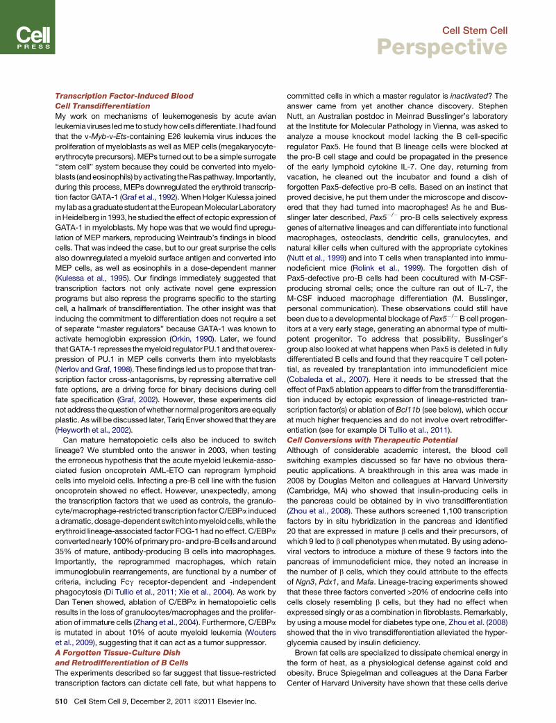
Transcription Factor-Induced Blood
Cell Transdifferentiation
My work on mechanisms of leukemogenesis by acute avian
leukemiaviruses ledme tostudyhowcellsdifferentiate. I had found
that the v-Myb-v-Ets-containing E26 leukemia virus induces the
proliferation of myeloblasts as well as MEP cells (megakaryocyte-
erythrocyte precursors). MEPs turned out to be a simple surrogate
‘‘stem cell’’ system because they could be converted into myelo-
blasts (andeosinophils)byactivating theRaspathway. Importantly,
during this process, MEPs downregulated the erythroid transcrip-
tion factor GATA-1 (Graf et al., 1992). When Holger Kulessa joined
my labasagraduatestudentat theEuropeanMolecularLaboratory
inHeidelberg in1993, hestudied the effect of ectopic expressionof
GATA-1 in myeloblasts. My hope was that we would find upregu-
lation of MEP markers, reproducing Weintraub’s findings in blood
cells. That was indeed the case, but to our great surprise the cells
also downregulated a myeloid surface antigen and converted into
MEP cells, as well as eosinophils in a dose-dependent manner
(Kulessa et al., 1995). Our findings immediately suggested that
transcription factors not only activate novel gene expression
programs but also repress the programs specific to the starting
cell, a hallmark of transdifferentiation. The other insight was that
inducing the commitment to differentiation does not require a set
of separate ‘‘master regulators’’ because GATA-1 was known to
activate hemoglobin expression (Orkin, 1990). Later, we found
thatGATA-1 represses themyeloid regulatorPU.1and thatoverex-
pression of PU.1 in MEP cells converts them into myeloblasts
(Nerlov andGraf, 1998). These findings led us to propose that tran-
scription factor cross-antagonisms, by repressing alternative cell
fate options, are a driving force for binary decisions during cell
fate specification (Graf, 2002). However, these experiments did
not address thequestionofwhether normalprogenitors areequally
plastic. Aswill bediscussed later, Tariq Enver showed that they are
(Heyworth et al., 2002).
Can mature hematopoietic cells also be induced to switch
lineage? We stumbled onto the answer in 2003, when testing
the erroneous hypothesis that the acute myeloid leukemia-asso-
ciated fusion oncoprotein AML-ETO can reprogram lymphoid
cells into myeloid cells. Infecting a pre-B cell line with the fusion
oncoprotein showed no effect. However, unexpectedly, among
the transcription factors that we used as controls, the granulo-
cyte/macrophage-restricted transcription factor C/EBPa induced
adramatic,dosage-dependent switch intomyeloidcells,while the
erythroid lineage-associated factor FOG-1 had no effect. C/EBPa
convertednearly100%ofprimarypro-andpre-Bcellsandaround
35% of mature, antibody-producing B cells into macrophages.
Importantly, the reprogrammed macrophages, which retain
immunoglobulin rearrangements, are functional by a number of
criteria, including Fcg receptor-dependent and -independent
phagocytosis (Di Tullio et al., 2011; Xie et al., 2004). As work by
Dan Tenen showed, ablation of C/EBPa in hematopoietic cells
results in the loss of granulocytes/macrophages and the prolifer-
ation of immature cells (Zhang et al., 2004). Furthermore, C/EBPa
is mutated in about 10% of acute myeloid leukemia (Wouters
et al., 2009), suggesting that it can act as a tumor suppressor.
A Forgotten Tissue-Culture Dish
and Retrodifferentiation of B Cells
The experiments described so far suggest that tissue-restricted
transcription factors can dictate cell fate, but what happens to
510 Cell Stem Cell 9, December 2, 2011 ª2011 Elsevier Inc.
committed cells in which a master regulator is inactivated? The
answer came from yet another chance discovery. Stephen
Nutt, an Australian postdoc in Meinrad Busslinger’s laboratory
at the Institute for Molecular Pathology in Vienna, was asked to
analyze a mouse knockout model lacking the B cell-specific
regulator Pax5. He found that B lineage cells were blocked at
the pro-B cell stage and could be propagated in the presence
of the early lymphoid cytokine IL-7. One day, returning from
vacation, he cleaned out the incubator and found a dish of
forgotten Pax5-defective pro-B cells. Based on an instinct that
proved decisive, he put them under the microscope and discov-
ered that they had turned into macrophages! As he and Bus-
slinger later described, Pax5�/� pro-B cells selectively express
genes of alternative lineages and can differentiate into functional
macrophages, osteoclasts, dendritic cells, granulocytes, and
natural killer cells when cultured with the appropriate cytokines
(Nutt et al., 1999) and into T cells when transplanted into immu-
nodeficient mice (Rolink et al., 1999). The forgotten dish of
Pax5-defective pro-B cells had been cocultured with M-CSF-
producing stromal cells; once the culture ran out of IL-7, the
M-CSF induced macrophage differentiation (M. Busslinger,
personal communication). These observations could still have
been due to a developmental blockage of Pax5�/�B cell progen-
itors at a very early stage, generating an abnormal type of multi-
potent progenitor. To address that possibility, Busslinger’s
group also looked at what happens when Pax5 is deleted in fully
differentiated B cells and found that they reacquire T cell poten-
tial, as revealed by transplantation into immunodeficient mice
(Cobaleda et al., 2007). Here it needs to be stressed that the
effect of Pax5 ablation appears to differ from the transdifferentia-
tion induced by ectopic expression of lineage-restricted tran-
scription factor(s) or ablation of Bcl11b (see below), which occur
at much higher frequencies and do not involve overt retrodiffer-
entiation (see for example Di Tullio et al., 2011).
Cell Conversions with Therapeutic Potential
Although of considerable academic interest, the blood cell
switching examples discussed so far have no obvious thera-
peutic applications. A breakthrough in this area was made in
2008 by Douglas Melton and colleagues at Harvard University
(Cambridge, MA) who showed that insulin-producing cells in
the pancreas could be obtained by in vivo transdifferentiation
(Zhou et al., 2008). These authors screened 1,100 transcription
factors by in situ hybridization in the pancreas and identified
20 that are expressed in mature b cells and their precursors, of
which 9 led to b cell phenotypes whenmutated. By using adeno-
viral vectors to introduce a mixture of these 9 factors into the
pancreas of immunodeficient mice, they noted an increase in
the number of b cells, which they could attribute to the effects
of Ngn3, Pdx1, and Mafa. Lineage-tracing experiments showed
that these three factors converted >20% of endocrine cells into
cells closely resembling b cells, but they had no effect when
expressed singly or as a combination in fibroblasts. Remarkably,
by using a mouse model for diabetes type one, Zhou et al. (2008)
showed that the in vivo transdifferentiation alleviated the hyper-
glycemia caused by insulin deficiency.
Brown fat cells are specialized to dissipate chemical energy in
the form of heat, as a physiological defense against cold and
obesity. Bruce Spiegelman and colleagues at the Dana Farber
Center of Harvard University have shown that these cells derive
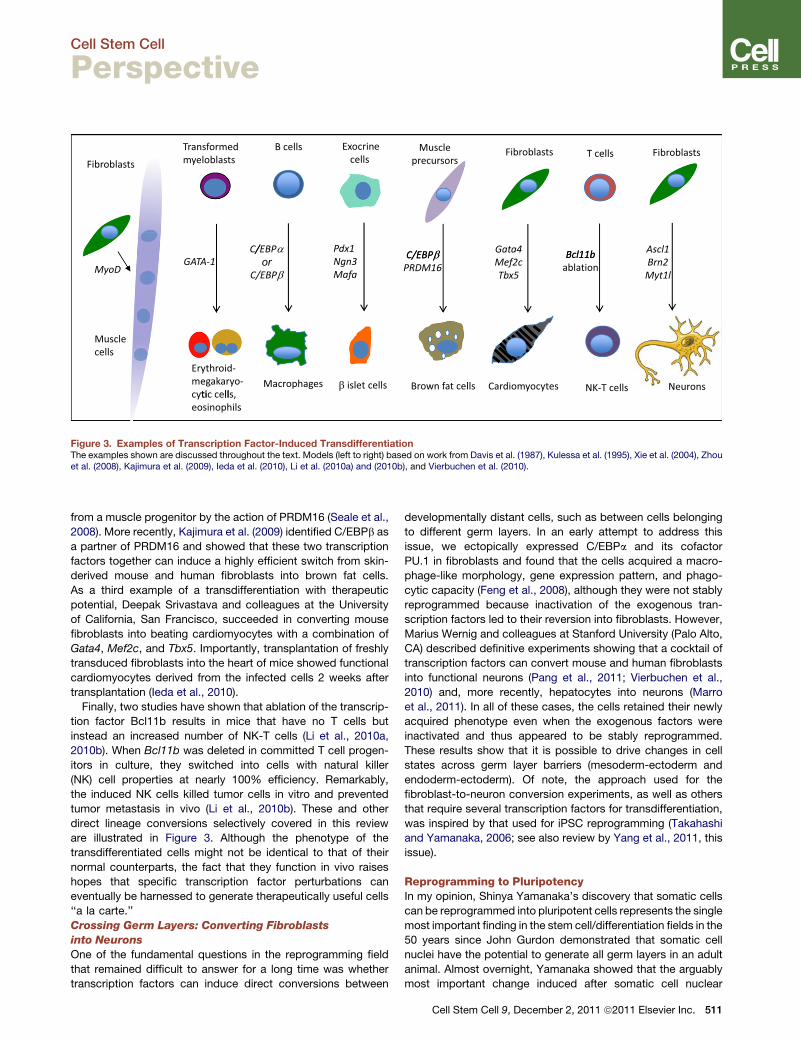
Figure 3. Examples of Transcription Factor-Induced TransdifferentiationThe examples shown are discussed throughout the text. Models (left to right) based on work from Davis et al. (1987), Kulessa et al. (1995), Xie et al. (2004), Zhouet al. (2008), Kajimura et al. (2009), Ieda et al. (2010), Li et al. (2010a) and (2010b), and Vierbuchen et al. (2010).
Cell Stem Cell
Perspective
from a muscle progenitor by the action of PRDM16 (Seale et al.,
2008). More recently, Kajimura et al. (2009) identified C/EBPb as
a partner of PRDM16 and showed that these two transcription
factors together can induce a highly efficient switch from skin-
derived mouse and human fibroblasts into brown fat cells.
As a third example of a transdifferentiation with therapeutic
potential, Deepak Srivastava and colleagues at the University
of California, San Francisco, succeeded in converting mouse
fibroblasts into beating cardiomyocytes with a combination of
Gata4, Mef2c, and Tbx5. Importantly, transplantation of freshly
transduced fibroblasts into the heart of mice showed functional
cardiomyocytes derived from the infected cells 2 weeks after
transplantation (Ieda et al., 2010).
Finally, two studies have shown that ablation of the transcrip-
tion factor Bcl11b results in mice that have no T cells but
instead an increased number of NK-T cells (Li et al., 2010a,
2010b). When Bcl11b was deleted in committed T cell progen-
itors in culture, they switched into cells with natural killer
(NK) cell properties at nearly 100% efficiency. Remarkably,
the induced NK cells killed tumor cells in vitro and prevented
tumor metastasis in vivo (Li et al., 2010b). These and other
direct lineage conversions selectively covered in this review
are illustrated in Figure 3. Although the phenotype of the
transdifferentiated cells might not be identical to that of their
normal counterparts, the fact that they function in vivo raises
hopes that specific transcription factor perturbations can
eventually be harnessed to generate therapeutically useful cells
‘‘a la carte.’’
Crossing Germ Layers: Converting Fibroblasts
into Neurons
One of the fundamental questions in the reprogramming field
that remained difficult to answer for a long time was whether
transcription factors can induce direct conversions between
developmentally distant cells, such as between cells belonging
to different germ layers. In an early attempt to address this
issue, we ectopically expressed C/EBPa and its cofactor
PU.1 in fibroblasts and found that the cells acquired a macro-
phage-like morphology, gene expression pattern, and phago-
cytic capacity (Feng et al., 2008), although they were not stably
reprogrammed because inactivation of the exogenous tran-
scription factors led to their reversion into fibroblasts. However,
Marius Wernig and colleagues at Stanford University (Palo Alto,
CA) described definitive experiments showing that a cocktail of
transcription factors can convert mouse and human fibroblasts
into functional neurons (Pang et al., 2011; Vierbuchen et al.,
2010) and, more recently, hepatocytes into neurons (Marro
et al., 2011). In all of these cases, the cells retained their newly
acquired phenotype even when the exogenous factors were
inactivated and thus appeared to be stably reprogrammed.
These results show that it is possible to drive changes in cell
states across germ layer barriers (mesoderm-ectoderm and
endoderm-ectoderm). Of note, the approach used for the
fibroblast-to-neuron conversion experiments, as well as others
that require several transcription factors for transdifferentiation,
was inspired by that used for iPSC reprogramming (Takahashi
and Yamanaka, 2006; see also review by Yang et al., 2011, this
issue).
Reprogramming to PluripotencyIn my opinion, Shinya Yamanaka’s discovery that somatic cells
can be reprogrammed into pluripotent cells represents the single
most important finding in the stem cell/differentiation fields in the
50 years since John Gurdon demonstrated that somatic cell
nuclei have the potential to generate all germ layers in an adult
animal. Almost overnight, Yamanaka showed that the arguably
most important change induced after somatic cell nuclear
Cell Stem Cell 9, December 2, 2011 ª2011 Elsevier Inc. 511
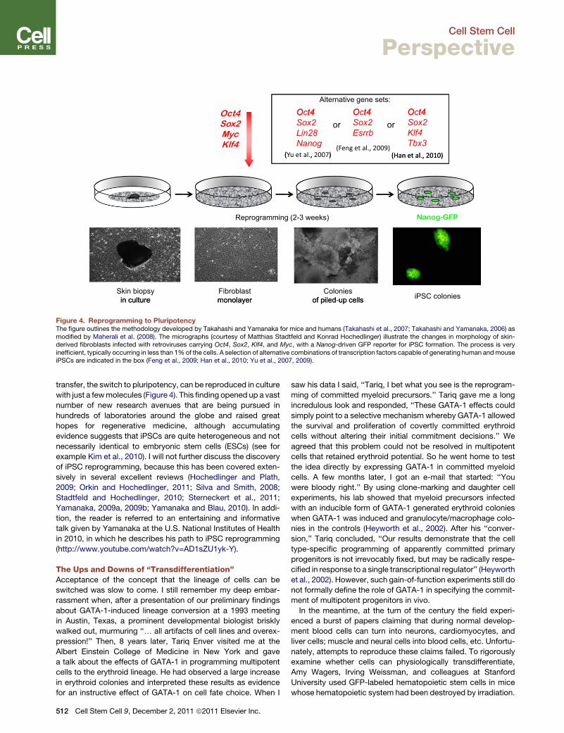
O t4 O t4
Alternative gene sets:
O t4Oct4Sox2MycKlf4
Oct4
Sox2
Lin28
Nanog
Oct4
Sox2
Esrrb
or
(Yu et al., 2007)(Feng et al., 2009)
Oct4
Sox2
Klf4
Tbx3
or
(Han et al., 2010)( , ) (Han et al., 2010)
Reprogramming (2-3 weeks) Nanog-GFP
Skin biopsyin culture iPSC colonies
Fibroblastmonolayer
Coloniesof piled up cellsin culture monolayer of piled-up cells
Figure 4. Reprogramming to PluripotencyThe figure outlines the methodology developed by Takahashi and Yamanaka for mice and humans (Takahashi et al., 2007; Takahashi and Yamanaka, 2006) asmodified by Maherali et al. (2008). The micrographs (courtesy of Matthias Stadtfeld and Konrad Hochedlinger) illustrate the changes in morphology of skin-derived fibroblasts infected with retroviruses carrying Oct4, Sox2, Klf4, and Myc, with a Nanog-driven GFP reporter for iPSC formation. The process is veryinefficient, typically occurring in less than 1%of the cells. A selection of alternative combinations of transcription factors capable of generating human andmouseiPSCs are indicated in the box (Feng et al., 2009; Han et al., 2010; Yu et al., 2007, 2009).
Cell Stem Cell
Perspective
transfer, the switch to pluripotency, can be reproduced in culture
with just a fewmolecules (Figure 4). This finding opened up a vast
number of new research avenues that are being pursued in
hundreds of laboratories around the globe and raised great
hopes for regenerative medicine, although accumulating
evidence suggests that iPSCs are quite heterogeneous and not
necessarily identical to embryonic stem cells (ESCs) (see for
example Kim et al., 2010). I will not further discuss the discovery
of iPSC reprogramming, because this has been covered exten-
sively in several excellent reviews (Hochedlinger and Plath,
2009; Orkin and Hochedlinger, 2011; Silva and Smith, 2008;
Stadtfeld and Hochedlinger, 2010; Sterneckert et al., 2011;
Yamanaka, 2009a, 2009b; Yamanaka and Blau, 2010). In addi-
tion, the reader is referred to an entertaining and informative
talk given by Yamanaka at the U.S. National Institutes of Health
in 2010, in which he describes his path to iPSC reprogramming
(http://www.youtube.com/watch?v=AD1sZU1yk-Y).
The Ups and Downs of ‘‘Transdifferentiation’’Acceptance of the concept that the lineage of cells can be
switched was slow to come. I still remember my deep embar-
rassment when, after a presentation of our preliminary findings
about GATA-1-induced lineage conversion at a 1993 meeting
in Austin, Texas, a prominent developmental biologist briskly
walked out, murmuring ‘‘. all artifacts of cell lines and overex-
pression!’’ Then, 8 years later, Tariq Enver visited me at the
Albert Einstein College of Medicine in New York and gave
a talk about the effects of GATA-1 in programming multipotent
cells to the erythroid lineage. He had observed a large increase
in erythroid colonies and interpreted these results as evidence
for an instructive effect of GATA-1 on cell fate choice. When I
512 Cell Stem Cell 9, December 2, 2011 ª2011 Elsevier Inc.
saw his data I said, ‘‘Tariq, I bet what you see is the reprogram-
ming of committed myeloid precursors.’’ Tariq gave me a long
incredulous look and responded, ‘‘These GATA-1 effects could
simply point to a selective mechanism whereby GATA-1 allowed
the survival and proliferation of covertly committed erythroid
cells without altering their initial commitment decisions.’’ We
agreed that this problem could not be resolved in multipotent
cells that retained erythroid potential. So he went home to test
the idea directly by expressing GATA-1 in committed myeloid
cells. A few months later, I got an e-mail that started: ‘‘You
were bloody right.’’ By using clone-marking and daughter cell
experiments, his lab showed that myeloid precursors infected
with an inducible form of GATA-1 generated erythroid colonies
when GATA-1 was induced and granulocyte/macrophage colo-
nies in the controls (Heyworth et al., 2002). After his ‘‘conver-
sion,’’ Tariq concluded, ‘‘Our results demonstrate that the cell
type-specific programming of apparently committed primary
progenitors is not irrevocably fixed, but may be radically respe-
cified in response to a single transcriptional regulator’’ (Heyworth
et al., 2002). However, such gain-of-function experiments still do
not formally define the role of GATA-1 in specifying the commit-
ment of multipotent progenitors in vivo.
In the meantime, at the turn of the century the field experi-
enced a burst of papers claiming that during normal develop-
ment blood cells can turn into neurons, cardiomyocytes, and
liver cells; muscle and neural cells into blood cells, etc. Unfortu-
nately, attempts to reproduce these claims failed. To rigorously
examine whether cells can physiologically transdifferentiate,
Amy Wagers, Irving Weissman, and colleagues at Stanford
University used GFP-labeled hematopoietic stem cells in mice
whose hematopoietic system had been destroyed by irradiation.
800
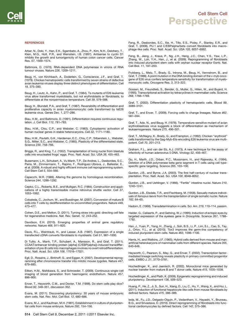
700Transdifferen�a�on
transdifferen�a�on
500
600
iPSC reprogramming
300
400
Claims of physiological
100
200
0201120092007200520032001199919981995199319911989198719851983
B-M iPSF-M M-E F-N
Year
Figure 5. Time Course of PubMed-Listed Papers Containing theTerms ‘‘Transdifferentiation’’ and ‘‘iPSC Reprogramming’’The transient transdifferentiation peak may be attributed to papers claimingphysiological transdifferentiation. F-M, conversion of fibroblasts into musclecells; M-E, switch of transformed erythroid into myeloid precursors; B-M,conversion of B cells intomacrophages; iPS, reprogramming of fibroblasts intoiPSCs; F-N, conversion of fibroblasts into neurons.
Cell Stem Cell
Perspective
They concluded that transdifferentiation of circulating hemato-
poietic stem cells and/or their progeny into distantly related cells
is an extremely rare event, and whenever observed it is mostly
attributable to cell fusions (reviewed in Wagers and Weissman,
2004). By using lineage-tracing experiments, Matthias Stadtfeld
in my group reached similar conclusions (Stadtfeld and Graf,
2005). The claims of physiological conversions gave the term
‘‘transdifferentiation’’ somewhat shady connotations for the
years to follow and reinvigorated some of the old skeptics.
When in 2004 Rudolf Jaenisch visited me in New York, I asked
him, euphoric by our recent finding that B cells can be transdif-
ferentiated into macrophages, what he thought about the idea
of testing whether transcription factor combinations could repro-
gram fibroblasts into ESCs. Rudolf replied emphatically, ‘‘you
are totally out of your mind, that is never going to work.’’ Rudolf’s
main argument was that the two cell types are developmentally
so far apart that chromatin modifications would present an insur-
mountable barrier. Reflecting this attitude, after Yamanaka first
presented his results at a Keystone meeting in 2005, skepticism
prevailed among the colleagues I talked to. And during that same
year we submitted an article to a high-profile immunology journal
showing that C/EBPa and PU.1 can reprogram committed T cell
progenitors into macrophages and dendritic cells. The paper
came back unreviewed: ‘‘It is the editorial policy of the journal
not to publish articles that are based on overexpression experi-
ments.’’ That turned out to be bad timing, because only one year
later, after Yamanaka’s discovery of iPSCs, the gates were
opened and the journal started publishing lineage conversion
experiments. So, ironically perhaps, the tremendous impact of
the iPSC reprogramming discovery helped restore the reputation
of transcription factor-induced transdifferentiation (or ‘‘lineage
conversion,’’ as it was less controversially called [Zhou and
Melton, 2008]), leading to a renewed and sustained explosion
of papers reporting transcription factor-induced conversion of
one specialized cell type into another. The number of papers
listed in PubMed under the terms ‘‘transdifferentiation’’ and
‘‘iPS cell reprogramming’’ reflect these developments. The tran-
sient increase of transdifferentiation papers between 1999 and
2004 represent the ‘‘physiological transdifferentiation’’ bubble,
while the steep increase after 2006 is at least in part attributable
to the discovery of iPSC reprogramming (Figure 5). As a footnote
it should be added that there are credible examples of both
physiological and pathological transdifferentiation, such as the
endothelial-blood cell transition in embryonic development
(Eilken et al., 2009; Zovein et al., 2008) or the epithelial-mesen-
chymal transition during tumor formation (discussed in Slack,
2007; Yang and Weinberg, 2008).
The Future of ReprogrammingAt this point, new lineage conversions are reported almost
every month, for example the switch of fibroblasts into hepa-
tocytes (Huang et al., 2011; Sekiya and Suzuki, 2011).
However, the hurdles that remain for use in cell therapy are still
formidable, whether the cells are obtained by direct lineage
conversion or iPSC reprogramming, making it hard to predict
whether and when these approaches will enter the clinic and
which will ultimately prevail (Cohen and Melton, 2011). Today’s
explosion in cell reprogramming research recapitulates that of
cancer research between the late 1970s and the 1990s
propelled by the discovery of retroviral oncogenes. Although
oncogene research did not lead to the rapid development of
new cancer therapies, as had been widely expected, the
wealth of knowledge for the understanding of basic cellular
processes has been enormous and some of it has been trans-
lated into the clinic. Similarly, cell reprogramming experiments
have already dramatically increased our understanding of cell
differentiation and enabled the creation of tissue culture
models for human cell degenerative diseases that could not
be studied previously.
The discovery of transdifferentiation and iPSC reprogram-
ming is only a decade or so old and many basic questions
remain to be resolved. How do the mechanisms of transcrip-
tion factor-induced transdifferentiation and iPSC reprogram-
ming differ? How does the regulatory network of one cell
collapse while a new one is generated? How important is the
role of chromatin modifications in these processes? Why
does only a minority of the cells respond during iPSC reprog-
ramming and what happens to the others? Future research in
cellular reprogramming will undoubtedly generate new discov-
eries beyond our current imagination, with likely potential for
benefiting human health.
ACKNOWLEDGMENTS
This review is dedicated to Hartmut Beug (1945–2011). I would also like tothank Konrad Hochedlinger, Mark Ptashne, Matthias Stadtfeld, Helen Blau,Stephen Tapscott, Timm Schroeder, Andrew Lassar, Meinrad Busslinger,Rudolf Jaenisch, Tariq Enver, Denis Thieffry, Pura Munoz, Pia Cosma,Salvador Aznar Benitah, and my coworkers for comments, anecdotes, andmaterials. This work was supported by the Ministerio de Ciencia e Inovacion,SAF2007-63058.
Cell Stem Cell 9, December 2, 2011 ª2011 Elsevier Inc. 513
Cell Stem Cell
Perspective
REFERENCES
Arber, N., Doki, Y., Han, E.K., Sgambato, A., Zhou, P., Kim, N.H., Delohery, T.,Klein, M.G., Holt, P.R., and Weinstein, I.B. (1997). Antisense to cyclin D1inhibits the growth and tumorigenicity of human colon cancer cells. CancerRes. 57, 1569–1574.
Baltimore, D. (1970). RNA-dependent DNA polymerase in virions of RNAtumour viruses. Nature 226, 1209–1211.
Beug, H., von Kirchbach, A., Doderlein, G., Conscience, J.F., and Graf, T.(1979). Chicken hematopoietic cells transformed by seven strains of defectiveavian leukemia viruses display three distinct phenotypes of differentiation. Cell18, 375–390.
Beug, H., Leutz, A., Kahn, P., and Graf, T. (1984). Ts mutants of E26 leukemiavirus allow transformed myeloblasts, but not erythroblasts or fibroblasts, todifferentiate at the nonpermissive temperature. Cell 39, 579–588.
Beug, H., Blundell, P.A., and Graf, T. (1987). Reversibility of differentiation andproliferative capacity in avian myelomonocytic cells transformed by tsE26leukemia virus. Genes Dev. 1, 277–286.
Blau, H.M., and Baltimore, D. (1991). Differentiation requires continuous regu-lation. J. Cell Biol. 112, 781–783.
Blau, H.M., Chiu, C.P., and Webster, C. (1983). Cytoplasmic activation ofhuman nuclear genes in stable heterocaryons. Cell 32, 1171–1180.
Blau, H.M., Pavlath, G.K., Hardeman, E.C., Chiu, C.P., Silberstein, L.,Webster,S.G., Miller, S.C., and Webster, C. (1985). Plasticity of the differentiated state.Science 230, 758–766.
Briggs, R., and King, T.J. (1952). Transplantation of living nuclei from blastulacells into enucleated frogs’ eggs. Proc. Natl. Acad. Sci. USA 38, 455–463.
Bussmann, L.H., Schubert, A., VuManh, T.P., De Andres, L., Desbordes, S.C.,Parra, M., Zimmermann, T., Rapino, F., Rodriguez-Ubreva, J., Ballestar, E.,et al. (2009). A robust and highly efficient immune cell reprogramming system.Cell Stem Cell 5, 554–566.
Capecchi, M.R. (1989). Altering the genome by homologous recombination.Science 244, 1288–1292.
Cepko, C.L., Roberts, B.E., and Mulligan, R.C. (1984). Construction and appli-cations of a highly transmissible murine retrovirus shuttle vector. Cell 37,1053–1062.
Cobaleda, C., Jochum, W., and Busslinger, M. (2007). Conversion of mature Bcells into T cells by dedifferentiation to uncommitted progenitors. Nature 449,473–477.
Cohen, D.E., and Melton, D. (2011). Turning straw into gold: directing cell fatefor regenerative medicine. Nat. Rev. Genet. 12, 243–252.
Davidson, E.H. (2010). Emerging properties of animal gene regulatorynetworks. Nature 468, 911–920.
Davis, R.L., Weintraub, H., and Lassar, A.B. (1987). Expression of a singletransfected cDNA converts fibroblasts to myoblasts. Cell 51, 987–1000.
Di Tullio, A., Manh, T.P., Schubert, A., Mansson, R., and Graf, T. (2011).CCAAT/enhancer binding protein {alpha} (C/EBP{alpha})-induced transdiffer-entiation of pre-B cells into macrophages involves no overt retrodifferentiation.Proc. Natl. Acad. Sci. USA 108, 17016–17021.
Egli, D., Rosains, J., Birkhoff, G., and Eggan, K. (2007). Developmental reprog-ramming after chromosome transfer into mitotic mouse zygotes. Nature 447,679–685.
Eilken, H.M., Nishikawa, S., and Schroeder, T. (2009). Continuous single-cellimaging of blood generation from haemogenic endothelium. Nature 457,896–900.
Enver, T., Heyworth, C.M., and Dexter, T.M. (1998). Do stem cells play dice?Blood 92, 348–351, discussion 352.
Evans, M. (2011). Discovering pluripotency: 30 years of mouse embryonicstem cells. Nat. Rev. Mol. Cell Biol. 12, 680–686.
Evans, M.J., and Kaufman, M.H. (1981). Establishment in culture of pluripoten-tial cells from mouse embryos. Nature 292, 154–156.
514 Cell Stem Cell 9, December 2, 2011 ª2011 Elsevier Inc.
Feng, R., Desbordes, S.C., Xie, H., Tillo, E.S., Pixley, F., Stanley, E.R., andGraf, T. (2008). PU.1 and C/EBPalpha/beta convert fibroblasts into macro-phage-like cells. Proc. Natl. Acad. Sci. USA 105, 6057–6062.
Feng, B., Jiang, J., Kraus, P., Ng, J.H., Heng, J.C., Chan, Y.S., Yaw, L.P.,Zhang, W., Loh, Y.H., Han, J., et al. (2009). Reprogramming of fibroblastsinto induced pluripotent stem cells with orphan nuclear receptor Esrrb. Nat.Cell Biol. 11, 197–203.
Frykberg, L., Metz, T., Brady, G., Introna, M., Beug, H., Vennstrom, B., andGraf, T. (1988). A point mutation in the DNA binding domain of the v-myb onco-gene of E26 virus confers temperature sensitivity for transformation of myelo-monocytic cells. Oncogene Res. 3, 313–322.
Gossen, M., Freundlieb, S., Bender, G., Muller, G., Hillen, W., and Bujard, H.(1995). Transcriptional activation by tetracyclines in mammalian cells. Science268, 1766–1769.
Graf, T. (2002). Differentiation plasticity of hematopoietic cells. Blood 99,3089–3101.
Graf, T., and Enver, T. (2009). Forcing cells to change lineages. Nature 462,587–594.
Graf, T., Ade, N., and Beug, H. (1978). Temperature-sensitive mutant of avianerythroblastosis virus suggests a block of differentiation as mechanism ofleukaemogenesis. Nature 275, 496–501.
Graf, T., McNagny, K., Brady, G., and Frampton, J. (1992). Chicken ‘‘erythroid’’cells transformed by the Gag-Myb-Ets-encoding E26 leukemia virus are multi-potent. Cell 70, 201–213.
Graham, F.L., and van der Eb, A.J. (1973). A new technique for the assay ofinfectivity of human adenovirus 5 DNA. Virology 52, 456–467.
Gu, H., Marth, J.D., Orban, P.C., Mossmann, H., and Rajewsky, K. (1994).Deletion of a DNA polymerase beta gene segment in T cells using cell type-specific gene targeting. Science 265, 103–106.
Gurdon, J.B., and Byrne, J.A. (2003). The first half-century of nuclear trans-plantation. Proc. Natl. Acad. Sci. USA 100, 8048–8052.
Gurdon, J.B., and Uehlinger, V. (1966). ‘‘Fertile’’ intestine nuclei. Nature 210,1240–1241.
Gurdon, J.B., Elsdale, T.R., and Fischberg, M. (1958). Sexually mature individ-uals of Xenopus laevis from the transplantation of single somatic nuclei. Nature182, 64–65.
Hadorn, E. (1968). Transdetermination in cells. Sci. Am. 219, 110–114, passim.
Halder, G., Callaerts, P., andGehring,W.J. (1995). Induction of ectopic eyes bytargeted expression of the eyeless gene in Drosophila. Science 267, 1788–1792.
Han, J., Yuan, P., Yang, H., Zhang, J., Soh, B.S., Li, P., Lim, S.L., Cao, S., Tay,J., Orlov, Y.L., et al. (2010). Tbx3 improves the germ-line competency ofinduced pluripotent stem cells. Nature 463, 1096–1100.
Harris, H., andWatkins, J.F. (1965). Hybrid cells derived frommouse and man:artificial heterokaryons of mammalian cells from different species. Nature 205,640–646.
Heyworth, C., Pearson, S., May, G., and Enver, T. (2002). Transcription factor-mediated lineage switching reveals plasticity in primary committed progenitorcells. EMBO J. 21, 3770–3781.
Hochedlinger, K., and Jaenisch, R. (2002). Monoclonal mice generated bynuclear transfer from mature B and T donor cells. Nature 415, 1035–1038.
Hochedlinger, K., and Plath, K. (2009). Epigenetic reprogramming and inducedpluripotency. Development 136, 509–523.
Huang, P., He, Z., Ji, S., Sun, H., Xiang, D., Liu, C., Hu, Y., Wang, X., and Hui, L.(2011). Induction of functional hepatocyte-like cells from mouse fibroblasts bydefined factors. Nature 475, 386–389.
Ieda, M., Fu, J.D., Delgado-Olguin, P., Vedantham, V., Hayashi, Y., Bruneau,B.G., and Srivastava, D. (2010). Direct reprogramming of fibroblasts into func-tional cardiomyocytes by defined factors. Cell 142, 375–386.
Cell Stem Cell
Perspective
Introna,M., Golay, J., Frampton, J., Nakano, T., Ness, S.A., andGraf, T. (1990).Mutations in v-myb alter the differentiation of myelomonocytic cells trans-formed by the oncogene. Cell 63, 1289–1297.
Jacob, F., and Monod, J. (1961). Genetic regulatory mechanisms in thesynthesis of proteins. J. Mol. Biol. 3, 318–356.
Jaenisch, R. (1976). Germ line integration and Mendelian transmission of theexogenousMoloney leukemia virus. Proc. Natl. Acad. Sci. USA 73, 1260–1264.
Jain, M., Arvanitis, C., Chu, K., Dewey, W., Leonhardt, E., Trinh, M., Sundberg,C.D., Bishop, J.M., and Felsher, D.W. (2002). Sustained loss of a neoplasticphenotype by brief inactivation of MYC. Science 297, 102–104.
Jones, P. (2011). Out of Africa and into epigenetics: discovering reprogram-ming drugs. Nat. Cell Biol. 13, 2.
Jones, P.A., and Taylor, S.M. (1980). Cellular differentiation, cytidine analogsand DNA methylation. Cell 20, 85–93.
Jones, D.L., and Wagers, A.J. (2008). No place like home: anatomy and func-tion of the stem cell niche. Nat. Rev. Mol. Cell Biol. 9, 11–21.
Kajimura, S., Seale, P., Kubota, K., Lunsford, E., Frangioni, J.V., Gygi, S.P.,and Spiegelman, B.M. (2009). Initiation of myoblast to brown fat switch bya PRDM16-C/EBP-beta transcriptional complex. Nature 460, 1154–1158.
Kim, K., Doi, A., Wen, B., Ng, K., Zhao, R., Cahan, P., Kim, J., Aryee, M.J., Ji,H., Ehrlich, L.I., et al. (2010). Epigenetic memory in induced pluripotent stemcells. Nature 467, 285–290.
King, T.J., and Briggs, R. (1955). Changes in the nuclei of differentiatinggastrula cells, as demonstrated by nuclear transplantation. Proc. Natl. Acad.Sci. USA 41, 321–325.
Kleinsmith, L.J., and Pierce, G.B., Jr. (1964). Multipotentiality of single embry-onal carcinoma cells. Cancer Res. 24, 1544–1551.
Koller, B.H., and Smithies, O. (1989). Inactivating the beta 2-microglobulinlocus in mouse embryonic stem cells by homologous recombination. Proc.Natl. Acad. Sci. USA 86, 8932–8935.
Kondo, M., Scherer, D.C., Miyamoto, T., King, A.G., Akashi, K., Sugamura, K.,and Weissman, I.L. (2000). Cell-fate conversion of lymphoid-committedprogenitors by instructive actions of cytokines. Nature 407, 383–386.
Kulessa, H., Frampton, J., and Graf, T. (1995). GATA-1 reprograms avian mye-lomonocytic cell lines into eosinophils, thromboblasts, and erythroblasts.Genes Dev. 9, 1250–1262.
Lassar, A.B., Paterson, B.M., and Weintraub, H. (1986). Transfection of a DNAlocus that mediates the conversion of 10T1/2 fibroblasts to myoblasts. Cell 47,649–656.
Levi-Montalcini, R., and Hamburger, V. (1951). Selective growth stimulatingeffects of mouse sarcoma on the sensory and sympathetic nervous systemof the chick embryo. J. Exp. Zool. 116, 321–361.
Li, L., Leid, M., and Rothenberg, E.V. (2010a). An early T cell lineage commit-ment checkpoint dependent on the transcription factor Bcl11b. Science 329,89–93.
Li, P., Burke, S., Wang, J., Chen, X., Ortiz, M., Lee, S.C., Lu, D., Campos, L.,Goulding, D., Ng, B.L., et al. (2010b). Reprogramming of T cells to naturalkiller-like cells upon Bcl11b deletion. Science 329, 85–89.
Maherali, N., Ahfeldt, T., Rigamonti, A., Utikal, J., Cowan, C., and Hochedlin-ger, K. (2008). A high-efficiency system for the generation and study of humaninduced pluripotent stem cells. Cell Stem Cell 3, 340–345.
Marro, S., Pang, Z.P., Yang, N., Tsai, M.C., Qu, K., Chang, H.Y., Sudhof, T.C.,and Wernig, M. (2011). Direct lineage conversion of terminally differentiatedhepatocytes to functional neurons. Cell Stem Cell 9, 374–382.
Martin, G.R. (1981). Isolation of a pluripotent cell line from early mouseembryos cultured in medium conditioned by teratocarcinoma stem cells.Proc. Natl. Acad. Sci. USA 78, 7634–7638.
Maves, L., and Schubiger, G. (2003). Transdetermination in Drosophila imag-inal discs: a model for understanding pluripotency and selector gene mainte-nance. Curr. Opin. Genet. Dev. 13, 472–479.
McGrath, J., and Solter, D. (1984). Inability of mouse blastomere nuclei trans-ferred to enucleated zygotes to support development in vitro. Science 226,1317–1319.
Metcalf, D. (1998). Lineage commitment and maturation in hematopoieticcells: the case for extrinsic regulation. Blood 92, 345–347, discussion 352.
Metz, T., and Graf, T. (1991). v-myb and v-ets transform chicken erythroid cellsand cooperate both in trans and in cis to induce distinct differentiation pheno-types. Genes Dev. 5, 369–380.
Miller, R.A., and Ruddle, F.H. (1976). Pluripotent teratocarcinoma-thymussomatic cell hybrids. Cell 9, 45–55.
Mucenski, M.L., McLain, K., Kier, A.B., Swerdlow, S.H., Schreiner, C.M.,Miller, T.A., Pietryga, D.W., Scott, W.J., Jr., and Potter, S.S. (1991). A func-tional c-myb gene is required for normal murine fetal hepatic hematopoiesis.Cell 65, 677–689.
Nagy, A., Gocza, E., Diaz, E.M., Prideaux, V.R., Ivanyi, E., Markkula, M., andRossant, J. (1990). Embryonic stem cells alone are able to support fetal devel-opment in the mouse. Development 110, 815–821.
Naldini, L., Blomer, U., Gallay, P., Ory, D., Mulligan, R., Gage, F.H., Verma,I.M., and Trono, D. (1996). In vivo gene delivery and stable transduction ofnondividing cells by a lentiviral vector. Science 272, 263–267.
Nerlov, C., and Graf, T. (1998). PU.1 induces myeloid lineage commitment inmultipotent hematopoietic progenitors. Genes Dev. 12, 2403–2412.
Ness, S.A., Beug, H., and Graf, T. (1987). v-myb dominance over v-myc indoubly transformed chick myelomonocytic cells. Cell 51, 41–50.
Nichols, J., Zevnik, B., Anastassiadis, K., Niwa, H., Klewe-Nebenius, D.,Chambers, I., Scholer, H., and Smith, A. (1998). Formation of pluripotentstem cells in the mammalian embryo depends on the POU transcription factorOct4. Cell 95, 379–391.
Noggle, S., Fung, H.L., Gore, A., Martinez, H., Satriani, K.C., Prosser, R., Oum,K., Paull, D., Druckenmiller, S., Freeby, M., et al. (2011). Human oocytes repro-gram somatic cells to a pluripotent state. Nature 478, 70–75.
Nutt, S.L., Heavey, B., Rolink, A.G., and Busslinger, M. (1999). Commitment tothe B-lymphoid lineage depends on the transcription factor Pax5. Nature 401,556–562.
Orkin, S.H. (1990). Globin gene regulation and switching: circa 1990. Cell 63,665–672.
Orkin, S.H., and Hochedlinger, K. (2011). Chromatin connections to pluripo-tency and cellular reprogramming. Cell 145, 835–850.
Pang, Z.P., Yang, N., Vierbuchen, T., Ostermeier, A., Fuentes, D.R., Yang,T.Q., Citri, A., Sebastiano, V., Marro, S., Sudhof, T.C., et al. (2011). Inductionof human neuronal cells by defined transcription factors. Nature 476, 220–223.
Picard, D., Salser, S.J., and Yamamoto, K.R. (1988). A movable and regulableinactivation function within the steroid binding domain of the glucocorticoidreceptor. Cell 54, 1073–1080.
Piccolo, F.M., Pereira, C.F., Cantone, I., Brown, K., Tsubouchi, T., Soza-Ried,J., Merkenschlager, M., and Fisher, A.G. (2011). Using heterokaryons to under-stand pluripotency and reprogramming. Philos. Trans. R. Soc. Lond. B Biol.Sci. 366, 2260–2265.
Ptashne, M. (1967). Specific binding of the lambda phage repressor to lambdaDNA. Nature 214, 232–234.
Ptashne, M. (2011). Principles of a switch. Nat. Chem. Biol. 7, 484–487.
Rieger, M.A., Hoppe, P.S., Smejkal, B.M., Eitelhuber, A.C., and Schroeder, T.(2009). Hematopoietic cytokines can instruct lineage choice. Science 325,217–218.
Rolink, A.G., Nutt, S.L., Melchers, F., and Busslinger, M. (1999). Long-termin vivo reconstitution of T-cell development by Pax5-deficient B-cell progeni-tors. Nature 401, 603–606.
Roussel, M., Saule, S., Lagrou, C., Rommens, C., Beug, H., Graf, T., and Ste-helin, D. (1979). Three new types of viral oncogene of cellular origin specific forhaematopoietic cell transformation. Nature 281, 452–455.
Cell Stem Cell 9, December 2, 2011 ª2011 Elsevier Inc. 515
Cell Stem Cell
Perspective
Scholer,H.R.,Ruppert,S.,Suzuki,N.,Chowdhury,K., andGruss,P. (1990).Newtype of POU domain in germ line-specific protein Oct-4. Nature 344, 435–439.
Seale, P., Bjork, B., Yang, W., Kajimura, S., Chin, S., Kuang, S., Scime, A.,Devarakonda, S., Conroe, H.M., Erdjument-Bromage, H., et al. (2008).PRDM16 controls a brown fat/skeletal muscle switch. Nature 454, 961–967.
Sekiya, S., and Suzuki, A. (2011). Direct conversion of mouse fibroblasts tohepatocyte-like cells by defined factors. Nature 475, 390–393.
Silva, J., and Smith, A. (2008). Capturing pluripotency. Cell 132, 532–536.
Slack, J.M. (2007). Metaplasia and transdifferentiation: from pure biology tothe clinic. Nat. Rev. Mol. Cell Biol. 8, 369–378.
Smith, A.G., Heath, J.K., Donaldson, D.D., Wong, G.G., Moreau, J., Stahl, M.,and Rogers, D. (1988). Inhibition of pluripotential embryonic stem cell differen-tiation by purified polypeptides. Nature 336, 688–690.
Soriano, P. (1999). Generalized lacZ expression with the ROSA26 Cre reporterstrain. Nat. Genet. 21, 70–71.
Stadtfeld, M., and Graf, T. (2005). Assessing the role of hematopoietic plas-ticity for endothelial and hepatocyte development by non-invasive lineagetracing. Development 132, 203–213.
Stadtfeld, M., and Hochedlinger, K. (2010). Induced pluripotency: history,mechanisms, and applications. Genes Dev. 24, 2239–2263.
Stadtfeld, M., Maherali, N., Breault, D.T., and Hochedlinger, K. (2008). Definingmolecular cornerstones during fibroblast to iPS cell reprogramming in mouse.Cell Stem Cell 2, 230–240.
Stadtfeld, M., Maherali, N., Borkent, M., and Hochedlinger, K. (2010). A re-programmable mouse strain from gene-targeted embryonic stem cells. Nat.Methods 7, 53–55.
Stehelin, D., Varmus, H.E., Bishop, J.M., and Vogt, P.K. (1976). DNA related tothe transforming gene(s) of avian sarcoma viruses is present in normal avianDNA. Nature 260, 170–173.
Sterneckert, J., Hoing, S., and Scholer, H.R. (2011). Oct4 and more: the re-programming expressway. Stem Cells, in press. Published online October18, 2011. 10.1002/stem.765.
Tada, M., Takahama, Y., Abe, K., Nakatsuji, N., and Tada, T. (2001). Nuclearreprogramming of somatic cells by in vitro hybridization with ES cells. Curr.Biol. 11, 1553–1558.
Takahashi, K., and Yamanaka, S. (2006). Induction of pluripotent stem cellsfrom mouse embryonic and adult fibroblast cultures by defined factors. Cell126, 663–676.
Takahashi, K., Okita, K., Nakagawa, M., and Yamanaka, S. (2007). Induction ofpluripotent stem cells from fibroblast cultures. Nat. Protoc. 2, 3081–3089.
Tapscott, S.J. (2005). The circuitry of a master switch: Myod and the regulationof skeletal muscle gene transcription. Development 132, 2685–2695.
Taylor, S.M., and Jones, P.A. (1979). Multiple new phenotypes induced in10T1/2 and 3T3 cells treated with 5-azacytidine. Cell 17, 771–779.
Temin, H.M., and Mizutani, S. (1970). RNA-dependent DNA polymerase invirions of Rous sarcoma virus. Nature 226, 1211–1213.
Thomson, J.A., Itskovitz-Eldor, J., Shapiro, S.S., Waknitz, M.A., Swiergiel, J.J.,Marshall, V.S., and Jones, J.M. (1998). Embryonic stem cell lines derived fromhuman blastocysts. Science 282, 1145–1147.
Ursprung, H., and Hadorn, E. (1962). [Further research on model growth incombination with partly dissociated wing imaginal disks of Drosophila mela-nogaster]. Dev. Biol. 4, 40–66.
Vierbuchen, T., Ostermeier, A., Pang, Z.P., Kokubu, Y., Sudhof, T.C., andWer-nig, M. (2010). Direct conversion of fibroblasts to functional neurons by definedfactors. Nature 463, 1035–1041.
Wagers, A.J., andWeissman, I.L. (2004). Plasticity of adult stem cells. Cell 116,639–648.
Wakayama, T., Perry, A.C., Zuccotti, M., Johnson, K.R., and Yanagimachi, R.(1998). Full-term development of mice from enucleated oocytes injected withcumulus cell nuclei. Nature 394, 369–374.
516 Cell Stem Cell 9, December 2, 2011 ª2011 Elsevier Inc.
Weintraub, H., Beug, H., Groudine, M., and Graf, T. (1982). Temperature-sensitive changes in the structure of globin chromatin in lines of red cell precur-sors transformed by ts-AEV. Cell 28, 931–940.
Weintraub, H., Tapscott, S.J., Davis, R.L., Thayer, M.J., Adam, M.A., Lassar,A.B., and Miller, A.D. (1989). Activation of muscle-specific genes in pigment,nerve, fat, liver, and fibroblast cell lines by forced expression of MyoD. Proc.Natl. Acad. Sci. USA 86, 5434–5438.
Wernig, M., Lengner, C.J., Hanna, J., Lodato, M.A., Steine, E., Foreman, R.,Staerk, J., Markoulaki, S., and Jaenisch, R. (2008). A drug-inducible transgenicsystem for direct reprogramming of multiple somatic cell types. Nat. Bio-technol. 26, 916–924.
Williams, R.L., Hilton, D.J., Pease, S., Willson, T.A., Stewart, C.L., Gearing,D.P., Wagner, E.F., Metcalf, D., Nicola, N.A., and Gough, N.M. (1988). Myeloidleukaemia inhibitory factor maintains the developmental potential of embry-onic stem cells. Nature 336, 684–687.
Wilmut, I., Schnieke, A.E., McWhir, J., Kind, A.J., and Campbell, K.H. (1997).Viable offspring derived from fetal and adult mammalian cells. Nature 385,810–813.
Wouters, B.J., Lowenberg, B., Erpelinck-Verschueren, C.A., van Putten, W.L.,Valk, P.J., and Delwel, R. (2009). Double CEBPA mutations, but not singleCEBPAmutations, define a subgroup of acutemyeloid leukemia with a distinc-tive gene expression that is uniquely associated with a favorable outcome.Blood 113, 3088–3091.
Xie, H., Ye, M., Feng, R., and Graf, T. (2004). Stepwise reprogramming of Bcells into macrophages. Cell 117, 663–676.
Yamanaka, S. (2009a). A fresh look at iPS cells. Cell 137, 13–17.
Yamanaka, S. (2009b). Elite and stochastic models for induced pluripotentstem cell generation. Nature 460, 49–52.
Yamanaka, S., and Blau, H.M. (2010). Nuclear reprogramming to a pluripotentstate by three approaches. Nature 465, 704–712.
Yang, J., and Weinberg, R.A. (2008). Epithelial-mesenchymal transition: at thecrossroads of development and tumor metastasis. Dev. Cell 14, 818–829.
Yang, N., Ng, Y.H., Pang, Z.P., Sudhof, T.C., and Wernig, M. (2011). Inducedneuronal cells: how to make and define a neuron. Cell Stem Cell 9, this issue,517–525.
Ying, Q.L., Wray, J., Nichols, J., Batlle-Morera, L., Doble, B., Woodgett, J., Co-hen, P., and Smith, A. (2008). The ground state of embryonic stem cell self-renewal. Nature 453, 519–523.
Yu, J., Vodyanik, M.A., Smuga-Otto, K., Antosiewicz-Bourget, J., Frane, J.L.,Tian, S., Nie, J., Jonsdottir, G.A., Ruotti, V., Stewart, R., et al. (2007). Inducedpluripotent stem cell lines derived from human somatic cells. Science 318,1917–1920.
Yu, J., Hu, K., Smuga-Otto, K., Tian, S., Stewart, R., Slukvin, I.I., and Thomson,J.A. (2009). Human induced pluripotent stem cells free of vector and transgenesequences. Science 324, 797–801.
Zhang, P., Iwasaki-Arai, J., Iwasaki, H., Fenyus, M.L., Dayaram, T., Owens,B.M., Shigematsu, H., Levantini, E., Huettner, C.S., Lekstrom-Himes, J.A.,et al. (2004). Enhancement of hematopoietic stem cell repopulating capacityand self-renewal in the absence of the transcription factor C/EBPa. Immunity21, 853–863.
Zhou, Q., and Melton, D.A. (2008). Extreme makeover: converting one cell intoanother. Cell Stem Cell 3, 382–388.
Zhou, Q., Brown, J., Kanarek, A., Rajagopal, J., andMelton, D.A. (2008). In vivoreprogramming of adult pancreatic exocrine cells to beta-cells. Nature 455,627–632.
Zinyk, D.L., Mercer, E.H., Harris, E., Anderson, D.J., and Joyner, A.L. (1998).Fate mapping of the mouse midbrain-hindbrain constriction using a site-specific recombination system. Curr. Biol. 8, 665–668.
Zovein, A.C., Hofmann, J.J., Lynch, M., French, W.J., Turlo, K.A., Yang, Y.,Becker, M.S., Zanetta, L., Dejana, E., Gasson, J.C., et al. (2008). Fate tracingreveals the endothelial origin of hematopoietic stem cells. Cell Stem Cell 3,625–636.