Embed Size (px)
Citation preview
Corresponding author: Cris Precup – Senior Lecturer, PhD MDDepartment of Anatomy and Embryology, Faculty of Medicine, Pharmacy and Dental Medicine“Vasile Goldis” Western University, Arad, Romania E-mail: [email protected]
REZUMAT
Dificultãåi de diagnostic histopatologic æi medico-legal la un pacient cu multiple comorbiditãåi æi traumatisme cranio-cerebrale multiple - prezentare de cazAceastã lucrare este o prezentare de caz. Cazul a fost ales deoarece a prezentat mai multe probleme referitoarela diagnosticul de certitudine al cauzei decesului æi, prin urmare, stabilirea mecanismului tanato-generator.Pacienta este de sex feminin dintr-un mediu rural care a decedat dupã internare (03 martie 2013), în SpitalulClinic de Urgenåã din Arad, Secåia de Neurologie. Pacienta este internatã prin Unitatea de Primire Urgenåe înurma unui traumatism cranio-cerebral în regiunea temporo-parietalã stânga, cu insuficienåã respiratorie ventilatã mecanic æi comã gradul II. Dificultãåile privind diagnosticul æi stabilirea legãturilor de cauzalitateapar atunci când coroborând datele clinice mai sus menåionate cu antecedentele personale patologice:hipertensiune arterialã, diabet zaharat de tip II, cardiopatie ischemicã cronicã, fibrilaåie atrialã cronicã. Dupãanaliza informaåiilor din arhiva Serviciului Judeåean de Medicinã Legalã Arad devine clar cã persoana a maifost victima unei agresiuni, în urma cãreia, de asemenea, a rezultat un traumatism cranio-cerebral în regiuneatemporo-parietalã stângã (februarie 2009), în urma unui traumatism produs prin loviturã de topor la nivelulextremitãåii cefalice . În urma traumatismului care a avut loc în februarie 2009 pacienta a suferit un trauma-tism cranio-cerebral pierderea temporarã a conætienåei æi a fost internatã pentru investigaåii æi tratament de specialitate. Dupã deces, s-a dispus efectuarea necropsiei medico-legale pentru lãmurirea tuturor detaliilormacroscopice æi microscopice legate de caz. Aspectele dificile în stabilirea cauzei de deces constau în exis-tenåa a douã traumatisme cranio-cerebrale care au avut loc la un interval de timp de patru ani æi comorbiditãåileasociate.
Cuvinte cheie: traumatism, autopsie, leziuni cerebrale
Modern Medicine. 2015, Vol. 22, No. 2 : 165- Copyright© Celsius
Case Report
Modern Medicine. 2015, Vol. 22, No. 2 : 165-171 Copyright© Celsius
Histopathology and Forensic Diagnostics Difficulties in aPatient with Multiple Affections and Double Cranio-CerebralInjuriesCris Precup, Mircea Ifrim, Ovidiu Bulzan, Csongor Toth, Gyori Zsolt, Hategan Ovidiu
Anatomy Department, Faculty of Medicine Pharmacy and Dental Medicine“Vasile Goldis” Western University of Arad, Romania
166 Cris Precup et al
INTRODUCTION
This paper is a case presentation. This case waschosen because it presented several problems withindicating, with certainty, the cause of death andtherefore establishing the tanato-generating mechanism. The patient is a female from the ruralenvironment who dies after a hospitalization (03March 2013) in the Clinical Emergency Hospital inArad, Department of Neurology. The patient isadmitted through the emergency unit due to acranio-cerebral trauma in the left parietal-temporalregion, assisted respiratory failure and second degreecoma.
CASE REPORT
Under the Ordinance issued by Arad City Police,a necropsy was conducted on G. V, age 67, a residentof the village of Fiscut (rural), Arad County. Theforensic objectives established in the ordinance were:whether the death was violent, if the body shows signsof violence and what were the causes of death.
The team has asked about the history and circumstances of death. Resolution of making theautopsy shows that on March 2013, the patient diedin the Arad County Hospital, Department of Neurology. The general clinical observation sheetfrom the Arad Emergency County Hospital, givesinformation on the reasons for hospitalization as wellas information regarding the period of hospitaliza-tion, between 03/08/2013 – 03/12/2013.
The patient was brought to the hospital by ambulance and from the observation sheet whichwas brought from the receptions unit - EmergencyHospital Arad County, we see why the patient wasbrought to hospital, II degree coma, with endo-tracheal intubation. From the medical history provided by her caregivers, the patient suffered fromchronic ischemic heart disease, hypertension, chronic atrial fibrillation and type II diabetes.
Corroborating the information provided by themedical team and the forensic investigation team werealize that the patient was found in the yard, athome, in a coma, and was sent by SMURD (emergency rescue service based in Romania) toTimisoara County Hospital for diagnosis and specialized treatment due to the deterioration ofneuro-logical status and the outcome of C.T. brainscan that shows a hypodense lesion in the left parietalregion. SMURD reaches Timisoara with the patientwith a referral diagnosis of mixed cerebral comagrade III. The diagnosis does not change for 72hours. Due to the socio-economic conditions of thepatient’s tutors, the patient and the medical team aretransferred from Timisoara County Hospital to AradCounty Hospital. The main diagnosis at the time ofdischarge is cardiac arrest, mixed cerebral coma,repeated ischemic stroke with sequelae in the basalnuclei, sequelae of head trauma in the left temporo-parietal region, acute respiratory failure requiringventilatory support, type II diabetes, exacerbatedchronic renal failure, chronic atrial fibrillation,ischemic heart disease and chronic hypertension. As
ABSTRACTThis paper is a case presentation. This case was chosen because it presented several problems with indicating,with certainty, the cause of death and therefore establishing the tanato-generating mechanism. The patient isa female from the rural environment who dies after a hospitalization (03 March 2013) in the Clinical Emergency Hospital in Arad, Department of Neurology. The patient is admitted through the emergency unitdue to a brain injury in the left parietal-temporal region, assisted respiratory failure and second degree coma.Diagnostic and causation difficulties occur when clinical data mentioned above is corroborated with previousmedical history: type II diabetes hypertension, chronic heart disease, chronic atrial fibrillation. After analyzing the information in the archive of the Forensic medicine service in Arad it becomes clear that theperson was the victim of an assault that also resulted in a head trauma in the left parietal-temporal region (February 2009), after being hit with an axe in the cephalic extremity. After the incident in 2009 the patientsuffered head trauma with temporary loss of consciousness and was admitted for investigation and specialisttreatment. Following the death, a necropsy was performed in order to clarify all the macroscopic and microscopic details. The difficulty of the case rests is the existence of two head traumas that have occurred 4years apart and the associated pathology.
Key words: trauma, autopsy, brain injury
Modern Medicine. 2015, Vol. 22, No. 2
it can be seen the patient had a body with multipleconditions.
After analyzing the information from the archiveof Arad County Department of Forensic Medicine itwas clear that our patient was the victim of an assaultthat also resulted in a head trauma in the left parietal-temporal region (February 2009) afterbeing hit with an axe in the cephalic extremity. Thepatient had, once more, suffered a head trauma withtemporary loss of consciousness, and was admittedfor investigation and specialist treatment, and she survived. This type of injury was studied byCychowska M in 2013 [1,2]
The next stage is the external examination of thecorpse by the rigorous autopsy protocol. [3,4,5,6,8]
The corpse submitted for autopsy belongs to afemale, aged 66, of medium height and with the muscular and adipose tissues proportionally repre-sented. The indices of the actual death are presentthrough the cadaveric lividity of a shade of purpleplaced on the dorsal declive of the trunk and limbs,which do not turn pale when applying pressure (imbi-bitions stage) and a cadaveric rigidity in the finalstage. There are no traumatic external injuriesdetected. The body shows signs of medical treat-ment: wound points in areas of choice for injectiontreatment (Right subclavicular region - three plaguespoint and on the elbow fold). A pearly white xifo-subombilical scar measuring 25 cm and a leg oedemaare noticed.
The internal examination of the body, at thecephalic extremity, reveals an area of 8/8 cm locatedin the left temporo-parietal region gray-white coloration (scar). The left temporo-parietal regionof the neurocranium shows a lack of bone in an areaof 4,5 / 4,5 cm (slightly irregular white edges). Leftparietal region of the duramater presents additionalto the underlying lack of bone described above awhitish gray scar of about 4.4 cm (the connective tissue is thickened and there is no bone covering).
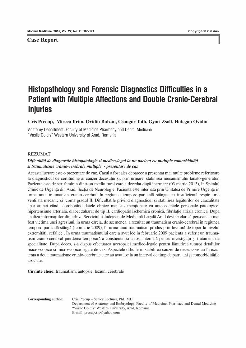
In the temporo-parietal region on the left side,on a surface measuring approximately 8sqcm, theleptomeningeal is thickened with a yellowish browncolor, without sheen, without substance (with pearlywhite edges) measuring 4/4 cm in the central area.(Fig. 1) The blood vessel at the base of the brainhave moderately thickened walls with a permeablelumen. In the left cerebral hemisphere [affectingcortex (with lack of substance on a surface area of 4.4 cm) and underlying white matter], the temporo-parietal region, the circumvolutions and the grooves
are missing and the nerve tissue has a yellowishbrown color with a low, brittle consistency. We analyzed the possible causes of this type of injuriesby surgical point of view. [9,10]
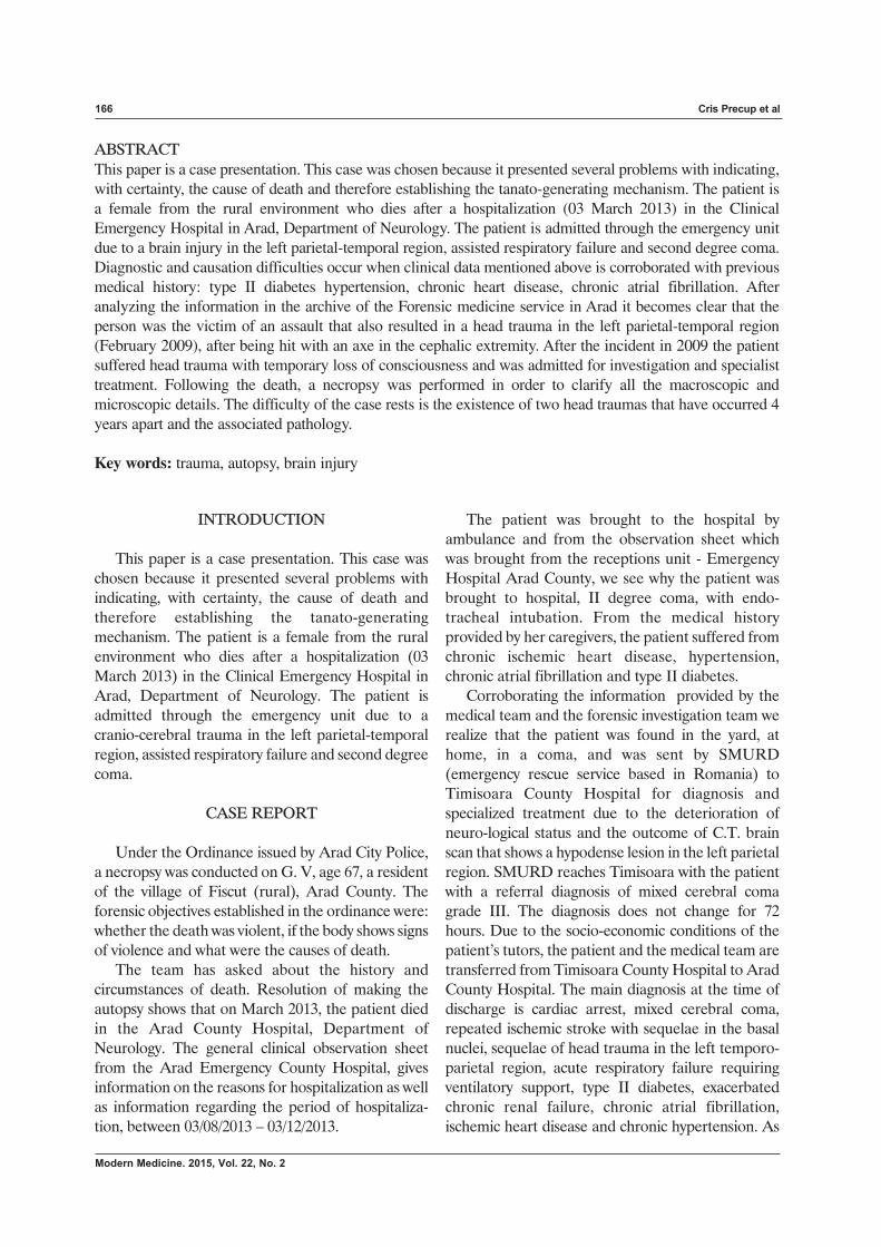
On the cross-section, the normal architecturaldesign is missing, the nerve tissue looks like a yellowish grey semi-solid mass. (Fig. 2 and 3) Rightcerebral hemisphere- with the convolutions widenedand the grooves between them erased, with a low,slightly brittle consistency and with the normal architectural design faded. On the cross-section thegrey matter is faintly delimitated from the whitematter which has a wet appearance with a mattedgloss. With multiple reddish dots which disappearwhen are being scraped with a knife. In the greybasal nuclei and the brainstem the nerve tissue has alow and brittle consistency with the erasure of thenormal architectural design. The diencephalon andcerebellum have a clear architectural design. The
Histopathology and Forensic Diagnostics Difficulties in a Patient with Multiple Affections and Double Cranio-Cerebral Injuries 167
Figure 1. Dissection of the head (external view)
Figure 2. Examination of the parietal and temporal bone (internal view)
Modern Medicine. 2015, Vol. 22, No. 2
168 Cris Precup et al
subarachnoid space and cerebral ventricles contain areddish fluid in a very small amount and the choroidplexus are reddish.
When internally examining the oropharyngealregion we found the lips with a reddish brown liningand the tongue with a gray velvet like lining on seriate sections of muscle mass without any traumatic injuries can be observed. The ovoid tonsilsare covered by reddish lining. The subcutaneous tissue in the cervical region does not have any traumatic injuries. The muscles of the neck do nothave any traumatic injuries. Both on the surface andin the cross-section, the thyroid gland have a reddishpurple color. The hyoid bone and the laryngeal cartilages are intact. The larynx exhibits a whitishgray lining. The pharynx exhibits a gray, wet andshiny lining. The cervical spine has no traumaticinjuries. The soft parts of the right hemithorax of theupper third part of the anterior region underlying thepoint wounds of the injection treatment described inthe external examination under “sings of medicaltreatment” exhibit a moderate dark hemorrhagicinfiltration al 4 sqcm surface. The pleuras are thickened, fibrosed, matte with the gloss weakened.The pleural cavities contain a minimum amount ofsero-citrine liquid. The trachea and the large bronchihave unreached walls, exhibit a reddish gray liningand an open lumen. The lungs occupy 2/3 of the thoracic cavity, are of a brownish gray color, with theantracotic design clear, they have a soft consistency,a diminished elasticity, with small, difured crepita-tion. On the cross-section they have an inhomoge-neous brown-gray color and a spontaneous large
blood drain. The pericardium in slim with the visceral foil wet, glossy and transparent. The pericar-dial cavity exhibits a minimum quantity of serocitrinliquid. The heart is firm in consistency and measures12/11/6 cm. The heart cavities contain liquid bloodand reddish-black, nonadherent clots. The valves areslightly thickened and transparent. The endocardiumexhibits a reddish brown color with a low consistencyand diminished elasticity. When observing seriatesections that were made along its thickness, we arepresented with an inhomogeneous surface and smallwhite areas on a reddish brown background; thethickness of the myocardium at the VS level is 1,7 cm.the coronary arteries present slightly thickened walls,with diminished elasticity and a permeable lumen.The aorta has walls that have not been breached,with diminished elasticity and the heart is yellowishwith rare hard, white plaques. The pulmonary arteries are free, the heart is smooth, of a yellowishwhite.
The abdominal wall does not exhibit any traumatic injuries. The peritoneum is thickened,exhibiting multiple fibrous adhesions through thesupramesocolic floor; the peritoneal cavity contains aminimum amount of serocitrine fluid. The stomachhad walls that have not been breached, a gray withblackish spots lining (easier to spot in the area of thelesser curvature and the pyloric channel) with pronounced folds and contains gray-green liquid. Theintestines exhibit walls that have not been breached, awhite and gray lining and content appropriate to thesegment.
The liver weighs 2200 grams, the visceral peri-
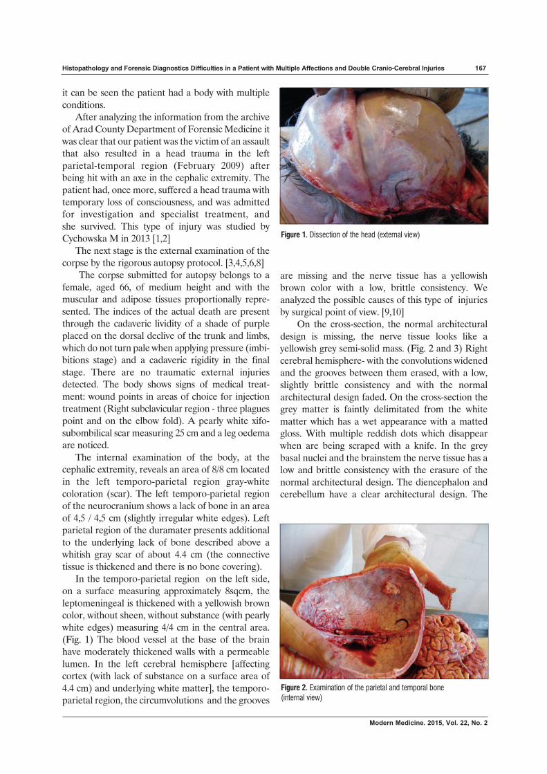
Figure 3. Old neurocranial injury with scar and dura mater condensation
a b
Modern Medicine. 2015, Vol. 22, No. 2
Histopathology and Forensic Diagnostics Difficulties in a Patient with Multiple Affections and Double Cranio-Cerebral Injuries 169
Modern Medicine. 2015, Vol. 22, No. 2
toneum and the capsule are smooth and transparentwith a dull gloss, the surface is smooth, yellowishbrown, with a hard consistency and slightly brittle.The cross-section is yellow, with a slightly grainyaspect and a small quantity of reddish brown blooddraining.
The cystic fossa of the lower face of the liver isfree (gall-bladder – absent), pearly white material ispresent (postcolecistectomie scar); the left and right hepatic ducts and the hepato-biliary duct arepermeable.
The spleen has a low, brittle consistency, weighs220 grams with a transparent, smooth and highlyglossy capsule. On the cross-section it is purple withwhite dots and streaks. The pancreas is yellowish graywith a high consistency. On the cross-section we arepresented with a glandular lobular design withwhitish glandular lobular and interlobular reddishspaces.
The kidneys exhibit diminished elasticity, a transparent capsule, with diminished shine, whichare hard to peel off and with the surface that hasbeen decapsulated exhibiting irregularities. On thecross-section, the cortical region is grayish brownwith red striations and the medular region is reddishbrown and the cortical-medular contrast is diminished. The abdominal aorta and the inferiorvena cava have walls that have not been breachedand a permeable lumen. The bladder exhibited gray-white lining is devoid of content. The internalgenitalia have no traumatic injuries. The pelvis boneexhibits no traumatic injuries.
The external examination of the perineum, theexternal genital organs and the anal region show thatthere are no traumatic injuries present. On a carefulexamination of the upper and lower limbs no traumatic injuries are present except for those causedduring the treatment.
After macroscopic examination the followingdiagnoses are established: Advanced autolysis withdecay of variable intensity; scarring on the soft sides ofthe left parieto-temporal region; lack of bone substance in the left parieto-temporal region; scarringon the duramater in the left parieto-temporal region;lack (old) of substance in the left left parieto-temporal region in the leptomeninges; minimal subarachnoid hemorrhage; left temporo-parietalcerebral softening; old lack of substance in the cerebral cortex in the left temporo-parietal region;moderate cerebral and coronary atherosclerosis; mio-cardofibrosis; bilateral pahipleuritis; emphysema,
liver dystrophy; renal dystrophy, old cholecystectomy,chronic pancreatitis and reactive spleen.
For diagnostic certainty tissue sampling is performed for the histopathological examination.The material taken for histopathological examina-tion included fragments of: cerebral hemisphere(left parieto-occipital), brainstem, thalamus andbasal nuclei, lung, heart, liver and kidneys.[8] Thefragments were prepared as per standard protocol ina solution containing 10% formaldehyde, paraffininclusion and application of hematoxylin-eosinstaining. [5]
The result of microscopic examination revealedthe following:
In the fragment of the cerebral hemisphereextravasated red blood cells in the subarachnoidspace were identified as well as the widening peri-cellular and perivascular spaces with dissecting thebrain substance, in the lumen of cerebral vesselsmore red blood cells and a few leukocytes are seenas well as small extravased perivascular blood cells inthe cerebral nerve substance, areas with irregularedges (made out of a fibrillar material produced byan eosinophil together with fibroblasts, fibroblasts,macrophages, and glial cells) where the normalarchitectural design is missing, the nerve substancelooks irregular with an areolar appearance, theneural silhouettes can no longer be identified; asmall amount of amorphic material, with abasophilic tint, some red blood cells many glial cellsand some macrophages and lymphocytes in thoseareas and moderate diffuse glial hyperplasia.
In the brainstem fragment, pericellular andperivascular visually empty spaces were identified,with the dissection of nerve substance; in the lumenof the vessels red blood cells and some white bloodcells can be observed, small scattered haematicextravasations perivascular in the cerebral nervesubstance, neurons with finely granulated cytoplasmand with difficult to see nuclei, in certain areas, andclusters of glial hyperplasia.
In the thalamus and basal nucleus fragmentspericellular and perivascular visually empty spaceswere identified, with the dissection of nerve sub-stance; in the lumen vessels a large number of redblood cells and a few white blood cells can be seen,small scattered haematic extravasations perivascularin the cerebral nerve substance, neurons with finelygranulated cytoplasm and with difficult to see nuclei,in certain areas, and clusters of glial hyperplasia.
In the lung fragment, a thickened visceral pleura
170 Cris Precup et al
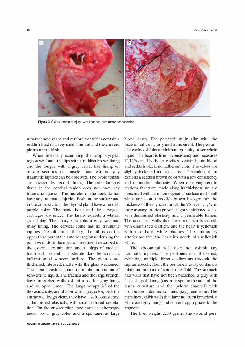
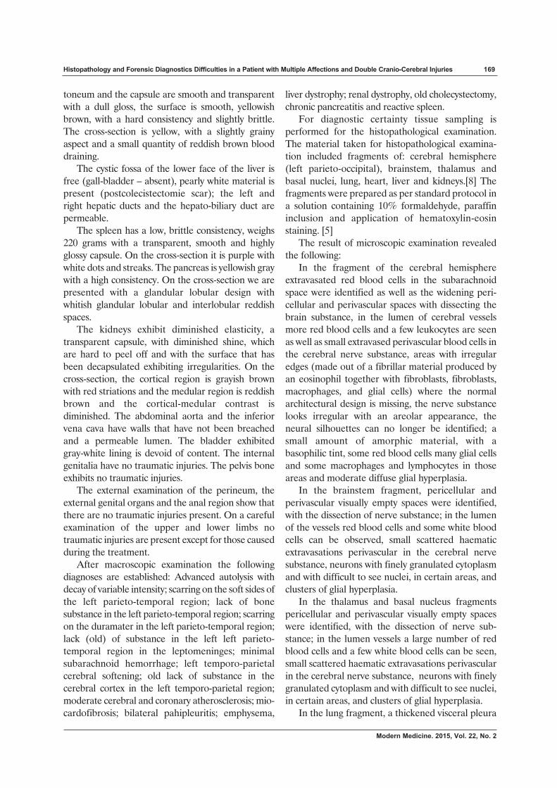
Figure 4. Microscopic view of the lung fragment, HE stain, X20, intraalveolar eosinophilic amorphous material contains macrophages lymphocytes, epithelial cells and some granulocytes and bronchi and bronchioles with leukocyte infiltration in the walls,
a b
can be seen, presenting parenchymal fibrosis withintensely eosinophilic interalveolar septa, thinned,some being fragmented, with visibly large voidspaces, with polygonal outline and enlarged inter-alveolar septa; in the enlarged lumen of the bloodcapillaries a large number of red blood cells and afew white blood cells can be seen; we can also seeareas where we can observe the presence of aeosinophilic fibrillar material accompanied by fibro-cytes, fibroblasts, and some macrophages (intracyto-plasmic dark brown pigment) and lymphocytes,areas where intraalveolar eosinophilic amorphousmaterial contains macrophages, lymphocytes,epithelial cells and some granulocytes, there are alsobronchi and bronchioles with leukocyte infiltrationin the walls; the lumen of the bronchiolar is an amorphous eosinophilic material with lymphocytes,macrophages, granulocytes and some epithelial cellspresent. (Fig. 4)
In the heart muscle fragment was observed smallhaematic extravased at the submesotelial connectivetissue level of the visceral serous pericardium. At thelevel of myocardium it was found a large inter-myocites space, in the lumenum of the heart bloodvessels it was found lots of hematia and a few leuco-cytes. At the subendocardial region it was found aneosinofil fibrilar tissue (with fibrocytes, fibroblastsand a few monocytes) and myocytes with small eosi-nofil variation of the sarcoplasma, faded striationsand in some areas with hardly observable nucleus.
In the liver fragment it was observed a wideningof the sinusoid capillary lumenum at the level of thePorta vein branches and at the centro-lobular veins.The lumenum contains lots of hematia and a fewleucocytes small haematic extravased at the Porta
branches and hepatic lobules level, hepatocytes withvacuolar cytoplasm, poorly outlined cell membraneand well structured nucleus
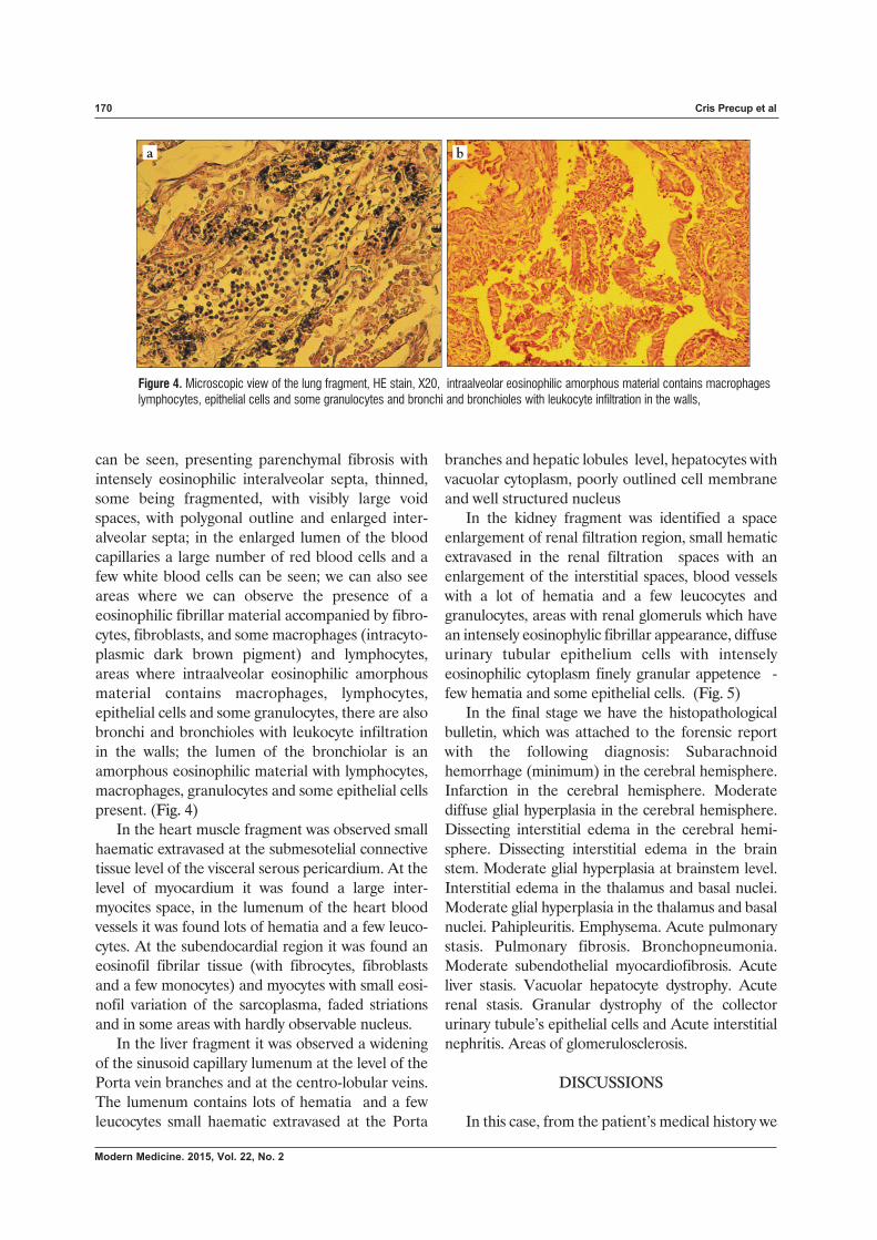
In the kidney fragment was identified a spaceenlargement of renal filtration region, small hematicextravased in the renal filtration spaces with anenlargement of the interstitial spaces, blood vesselswith a lot of hematia and a few leucocytes and granulocytes, areas with renal glomeruls which havean intensely eosinophylic fibrillar appearance, diffuseurinary tubular epithelium cells with intenselyeosinophilic cytoplasm finely granular appetence -few hematia and some epithelial cells. (Fig. 5)
In the final stage we have the histopathologicalbulletin, which was attached to the forensic reportwith the following diagnosis: Subarachnoid hemorrhage (minimum) in the cerebral hemisphere.Infarction in the cerebral hemisphere. Moderate diffuse glial hyperplasia in the cerebral hemisphere.Dissecting interstitial edema in the cerebral hemi-sphere. Dissecting interstitial edema in the brainstem. Moderate glial hyperplasia at brainstem level.Interstitial edema in the thalamus and basal nuclei.Moderate glial hyperplasia in the thalamus and basalnuclei. Pahipleuritis. Emphysema. Acute pulmonarystasis. Pulmonary fibrosis. Bronchopneumonia.Moderate subendothelial myocardiofibrosis. Acuteliver stasis. Vacuolar hepatocyte dystrophy. Acuterenal stasis. Granular dystrophy of the collector urinary tubule’s epithelial cells and Acute interstitialnephritis. Areas of glomerulosclerosis.
DISCUSSIONS
In this case, from the patient’s medical history we
Modern Medicine. 2015, Vol. 22, No. 2
Histopathology and Forensic Diagnostics Difficulties in a Patient with Multiple Affections and Double Cranio-Cerebral Injuries 171
keep in mind that she was the victim of an aggressionfrom a known individual, which happened in theevening of 02.02.2009, in Fiscut, when she was hitwith an ax in the cephalic extremity. She suffers acranio- cerebral injury with a temporary loss of consciousness and is admitted in the emergencyroom for medical investigations and specializedtreatment.
After a rigorous anamnesis we succeeded tounlock this forensic problem. There was a criminaltrial in the first aggression case; the tutors wantedthe actual hospital costs to be paid by the aggressor.For this purpose they asked if there is or not a causallink between the first case and the second.
We concluded, according to the identified infor-mation that four years later, (2013) she suffers adecompensation episode due to type II diabetes,after which she is admitted through the ambulanceservices. The 67 year old patient, who was admittedto the Diabetes and Nutrition ward, is showing adeterioration of her neurological condition, which iswhy a CT scan is performed which shows a hypo-dense lesion left parietal region.
The patient, having acute TCC sequelae due toaggression, insulin-dependent type II diabetes,chronic renal failure and atrial fibrillation is imme-diately admitted to the Diabetes ward in 04.03.2013,in a I degree comatose state and metabolic decom-pensation. Taking into consideration the CT findings with the disabling ischemic changes and suspected brain tumor she is sent, in a comatosestate and metabolically unbalanced, to Timisoara toATI – Neurosurgery where she is sent to Arad- ATIat the caregivers request and because she had noneurosurgical indication. The patient is transportedwith a blood glucose level of mg. on the ATI ward,
Arad, she is admitted through the Department ofNeurology although sent to ATI – diabetes ward.
Murty OP in 2009 concluded also that type II dia-betes raise the diagnostic difficulties to give a clearforensic diagnosis. [7,8]
CONCLUSIONS
Death was due to bronchopneumonia and acuteinterstitial nephritis, subsequent to prolonged meta-bolic diabetic coma (with diabetic hyperosmolar syndrome and diabetic keton-acidosis). There is ahigh degree of difficulty in terms of causal link,between the injury and the death causing mecha-nism in patients, with multiple affections.
REFERENCES
1. Cychowska M, Bloch-Boguslawska E. Cases of non-fatal chopwounds to the head. Arch Med Sadowej Kryminol. 2013 Oct-Dec;63(4):283-7
2. Gregory Murrey, Donald Starzinski The Forensic Evaluation ofTraumatic Brain Injury: A Handbook for Clinicians and Attorneys,Second Edition CRC Press; 2 edition 2007
3. Vladimir Belis, Medicina Legala, Teora, Bucuresti 19924. Dan Dermengiu, Patologie Medico-legala, Ed Viata Medicala
Romaneasca, Bucuresti. 20025. Dan Dermengiu, Gheorghe Alexandrescu, Medicina Legala
Prosecturala, Ed Viata Medicala Romaneasca, Bucuresti. 20116. Valentin Iftenie, Dan Dermengiu, Medicina legala, C.H. BECK,
20097. Murty OP. Diabetic artefacts in forensic practice. J Forensic Leg
Med. 2009 May;16(4):218-23. doi: 10.1016/j.jflm.2008.07.010. Epub2008 Nov 11
8. Reinhard Dettmeyer Forensic Histopathology: Fundamentals andPerspectives Publisher Springer, ISBN-13: 978-3642206580 ISBN-10: 3642206581 Edition: 1st ed. 2011
9. Juan Rosai MD Rosai and Ackerman’s Surgical Pathology - 2 Volume Set: Expert Consult: Online and Print, 10e Ed Elsevier 2011
10. Darryl Carter, Joel K. Greenson, Victor E. Reuter, Mark H. Stoler, Stacey E. Mills Sternberg’s Diagnostic Surgical PathologyPublisher Lippincott Wiliams and Wilkins ISBN-13: 978-0781779425 ISBN-10: 0781779421 Edition: 5th 2009
Figure 5. Microscopic view of the kidney fragment, HE stain, X20, glomerulosclerosis and granular distrophy of urinary tubes,amorphous material contains macrophages, and few lymphocytes
a b
Modern Medicine. 2015, Vol. 22, No. 2