Embed Size (px)
Citation preview
OPEN ACCESSAquaculture, Aquarium, Conservation & LegislationInternational Journal of the Bioflux Society Research Article
Volume 5 | Issue 1 Page 88 AACL Bioflux
http://www.bioflux.com.ro/aacl
Histopathological study of parasitic infestation of skin and gill on Oscar (Astronotus ocellatus) and
discus (Symphysodon discus) 1Forough Mohammadi, 1Seyed M. Mousavi, 2Annahita Rezaie
1 Department of Fisheries, Faculty of Marine Natural Resources, Khorramshahr University of Marine Science and Technology, Khorramshahr, Iran; 2 Department of Pathobiology, Faculty of Veterinary Medicine, Shahid Chamran University of Ahvaz, Ahvaz, Iran.
Abstract. Histopathology provides a rapid method to detect effects of irritants and pathogens in different organs and it can be considered as the indicator for abnormal condition for fish environment. The present study was initiated to record the histopathological lesions of gill and skin associated with external parasites in two common aquarium fish, Oscar (Astronotus ocellatus) and Discus (Symphysodon discus) fish. Twenty Oscar and twenty Discus were obtained from aquarium shops and wet mount was prepared from skin and gill mucosa and histopathological study was performed on tissue samples of gills and skin on tissue sections which were stained with haematoxilin-eosin. Based on the results, Dactylogyrus sp. was the most prevalent parasites in Oscar and Discus fish. Ichthyophthirius multifliis, Trichodina sp. and Gyrodactylus sp., Epistylis sp. and Vorticella sp. were seen in the skin and gill of fish. In histopathological examination, fusion of secondary lamella associated with hyperplasia, aneurysm, edema, purulent bronchitis were seen. Sections of Ichthyobodo sp. and purulent bronchitis are rare and in skin.Dermatitis was observed. Histopathological lesions in Oscar were in high rate in comparison with lesions which were seen in Discus and they are in relation to parasitic infestation.
Key Words: histopathology, Oscar fish, Discus fish, gill, skin.
Copyright: This is an open-access article distributed under the terms of the Creative Commons Attribution License, which permits unrestricted use, distribution, and reproduction in any medium, provided the original author and source are credited.
Corresponding Authors: S. M. Mousavi, [email protected]
IntroductionOrnamental fish culture has rapidly developed in different countries. Parasitic infestation is the most important disease affecting ornamental fish and it causes economical losses for this growing industry in intensive culture systems. Fish may be infected by the parasites as final or intermediate hosts in a parasitic life cycle (Hoffman 1999; Smith & Roberts 2010). Parasites of fish can either be external or internal. Parasitic infections often give an indication of the quality of water, since parasites generally increase in abundance and diversity in more polluted waters (Poulin 1992; Noga 2010). Parasites are capable to cause damage to the fish through injury to the tissues or organs. Fish parasites resulted in economic losses or mortality, treatment expenses, growth reduction during and after outbreak of disease and these interact with expanding of ornamental fish culture. There are no specific pathogonomic clinical signs for parasitic diseases in fish, although a group of clinical signs may be specific for some parasitic infesta-tion (Reavill & Roberts 2007). Most external parasites can be readily identified on direct observation and wet mount prepa-rations. But some parasitic infections need another paraclini-cal examination to be confirmed. Histopathology provides a rapid method to detect effects of irritants and pathogens in different organs (Johnson et al 1993) and it can be consid-ered as the indicator for abnormal condition for fish environ-ment (Roberts 2001). Oscar (Astronotus ocellatus (Agassiz,
1831)) and Discus (Symphysodon discus Heckel, 1840) belong to Cichlidae family and they have been kept in aquariums for many years and they are considered the “Kings and Queens” of the tank. The present study was initiated to record the his-topathological lesions of gill and skin associated with external parasites in these two common aquarium fish, Discus and Oscar.
Material and MethodsThis research was conducted as from april 2010 to may 2011. Twenty Oscar and Twenty Discus were obtained from aquari-um shops and transferred to the Fisheries laboratory. Biometry was performed and wet mount was prepared from skin and gill mucosa. For histopathological study, the fish were euthanized and tissue specimens of gills and skin were excised, rinsed in normal saline and fixed in formalin buffer 10% for 24 h. After fixation, the tissues were dehydrated in an alcohol series of ascending concentration (70%, 80%, 90% and 100%, respec-tively), embedded in paraffin and sectioned at 5 µm. The tissue sections were stained with haematoxilin-eosin (H&E) and were examined by light microscope.
ResultsTable 1 shows the frequency of parasites which were observed on wet mount in Oscar fish and Discus fish. Based on the results, Dactylogyrus sp. was the most prevalent in Oscar fish (Figure
Mousavi et al 2012
Volume 5 | Issue 2 Page 89 AACL Bioflux
http://www.bioflux.com.ro/aacl
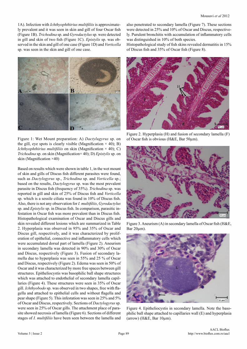
1A). Infection with Ichthyophthirius multifiliis is approximate-ly prevalent and it was seen in skin and gill of four Oscar fish (Figure 1B). Trichodina sp. and Gyrodactylus sp. were detected in gill and skin of two fish (Figure 1C). Epistylis sp. was ob-served in the skin and gill of one case (Figure 1D) and Vorticella sp. was seen in the skin and gill of one case.
Figure 1: Wet Mount preparation: A) Dactylugyrus sp. on the gill, eye spots is clearly visible (Magnification × 40); B) Ichthyophthirius multifiliis on skin (Magnification × 40); C) Trichodina sp. on skin (Magnification× 40); D) Epistylis sp. on skin (Magnification ×40)
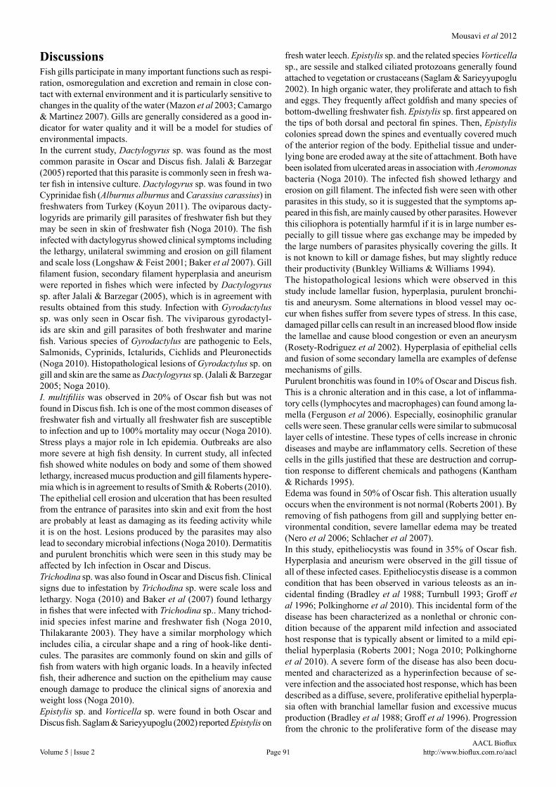
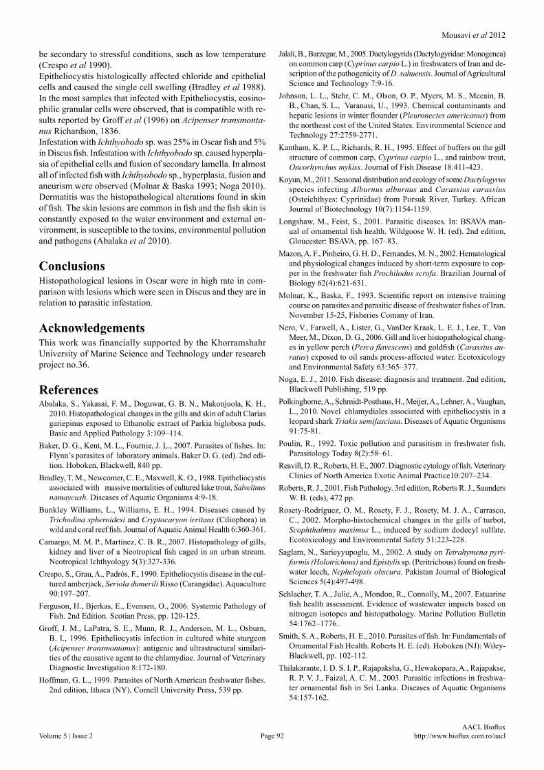
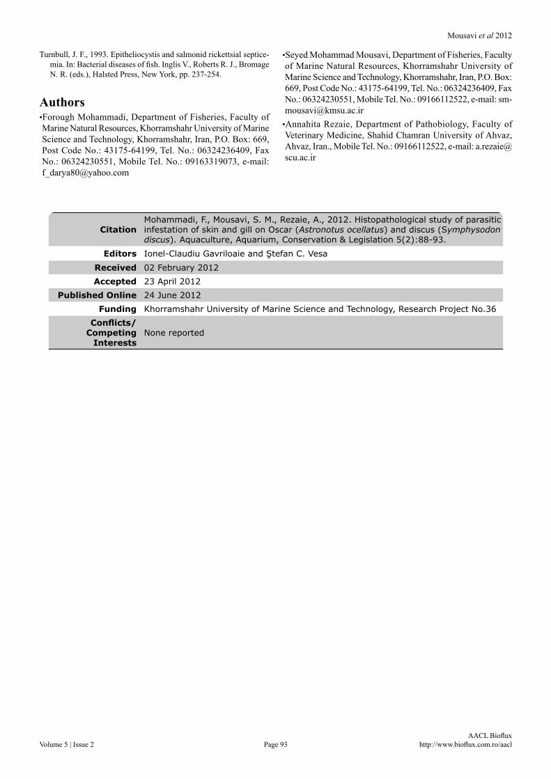
Based on results which were shown in table 1, in the wet mount of skin and gills of Discus fish different parasites were found, such as Dactylogyrus sp., Trichodina sp. and Vorticella sp.; based on the results, Dactylogyrus sp. was the most prevalent parasite in Discus fish (frequency of 35%). Trichodina sp. was reported in gill and skin of 25% of Discus fish and Vorticella sp. which is a sessile ciliata was found in 10% of Discus fish. Also, there is not any observation for I. multifiliis, Gyrodactylus sp. and Epistylis sp. in Discus fish. In comparison, parasitic in-festation in Oscar fish was more prevalent than in Discus fish. Histopathological examination of Oscar and Discus gills and skin revealed different lesions which are summarized in Table 2. Hyperplasia was observed in 95% and 35% of Oscar and Discus gill, respectively, and it was characterized by prolif-eration of epithelial, connective and inflammatory cells which were accumulated dorsal part of lamella (Figure 2). Aneurism in secondary lamella was detected in 90% and 30% of Oscar and Discus, respectively (Figure 3). Fusion of secondary la-mella due to hyperplasia was seen in 55% and 25 % of Oscar and Discus, respectively (Figure 2). Edema was seen in 50% of Oscar and it was characterized by more free spaces between gill structures. Epitheliocystis was basophilic ball shape structures which was attached to endothelial of secondary lamella capil-laries (Figure 4). These structures were seen in 35% of Oscar gill. Ichthyobodo sp. was observed in two shapes, free with fla-gella and attached to epithelial cells and without flagella and pear shape (Figure 5). This infestation was seen in 25% and 5% of Oscar and Discus, respectively. Sections of Dactylogyrus sp. were seen in 25% of Oscar gills. The attachment place of para-site showed necrosis of lamella (Figure 6). Sections of different stages of I. multifiliis have been seen between the lamella and
also penetrated to secondary lamella (Figure 7). These sections were detected in 25% and 10% of Oscar and Discus, respective-ly. Purulent bronchitis with accumulation of inflammatory cells was distinguished in 10% of both species. Histopathological study of fish skins revealed dermatitis in 15% of Discus fish and 35% of Oscar fish (Figure 8).
Figure 2. Hyperplasia (H) and fusion of secondary lamella (F) of Oscar fish is obvious (H&E, Bar 50µm).
Figure 3. Aneurism (A) in secondary lamella of Oscar fish (H&E, Bar 20µm).
Figure 4. Epitheliocystis in secondary lamella. Note the baso-philic ball shape attached to capillaries wall (E) and hyperplasia (arrow) (H&E, Bar 10µm).

Mousavi et al 2012
Volume 5 | Issue 2 Page 90 AACL Bioflux
http://www.bioflux.com.ro/aacl
Figure 5. Ichtiobodo sp. attached to epithelial cells of second-ary lamella (arrows) and they are pear shape. Also hyperplasia (H) is obvious (H&E, Bar 10µm)
Figure 6. Section of Dactylogyrus sp. in gill of Oscar. Note the necrosis of attachment place of parasite (H&E, Bar 50µm)
Figure 7. Trophont stage of I. multifiliis (arrows) in Oscar fish (H&E, Bar 20µm)
Figure 8. Dermatitis in Oscar. Note to dermal edema and vacu-oles in epidermal cells (blue arrows) and penetration of inflam-matory cells between epithelial and dermal cells (black arrows) (H&E, Bar 50µm)
Table 1. Frequency of parasitic infestation in gill and skin in Oscar and Discus (%)
Parasite infestation Oscar gill Oscar skin Discus gill Discus skin
Dactylogyrus sp. 50 20 35 35
Ichthyophthirius multifiliis 20 20 0 0
Gyrodactylus sp. 5 10 0 0
Trichodina sp. 5 10 25 25
Epistylis sp. 5 5 0 0
Vorticella sp. 5 5 10 10
Table 2. Frequency of histopathological effects on gill and skin in Oscar and Discus (%)
Gill and skin histopathological lesion Oscar frequency Discus frequency
Hyperplasia 95 35
Aneurism 90 30
Fusion of secondary lamella 55 25
Edema 50 0
Epitheliocystis 35 0
Ichthyobodo sp. 25 5
Dactylogyrus sp. section 25 0
Ichthyophthirius multifiliis 25 10
Purulent bronchitis 10 10
Dermatitis 35 15
Mousavi et al 2012
Volume 5 | Issue 2 Page 91 AACL Bioflux
http://www.bioflux.com.ro/aacl
DiscussionsFish gills participate in many important functions such as respi-ration, osmoregulation and excretion and remain in close con-tact with external environment and it is particularly sensitive to changes in the quality of the water (Mazon et al 2003; Camargo & Martinez 2007). Gills are generally considered as a good in-dicator for water quality and it will be a model for studies of environmental impacts.In the current study, Dactylogyrus sp. was found as the most common parasite in Oscar and Discus fish. Jalali & Barzegar (2005) reported that this parasite is commonly seen in fresh wa-ter fish in intensive culture. Dactylogyrus sp. was found in two Cyprinidae fish (Alburnus alburnus and Carassius carassius) in freshwaters from Turkey (Koyun 2011). The oviparous dacty-logyrids are primarily gill parasites of freshwater fish but they may be seen in skin of freshwater fish (Noga 2010). The fish infected with dactylogyrus showed clinical symptoms including the lethargy, unilateral swimming and erosion on gill filament and scale loss (Longshaw & Feist 2001; Baker et al 2007). Gill filament fusion, secondary filament hyperplasia and aneurism were reported in fishes which were infected by Dactylogyrus sp. after Jalali & Barzegar (2005), which is in agreement with results obtained from this study. Infection with Gyrodactylus sp. was only seen in Oscar fish. The viviparous gyrodactyl-ids are skin and gill parasites of both freshwater and marine fish. Various species of Gyrodactylus are pathogenic to Eels, Salmonids, Cyprinids, Ictalurids, Cichlids and Pleuronectids (Noga 2010). Histopathological lesions of Gyrodactylus sp. on gill and skin are the same as Dactylogyrus sp. (Jalali & Barzegar 2005; Noga 2010). I. multifiliis was observed in 20% of Oscar fish but was not found in Discus fish. Ich is one of the most common diseases of freshwater fish and virtually all freshwater fish are susceptible to infection and up to 100% mortality may occur (Noga 2010). Stress plays a major role in Ich epidemia. Outbreaks are also more severe at high fish density. In current study, all infected fish showed white nodules on body and some of them showed lethargy, increased mucus production and gill filaments hypere-mia which is in agreement to results of Smith & Roberts (2010). The epithelial cell erosion and ulceration that has been resulted from the entrance of parasites into skin and exit from the host are probably at least as damaging as its feeding activity while it is on the host. Lesions produced by the parasites may also lead to secondary microbial infections (Noga 2010). Dermatitis and purulent bronchitis which were seen in this study may be affected by Ich infection in Oscar and Discus.Trichodina sp. was also found in Oscar and Discus fish. Clinical signs due to infestation by Trichodina sp. were scale loss and lethargy. Noga (2010) and Baker et al (2007) found lethargy in fishes that were infected with Trichodina sp.. Many trichod-inid species infest marine and freshwater fish (Noga 2010, Thilakarante 2003). They have a similar morphology which includes cilia, a circular shape and a ring of hook-like denti-cules. The parasites are commonly found on skin and gills of fish from waters with high organic loads. In a heavily infected fish, their adherence and suction on the epithelium may cause enough damage to produce the clinical signs of anorexia and weight loss (Noga 2010).Epistylis sp. and Vorticella sp. were found in both Oscar and Discus fish. Saglam & Sarieyyupoglu (2002) reported Epistylis on
fresh water leech. Epistylis sp. and the related species Vorticella sp., are sessile and stalked ciliated protozoans generally found attached to vegetation or crustaceans (Saglam & Sarieyyupoglu 2002). In high organic water, they proliferate and attach to fish and eggs. They frequently affect goldfish and many species of bottom-dwelling freshwater fish. Epistylis sp. first appeared on the tips of both dorsal and pectoral fin spines. Then, Epistylis colonies spread down the spines and eventually covered much of the anterior region of the body. Epithelial tissue and under-lying bone are eroded away at the site of attachment. Both have been isolated from ulcerated areas in association with Aeromonas bacteria (Noga 2010). The infected fish showed lethargy and erosion on gill filament. The infected fish were seen with other parasites in this study, so it is suggested that the symptoms ap-peared in this fish, are mainly caused by other parasites. However this ciliophora is potentially harmful if it is in large number es-pecially to gill tissue where gas exchange may be impeded by the large numbers of parasites physically covering the gills. It is not known to kill or damage fishes, but may slightly reduce their productivity (Bunkley Williams & Williams 1994).The histopathological lesions which were observed in this study include lamellar fusion, hyperplasia, purulent bronchi-tis and aneurysm. Some alternations in blood vessel may oc-cur when fishes suffer from severe types of stress. In this case, damaged pillar cells can result in an increased blood flow inside the lamellae and cause blood congestion or even an aneurysm (Rosety-Rodriguez et al 2002). Hyperplasia of epithelial cells and fusion of some secondary lamella are examples of defense mechanisms of gills. Purulent bronchitis was found in 10% of Oscar and Discus fish. This is a chronic alteration and in this case, a lot of inflamma-tory cells (lymphocytes and macrophages) can found among la-mella (Ferguson et al 2006). Especially, eosinophilic granular cells were seen. These granular cells were similar to submucosal layer cells of intestine. These types of cells increase in chronic diseases and maybe are inflammatory cells. Secretion of these cells in the gills justified that these are destruction and corrup-tion response to different chemicals and pathogens (Kantham & Richards 1995). Edema was found in 50% of Oscar fish. This alteration usually occurs when the environment is not normal (Roberts 2001). By removing of fish pathogens from gill and supplying better en-vironmental condition, severe lamellar edema may be treated (Nero et al 2006; Schlacher et al 2007).In this study, epitheliocystis was found in 35% of Oscar fish. Hyperplasia and aneurism were observed in the gill tissue of all of these infected cases. Epitheliocystis disease is a common condition that has been observed in various teleosts as an in-cidental finding (Bradley et al 1988; Turnbull 1993; Groff et al 1996; Polkinghorne et al 2010). This incidental form of the disease has been characterized as a nonlethal or chronic con-dition because of the apparent mild infection and associated host response that is typically absent or limited to a mild epi-thelial hyperplasia (Roberts 2001; Noga 2010; Polkinghorne et al 2010). A severe form of the disease has also been docu-mented and characterized as a hyperinfection because of se-vere infection and the associated host response, which has been described as a diffuse, severe, proliferative epithelial hyperpla-sia often with branchial lamellar fusion and excessive mucus production (Bradley et al 1988; Groff et al 1996). Progression from the chronic to the proliferative form of the disease may
Mousavi et al 2012
Volume 5 | Issue 2 Page 92 AACL Bioflux
http://www.bioflux.com.ro/aacl
be secondary to stressful conditions, such as low temperature (Crespo et al 1990). Epitheliocystis histologically affected chloride and epithelial cells and caused the single cell swelling (Bradley et al 1988). In the most samples that infected with Epitheliocystis, eosino-philic granular cells were observed, that is compatible with re-sults reported by Groff et al (1996) on Acipenser transmonta-nus Richardson, 1836. Infestation with Ichthyobodo sp. was 25% in Oscar fish and 5% in Discus fish. Infestation with Ichthyobodo sp. caused hyperpla-sia of epithelial cells and fusion of secondary lamella. In almost all of infected fish with Ichthyobodo sp., hyperplasia, fusion and aneurism were observed (Molnar & Baska 1993; Noga 2010). Dermatitis was the histopathological alterations found in skin of fish. The skin lesions are common in fish and the fish skin is constantly exposed to the water environment and external en-vironment, is susceptible to the toxins, environmental pollution and pathogens (Abalaka et al 2010).
ConclusionsHistopathological lesions in Oscar were in high rate in com-parison with lesions which were seen in Discus and they are in relation to parasitic infestation.
AcknowledgementsThis work was financially supported by the Khorramshahr University of Marine Science and Technology under research project no.36.
ReferencesAbalaka, S., Yakasai, F. M., Doguwar, G. B. N., Makonjuola, K. H.,
2010. Histopathological changes in the gills and skin of adult Clarias gariepinus exposed to Ethanolic extract of Parkia biglobosa pods. Basic and Applied Pathology 3:109–114.
Baker, D. G., Kent, M. L., Fournie, J. L., 2007. Parasites of fishes. In: Flynn’s parasites of laboratory animals. Baker D. G. (ed). 2nd edi-tion. Hoboken, Blackwell, 840 pp.
Bradley, T. M., Newcomer, C. E., Maxwell, K. O., 1988. Epitheliocystis associated with massive mortalities of cultured lake trout, Salvelinus namaycush. Diseases of Aquatic Organisms 4:9-18.
Bunkley Williams, L., Williams, E. H., 1994. Diseases caused by Trichodina spheroidesi and Cryptocaryon irritans (Ciliophora) in wild and coral reef fish. Journal of Aquatic Animal Health 6:360-361.
Camargo, M. M. P., Martinez, C. B. R., 2007. Histopathology of gills, kidney and liver of a Neotropical fish caged in an urban stream. Neotropical Ichthyology 5(3):327-336.
Crespo, S., Grau, A., Padrós, F., 1990. Epitheliocystis disease in the cul-tured amberjack, Seriola dumerili Risso (Carangidae). Aquaculture 90:197–207.
Ferguson, H., Bjerkas, E., Evensen, O., 2006. Systemic Pathology of Fish. 2nd Edition. Scotian Press, pp. 120-125.
Groff, J. M., LaPatra, S. E., Munn, R. J., Anderson, M. L., Osburn, B. I., 1996. Epitheliocystis infection in cultured white sturgeon (Acipenser transmontanus): antigenic and ultrastructural similari-ties of the causative agent to the chlamydiae. Journal of Veterinary Diagnostic Investigation 8:172-180.
Hoffman, G. L., 1999. Parasites of North American freshwater fishes. 2nd edition, Ithaca (NY), Cornell University Press, 539 pp.
Jalali, B., Barzegar, M., 2005. Dactylogyrids (Dactylogyridae: Monogenea) on common carp (Cyprinus carpio L.) in freshwaters of Iran and de-scription of the pathogenicity of D. sahuensis. Journal of Agricultural Science and Technology 7:9-16.
Johnson, L. L., Stehr, C. M., Olson, O. P., Myers, M. S., Mccain, B. B., Chan, S. L., Varanasi, U., 1993. Chemical contaminants and hepatic lesions in winter flounder (Pleuronectes americanus) from the northeast cost of the United States. Environmental Science and Technology 27:2759-2771.
Kantham, K. P. L., Richards, R. H., 1995. Effect of buffers on the gill structure of common carp, Cyprinus carpio L., and rainbow trout, Oncorhynchus mykiss. Journal of Fish Disease 18:411-423.
Koyun, M., 2011. Seasonal distribution and ecology of some Dactylogyrus species infecting Alburnus alburnus and Carassius carassius (Osteichthyes: Cyprinidae) from Porsuk River, Turkey. African Journal of Biotechnology 10(7):1154-1159.
Longshaw, M., Feist, S., 2001. Parasitic diseases. In: BSAVA man-ual of ornamental fish health. Wildgoose W. H. (ed). 2nd edition, Gloucester: BSAVA, pp. 167–83.
Mazon, A. F., Pinheiro, G. H. D., Fernandes, M. N., 2002. Hematological and physiological changes induced by short-term exposure to cop-per in the freshwater fish Prochilodus scrofa. Brazilian Journal of Biology 62(4):621-631.
Molnar, K., Baska, F., 1993. Scientific report on intensive training course on parasites and parasitic disease of freshwater fishes of Iran. November 15-25, Fisheries Comany of Iran.
Nero, V., Farwell, A., Lister, G., VanDer Kraak, L. E. J., Lee, T., Van Meer, M., Dixon, D. G., 2006. Gill and liver histopathological chang-es in yellow perch (Perca flavescens) and goldfish (Carassius au-ratus) exposed to oil sands process-affected water. Ecotoxicology and Environmental Safety 63:365–377.
Noga, E. J., 2010. Fish disease: diagnosis and treatment. 2nd edition, Blackwell Publishing, 519 pp.
Polkinghorne, A., Schmidt-Posthaus, H., Meijer, A., Lehner, A., Vaughan, L., 2010. Novel chlamydiales associated with epitheliocystis in a leopard shark Triakis semifasciata. Diseases of Aquatic Organisms 91:75-81.
Poulin, R., 1992. Toxic pollution and parasitism in freshwater fish. Parasitology Today 8(2):58–61.
Reavill, D. R., Roberts, H. E., 2007. Diagnostic cytology of fish. Veterinary Clinics of North America Exotic Animal Practice10:207–234.
Roberts, R. J., 2001. Fish Pathology. 3rd edition, Roberts R. J., Saunders W. B. (eds), 472 pp.
Rosety-Rodríguez, O. M., Rosety, F. J., Rosety, M. J. A., Carrasco, C., 2002. Morpho-histochemical changes in the gills of turbot, Scophthalmus maximus L., induced by sodium dodecyl sulfate. Ecotoxicology and Environmental Safety 51:223-228.
Saglam, N., Sarieyyupoglu, M., 2002. A study on Tetrahymena pyri-formis (Holotrichous) and Epistylis sp. (Peritrichous) found on fresh-water leech, Nephelopsis obscura. Pakistan Journal of Biological Sciences 5(4):497-498.
Schlacher, T. A., Julie, A., Mondon, R., Connolly, M., 2007. Estuarine fish health assessment. Evidence of wastewater impacts based on nitrogen isotopes and histopathology. Marine Pollution Bulletin 54:1762–1776.
Smith, S. A., Roberts, H. E., 2010. Parasites of fish. In: Fundamentals of Ornamental Fish Health. Roberts H. E. (ed). Hoboken (NJ): Wiley-Blackwell, pp. 102-112.
Thilakarante, I. D. S. I. P., Rajapaksha, G., Hewakopara, A., Rajapakse, R. P. V. J., Faizal, A. C. M., 2003. Parasitic infections in freshwa-ter ornamental fish in Sri Lanka. Diseases of Aquatic Organisms 54:157-162.
Mousavi et al 2012
Volume 5 | Issue 2 Page 93 AACL Bioflux
http://www.bioflux.com.ro/aacl
Turnbull, J. F., 1993. Epitheliocystis and salmonid rickettsial septice-mia. In: Bacterial diseases of fish. Inglis V., Roberts R. J., Bromage N. R. (eds.), Halsted Press, New York, pp. 237-254.
Authors•Forough Mohammadi, Department of Fisheries, Faculty of Marine Natural Resources, Khorramshahr University of Marine Science and Technology, Khorramshahr, Iran, P.O. Box: 669, Post Code No.: 43175-64199, Tel. No.: 06324236409, Fax No.: 06324230551, Mobile Tel. No.: 09163319073, e-mail: [email protected]
•Seyed Mohammad Mousavi, Department of Fisheries, Faculty of Marine Natural Resources, Khorramshahr University of Marine Science and Technology, Khorramshahr, Iran, P.O. Box: 669, Post Code No.: 43175-64199, Tel. No.: 06324236409, Fax No.: 06324230551, Mobile Tel. No.: 09166112522, e-mail: [email protected]
•Annahita Rezaie, Department of Pathobiology, Faculty of Veterinary Medicine, Shahid Chamran University of Ahvaz, Ahvaz, Iran., Mobile Tel. No.: 09166112522, e-mail: [email protected]
CitationMohammadi, F., Mousavi, S. M., Rezaie, A., 2012. Histopathological study of parasitic infestation of skin and gill on Oscar (Astronotus ocellatus) and discus (Symphysodon discus). Aquaculture, Aquarium, Conservation & Legislation 5(2):88-93.
Editors Ionel-Claudiu Gavriloaie and Ştefan C. VesaReceived 02 February 2012Accepted 23 April 2012
Published Online 24 June 2012Funding Khorramshahr University of Marine Science and Technology, Research Project No.36
Conflicts/ Competing
InterestsNone reported