Embed Size (px)
Citation preview

_____________________________________________________________________________________________________ *Corresponding author: E-mail: [email protected];
Annual Research & Review in Biology 11(1): 1-6, 2016, Article no.ARRB.28101
ISSN: 2347-565X, NLM ID: 101632869
SCIENCEDOMAIN international www.sciencedomain.org
Histopathological Studies on Liver Affected with Hydatidosis in One Humped Camel (Camelus
dromedarius) in Tampool Slaughterhouse, Sudan
Nuseba H. Mohammed1* and Halima M. Osman1
1Veterinary Research Institute, Animal Resources Corporation, P.O.Box 8067 (Al-Amarat), Khartoum,
Sudan.
Authors’ contributions
This work was carried out in collaboration between both authors. Both authors read and approved the final manuscript.
Article Information
DOI: 10.9734/ARRB/2016/28101
Editor(s): (1) George Perry, Dean and Professor of Biology, University of Texas at San Antonio, USA.
Reviewers: (1) S. Sivajothi, College of Veterinary Science, S.V.V.U., Andhra Pradesh, India.
(2) Layla Omran Elmajdoub, Misurata University, Libya. Complete Peer review History: http://www.sciencedomain.org/review-history/16013
Received 1st
July 2016 Accepted 8
th August 2016
Published 2nd September 2016
ABSTRACT
This study aimed to investigate the different pathological conditions reported in livers of slaughtered camels leading to their total or partial condemnation at abattoir in the present study, 80 liver samples collected randomly from Sudanese camels at Tampool slaughter-house. Gezira State, Sudan. This area is known to be heavily populated with camels (Camelus dromedarius) come from different parts of the Sudan. Placed in 10% formalin for histopathological studies. 16 out of 80 (20%) camel livers examined histopathologically, were infected with hydatid cyst the larval stage of Echinoccocus granulosus characterized by presence of distinct rostellar hook. microscopic studies of cysts were done. No doubt, hydatidosis is a public health problem that affects also the human welfare and economy. It is concluded that records from slaughterhouse are very important monitors for animals’ prevailing diseases and their control plan. Liver is one of the important organs found to be affected by different pathogens which are of much concern to human public health authorities and economist the preventive and control measures for man and animal were recommended.
Keywords: Camel; hydatidosis; abattoir; Sudan.
Original Research Article
Mohammed and Osman; ARRB, 11(1): 1-6, 2016; Article no.ARRB.28101
2
1. INTRODUCTION Slaughterhouses are valuable sources for information about food borne and zoonotic diseases epidemiology, actual losses in meat production and the economical impacts for condemnations [1] However, zoonotic threats prevention and control remains one of the major aspects in the developing countries [2]. Meat inspection at abattoirs for hygienic quality involves both ante and postmortem examination which include gross and microbiological investigation [3,4]. Liver is one of the most important organs as it is involved in many metabolic disorders and parasitic diseases [5]. It may harbor pathogens are dangerous for human consumption when passed with localized or mild infection. However, the most common liver lesions which are harmful to human health are fasciola, liver abscess and hydated cycts [6]. Hydatid liver disease is caused by the larval form of Echinococcus granulosus is one of the most important liver problems in animals and man worldwide [7]. It is more prone to occur in liver and lung than any other visceral organs. Its involvement in hepatobiliary system is both mechanical and toxic depending upon its developmental stage. The presence of large-size hydatid cysts is responsible for substantial economic loss on account of reduced productivity and condemnation of visceral organs and carcasses. Histopathological structures as per the age, stage of development and viability of the cysts, i.e., laminated hyaline layer surrounded by a thick coat of granulation tissue showing fibrosis, infiltration of leucocytes along with giant cells, lymphocytes, epitheloid cells… etc. Furthermore, the presence of the thick fibrous tissue layer and infiltration of lymphocytes along with some fibroblasts and eosinophils and the germinal layer with the presence of scolices in various stages of development in viable fertile cyst were quite common. The camel is regarded as one of the common intermediate hosts for the metacestodes of E. granulosus in Sudan [8]. The prevalence of hydatid cyst became one of the public health problems and economical loss due to condemnation of the edible organs infected by hydatid cyst [9]. Several studies
reported the prevalence of hydatidosis in Sudanese cattle and sheep [10,11,12]. In human, the diagnosis of hydatid cyst prevalence needs modern diagnostic facilities, however, high incidence of hydatid cyst in Maasia population were reported in Tansani [13] which was attributed to the large population of stray dogs and improper disposal of abattoir organs . In Sudan, meat and liver examination in the slaughterhouses are of much concern as eating raw meat and liver (Marara and umfitfit) is one of the Sudanese traditions. Therefore this study aimed to investigate the prevalence of different liver lesions, their zoonotic importance and economical impacts for slaughtered camels at Tampool abattoir during the period (2010-2011). A prevelance of 59.8% of hydatidosis was reported in the dromedary camels from different parts of Sudan [14].
2. MATERIALS AND METHODS During a 2-year period (2010- 2011) Postmortem examination was carried out immediately after slaughtering of the camels. 80 Livers were sampled randomly then examined for the presence of any pathological lesion and a portion from the affected areas was cut off and preserved in 10% formalin and transported to the laboratory and processed for histopathological examination using the conventional methods as follow: About one cm of the lesion was fixed in 10% formalin for preservation and fortifying the tissue the fixation extends for three days. Gradual dehydration of samples was done using different concentrations of ethyl alcohol starting from 50% to 99%. Xylene act intermediate between alcohol and paraffin wax. The impregnated tissues were transferred to paraffin wax in melting point, in a suitable size blocks and were allowed to cool and solidify. Microtetomy of the embedded tissues was performed using a rotator microtome. Sections were cut 4-6 um in thickness. Sections were fixed on a glass slides by egg albumin and dried at 37C till staining. Tissues were dewaxed using xylene and hydration using different descending grades of ethanol (99%-50%), followed by washing with tap water then stained with haematoxylin and eosin (H&E) and covered with a glass cover using canda blasma.
3. RESULTS 16 out of 80 (20%) camel livers examined histopathologically, were infected with hydatid cyst the larval stage of Echinococcus granulosus.

Mohammed and Osman; ARRB, 11(1): 1-6, 2016; Article no.ARRB.28101
3
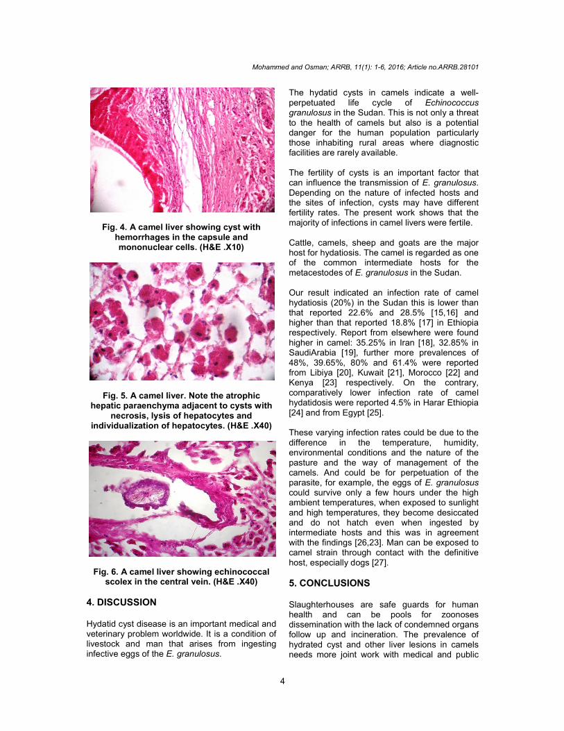
The general architeture of the infected livers was pale brown color and fragile. Multiple cysts were found in most of the cases. Some affected livers displayed severe damage of the parenchyma. The parenchyma around the cyst was hard due to the presence of a fibrous capsule. Hydatid cyst in the liver was recognized by the presence of varying size cysts protruding on the surface. The cysts were unilocular and also found deep in the liver parenchyma. Cysts were filled with clear watery fluid, and the cut surface of these cysts showed cavities lined by a smooth membrane which could be shelled out. Numerous cysts in late stage were embedded at different depths of the liver resulting in its gross enlargement of the organ. The lesions found in the liver depended upon the stage of development and the state of the cysts. In sections echinococcal scolexes were displayed in the capsule with negligible reaction in fertile cysts; there was a thick connective tissue layer, the outer part of which was densely infiltrated with lymphocytes; fibroblasts and eosinophils. While on the inner side there was a structure less hyaline layer followed by the germinal layer. It showed brood capsules containing scolices in varying stages of development along with hook lets (Fig. 1). Such cysts were also encountered in the hepatic parenchyma with slight hemorrhage, leukocyte infiltration and mild hepatocellular degeneration (Fig. 2). In most cysts, the capsule was thick from inside with a highly cellular zone rich in mononuclear cells, abundant fibroblasts and an outer thick fibrous zone of concentrically arranged collagen bundles. The other cyst wall was filled of an eosinophilic laminated hyaline structure (Fig. 3). A predominant infiltration with lymphocytes and macrophages, and occasionally neutrophils, eosinophils and giant cells were observed. The adjacent parenchyma was markedly congested and showed multiple small haemorrhagic areas. The junction between the hepatic parenchyma and the connective tissue capsule of the cyst revealed infiltration of mononuclear cells. Nearby hepatic tissue showed dilated sinusoids, collapsed hepatocytes and fibrosis (Fig. 4). In other section, the hepatocytes in the cyst wall showed atrophy, dissociation of the hepatocytes and disrupted hepatic cord. The cells became atrophic,
spherical to oval in shape with dark nuclei (Figs. 5 and 6).
Fig. 1. A camel liver showing Echinococcal scolices. (H&E .X10)
Fig. 2. A camel liver note cyst with eosinophilic laminated material, inflammatory
cells. (H&E .X10)
Fig. 3. A camel liver. Note the cyst wall formed of thick fibrous connective tissue with
inflammatory cellular reaction. (H&E .X40)
Mohammed and Osman; ARRB, 11(1): 1-6, 2016; Article no.ARRB.28101
4
Fig. 4. A camel liver showing cyst with hemorrhages in the capsule and mononuclear cells. (H&E .X10)
Fig. 5. A camel liver. Note the atrophic hepatic paraenchyma adjacent to cysts with
necrosis, lysis of hepatocytes and individualization of hepatocytes. (H&E .X40)
Fig. 6. A camel liver showing echinococcal scolex in the central vein. (H&E .X40)
4. DISCUSSION Hydatid cyst disease is an important medical and veterinary problem worldwide. It is a condition of livestock and man that arises from ingesting infective eggs of the E. granulosus.
The hydatid cysts in camels indicate a well-perpetuated life cycle of Echinococcus granulosus in the Sudan. This is not only a threat to the health of camels but also is a potential danger for the human population particularly those inhabiting rural areas where diagnostic facilities are rarely available. The fertility of cysts is an important factor that can influence the transmission of E. granulosus. Depending on the nature of infected hosts and the sites of infection, cysts may have different fertility rates. The present work shows that the majority of infections in camel livers were fertile. Cattle, camels, sheep and goats are the major host for hydatiosis. The camel is regarded as one of the common intermediate hosts for the metacestodes of E. granulosus in the Sudan. Our result indicated an infection rate of camel hydatiosis (20%) in the Sudan this is lower than that reported 22.6% and 28.5% [15,16] and higher than that reported 18.8% [17] in Ethiopia respectively. Report from elsewhere were found higher in camel: 35.25% in Iran [18], 32.85% in SaudiArabia [19], further more prevalences of 48%, 39.65%, 80% and 61.4% were reported from Libiya [20], Kuwait [21], Morocco [22] and Kenya [23] respectively. On the contrary, comparatively lower infection rate of camel hydatidosis were reported 4.5% in Harar Ethiopia [24] and from Egypt [25]. These varying infection rates could be due to the difference in the temperature, humidity, environmental conditions and the nature of the pasture and the way of management of the camels. And could be for perpetuation of the parasite, for example, the eggs of E. granulosus could survive only a few hours under the high ambient temperatures, when exposed to sunlight and high temperatures, they become desiccated and do not hatch even when ingested by intermediate hosts and this was in agreement with the findings [26,23]. Man can be exposed to camel strain through contact with the definitive host, especially dogs [27].
5. CONCLUSIONS Slaughterhouses are safe guards for human health and can be pools for zoonoses dissemination with the lack of condemned organs follow up and incineration. The prevalence of hydrated cyst and other liver lesions in camels needs more joint work with medical and public
Mohammed and Osman; ARRB, 11(1): 1-6, 2016; Article no.ARRB.28101
5
health researchers in human zoonosis in and out slaughterhouses. Public health extension awareness about the risks of eating raw livers and meat and dealing with infected and pet animals are highly required. The local authority's regulations should support animals’ owners and encourage meat industry [27].
COMPETING INTERESTS Authors have declared that no competing interests exist.
REFERENCES 1. Herenda D, Jackel O. Poultry abattoir
survey of carcass condemnation for standard, vegetarian and free range chickens. Can. Vet. J. 1994;35(5):293-296.
2. Stőhrk K, Melsin FX. The role of veterinary public health in the prevention of zoonoses. Arch. Virol. Suppl. 1997;13: 207-218.
3. Elhassan IM, Abdelgadir AE, Ibrahim AE. Microbiological assess-men of mutton intended for export from Alkadro export slaughterhouse. Sudan. J. Vet. Med. Anim. Prod. 2011;2(1):23-33.
4. Harhoura KH, Boukhors KT, Dahmani A, Zenia S, Aissi M. Survey of hygiene in ovine slaughterhouses of Algiers region by bacteriological analysis of Carcasses. African Journal of Microbiology Research. 2012;6(22):4722-4726.
5. Saleha AA. Liver fluke diseases (Fascioliasis): Epidemiology, economic impact and Public health significance. South East Asian J. Trop. Med. Pub. Health. 1991;(Suppl 22):361-364.
6. Mellau LSB, Nogna HE, Karimuribo ED. A slaughterhouse survey of liver lesions in slaughtered cattle, sheep and goat at Arusha, Tanzania. Res. J. Vet. Sci. 2010; 3(3):179-188.
7. Torgerson PR, Budke C M. Echinococcosis - An international public health challenge. Res. Vet. Sci. 2003; 74(3):191-202.
8. Saad MB, Zien Eldin EA, Tag El Din MH. Some observations on the prevalence and pathology of hydatidosis in Sudanese camels (Camelus dromedarius). Rev Elev Med Vet Pays Trop. 1983;6(4):359-63.
9. Alazlaf R, Dakkak A. Epidemiological study of the cystic echinococcosis in Morocco. Vet. Parasitol. 2006;137:83-93.
10. Elmahdi IE, Ali QM, Magzoub MM, Ibrahim AM, Saad MB, Romig T. Cystic echinoccosis of livestock and humans in central Sudan. Ann. Trop. Med. Parasitol. 2004;98:473-479.
11. Osman AMA. Pathological and bio molecular study on hydatid diseases in Camel, Cattle and Sheep. M.Vsc. Dep. Pathology, Faculty of Veterinary Medicine, University of Khartoum, Sudan; 2008.
12. Mohamadin SA, Elamin AA. Study on hydatid cyst infection in slaughterhouses in Khartoum state, Sudan. Archives of Applied Science Research. 2011;3(6):18.
13. Ernest E, Nonga HE, Kassuku AA, Kazwala RR. Hydatidosis of slaughtered animals in Ngorongoro district of Arusha region, Tanzania. Trop Anim. Health Prod. 2009;41:(7):1179.
14. Omer RA, Aradaib IE, Majid AA, Mukhtar O, Nahas AA. A survey of hydatid disease in camel, sheep and cattle in central Sudan. The proceeding of the 27
th
international congress of the world veterinary association, Ghartage, Tunisia; 2002.
15. Muskin S, Hailu D, Moti Y. Infection rates, cyst fertility and larval viability of hydatid disease in camels (Camelus dromedarius) from Borena, Kereyu and Harar Areas of Ethiopia. Global Veterinaria. 2011;7(6): 518-522.
16. Bitsat K. The prevalence of hydatidosis in Jijiga municipal abattoir, DVM Thesis. Jimma University, Ethiopia; 2009.
17. Woldemeskel M, Issa A, Mersie, Potgieter LND. Investigation of parasitic disease of one-humped camel (Camelus dromedarius) in eastern Ethiopia. J. Camel Pract. Res. 2001;8:77-81.
18. Ahmadi NA. Hydatidosis in camel (Camelus dromedaries) and their potential role in epidemiology of Echinococcus granulosus. Iran. J. Helminthol. 2005;79: 119-125.
19. Mohammed MI. Study of cystic echinococcosis in slaughtered animals in Al Baha region, Saudi Arabia: Interaction between some biotic factors. Acta Tropia. 2010;113:26-33.
20. Ibrahim MM, Craig PS. Prevalence of hydatidosis cystic echinococcus in camels (Camelus dromedaries) in Libya. J. Helminthol. 1998;72:27-31.
21. Abdul-salam JM, Farah MA. Hydatidosis in camels in Kuwait. Parasitology Res. 1988; 74:267-270.

Mohammed and Osman; ARRB, 11(1): 1-6, 2016; Article no.ARRB.28101
6
22. Pandev VS, Ouhell H, Ouchou M. Hydatidosis in sheep, goat and dromedaries in Morocco. Annals of Tropical Medicine and Parasitol. 1986;80: 525-529.
23. Njoroge EM, Mbithi PMF, Gathuma JM, Wachira TM, Gathura PB, Magamboc JK, Zeyhle E. A study of cystic echinococcosis in slaughter animals in three selection areas of northern Turkana, Kenya. Vet Parasitol. 2002;104:85-91.
24. Woubet M. A preliminary study of echinococcosis / hydatidosis in Hararghe region and the efficacy of Glinhs lotoidus seeds against Echinococcus granulosus in pups infected experimentally with hydatid material. DVM thesis, Addis Ababa University, Ethiopia; 1987.
25. Dyab KA, Hassanein R, Hussein AA, Metwally SE, Gaad HM. Hydatidosis among man and animals in Assiut and Aswan Governorates. J. Egypt. Soci. Para. 2005;35:157-166.
26. Wachira TM, Macpherson CNL, Gathuma JM. Release and survival of Echinococcus eggs in different environments in Turkana and their possible impact on the incidence of hydatidosis in man and livestock. J. Hemintholol. 1991;65:55-61.
27. Garba HS, Maigandi S. Diseases of camels (Camelus dromedarius) encountered at slaughter at the abattoir in Sokoto, Nigeria. Trop. Vet. 1995;13(3-4): 95-102.
_________________________________________________________________________________ © 2016 Mohammed and Osman; This is an Open Access article distributed under the terms of the Creative Commons Attribution License (http://creativecommons.org/licenses/by/4.0), which permits unrestricted use, distribution, and reproduction in any medium, provided the original work is properly cited.
Peer-review history: The peer review history for this paper can be accessed here:
http://sciencedomain.org/review-history/16013