Embed Size (px)
Citation preview

/ . Embryol. exp. Morph., Vol. 15, 3, pp. 271-279, June 1966 2 7 1With 2 plates
Printed in Great Britain
Histology of larval eye-antennal disks and cephalicganglia of Drosophila cultured in vitro
BylMOGENE SCHNEIDER1
From the Department of Biology, Yale University and theDepartment of Entomology, Walter Reed Army Institute of Research
INTRODUCTION
In the past decade, a relatively large number of reports have been publishedon the culture in vitro of organs of Drosophila melanogaster. The majority ofthese reports have been concerned with the development of the eye-antennalcomplex explanted, with or without the attached cephalic ganglia, from theprepupal stage (Denial, 1956) or from late second and third larval instar stages(Kuroda & Yamaguchi, 1956; Gottschewski, 1958, 1960, 1962; Gottschewski &Querner, 1961; Fugio, 1962; Schneider, 1964). With the exception of the workof Demal (1956), the above reports have been confined to descriptions of thedeveloping explants solely in morphological terms and, as such, are not whollyadequate for comparisons to be made between development in vivo and in vitro.If, however, such descriptions are supplemented with histological studies, amore valid appreciation can be gained of the potentialities as well as the limita-tions of such explants under conditions in vitro.
In a recent paper (Schneider, 1964) the day-by-day morphological develop-ment of late third instar eye-antennal disks of D. melanogaster cultured in vitrofor varying lengths of time was described. The present report deals with theextent of histological differentiation attained in these explants.
MATERIALS AND METHODS
Since the majority of explants in the previous study had been allowed toremain in culture for as long as 2-3 weeks, a second series were placed in culturein order to obtain explants that had been under in vitro conditions for relativelylimited periods, i.e. 24-96 h. The method of culturing was identical to thatdescribed previously, with the exception that the culture medium was slightlyrevised as follows: the concentrations of KH2PO4 and NaHCO3 were lowered to45 and 40 mg/100 ml respectively and Na2HPO4 was added in a concentrationof 70 mg/100 ml. The individual amino acids were used instead of lactalbumin
1 Author's address: Department of Entomology, Walter Reed Army Institute of Research,Washington, B.C., U.S.A.

272 I. SCHNEIDER
hydrolysate; and TC yeastolate in a concentration of 200 mg/100 ml was em-ployed instead of the individual B vitamins.
In addition, eye-antennal disks with cephalic ganglia were removed from intactlarvae, prepupae and pupae of D. melanogaster (raised under aseptic conditionsat 25° C and timed to ± 2 h after emergence) and fixed immediately to serve ascontrols.
The above were fixed in warm FAA (6:16:1 + 30 parts water), embeddedusing the Peterfi methyl benzoate-celloidin method, sectioned at 8 /i, and stainedwith Harris's hemotoxylin.
Although explants from both D. melanogaster and D. virilis were used in theprevious study, this report deals only with explants from the former.
RESULTSWith the exception of orange and red pigment deposition, differentiation of
the eye-antennal disks and the brain lobes reached its peak between the 7th and8th day in vitro. Thereafter the explants markedly deteriorated even thoughfurther pigment deposition took place.
In general, the antennal disks were least adversely affected by being placedin culture whereas the opposite held true for the ventral ganglion. The formershowed relatively few signs of degeneration even after 14 days in culture, whereasin the latter blebs and vacuoles as well as increasing opaqueness became evidentafter 24-48 h. The effects of culturing on the eye disks and paired brain lobeswere intermediate between these two extremes. Consequently, the histology ofthe developing antennal disks most closely approached that of the controls.The antennal disks were the only structures which consistently differentiated tothe point of approximating the adult organ. With few exceptions, both the eyedisks and the brain differentiated to an extent comparable to mid-pupal or latepupal controls.
Contrary to an earlier assumption, based solely on the morphologicalappearance of the explants, the eye-antennal disks did not evaginate but shiftedonly to a position normally attained in the late prepupal stage.
Histology of the developing eye disks
After one day in vitro, longitudinal sections through the eye disk show aseries of adjacent 'goblet-like' units (terminology of Steinberg, 1943) with eachunit having a two-layered appearance. The upper portion of these units showssome signs of organization within the presumptive cone area whereas the lowerlayer is a mass of unorganized cells. This effect is further heightened by the factthat the upper layer invariably stains less intensely than does the lower layer(Plate 1, fig. A). In cross-section the four-celled 'clusters' of the presumptiveommatidia are very prominent in the outer layer of the eye disks. On the thirdday the pre-ommatidia are more elongate and further development takes place

Histology of cultured explants 273
in the upper layer. The pseudocone, corneagenous cells and cone cells can beidentified although they are often somewhat displaced in comparison with thepupal controls. Beneath the cone cells are a number of oval-shaped cells, atleast four in number and possibly more. In a few instances an axial rod can beseen extending from the pseudocone throughout the length of the retinula tothe optic nerve. There is still little or no organization of the cells in the lowerlayer.
During the fifth and sixth days, the first indication of the cornea appears in theform of a darkened layer at the upper border of the ommatidia. Between theseventh and eighth days (see Plate 1, figs. B, C) the facets are delineated, althoughoccasionally one facet appears to overlap two or more ommatidia. The setiformcorneal hairs are also apparent and, in contrast to the normal controls in whichone seta is found at the intersection of three adjacent facets, as many as sevenhairs may project upward from the border of a single facet. Although the upperarea of the ommatidia (i.e. from the cone cells to the cornea) attains a reasonablynormal appearance, the retinulae are very much foreshortened and often aresituated at an oblique angle to the upper portion. In many instances the retinulaesimply taper off into an unrecognizable mass with the lower layer of cells, whichnever attains any degree of organization.
In addition to the eye proper, the ocelli as well as brown and black head cuticleand bristles differentiate from certain sectors within the eye disk. No attemptwas made to follow the course of this development in vitro other than to notethat both bristles and cuticle are present by the seventh day. The ocelli, if present,were not detected.
No further differentiation is apparent in explants 10-15 days old. On the con-trary, the structures are much less definitive in appearance and position due tothe advanced state of degeneration in the explants.
Histology of developing antennal disks
During the first 2 days in vitro the concentric folds within the antennal diskbecome increasingly thickened, especially the fold destined to become the thirdantennal segment. This third segment is the first to protrude from the antennal'center' and is followed shortly thereafter by the second segment or pedicel andthe small basal scape. In the next few days the second and third segments of theantenna increase both in length and width.
Whereas the cells in the basal segment of a 2-day-old explant are fairly uniformin size, those within the second and third segments show a gradation, being verylarge at the border and becoming progressively smaller toward the interior.These cells appear to be partially oriented into adjacent wedge-shaped rows,wide at the periphery, accommodating the larger cells, and tapering to a narrowshaft as the central lumen is reached.
Indication of a lumen is present in all three segments as early as the first day.In the basal segment the lumen is quite small, occupying perhaps a fifth of the

274 I. SCHNEIDER
total area. In the second and third segments the lumina are larger, in the formercovering as much as a third of the total area. By the fifth day (Plate 1, fig. D) thelumina have fused into a common duct serving all three segments.
Spanning the width of the lumen in the second segment is a diagonal cell'bridge' (occasionally two), a few cell layers thick, 1-5 or more cells in widthand from 8 to over 20 cells in length. These bridges are present by the first dayand become increasingly larger as well as more complex in structure on succes-sive days. This increase in cell number within the bridges may be due to division,but it seems more likely that at least part of the increase is due to a migration ofcells from certain sectors on the periphery of the lumen. As development con-tinues, it is apparent that this internal organization leads to the formation ofJohnston's organ.
The base of the arista (segments 4 and 5 of Ferris, 1950) becomes evidentbetween the fifth and eighth day in vitro. The arista itself is much foreshortenedand does not appear in all of the explants (Plate 2, fig. E). The antennal nerve istraceable from its origin in the deutocerebrum to its termination point in thethird segment by the sixth day. Formation of antennal muscles and bristles, aswell as sclerotization of the first and second segments, begins on the fifth day,with the organ essentially adult in appearance by the seventh day (Plate 2, fig. F).
Histology of the developing brain lobes
After 1 day in vitro, the brain lobes lose their more or less spherical appear-ance and elongate laterally. The fusion of the two lobes mediodorsally is alsonearly completed. Aside from these two developments the brain lobes are stillessentially late larval in character. The external optic glomerulus (Plate 2, fig. G)is closely apposed to the outer lateral edge of the optic lobe, where it is united
EXPLANATION OF PLATES
Abbreviations. A \-A 6, antennal segments 1-6; AR, axial rod of ommatidium; BM,basement membrane; E, eye disk; EEC, external optic glomerulus; F, facet; H, corneal hair;TEG, inner optic glomerulus; JO, Johnston's organ; MEG, middle optic glomerulus; LOR,lower ommatidial region; O, ommatidia; UOR, upper ommatidial region. The horizontalline under each figure presents 10 ji.
PLATE 1
Fig. A. Section of eye disk from 2-day explant. The more lightly staining upper half shows aseries of adjacent 'goblet-like' units, each composed of four cells. Little or no organizationis apparent in the darker staining lower half.Fig. B. Ommatidia of 7-day explant. Failure of head to evaginate is probably responsible forcrowding of ommatidia and overlapping of facets. Axial rod can be traced in ommatidiumto right of the corneal hair.Fig. C. Ommatidia of 8-day explant. Note difference in height of facets in this explant ascompared with flattened facets in fig. B. Ommatidia have attained their greatest length, lessthan two-thirds of that found in late pupal controls. Inverted facet at left may or may notbe an artifact.Fig. D. Antenna of 5-day explant. The individual lumina have fused into a common ductserving all three segments.

/ . Embryol. exp. Morph., Vol. 15, Part 3 PLATE 1
AR
I. SCHNEIDER facing p. 274

J. Embryol. exp. Morph., Vol. 15, Part 3 PLATE 2
Fig. E. Antenna of 7-day explant. Segments 4, 5 and 6 protrude from third segment. Thearista (segment 6) is not seen in its entirety.Fig. F. Fully formed antenna in 8-day explant. Johnston's organ is at top. Note the pro-nounced sclerotization of second segment and the numerous bristles bordering entire organ.Fig. G. Brain lobe of 2-day explant. External optic glomerulus is surrounded by outerimaginal epithelium.Fig. H. Nine-day explant. External optic glomerulus is now fan-shaped but does not liecompletely outside the rest of the brain. Facets border upper area of the unevaginated eye.
I. SCHNEIDER facing p. 275

Histology of cultured explants 275with the fibrous optic nerve. The former has differentiated into an inner andouter mass of cells divided by a lightly staining fibrous band. The middleglomerulus as well as the anterior and posterior portions of the inner glomerulusare well defined. The cells surrounding these optic glomeruli are very small ascontrasted with the much larger cells in the central portion of the brain.
In the next 24-48 h, fusion of the brain lobes is completed. The external opticglomerulus, previously within the brain lobe proper, now protrudes outward toa distance corresponding to approximately a third of its length. The middleglomerulus increases both in length and width, in contrast to the inner glomer-ulus which remains essentially the same.
Between the fifth and eighth day in vitro, the external optic glomerulusassumes a somewhat fan-shaped appearance and is in close apposition to theommatidia of the unevaginated eye (Plate 2, fig. H). This structure never attainsthe normal position of lying outside of the brain; it is always bounded on itsanterior and outer lateral sides by a thin cellular layer of the outer imaginalepithelium.
By the fifth day in vitro, all of the numerous components of central portionof the adult brain as described by Miller (1950) can readily be identified in theexplants. Nonetheless, there are a number of characteristics by which the ex-plants differ from the controls. In addition to the essentially mid-pupal positionof the external optic glomerulus, the entire brain appears flattened so that infrontal sections the deutocerebrum does not assume its normally elevatedposition but is instead strongly compressed against the protocerebrum. Finally,although the nervous tissue (excluding the ventral ganglion) shows few signsof degeneration, the periphery of the brain lobes, especially those portions lyingdirectly beneath the middle and posterior inner glomeruli, becomes increasinglyvacuolated after the third or fourth day in culture.
A summary of the results together with an in vivo timetable is given in Table 1.
DISCUSSION
The foregoing description was based on a histological study of approximately140 explants. The rate and the extent of differentiation within these explants wasby no means uniform and consequently the description given is a more or lessgeneralized one.
As a rule, if a developmental sequence takes place within 24 h after pupariumformation this same sequence will occur in vitro without a serious time-lag. Ifsuch a sequence normally takes place after this period, the time-lag not onlyincreases considerably but there is also much greater diversity among theexplants with respect to their degree of development (see Table 1). Undoubtedly,an explant when first placed in culture has sufficient nutritional and hormonalreserves to continue normal or near-normal development for a limited time. Oncesuch reserves have been greatly diminished or exhausted, the explant must rely
276 I. SCHNEIDER
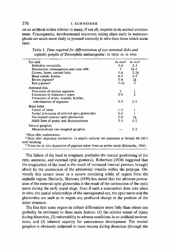
on an artificial milieu inferior in many, if not all, respects to its normal environ-ment. Consequently, developmental sequences taking place early in metamor-phosis are much more likely to proceed normally in vitro than those which occurlater.
Table 1. Time required for differentiation of eye-antennal disks andcephalic ganglia 0/Drosophila melanogaster: in vitro vs. in vivo
Eye diskDefinitive ommatidiaPseudocone, corneagenous and cone cellsCornea, facets, corneal hairsHead cuticle, bristlesBrown pigment3
Red pigment3
Antennal diskProtrusion of distinct segmentsFormation of Johnston's organFormation of arista, muscles, bristles;sclerotization of segments
Brain lobesFusion of lobesPartial protrusion of external optic glomerulusFan-shaped external optic glomerulusAdult form of proto- and deutocerebrum
In vitro1
5-83
5-86-75-87-12
23-5
5-7
1-32-35-83-5
In vivo2
2-3l±-22-2*2-32*3
12
2-3
111*1-2
Ventral ganglionMetamorphosis into imaginal ganglion — 2-3
1 Days after explantation.2 Days after puparium formation: in aseptic cultures the puparium is formed 98-100 h
post hatching.3 Times for in vitro deposition of pigment taken from an earlier study (Schneider, 1964).
The failure of the head to evaginate precludes the normal positioning of theeyes, antennae, and external optic glomeruli. Robertson (1936) suggested thatthe evagination of the head is the result of increased internal pressure broughtabout by the contraction of the abdominal muscles within the prepupa. Ob-viously this cannot occur in a system consisting solely of organs from thecephalic region. Similarly, Shatoury (1956) has stated that the ultimate protru-sion of the external optic glomerulus is the result of the contraction of the opticnerve during the early pupal stage. Even if such a contraction does take placein vitro, the spatial relationships of the unevaginated eye, the optic nerve and theglomerulus are such as to negate any profound change in the position of thelatter structure.
The fact that some organs in culture differentiate more fully than others canprobably be attributed to three main factors: (1) the relative extent of injuryduring dissection, (2) vulnerability to adverse conditions in an artificial environ-ment, and (3) inherent capacity for autonomous development. The ventralganglion is obviously subjected to more trauma during dissection (through the

Histology of cultured explants 277necessity of severing the multitude of nervous fibers emanating along its entirelength) than are the eye disks and brain lobes, which are virtually untouched, orthe antennal disks, which are severed only at their apex. Furthermore, the ventralganglion is completely vulnerable to any adverse environmental conditions incontrast to the rest of the brain, which is 'protected' to some extent by an outerepithelium, or to the antenna, which is completely surrounded by other organs.This factor may also account for the difference in the extent of differentiationfound in the upper and lower areas of the ommatidia. Finally, the culture invitro of isolated eye-antennal disks has shown that they display a considerabledegree of autonomous development if explanted at the end of the third larvalstage. It is evident that the ventral ganglion either lacks this degree of autonomyor that it cannot be expressed under the conditions imposed by the environmentin vitro.
The results reported here as well as those of an earlier study (Schneider, 1964)are at variance in a number of respects with those of Gottschewski (1960). Inthe latter study eye-antennal disks with cephalic ganglia were explanted at theend of the third larval instar (exact age and temperature not given), and differ-entiation of the eye disks, including the evagination movement, was completeby 96 h, a time-lag of not more than 12 h compared with in vivo controls.Possible explanations for the great disparity in the rate of differentiation ob-served in the two studies are: (1) difference in age of the explants cultured,(2) difference in the volume of culture medium employed, and (3) differencesbetween the media.
Larvae grown under aseptic conditions tend to mature at a somewhat slowerpace than normal and therefore an explant may be slightly younger develop-mentally than the chronological age of the donor would indicate. In order toeliminate this factor, explants from larvae grown on standard food were alsocultured (the concentration of antibiotics suppressed but did not eliminatecontamination from bacteria and/or yeast). Little improvement was noted inthe time required for differentiation. However, when explants were taken fromdonors at the end of the prepupal stage they developed at a rate comparable tothat found by Gottschewski, the time-lag rarely exceeding 24 h.
Reducing the size of the hanging drop from 0-008 to 0-005 ml resulted in aconsiderable reduction in differentiation time: from 8-12 days to 5-6 days.However, the accumulation of detrimental metabolic wastes apparently in-creased at an even faster rate since an explant kept for four days in a smallerdrop usually showed more signs of degeneration than an 8 or 9-day explant ina larger drop. The above also held true for explants cultured in Gottschewski'smedium. (It should be noted here that partial renewal of the culture mediumevery third or fourth day increases the survival time of an explant by as muchas a week or more; however, differentiation is greatly retarded or ceases alto-gether if such a renewal is made.) Since there is no evidence whatsoever forevagination of eye disks explanted in vitro at the end of the third larval instar,
l8 JEEM 15

278 I. SCHNEIDER
nor any indications from the extensive work on transplantation that trans-planted disks can evert in vivo, reports of evagination of larval disks explantedin vitro must be regarded with skepticism. On the other hand it is possible thatdisks explanted at the close of the prepupal period, near the time of normaleversion, might be found in an evaginated condition when observed in vitro.
SUMMARY
1. A description is given of the histological development of eye-antennaldisks and cephalic ganglia of Drosophila melanogaster cultured in vitro forvarying lengths of time.
2. The eye-antennal disks do not evaginate but move only to a positionnormally attained in the late prepupal stage.
3. The relative competence of the various cephalic organs to undergodifferentiation is as follows (descending order): antennal disks, eye disks, brainlobes, ventral ganglion. Possible explanations for this difference in differentia-tion capacity are given.
4. In general, if a developmental sequence takes place in vivo within 24 hafter puparium formation, the same sequence occurs in vitro without a serioustime-lag. After this period, however, an interval of from 3 to 9 days is necessaryfor comparable development.
RESUME
Histologie des disques oculo-antennaires et des ganglions cephaliquesde la larve de Drosophile, en culture in vitro
1. Le developpement histologique des disques oculo-antennaires et desganglions cephaliques, cultives in vitro pendant des temps varies, est decrit chezDrosophila melanogaster.
2. Les disques oculo-antennaires n'accomplissent pas l'evagination maisn'atteignent que la position normalement acquise au stade prepupe avancee.
3. La capacite relative de differenciation des differents organes cephaliquesdecroit dans l'ordre suivant: disques antennaires, disques oculaires, lobescerebraux, ganglion ventral. Des explications possibles de cette difference dansla capacite de differenciation sont fournies.
4. En general, si une sequence du developpement se produit in vivo dans les24 heures suivant la formation de la pupe, cette meme sequence apparait invitro sans retard notable. Cependant, apres ce stade du developpement, unretard de 3 a 9 jours est observe pour obtenir un developpement comparable.
I wish to thank Prof. D. F. Poulson and Drs S. J. Counce and W. W. Doane for theircomments and criticisms of the manuscript. The technical assistance of Mrs Betsy Jeavons isalso acknowledged. The major part of this investigation was supported by N.S.F. grantGB 1718.

Histology of cultured explants 279
REFERENCES
DEMAL, J. (1956). Culture in vitro d'ebauches imaginales de Dipteres. Ann. des Sc. Nat. Zool.lleserie, 18, 155-61.
FERRIS, G. F. (1950). External morphology of the adult. In The Biology of Drosophila, ed.M. Demerec. New York: Wiley.
FUGIO, Y. (1962). Studies on the development of eye-antennal discs of Drosophila melano-gaster in tissue culture. II. Effects of substances secreted from cephalic complexes uponeye-antennal discs of eye mutant strains. Jap. J. Genet. 37, 110-17.
GOTTSCHEWSKI, G. H. M. (1958). Ober das Wachstum von Drosophila-Augen-ImsLginal-scheiben in vitro. Naturwissenschaften, 45, 400.
GOTTSCHEWSKI, G. H. M. (1960). Morphogenetische Untersuchungen an in vitro wachsendenAugenanlagen von Drosophila melanogaster. Wilhelm Roux Arch. EntwMech. Org. 152,204-29.
GOTTSCHEWSKI, G. H. M. (1962). Die Entwicklung des Chiasma externum bei Drosophilamelanogaster. 1. Europdischen Anatomen-Kongresses, Strassburg 1960. Jena: GustavFischer.
GOTTSCHEWSKI, G. H. M., & QUERNER, W. (1961). Beobachtungen an explantierten friihenEntwicklungsstaden der Augenanlage von Drosophila melanogaster. Wilhelm Roux Arch.EntwMech. Org. 153, 168-75.
KURODA, Y. & YAMAGUCHI, K. (1956). The effects of the cephalic complex upon the eye discsof Drosophila melanogaster. Jap. J. Genet. 31, 98-103.
MILLER, A. (1950). The internal anatomy and histology of the imago of Drosophila melano-gaster. In The Biology of Drosophila, ed. M. Demerec. New York: Wiley.
ROBERTSON, C. W. (1936). The metamorphosis of Drosophila melanogaster, including anaccurately timed account of the principal morphological changes. / . Morph. 59, 351-99.
SCHNEIDER, I. (1964). Differentiation of larval Drosophila eye-antennal discs in vitro. J. exp.Zool. 156, 91-104.
SHATOURY, H. H. El (1956). Differentiation and metamorphosis of the imaginal opticglomeruli in Drosophila. J. Embryol. exp. Morph. 4, 240-7.
STErNBERG, A. G. (1943). The development of the wild type and Bar eyes of Drosophilamelanogaster. Canad. J. Res., Sec. D., 21, 277-83.
{Manuscript received 10 November 1965)
18-2