Embed Size (px)
Citation preview
Gut, 1964, 5, 510
Histology of Crohn's syndromeW. JONES WILLIAMS
From the Institute ofPathology, Welsh National School of Medicine, Cardiff
EDITORIAL SYNOPSIS Three types of inflammatory reactions are described in an analysis of 61patients diagnosed clinically as cases of Crohn'sfactors is raised.
The term 'Crohn's syndrome' is preferred to that ofCrohn's disease as this is a condition of unknown,single, or possibly multiple aetiology. There are alarge number of synonyms, including terminalileitis, regional ileitis, regional or segmented colitis.It affects ileum or colon, alone or in combination.The disease manifests itself by well-demarcatedareas of thickened bowel, stenosis, often by ad-hesions, frequently by local lympho-adenopathy,and sometimes by fistulae. It frequently occurs in,but is not confined to, young adults. It is furthercharacterized by a tendency to relapse and to take achronic course but is seldom fatal.
MATERIAL AND METHODS
Sixty-one surgical specimens were available for studyincluding 45 with lymph glands. In many cases the wholespecimen was available and in the others numerousparaffin blocks. All cases were diagnosed on clinical,radiological, and macroscopic grounds as of Crohn'ssyndrome, the illness varying in duration from a fewweeks to 10 years. Sections were stained routinely byhaematoxylin and eosin and many with van Gieson,alcian blue, mucicarmine, periodic-acid Schiff, von Kossa,phosphotungstic acid haematoxylin, and Weigert reti-culin. Frozen sections were also examined and stainedwith oil red 0 and nile blue sulphate. All were examinedunder ordinary and polarized light.
In 24 cases the lesion was confined to the terminal ileum;the next most frequent sites were the terminal ileum andcolon, in which lesions were found in 20 cases and in 15the colon alone was affected. In only two cases did thelesion affect the ileum proximal to the terminal 2 feet, andthen was within 6 feet of the ileo-caecal valve.
HISTOLOGICAL FEATURES
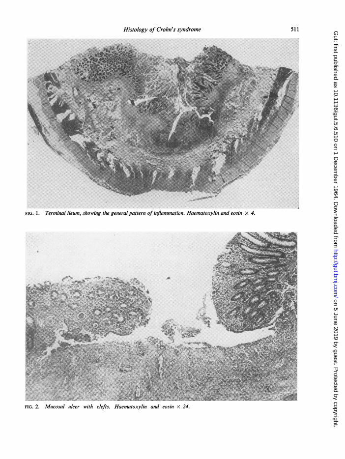
Figure 1 shows the general pattern of the inflam-mation. It is noted that all coats are affected. inparticular the submucosa. The changes are similar inboth the ileum and the colon.
disease. The possibility of different aetiological
TABLE IHISTOLOGICAL DATA
Type of Inflammation
Non-specific aloneDiffuse granulomatousFocal granulomataSchaumann bodies
Intestine LymphGlands
% No. % No.
1317316
100
209
21275010
45
94
The histological features fall into one of threetypes of inflammation, namely, non-specific, diffusegranulomatous, and focal granulomatous (Table I).The non-specific diffuse granulomatous inflam-mations may be grouped as the non-focal granulomaas distinct from focal granuloma.
NON-SPECIFIC INFLAMMATION This is present in everycase and in 13 of the 61 (21 %) it is the only finding.It is most prominent in the mucosa and submucosa,tending to be patchy rather than diffuse and withtwo components, cellular and fluid. Although thecellular exudate in itself is not specific, certainfeatures are of value when the colon is affected indistinguishing it from ulcerative colitis, namely, theinvolvement of all coats, angular fissured ulcers, andprominent lymphoid tissue. The cellular componentconsists mainly of lymphocytes, some plasma cells,and scanty polymorphs. Eosinophils may be presentbut are inconspicuous. Polymorphs are most frequentin areas of mucosal ulceration and at the base ofmucosal clefts. Histiocytes are scanty and giantcells are absent. The fluid component consists oflymph and serum produced from the engorged,distended capillaries and lymphatics, and associatedis considerable activity of, and probably increase in,lymphoid follicles with prominent reticulum centres.These features produce the typical macroscopiccobblestone appearance of the mucosa. Superficial
510
on 5 June 2019 by guest. Protected by copyright.
http://gut.bmj.com
/G
ut: first published as 10.1136/gut.5.6.510 on 1 Decem
ber 1964. Dow
nloaded from
Histology of Crohn's syndrome
.W .._
FIG. 1. Terminal ileum, showing the general pattern of inflammation. Haematoxylin and eosin x 4.
FIG. 2. Mucosal ulcer with clefts. Haematoxylin and eosin x 24.
511
on 5 June 2019 by guest. Protected by copyright.
http://gut.bmj.com
/G
ut: first published as 10.1136/gut.5.6.510 on 1 Decem
ber 1964. Dow
nloaded from
W. Jones Williams
ulceration is common. In 25% of cases clefts orfissures are seen, often in relation to the overhangingedge of an ulcer (Fig. 2), and the clefts oftenterminate in a lymphoid follicle (Fig. 1). Occasionallythey penetrate the muscle layer and even extend intothe serosa, and are thought to be the basis of fistulaformation. The superficial parts of the clefts are oftenlined by ingrowing columnar cells but the deeperparts are lined by inflammatory cells. The base ordeepest angle frequently shows microabscesses.Fibrosis is seldom marked and dense collagen withdisappearance of the cellular infiltration is neverseen. The general thickening is due to the cellularinfiltration and oedema rather than to fibrosis.Lymphatic and vascular block is not seen and thereis no arteritis. There is no evidence of muscle ornerve fibre proliferation or hyperplasia. Ganglioncells are frequently conspicuous in both internaland external plexi but there is no definite increase orabnormality of these cells.
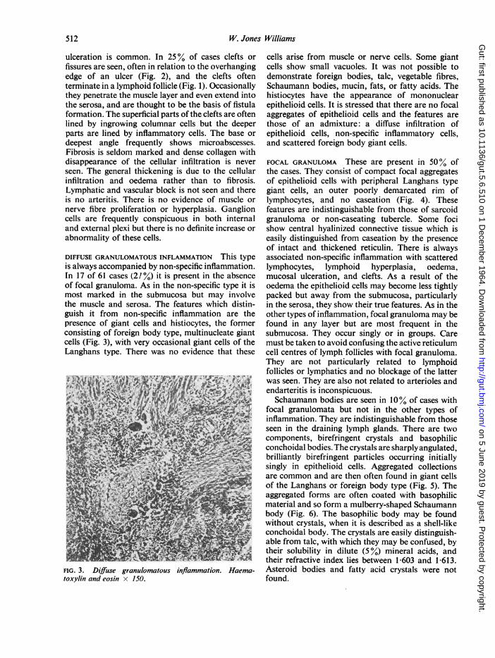
DIFFUSE GRANULOMATOUS INFLAMMATION This typeis always accompanied by non-specific inflammation.In 17 of 61 cases (21%) it is present in the absenceof focal granuloma. As in the non-specific type it ismost marked in the submucosa but may involvethe muscle and serosa. The features which distin-guish it from non-specific inflammation are thepresence of giant cells and histiocytes, the formerconsisting of foreign body type, multinucleate giantcells (Fig. 3), with very occasional giant cells of theLanghans type. There was no evidence that these
FIG. 3. Diffuse granulomatous inflammation. Haema-toxylin and eosin x 150.
cells arise from muscle or nerve cells. Some giantcells show small vacuoles. It was not possible todemonstrate foreign bodies, talc, vegetable fibres,Schaumann bodies, mucin, fats, or fatty acids. Thehistiocytes have the appearance of mononuclearepithelioid cells. It is stressed that there are no focalaggregates of epithelioid cells and the features arethose of an admixture: a diffuse infiltration ofepithelioid cells, non-specific inflammatory cells,and scattered foreign body giant cells.
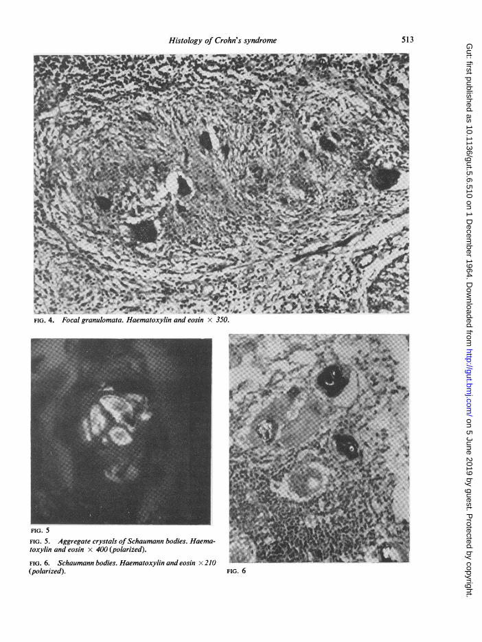
FOCAL GRANULOMA These are present in 50% ofthe cases. They consist of compact focal aggregatesof epithelioid cells with peripheral Langhans typegiant cells, an outer poorly demarcated rim oflymphocytes, and no caseation (Fig. 4). Thesefeatures are indistinguishable from those of sarcoidgranuloma or non-caseating tubercle. Some focishow central hyalinized connective tissue which iseasily distinguished from caseation by the presenceof intact and thickened reticulin. There is alwaysassociated non-specific inflammation with scatteredlymphocytes, lymphoid hyperplasia, oedema,mucosal ulceration, and clefts. As a result of theoedema the epithelioid cells may become less tightlypacked but away from the submucosa, particularlyin the serosa, they show their true features. As in theother types of inflammation, focal granuloma may befound in any layer but are most frequent in thesubmucosa. They occur singly or in groups. Caremust be taken to avoid confusing the active reticulumcell centres of lymph follicles with focal granuloma.They are not particularly related to lymphoidfollicles or lymphatics and no blockage of the latterwas seen. They are also not related to arterioles andendarteritis is inconspicuous.Schaumann bodies are seen in 10% of cases with
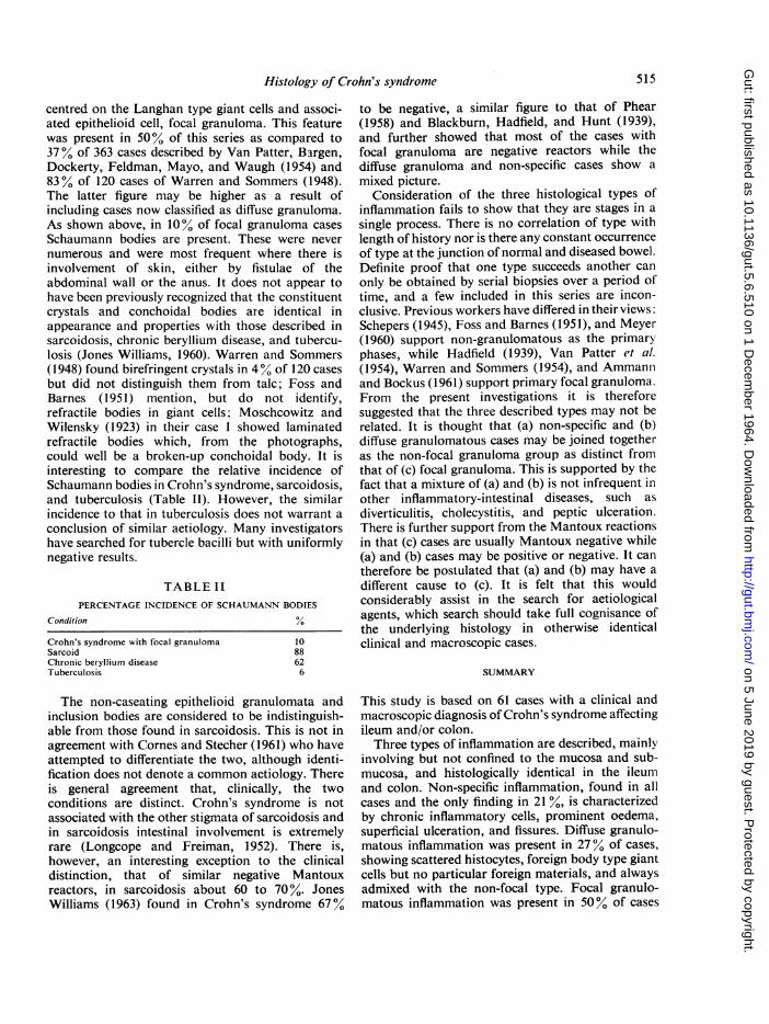
focal granulomata but not in the other types ofinflammation. They are indistinguishable from thoseseen in the draining lymph glands. There are twocomponents, birefringent crystals and basophilicconchoidal bodies. The crystals are sharply angulated,brilliantly birefringent particles occurring initiallysingly in epithelioid cells. Aggregated collectionsare common and are then often found in giant cellsof the Langhans or foreign body type (Fig. 5). Theaggregated forms are often coated with basophilicmaterial and so form a mulberry-shaped Schaumannbody (Fig. 6). The basophilic body may be foundwithout crystals, when it is described as a shell-likeconchoidal body. The crystals are easily distinguish-able from talc, with which they may be confused, bytheir solubility in dilute (5%) mineral acids, andtheir refractive index lies between 1-603 and 1613.Asteroid bodies and fatty acid crystals were notfound.
512
on 5 June 2019 by guest. Protected by copyright.
http://gut.bmj.com
/G
ut: first published as 10.1136/gut.5.6.510 on 1 Decem
ber 1964. Dow
nloaded from
Histology of Crohn's syndrome
FIG. 4. Focal granulomata. Haematoxylin and eosin x 350.
FIG. D
FIG. 5. Aggregate crystals ofSchaumann bodies. Haema-toxylin and eosin x 400 (polarized).FIG. 6. Schaumann bodies. Haematoxylin and eosin x 210(polarized). FIG. 6
513
on 5 June 2019 by guest. Protected by copyright.
http://gut.bmj.com
/G
ut: first published as 10.1136/gut.5.6.510 on 1 Decem
ber 1964. Dow
nloaded from

W. Jones Williams
. . ,.
FIG. 7. Focal granuloma in lymph gland. Haematoxylinand eosin x 140.
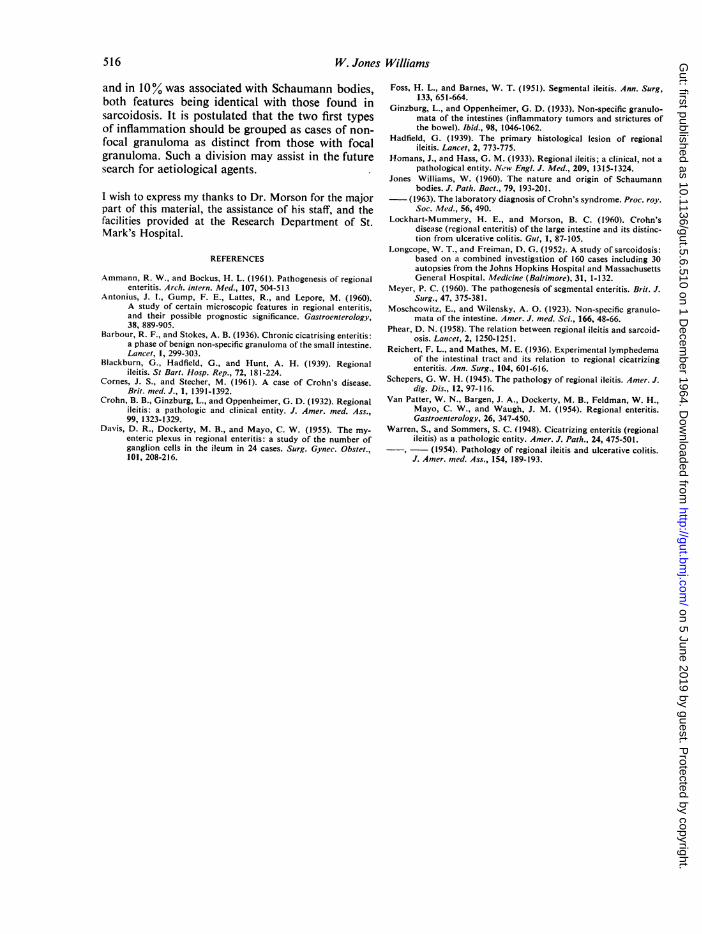
LYMPH GLANDS All the glands examined show non-
specific reactive hyperplasia, enlarged follicles withprominent reticulum centres, littoral cell hyperplasia,and prominent sinusoids distended with faintlyeosinophilic lymph. Diffuse granulomatous inflam-mation is not found. In nine cases (20 %) focalgranulomata are present, indistinguishable fromthose described in the intestines (Fig. 7). These are
never very numerous and are scattered at randomwith no particular relationship to the gland archi-tecture. Of these nine cases with focal granulomata,four show similar Schaumann bodies to thosedescribed above but they are not seen in the absenceof focal granulomata.
DISCUSSION
It is important to realize that this condition is a
syndrome and as such the histological diagnosis ispartly dependent on clinical and macroscopicappearances. It is further noted that the threehistological types are indistinguishable in ileum andcolon.When focal, sarcoid-like, granuloma are present
(50%) the diagnosis is rarely in doubt. Similarly
those cases (27 %) with diffuse granulomatousinflammation present no real difficulty. The mostconfusion is accounted for by the cases with non-specific inflammation (21 %). This is especially sowhen the colon alone is affected, when the conditionhas to be distinguished from segmental ulcerativecolitis. In ulcerative colitis the inflammation isusually superficial, involving only the mucosa,except in acute cases when there is extensive purulentinflammation and necrosis (Lockhart-Mummery andMorson, 1960). Ulcers and clefts are common andthe latter can be distinguished from diverticula bytheir superficial situation, their relationship to ulcers,and part mucosal lining. Hadfield (1939) consideredulceration to be secondary to underlying lymphoidhyperplasia but this could not be confirmed. Theonly possible association is that the clefts oftenterminate in lymph follicles. The presence of theseulcers and clefts, together with the unusual combin-ation of lymphocytic proliferation and oedema withrelative paucity of polymorphs, is of considerablediagnostic value, and, when combined with the grossappearances, then the diagnosis is certain. Theclefts may be the result of traumatic tearing of theoedematous, cellular, bulging mucosa, but this isunlikely, as they are not more common in thecolon than in the ileum. They may result fromuncoordinated muscle contractions or spasm.Meyer (1960) described focal hypertrophy of themuscularis mucosa, but this feature was not seen.Barbour and Stokes (1936) and Davis, Dockerty,and Mayo (1955) incriminate involvement andincrease of nerve cells, and Antonius, Gump, Lattes,and Lepore (1960) described 'neuromatous' lesions;these findings could not be confirmed. However,the absence of anatomical changes does not disprovea functional disturbance.
Considerable interest has been centred on the twotypes of giant cells, foreign body cells and Langhans.To explain the foreign body type, various exogenousparticles have been considered, e.g., food particles(Crohn, Ginzburg, and Oppenheimer, 1932; Ginz-burg and Oppenheimer, 1933) and talc (Reichertand Mathes, 1936) but these could not be demon-strated in the present series. In those patients withprevious operations glove powder was looked forbut was not found. Homans and Hass (1933) andWarren and Sommers (1954) suggested that theywere a reaction to lipids and fats, but this could notbe confirmed. Displaced mucins free in the tissueswere sought for, as the author has seen a similarforeign body type granulomatous reaction indiverticulitis and cholecystitis but again the resultswere negative. It is, however, possible that thereaction could occur to altered mucin which has lostits staining properties. Even more interest has been
514
on 5 June 2019 by guest. Protected by copyright.
http://gut.bmj.com
/G
ut: first published as 10.1136/gut.5.6.510 on 1 Decem
ber 1964. Dow
nloaded from
Histology of Crohn's syndrome
centred on the Langhan type giant cells and associ-ated epithelioid cell, focal granuloma. This featurewas present in 500% of this series as compared to37% of 363 cases described by Van Patter, Bargen,Dockerty, Feldman, Mayo, and Waugh (1954) and83% of 120 cases of Warren and Sommers (1948).The latter figure may be higher as a result ofincluding cases now classified as diffuse granuloma.As shown above, in 10% of focal granuloma casesSchaumann bodies are present. These were nevernumerous and were most frequent where there isinvolvement of skin, either by fistulae of theabdominal wall or the anus. It does not appear tohave been previously recognized that the constituentcrystals and conchoidal bodies are identical inappearance and properties with those described insarcoidosis, chronic beryllium disease, and tubercu-losis (Jones Williams, 1960). Warren and Sommers(1948) found birefringent crystals in 40% of 120 casesbut did not distinguish them from talc; Foss andBarnes (1951) mention, but do not identify,refractile bodies in giant cells; Moschcowitz andWilensky (1923) in their case 1 showed laminatedrefractile bodies which, from the photographs,could well be a broken-up conchoidal body. It isinteresting to compare the relative incidence ofSchaumann bodies in Crohn's syndrome, sarcoidosis,and tuberculosis (Table II). However, the similarincidence to that in tuberculosis does not warrant aconclusion of similar aetiology. Many investigatorshave searched for tubercle bacilli but with uniformlynegative results.
TABLE IIPERCENTAGE INCIDENCE OF SCHAUMANN BODIES
Condition %
Crohn's syndrome with focal granulomaSarcoidChronic beryllium diseaseTuberculosis
1088626
The non-caseating epithelioid granulomata andinclusion bodies are considered to be indistinguish-able from those found in sarcoidosis. This is not inagreement with Cornes and Stecher (1961) who haveattempted to differentiate the two, although identi-fication does not denote a common aetiology. Thereis general agreement that, clinically, the twoconditions are distinct. Crohn's syndrome is notassociated with the other stigmata of sarcoidosis andin sarcoidosis intestinal involvement is extremelyrare (Longcope and Freiman, 1952). There is,however, an interesting exception to the clinicaldistinction, that of similar negative Mantouxreactors, in sarcoidosis about 60 to 70%. JonesWilliams (1963) found in Crohn's syndrome 67%
to be negative, a similar figure to that of Phear(1958) and Blackburn, Hadfield, and Hunt (1939),and further showed that most of the cases withfocal granuloma are negative reactors while thediffuse granuloma and non-specific cases show amixed picture.
Consideration of the three histological types ofinflammation fails to show that they are stages in asingle process. There is no correlation of type withlength of history nor is there any constant occurrenceof type at the junction of normal and diseased bowel.Definite proof that one type succeeds another canonly be obtained by serial biopsies over a period oftime, and a few included in this series are incon-clusive. Previous workers have differed in their views,Schepers (1945), Foss and Barnes (1951), and Meyer(1960) support non-granulomatous as the primaryphases, while Hadfield (1939), Van Patter et al.(1954), Warren and Sommers (1954), and Ammannand Bockus (1961) support primary focal granuloma.From the present investigations it is thereforesuggested that the three described types may not berelated. It is thought that (a) non-specific and (b)diffuse granulomatous cases may be joined togetheras the non-focal granuloma group as distinct fromthat of (c) focal granuloma. This is supported by thefact that a mixture of (a) and (b) is not infrequent inother inflammatory-intestinal diseases, such asdiverticulitis, cholecystitis, and peptic ulceration.There is further support from the Mantoux reactionsin that (c) cases are usually Mantoux negative while(a) and (b) cases may be positive or negative. It cantherefore be postulated that (a) and (b) may have adifferent cause to (c). It is felt that this wouldconsiderably assist in the search for aetiologicalagents, which search should take full cognisance ofthe underlying histology in otherwise identicalclinical and macroscopic cases.
SUMMARY
This study is based on 61 cases with a clinical andmacroscopic diagnosis of Crohn's syndrome affectingileum and/or colon.Three types of inflammation are described, mainly
involving but not confined to the mucosa and sub-mucosa, and histologically identical in the ileumand colon. Non-specific inflammation, found in allcases and the only finding in 21%, is characterizedby chronic inflammatory cells, prominent oedema,superficial ulceration, and fissures. Diffuse granulo-matous inflammation was present in 27% of cases,showing scattered histocytes, foreign body type giantcells but no particular foreign materials, and alwaysadmixed with the non-focal type. Focal granulo-matous inflammation was present in 500% of cases
515
on 5 June 2019 by guest. Protected by copyright.
http://gut.bmj.com
/G
ut: first published as 10.1136/gut.5.6.510 on 1 Decem
ber 1964. Dow
nloaded from
516 W. Jones Williams
and in 10% was associated with Schaumann bodies,both features being identical with those found insarcoidosis. It is postulated that the two first typesof inflammation should be grouped as cases of non-focal granuloma as distinct from those with focalgranuloma. Such a division may assist in the futuresearch for aetiological agents.
I wish to express my thanks to Dr. Morson for the majorpart of this material, the assistance of his staff, and thefacilities provided at the Research Department of St.Mark's Hospital.
REFERENCES
Ammann, R. W., and Bockus, H. L. (1961). Pathogenesis of regionalenteritis. Arch. intern. Med., 107, 504-513
Antonius, J. 1., Gump, F. E., Lattes, R., and Lepore, M. (1960).A study of certain microscopic features in regional enteritis,and their possible prognostic significance. Gastroenterology,38, 889-905.
Barbour, R. F., and Stokes, A. B. (1936). Chronic cicatrising enteritis:a phase of benign non-specific granuloma of the small intestine.Lancet, 1, 299-303.
Blackburn, G., Hadfield, G., and Hunt, A. H. (1939). Regionalileitis. St Bart. Hosp. Rep., 72, 181-224.
Cornes, J. S., and Stecher, M. (1961). A case of Crohn's disease.Brit. med. J., 1, 1391-1392.
Crohn, B. B., Ginzburg, L., and Oppenheimer, G. D. (1932). Regionalileitis: a pathologic and clinical entity. J. Amer. med. Ass.,99, 1323-1329.
Davis, D. R., Dockerty, M. B., and Mayo, C. W. (1955). The my-enteric plexus in regional enteritis: a study of the number ofganglion cells in the i!eum in 24 cases. Surg. Gynec. Obstet.,101, 208-216.
Foss, H. L., and Barnes, W. T. (1951). Segmental ileitis. Ann. Surg,133, 651-664.
Ginzburg, L., and Oppenheimer, G. D. (1933). Non-specific granulo-mata of the intestines (inflammatory tumors and strictures ofthe bowel). Ibid., 98, 1046-1062.
Hadfield, G. (1939). The primary histological lesion of regionalileitis. Lancet, 2, 773-775.
Homans, J., and Hass, G. M. (1933). Regional ileitis; a clinical, not apathological entity. New Engl. J. Med., 209, 1315-1324.
Jones Williams, W. (1960). The nature and origin of Schaumannbodies. J. Path. Bact., 79, 193-201.
(1963). The laboratory diagnosis of Crohn's syndrome. Proc. roy.Soc. Med., 56, 490.
Lockhart-Mummery, H. E., and Morson, B. C. (1960). Crohn'sdisease (regional enteritis) of the large intestine and its distinc-tion from ulcerative colitis. Gut, 1, 87-105.
Longcope, W. T., and Freiman, D. 0. (1952). A study of sarcoidosis:based on a combined investigation of 160 cases including 30autopsies from the Johns Hopkins Hospital and MassachusettsGeneral Hospital. Medicine (Baltimore), 31, 1-132.
Meyer, P. C. (1960). The pathogenesis of segmental enteritis. Brit. J.Surg., 47, 375-381.
Moschcowitz, E., and Wilensky, A. 0. (1923). Non-specific granulo-mata of the intestine. Amer. J. med. Sci., 166, 48-66.
Phear, D. N. (1958). The relation between regional ileitis and sarcoid-osis. Lancet, 2, 1250-1251.
Reichert, F. L., and Mathes, M. E. (1936). Experimental lymphedemaof the intestinal tract and its relation to regional cicatrizingenteritis. Ann. Surg., 104, 601-616.
Schepers, G. W. H. (1945). The pathology of regional ileitis. Amer. J.dig. Dis., 12, 97-116.
Van Patter, W. N., Bargen, J. A., Dockerty, M. B., Feldman, W. H.,Mayo, C. W., and Waugh, J. M. (1954). Regional enteritis.Gastroenterology, 26, 347-450.
Warren, S., and Sommers, S. C. (1948). Cicatrizing enteritis (regionalileitis) as a pathologic entity. Amer. J. Path., 24, 475-501.
-, (1954). Pathology of regional ileitis and ulcerative colitis.J. Amer. med. Ass., 154, 189-193.
on 5 June 2019 by guest. Protected by copyright.
http://gut.bmj.com
/G
ut: first published as 10.1136/gut.5.6.510 on 1 Decem
ber 1964. Dow
nloaded from


![Histology Slides - mediconotes.commediconotes.com/freenotes/basic/histology_laboratory_slides.pdf[Histology] Histology Slides MedicoNotes provides real laboratory Histological slides](https://img.pdfslide.us/doc/110x75/5ae110e87f8b9a5a668e6aa3/histology-slides-histology-histology-slides-mediconotes-provides-real-laboratory.jpg)