Embed Size (px)
Citation preview

HISTOLOGY OF ACCESSORY
ORGANS OF GIT

SALIVARY GLANDS
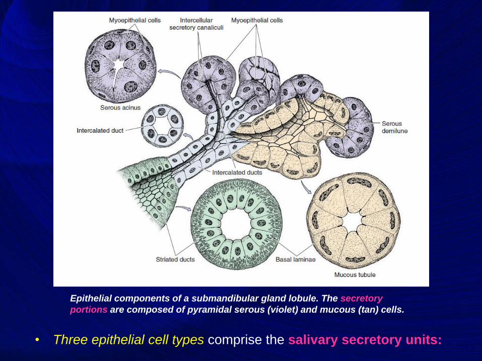
• Three epithelial cell types comprise the salivary secretory units:
Epithelial components of a submandibular gland lobule. The secretory
portions are composed of pyramidal serous (violet) and mucous (tan) cells.
4
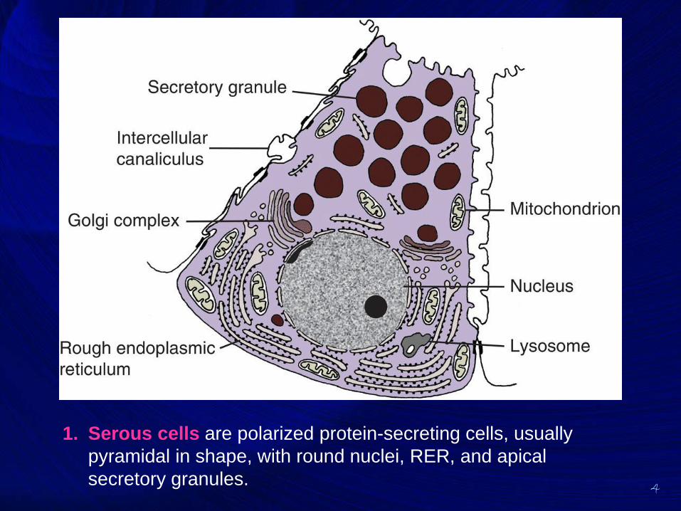
1. Serous cells are polarized protein-secreting cells, usually
pyramidal in shape, with round nuclei, RER, and apical
secretory granules.
5
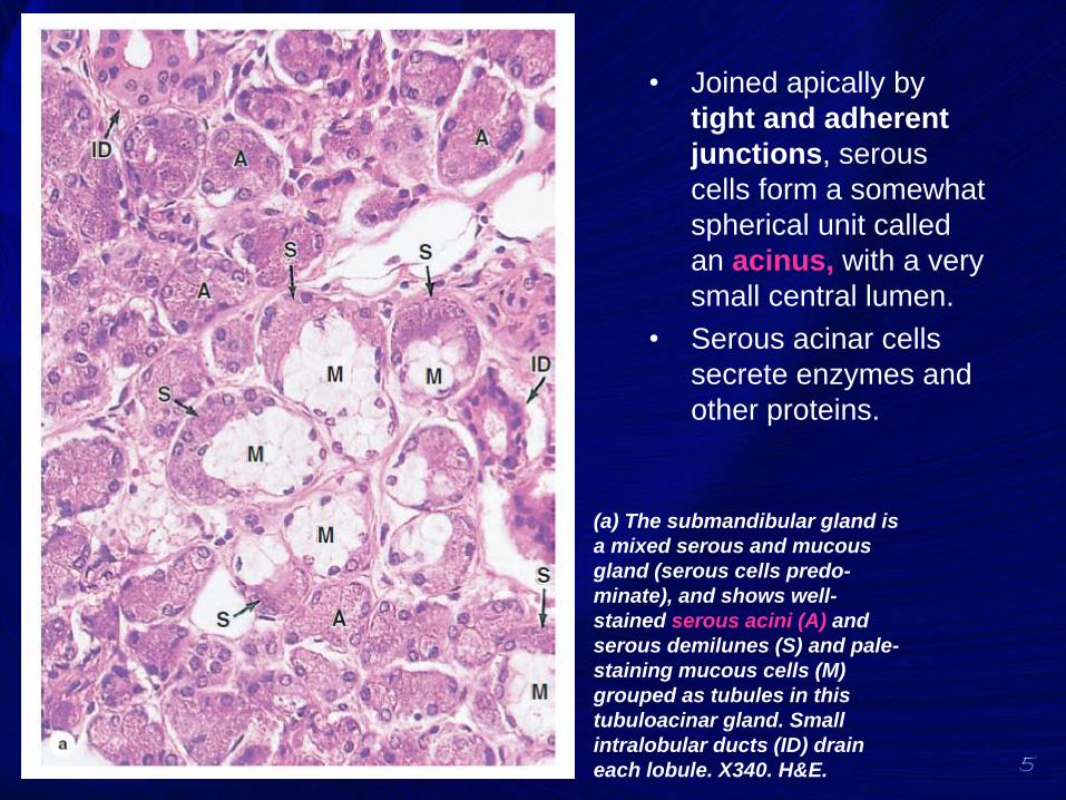
• Joined apically by
tight and adherent
junctions, serous
cells form a somewhat
spherical unit called
an acinus, with a very
small central lumen.
• Serous acinar cells
secrete enzymes and
other proteins.
(a) The submandibular gland is
a mixed serous and mucous
gland (serous cells predo-
minate), and shows well-
stained serous acini (A) and
serous demilunes (S) and pale-
staining mucous cells (M)
grouped as tubules in this
tubuloacinar gland. Small
intralobular ducts (ID) drain
each lobule. X340. H&E.
6
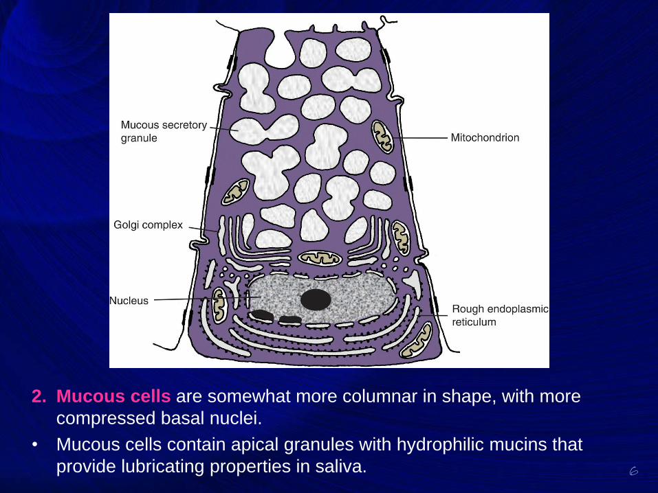
2. Mucous cells are somewhat more columnar in shape, with more
compressed basal nuclei.
• Mucous cells contain apical granules with hydrophilic mucins that
provide lubricating properties in saliva.
7
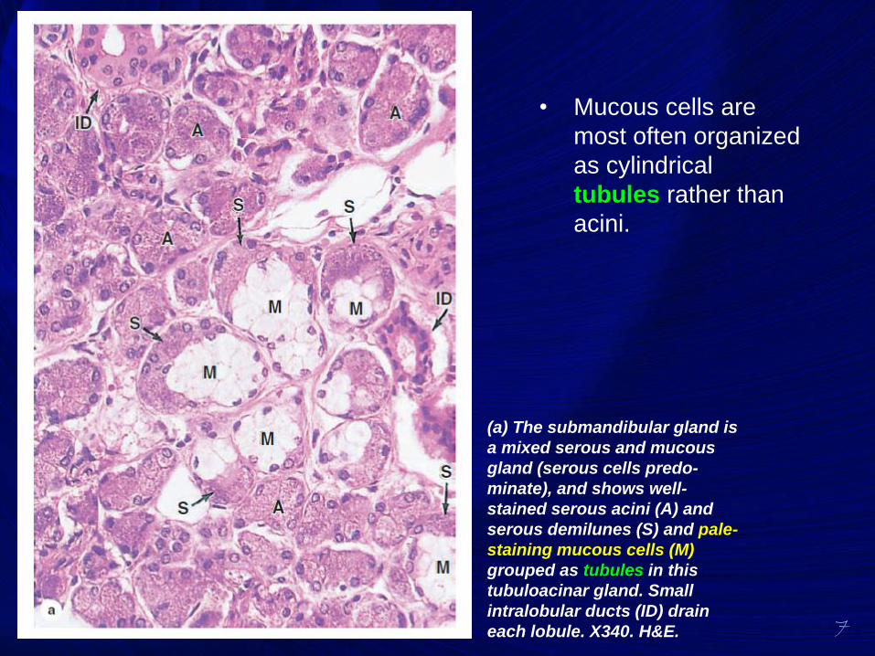
• Mucous cells are
most often organized
as cylindrical
tubules rather than
acini.
(a) The submandibular gland is
a mixed serous and mucous
gland (serous cells predo-
minate), and shows well-
stained serous acini (A) and
serous demilunes (S) and pale-
staining mucous cells (M)
grouped as tubules in this
tubuloacinar gland. Small
intralobular ducts (ID) drain
each lobule. X340. H&E.
8
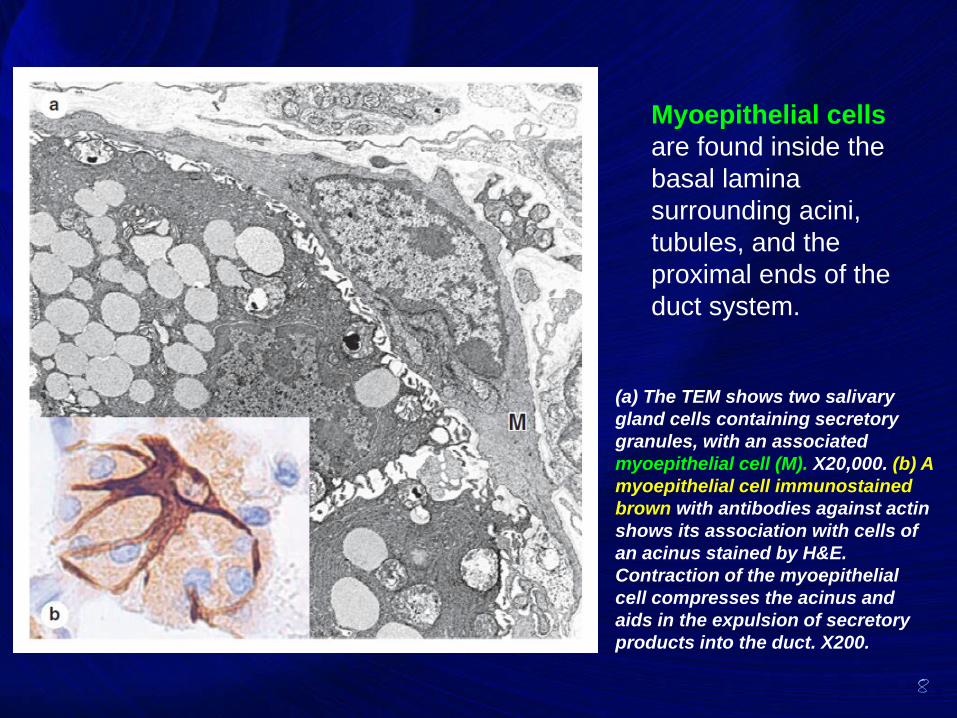
Myoepithelial cells
are found inside the
basal lamina
surrounding acini,
tubules, and the
proximal ends of the
duct system.
(a) The TEM shows two salivary
gland cells containing secretory
granules, with an associated
myoepithelial cell (M). X20,000. (b) A
myoepithelial cell immunostained
brown with antibodies against actin
shows its association with cells of
an acinus stained by H&E.
Contraction of the myoepithelial
cell compresses the acinus and
aids in the expulsion of secretory
products into the duct. X200.
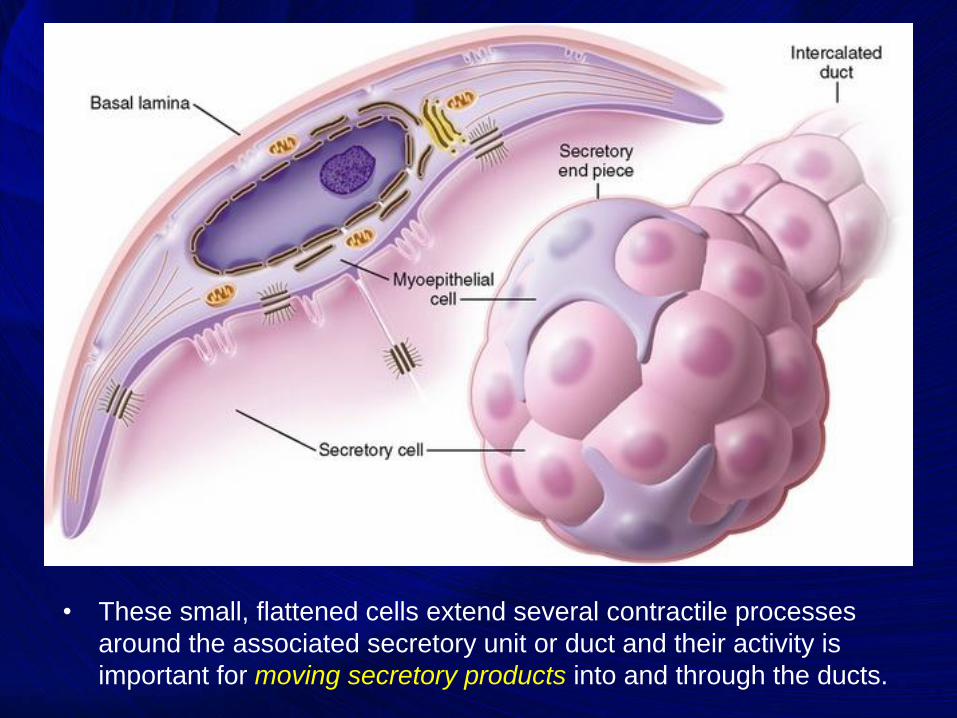
• These small, flattened cells extend several contractile processes
around the associated secretory unit or duct and their activity is
important for moving secretory products into and through the ducts.
10
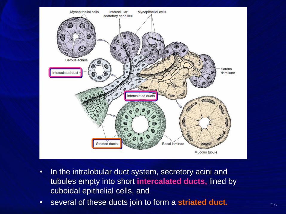
• In the intralobular duct system, secretory acini and
tubules empty into short intercalated ducts, lined by
cuboidal epithelial cells, and
• several of these ducts join to form a striated duct.
11
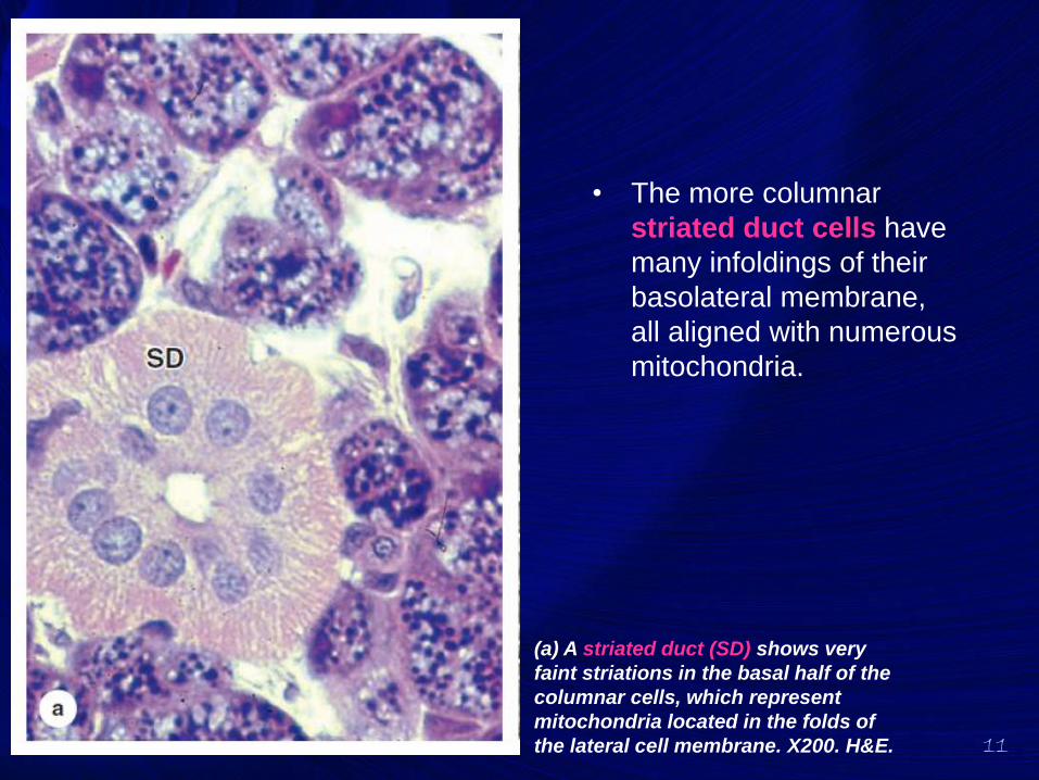
• The more columnar
striated duct cells have
many infoldings of their
basolateral membrane,
all aligned with numerous
mitochondria.
(a) A striated duct (SD) shows very
faint striations in the basal half of the
columnar cells, which represent
mitochondria located in the folds of
the lateral cell membrane. X200. H&E.
12
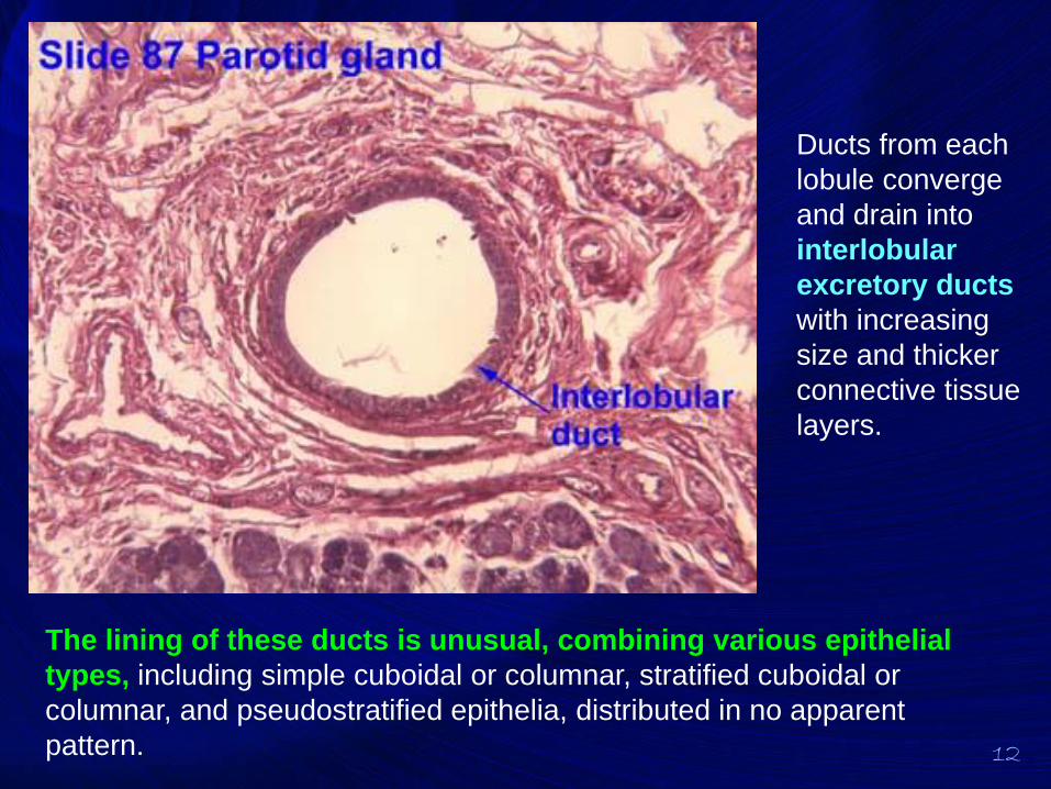
Ducts from each
lobule converge
and drain into
interlobular
excretory ducts
with increasing
size and thicker
connective tissue
layers.
The lining of these ducts is unusual, combining various epithelial
types, including simple cuboidal or columnar, stratified cuboidal or
columnar, and pseudostratified epithelia, distributed in no apparent
pattern.
13
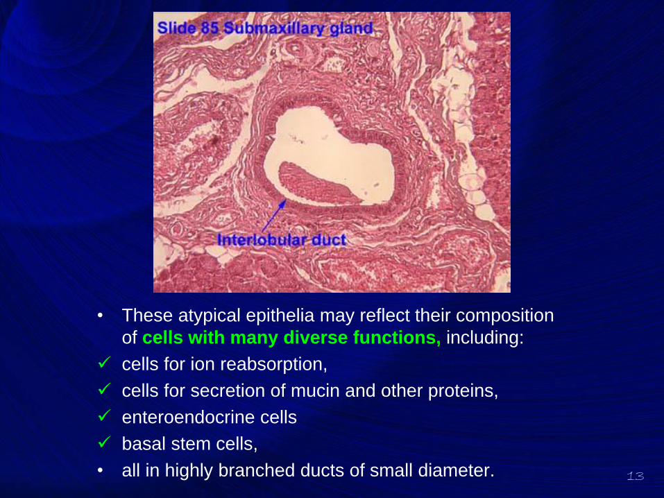
• These atypical epithelia may reflect their composition
of cells with many diverse functions, including:
✓ cells for ion reabsorption,
✓ cells for secretion of mucin and other proteins,
✓ enteroendocrine cells
✓ basal stem cells,
• all in highly branched ducts of small diameter.
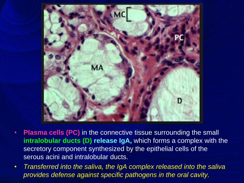
• Plasma cells (PC) in the connective tissue surrounding the small
intralobular ducts (D) release IgA, which forms a complex with the
secretory component synthesized by the epithelial cells of the
serous acini and intralobular ducts.
• Transferred into the saliva, the IgA complex released into the saliva
provides defense against specific pathogens in the oral cavity.
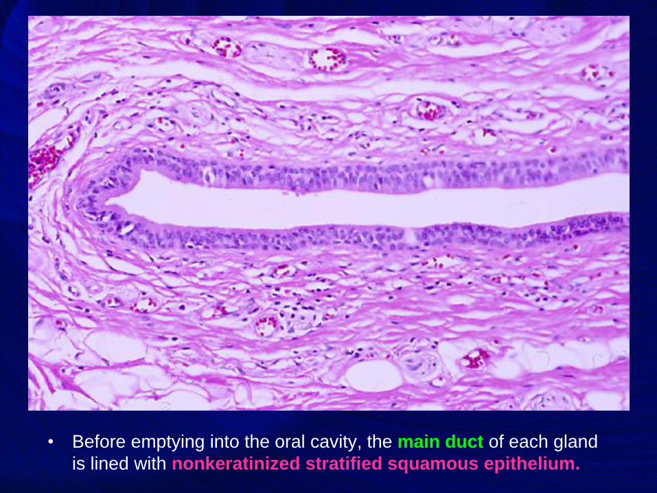
• Before emptying into the oral cavity, the main duct of each gland
is lined with nonkeratinized stratified squamous epithelium.
16
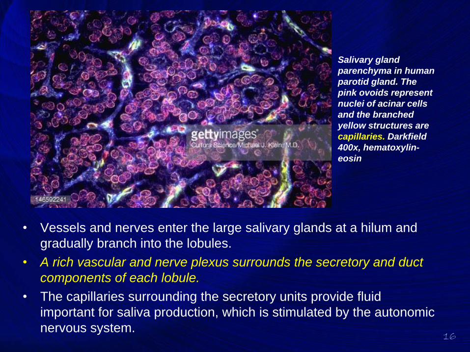
• Vessels and nerves enter the large salivary glands at a hilum and
gradually branch into the lobules.
• A rich vascular and nerve plexus surrounds the secretory and duct
components of each lobule.
• The capillaries surrounding the secretory units provide fluid
important for saliva production, which is stimulated by the autonomic
nervous system.
Salivary gland
parenchyma in human
parotid gland. The
pink ovoids represent
nuclei of acinar cells
and the branched
yellow structures are
capillaries. Darkfield
400x, hematoxylin-
eosin
Parotid Glands
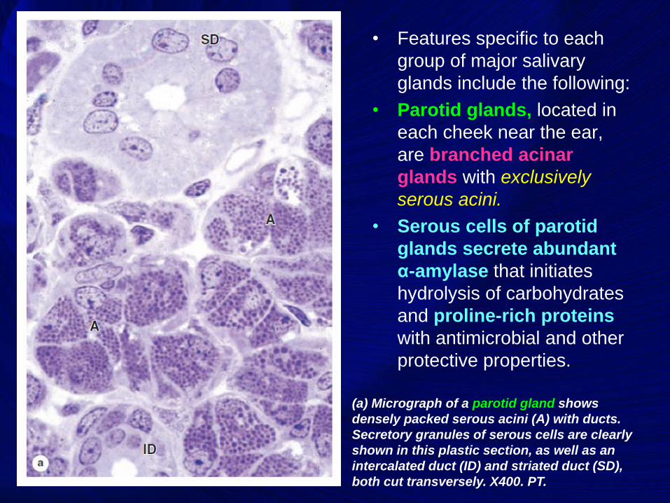
• Features specific to each
group of major salivary
glands include the following:
• Parotid glands, located in
each cheek near the ear,
are branched acinar
glands with exclusively
serous acini.
• Serous cells of parotid
glands secrete abundant
α-amylase that initiates
hydrolysis of carbohydrates
and proline-rich proteins
with antimicrobial and other
protective properties.
(a) Micrograph of a parotid gland shows
densely packed serous acini (A) with ducts.
Secretory granules of serous cells are clearly
shown in this plastic section, as well as an
intercalated duct (ID) and striated duct (SD),
both cut transversely. X400. PT.

Submandibular Glands
20
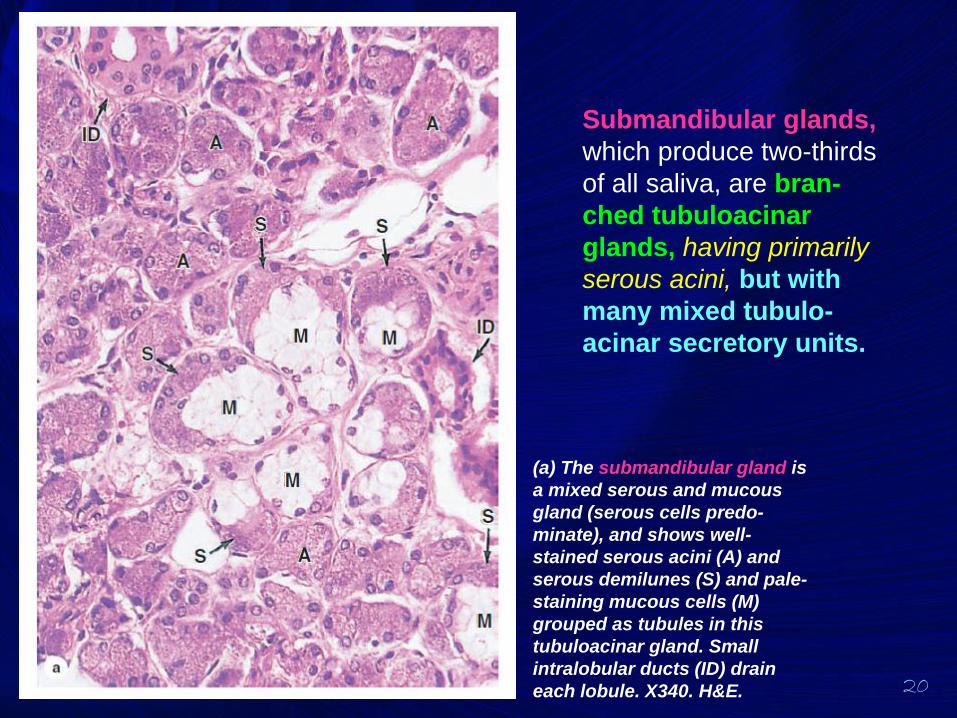
Submandibular glands,
which produce two-thirds
of all saliva, are bran-
ched tubuloacinar
glands, having primarily
serous acini, but with
many mixed tubulo-
acinar secretory units.
(a) The submandibular gland is
a mixed serous and mucous
gland (serous cells predo-
minate), and shows well-
stained serous acini (A) and
serous demilunes (S) and pale-
staining mucous cells (M)
grouped as tubules in this
tubuloacinar gland. Small
intralobular ducts (ID) drain
each lobule. X340. H&E.
21
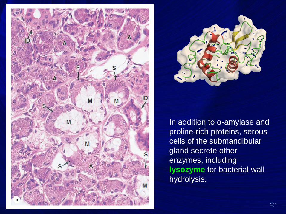
In addition to α-amylase and
proline-rich proteins, serous
cells of the submandibular
gland secrete other
enzymes, including
lysozyme for bacterial wall
hydrolysis.

Sublingual Glands
23
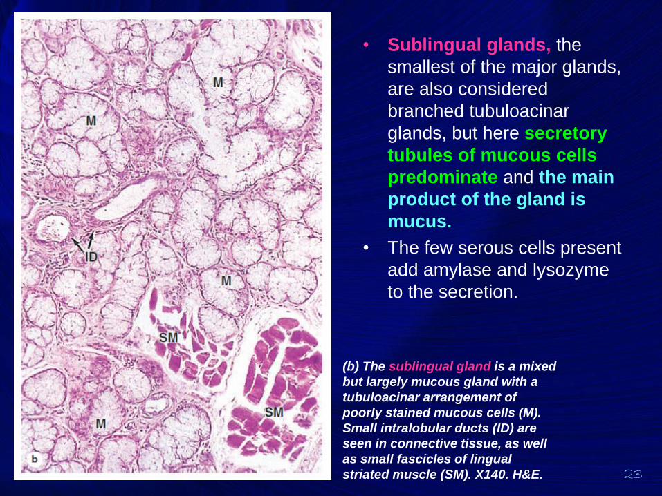
• Sublingual glands, the
smallest of the major glands,
are also considered
branched tubuloacinar
glands, but here secretory
tubules of mucous cells
predominate and the main
product of the gland is
mucus.
• The few serous cells present
add amylase and lysozyme
to the secretion.
(b) The sublingual gland is a mixed
but largely mucous gland with a
tubuloacinar arrangement of
poorly stained mucous cells (M).
Small intralobular ducts (ID) are
seen in connective tissue, as well
as small fascicles of lingual
striated muscle (SM). X140. H&E.

Nonencapsulated Salivary
Glands
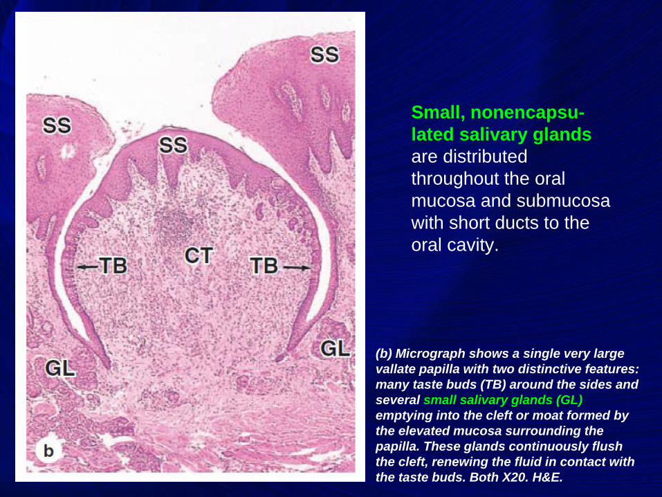
Small, nonencapsu-
lated salivary glands
are distributed
throughout the oral
mucosa and submucosa
with short ducts to the
oral cavity.
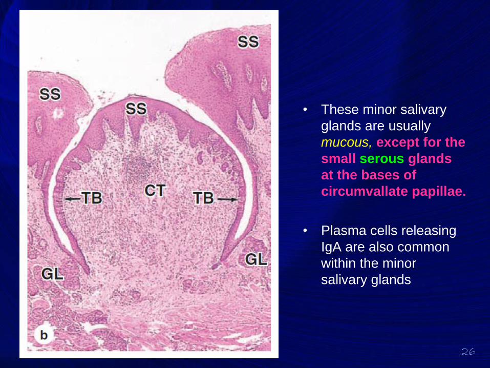
(b) Micrograph shows a single very large
vallate papilla with two distinctive features:
many taste buds (TB) around the sides and
several small salivary glands (GL)
emptying into the cleft or moat formed by
the elevated mucosa surrounding the
papilla. These glands continuously flush
the cleft, renewing the fluid in contact with
the taste buds. Both X20. H&E.
26
• These minor salivary
glands are usually
mucous, except for the
small serous glands
at the bases of
circumvallate papillae.
• Plasma cells releasing
IgA are also common
within the minor
salivary glands
Pancreas
28
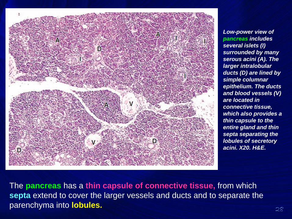
The pancreas has a thin capsule of connective tissue, from which
septa extend to cover the larger vessels and ducts and to separate the
parenchyma into lobules.
Low-power view of
pancreas includes
several islets (I)
surrounded by many
serous acini (A). The
larger intralobular
ducts (D) are lined by
simple columnar
epithelium. The ducts
and blood vessels (V)
are located in
connective tissue,
which also provides a
thin capsule to the
entire gland and thin
septa separating the
lobules of secretory
acini. X20. H&E.
29
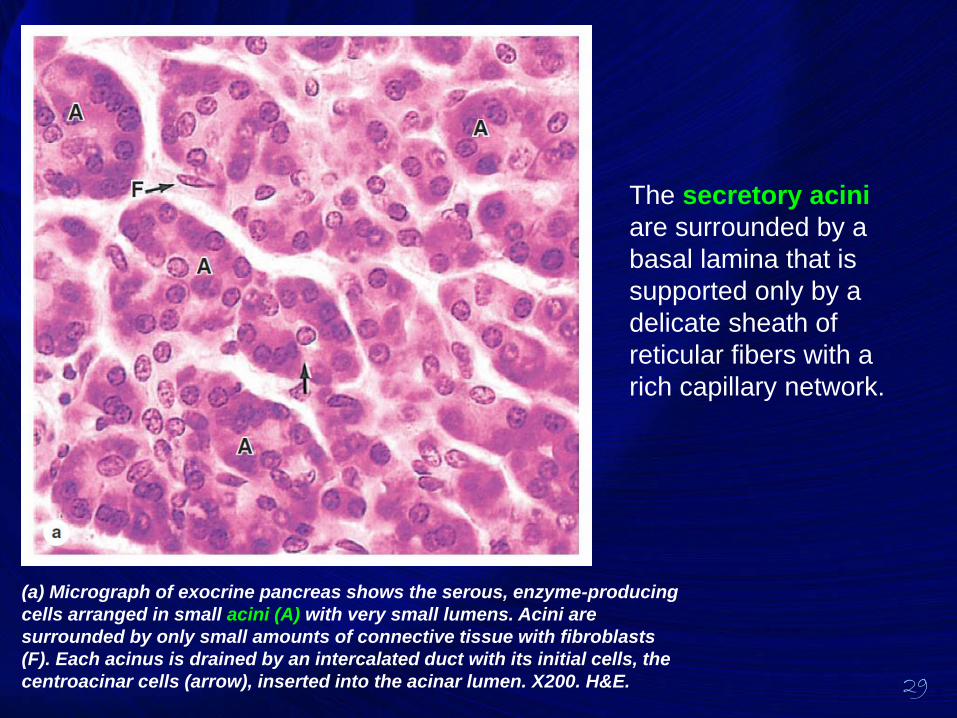
The secretory acini
are surrounded by a
basal lamina that is
supported only by a
delicate sheath of
reticular fibers with a
rich capillary network.
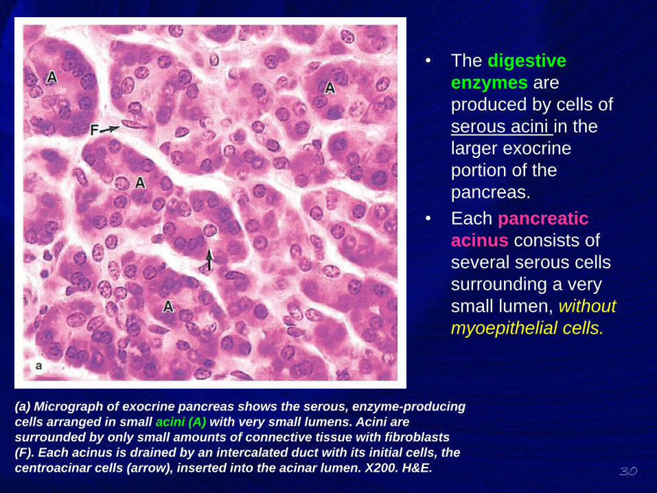
(a) Micrograph of exocrine pancreas shows the serous, enzyme-producing
cells arranged in small acini (A) with very small lumens. Acini are
surrounded by only small amounts of connective tissue with fibroblasts
(F). Each acinus is drained by an intercalated duct with its initial cells, the
centroacinar cells (arrow), inserted into the acinar lumen. X200. H&E.
30
• The digestive
enzymes are
produced by cells of
serous acini in the
larger exocrine
portion of the
pancreas.
• Each pancreatic
acinus consists of
several serous cells
surrounding a very
small lumen, without
myoepithelial cells.
(a) Micrograph of exocrine pancreas shows the serous, enzyme-producing
cells arranged in small acini (A) with very small lumens. Acini are
surrounded by only small amounts of connective tissue with fibroblasts
(F). Each acinus is drained by an intercalated duct with its initial cells, the
centroacinar cells (arrow), inserted into the acinar lumen. X200. H&E.
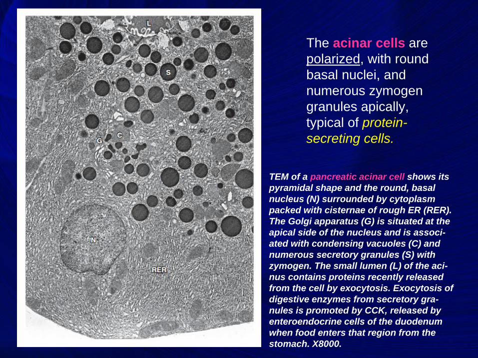
The acinar cells are
polarized, with round
basal nuclei, and
numerous zymogen
granules apically,
typical of protein-
secreting cells.
TEM of a pancreatic acinar cell shows its
pyramidal shape and the round, basal
nucleus (N) surrounded by cytoplasm
packed with cisternae of rough ER (RER).
The Golgi apparatus (G) is situated at the
apical side of the nucleus and is associ-
ated with condensing vacuoles (C) and
numerous secretory granules (S) with
zymogen. The small lumen (L) of the aci-
nus contains proteins recently released
from the cell by exocytosis. Exocytosis of
digestive enzymes from secretory gra-
nules is promoted by CCK, released by
enteroendocrine cells of the duodenum
when food enters that region from the
stomach. X8000.
32
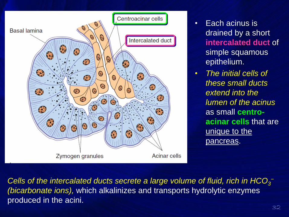
• Each acinus is
drained by a short
intercalated duct of
simple squamous
epithelium.
• The initial cells of
these small ducts
extend into the
lumen of the acinus
as small centro-
acinar cells that are
unique to the
pancreas.
Cells of the intercalated ducts secrete a large volume of fluid, rich in HCO3−
(bicarbonate ions), which alkalinizes and transports hydrolytic enzymes
produced in the acini.
33
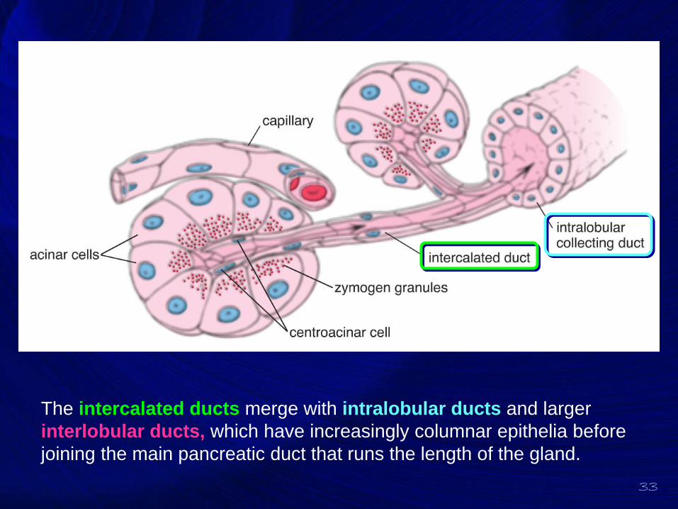
The intercalated ducts merge with intralobular ducts and larger
interlobular ducts, which have increasingly columnar epithelia before
joining the main pancreatic duct that runs the length of the gland.
LIVER

Hepatocytes &
Hepatic Lobules
36
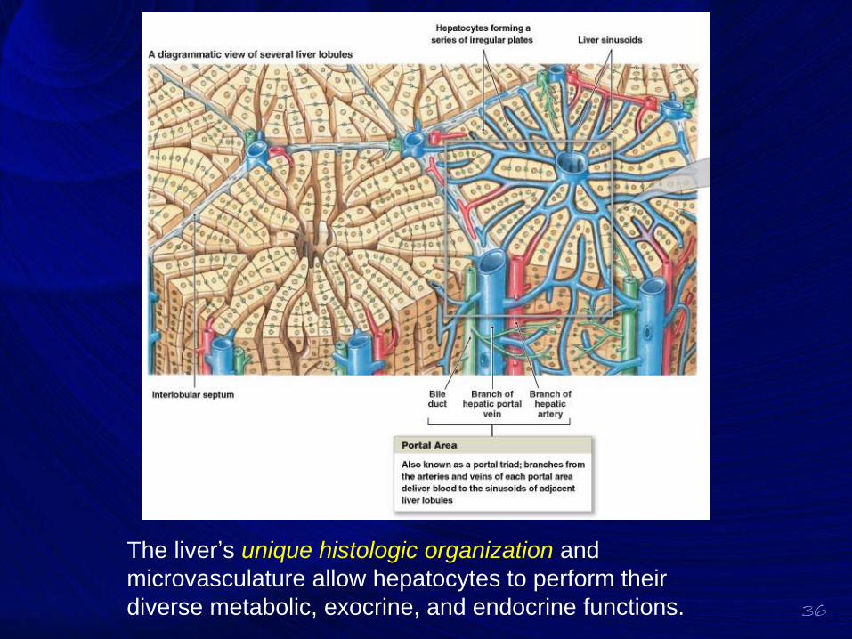
The liver’s unique histologic organization and
microvasculature allow hepatocytes to perform their
diverse metabolic, exocrine, and endocrine functions.
37
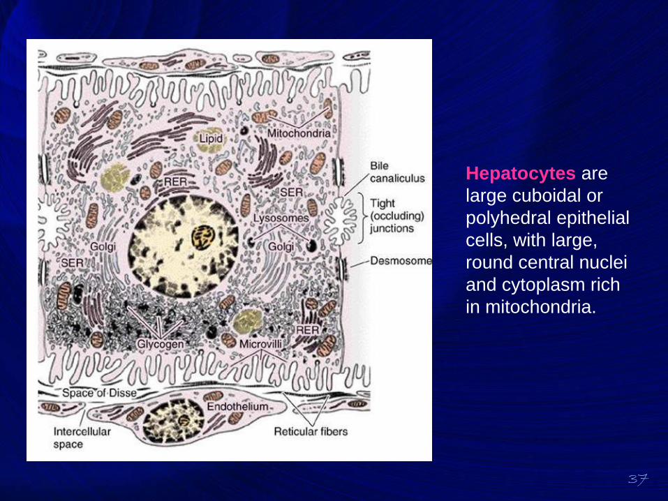
Hepatocytes are
large cuboidal or
polyhedral epithelial
cells, with large,
round central nuclei
and cytoplasm rich
in mitochondria.
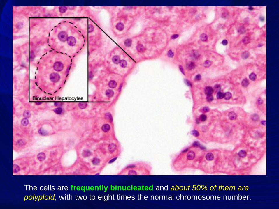
The cells are frequently binucleated and about 50% of them are
polyploid, with two to eight times the normal chromosome number.
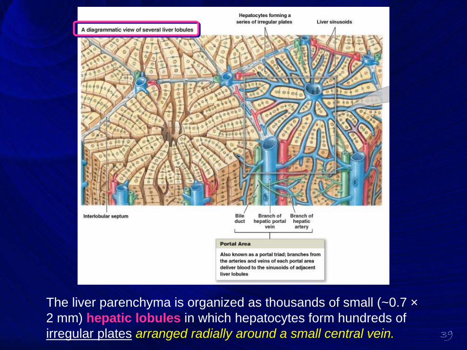
39
The liver parenchyma is organized as thousands of small (~0.7 ×
2 mm) hepatic lobules in which hepatocytes form hundreds of
irregular plates arranged radially around a small central vein.
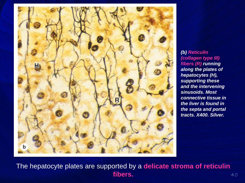
40
The hepatocyte plates are supported by a delicate stroma of reticulin
fibers.
(b) Reticulin
(collagen type III)
fibers (R) running
along the plates of
hepatocytes (H),
supporting these
and the intervening
sinusoids. Most
connective tissue in
the liver is found in
the septa and portal
tracts. X400. Silver.
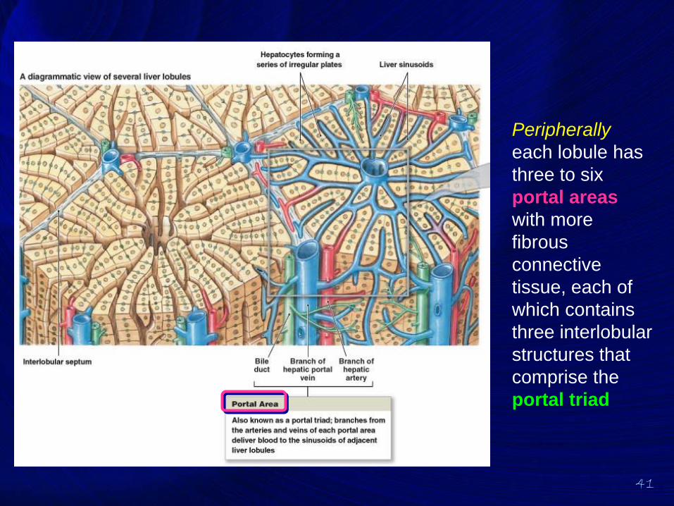
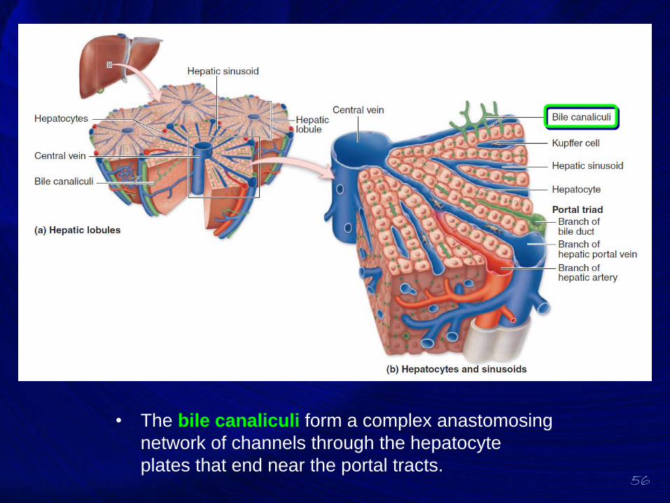
41
Peripherally
each lobule has
three to six
portal areas
with more
fibrous
connective
tissue, each of
which contains
three interlobular
structures that
comprise the
portal triad
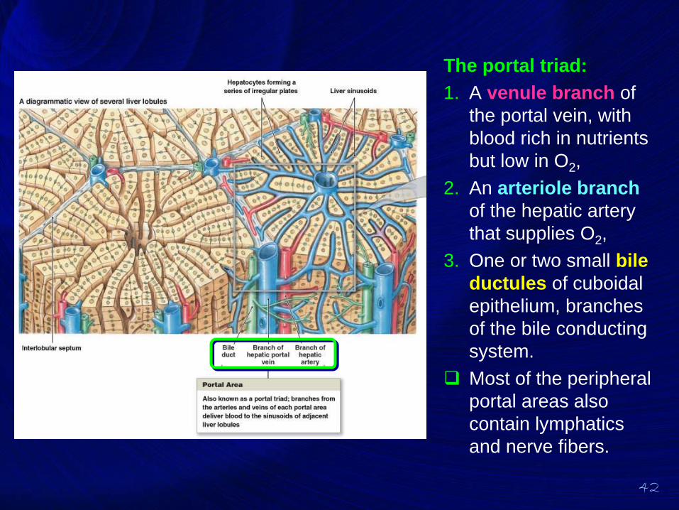
42
The portal triad:
1. A venule branch of
the portal vein, with
blood rich in nutrients
but low in O2,
2. An arteriole branch
of the hepatic artery
that supplies O2,
3. One or two small bile
ductules of cuboidal
epithelium, branches
of the bile conducting
system.
❑ Most of the peripheral
portal areas also
contain lymphatics
and nerve fibers.
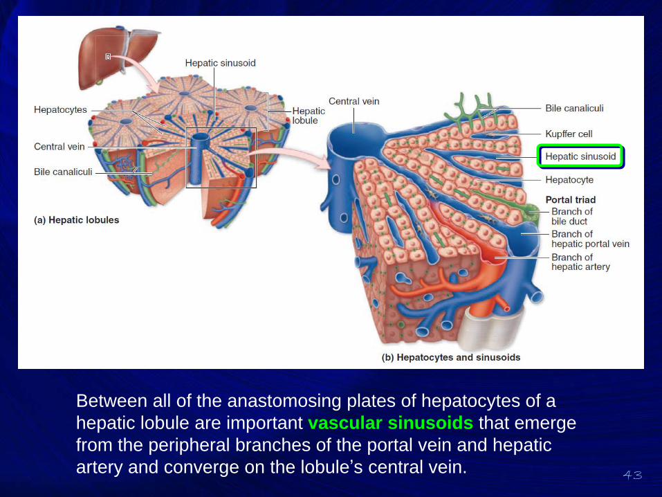
43
Between all of the anastomosing plates of hepatocytes of a
hepatic lobule are important vascular sinusoids that emerge
from the peripheral branches of the portal vein and hepatic
artery and converge on the lobule’s central vein.
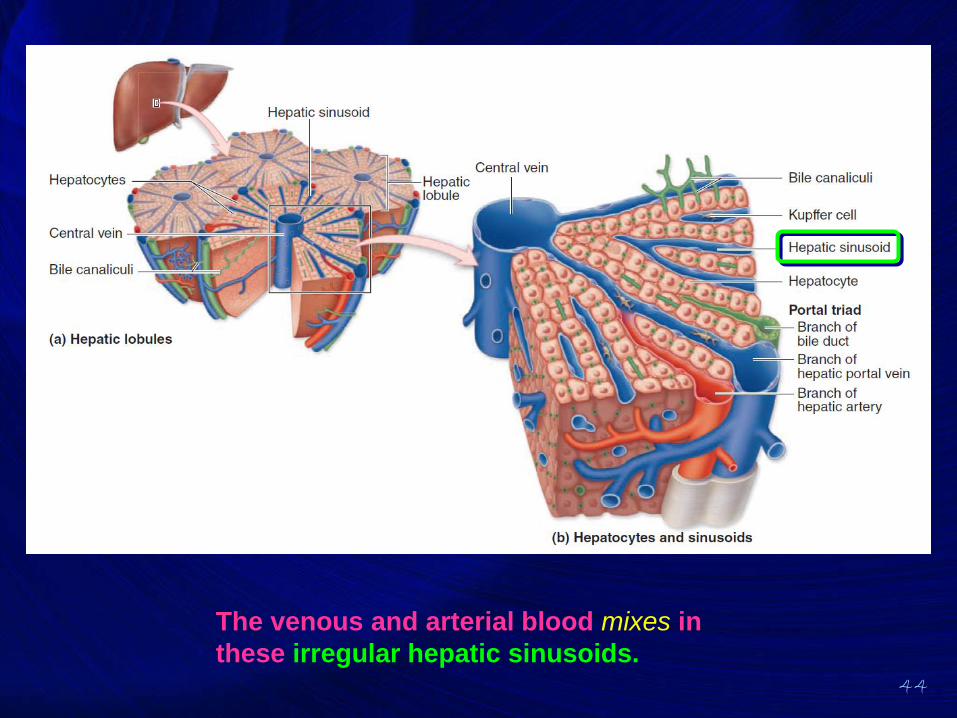
44
The venous and arterial blood mixes in
these irregular hepatic sinusoids.
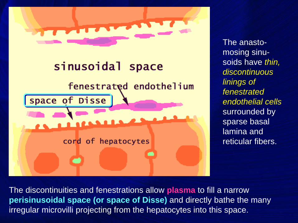
The anasto-
mosing sinu-
soids have thin,
discontinuous
linings of
fenestrated
endothelial cells
surrounded by
sparse basal
lamina and
reticular fibers.
The discontinuities and fenestrations allow plasma to fill a narrow
perisinusoidal space (or space of Disse) and directly bathe the many
irregular microvilli projecting from the hepatocytes into this space.

46
This direct contact between hepatocytes and plasma
facilitates most key hepatocyte functions that involve uptake
and release of nutrients, proteins, and potential toxins.
The numerous small structures in the space of Disse are microvilli on the surface of the hepatocyte.
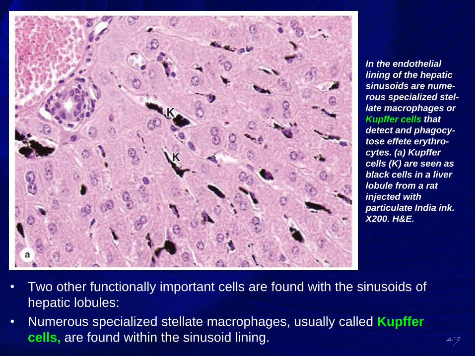
47
• Two other functionally important cells are found with the sinusoids of
hepatic lobules:
• Numerous specialized stellate macrophages, usually called Kupffer
cells, are found within the sinusoid lining.
In the endothelial
lining of the hepatic
sinusoids are nume-
rous specialized stel-
late macrophages or
Kupffer cells that
detect and phagocy-
tose effete erythro-
cytes. (a) Kupffer
cells (K) are seen as
black cells in a liver
lobule from a rat
injected with
particulate India ink.
X200. H&E.
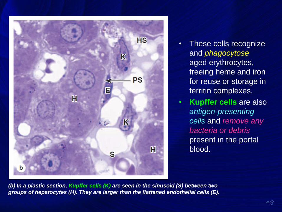
48
• These cells recognize
and phagocytose
aged erythrocytes,
freeing heme and iron
for reuse or storage in
ferritin complexes.
• Kupffer cells are also
antigen-presenting
cells and remove any
bacteria or debris
present in the portal
blood.
(b) In a plastic section, Kupffer cells (K) are seen in the sinusoid (S) between two
groups of hepatocytes (H). They are larger than the flattened endothelial cells (E).
49
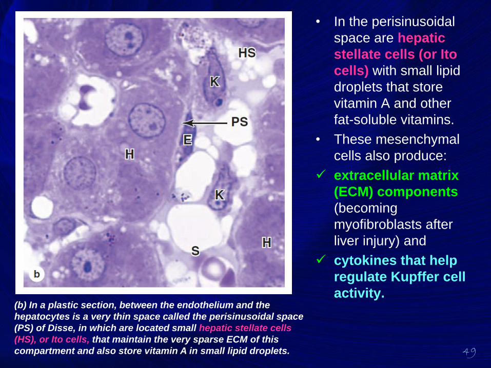
• In the perisinusoidal
space are hepatic
stellate cells (or Ito
cells) with small lipid
droplets that store
vitamin A and other
fat-soluble vitamins.
• These mesenchymal
cells also produce:
✓ extracellular matrix
(ECM) components
(becoming
myofibroblasts after
liver injury) and
✓ cytokines that help
regulate Kupffer cell
activity.(b) In a plastic section, between the endothelium and the
hepatocytes is a very thin space called the perisinusoidal space
(PS) of Disse, in which are located small hepatic stellate cells
(HS), or Ito cells, that maintain the very sparse ECM of this
compartment and also store vitamin A in small lipid droplets.
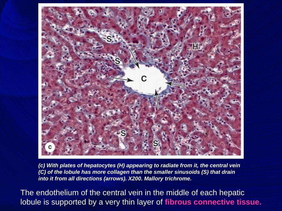
The endothelium of the central vein in the middle of each hepatic
lobule is supported by a very thin layer of fibrous connective tissue.
(c) With plates of hepatocytes (H) appearing to radiate from it, the central vein
(C) of the lobule has more collagen than the smaller sinusoids (S) that drain
into it from all directions (arrows). X200. Mallory trichrome.
51
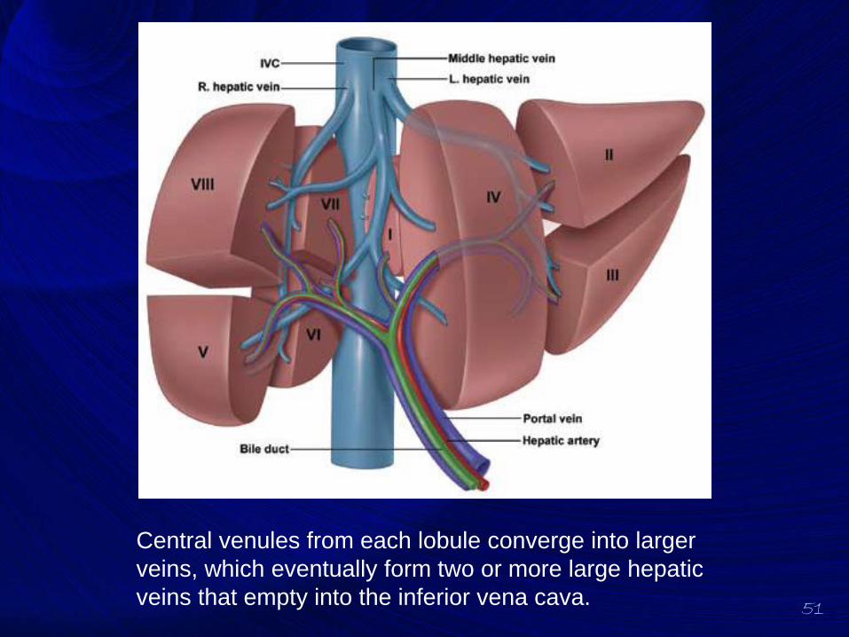
Central venules from each lobule converge into larger
veins, which eventually form two or more large hepatic
veins that empty into the inferior vena cava.
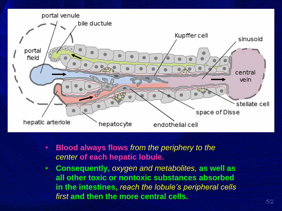
52
• Blood always flows from the periphery to the
center of each hepatic lobule.
• Consequently, oxygen and metabolites, as well as
all other toxic or nontoxic substances absorbed
in the intestines, reach the lobule’s peripheral cells
first and then the more central cells.
53
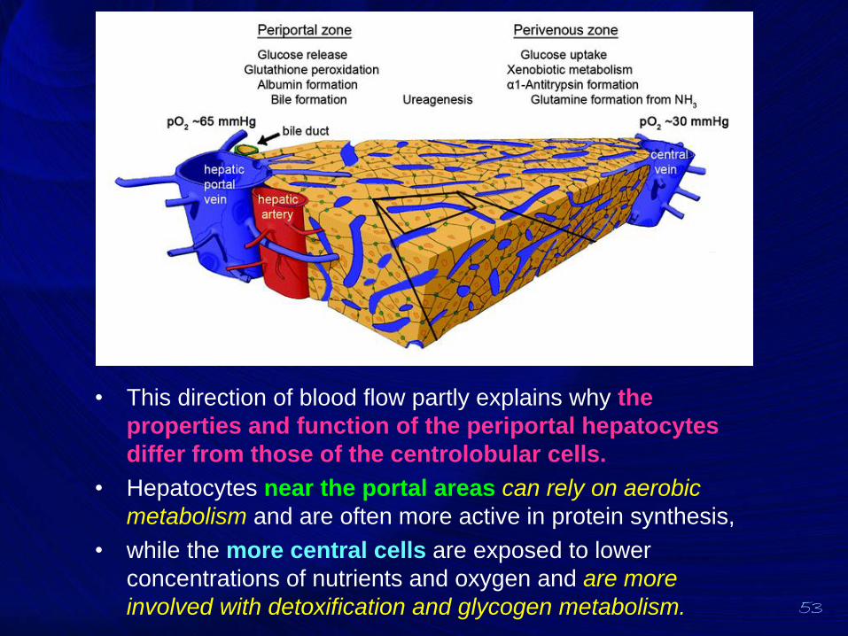
• This direction of blood flow partly explains why the
properties and function of the periportal hepatocytes
differ from those of the centrolobular cells.
• Hepatocytes near the portal areas can rely on aerobic
metabolism and are often more active in protein synthesis,
• while the more central cells are exposed to lower
concentrations of nutrients and oxygen and are more
involved with detoxification and glycogen metabolism.
54
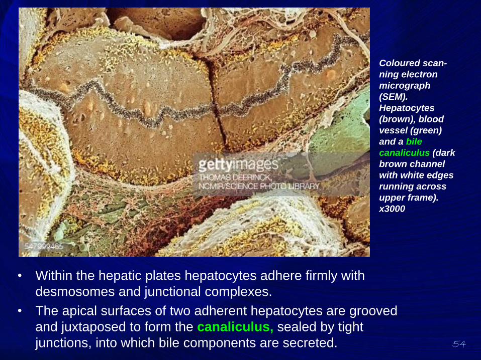
• Within the hepatic plates hepatocytes adhere firmly with
desmosomes and junctional complexes.
• The apical surfaces of two adherent hepatocytes are grooved
and juxtaposed to form the canaliculus, sealed by tight
junctions, into which bile components are secreted.
Coloured scan-
ning electron
micrograph
(SEM).
Hepatocytes
(brown), blood
vessel (green)
and a bile
canaliculus (dark
brown channel
with white edges
running across
upper frame).
x3000
55
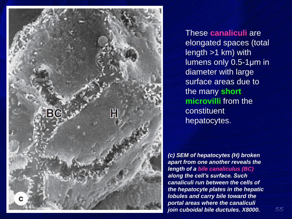
These canaliculi are
elongated spaces (total
length >1 km) with
lumens only 0.5-1μm in
diameter with large
surface areas due to
the many short
microvilli from the
constituent
hepatocytes.
(c) SEM of hepatocytes (H) broken
apart from one another reveals the
length of a bile canaliculus (BC)
along the cell’s surface. Such
canaliculi run between the cells of
the hepatocyte plates in the hepatic
lobules and carry bile toward the
portal areas where the canaliculi
join cuboidal bile ductules. X8000.
56
• The bile canaliculi form a complex anastomosing
network of channels through the hepatocyte
plates that end near the portal tracts.
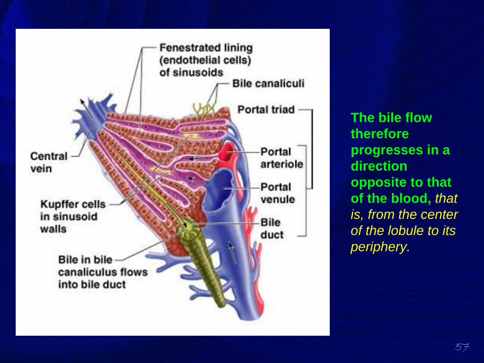
57
The bile flow
therefore
progresses in a
direction
opposite to that
of the blood, that
is, from the center
of the lobule to its
periphery.
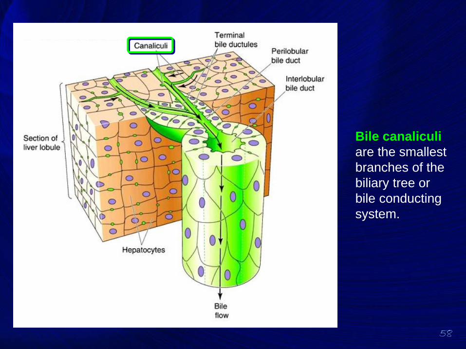
58
Bile canaliculi
are the smallest
branches of the
biliary tree or
bile conducting
system.
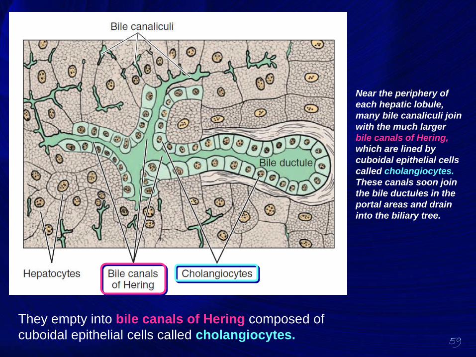
59
Near the periphery of
each hepatic lobule,
many bile canaliculi join
with the much larger
bile canals of Hering,
which are lined by
cuboidal epithelial cells
called cholangiocytes.
These canals soon join
the bile ductules in the
portal areas and drain
into the biliary tree.
They empty into bile canals of Hering composed of
cuboidal epithelial cells called cholangiocytes.
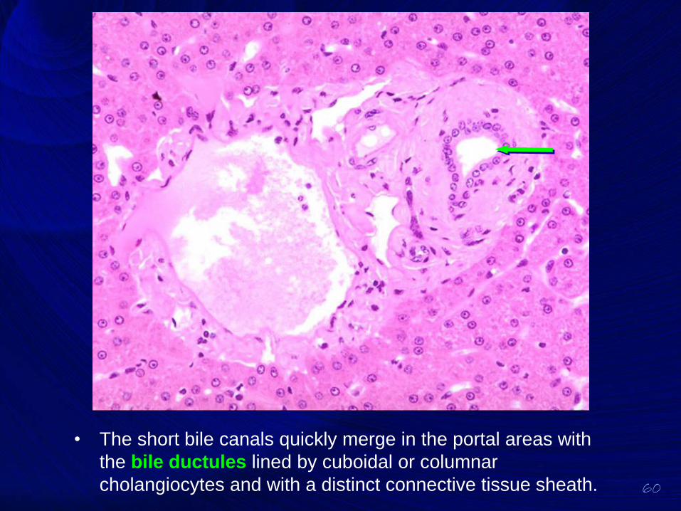
60
• The short bile canals quickly merge in the portal areas with
the bile ductules lined by cuboidal or columnar
cholangiocytes and with a distinct connective tissue sheath.
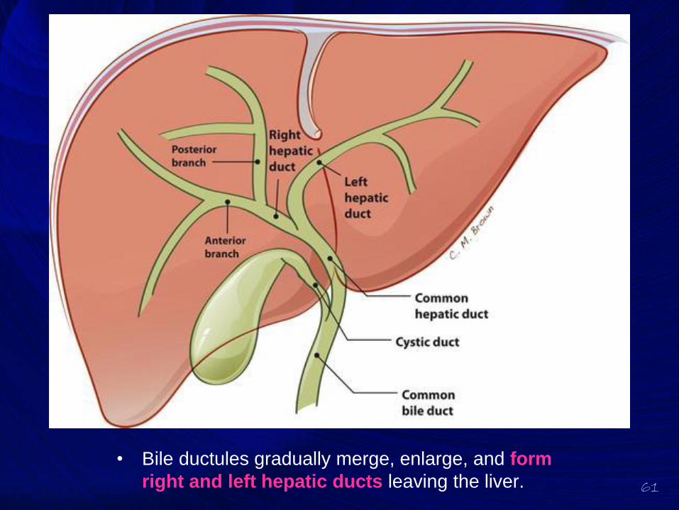
61
• Bile ductules gradually merge, enlarge, and form
right and left hepatic ducts leaving the liver.

BILIARY TRACT &
GALLBLADDER
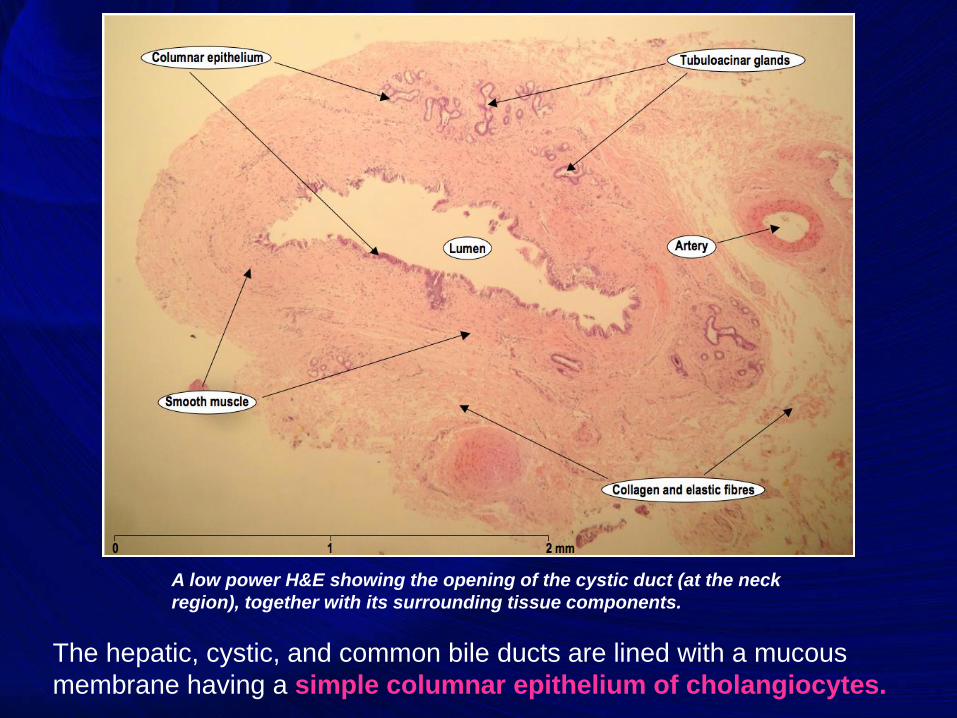
The hepatic, cystic, and common bile ducts are lined with a mucous
membrane having a simple columnar epithelium of cholangiocytes.
A low power H&E showing the opening of the cystic duct (at the neck
region), together with its surrounding tissue components.
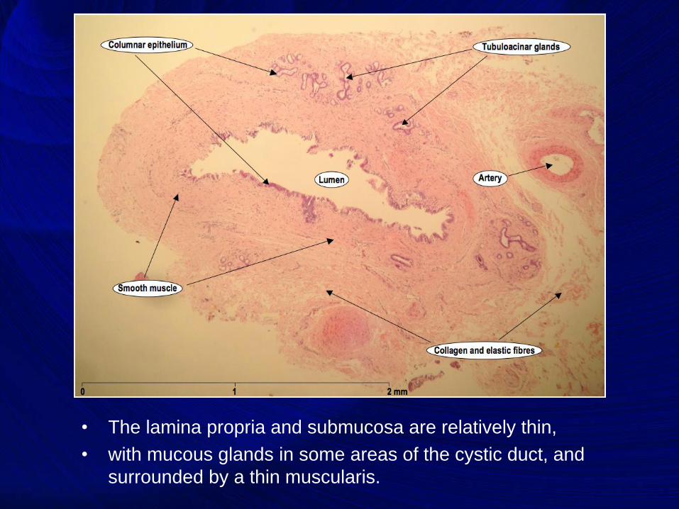
• The lamina propria and submucosa are relatively thin,
• with mucous glands in some areas of the cystic duct, and
surrounded by a thin muscularis.
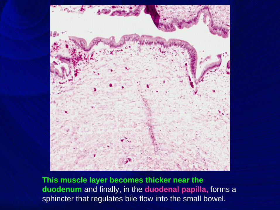
This muscle layer becomes thicker near the
duodenum and finally, in the duodenal papilla, forms a
sphincter that regulates bile flow into the small bowel.
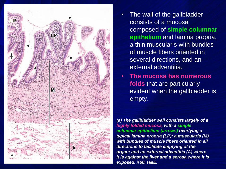
• The wall of the gallbladder
consists of a mucosa
composed of simple columnar
epithelium and lamina propria,
a thin muscularis with bundles
of muscle fibers oriented in
several directions, and an
external adventitia.
• The mucosa has numerous
folds that are particularly
evident when the gallbladder is
empty.
(a) The gallbladder wall consists largely of a
highly folded mucosa, with a simple
columnar epithelium (arrows) overlying a
typical lamina propria (LP); a muscularis (M)
with bundles of muscle fibers oriented in all
directions to facilitate emptying of the
organ; and an external adventitia (A) where
it is against the liver and a serosa where it is
exposed. X60. H&E.
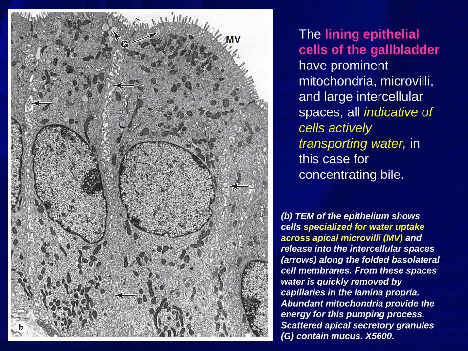
The lining epithelial
cells of the gallbladder
have prominent
mitochondria, microvilli,
and large intercellular
spaces, all indicative of
cells actively
transporting water, in
this case for
concentrating bile.
(b) TEM of the epithelium shows
cells specialized for water uptake
across apical microvilli (MV) and
release into the intercellular spaces
(arrows) along the folded basolateral
cell membranes. From these spaces
water is quickly removed by
capillaries in the lamina propria.
Abundant mitochondria provide the
energy for this pumping process.
Scattered apical secretory granules
(G) contain mucus. X5600.

Good Luck