Embed Size (px)
Citation preview
Histology
The study of cells and tissues
Histology – Objectives
what are the diagnostic characteristics of each of the four main types of tissues
what are the diagnostic characteristics of each of the subtypes of tissues
be able to give examples of each tissue type
are organs made of one or more tissues?
Tissues – 4 basic types epithelial connective muscular nervous
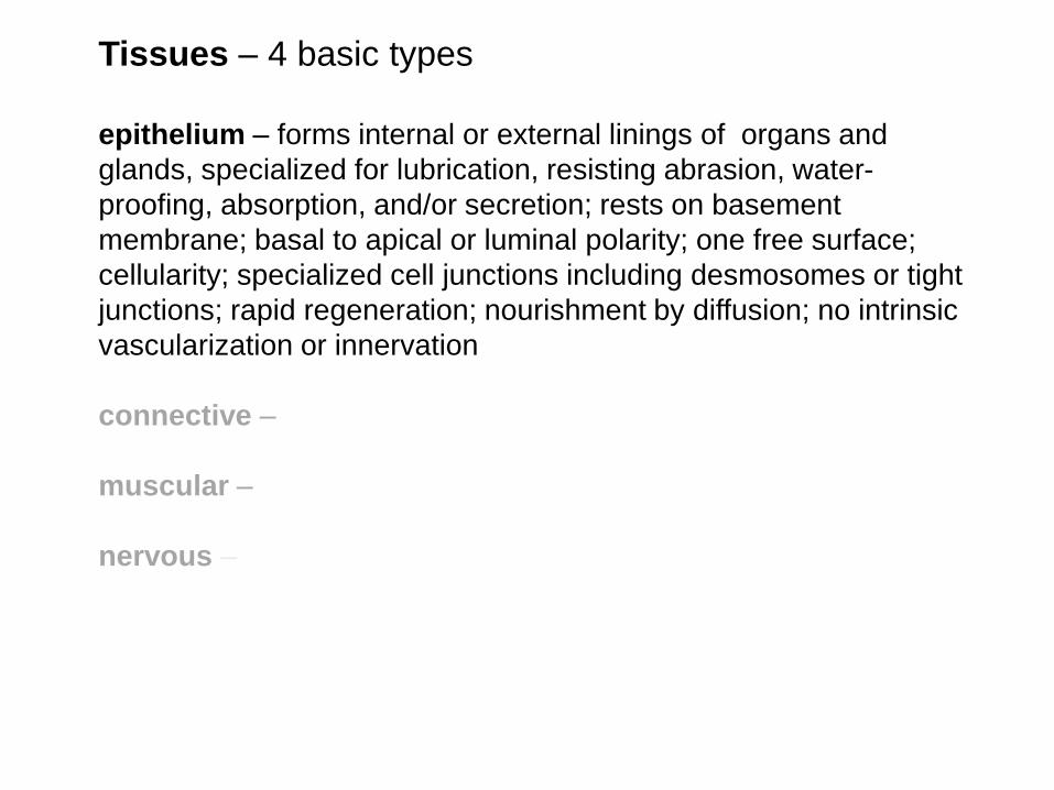
Tissues – 4 basic types
epithelium – forms internal or external linings of organs and
glands, specialized for lubrication, resisting abrasion, water-
proofing, absorption, and/or secretion; rests on basement
membrane; basal to apical or luminal polarity; one free surface;
cellularity; specialized cell junctions including desmosomes or tight
junctions; rapid regeneration; nourishment by diffusion; no intrinsic
vascularization or innervation
connective –
muscular –
nervous –
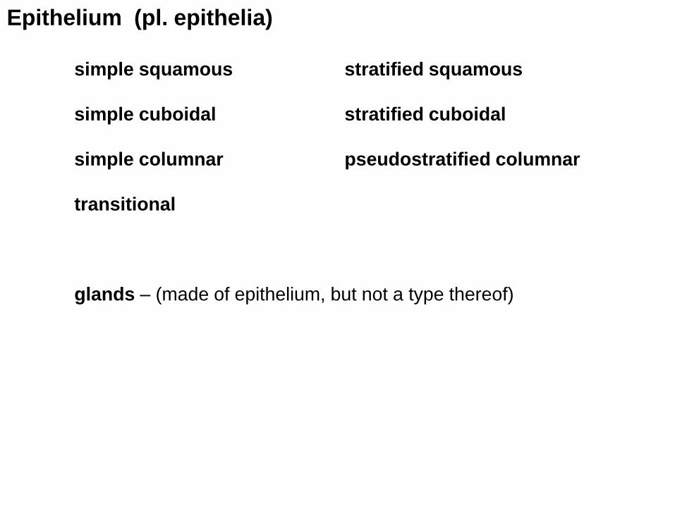
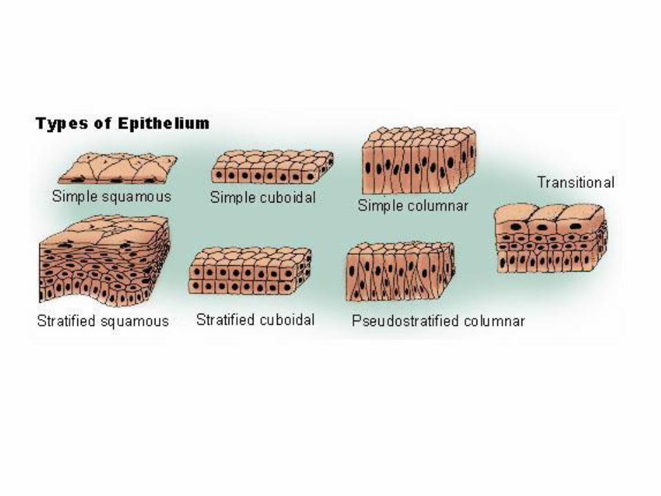
Epithelium (pl. epithelia)
simple squamous stratified squamous
simple cuboidal stratified cuboidal
simple columnar pseudostratified columnar
transitional
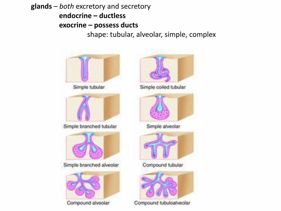
glands – (made of epithelium, but not a type thereof)
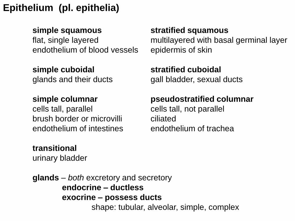
Epithelium (pl. epithelia)
simple squamous stratified squamous
flat, single layered multilayered with basal germinal layer
endothelium of blood vessels epidermis of skin
simple cuboidal stratified cuboidal
glands and their ducts gall bladder, sexual ducts
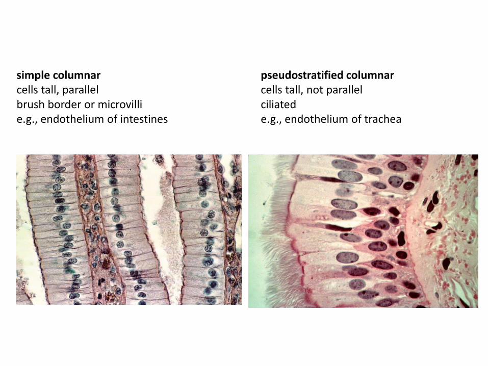
simple columnar pseudostratified columnar
cells tall, parallel cells tall, not parallel
brush border or microvilli ciliated
endothelium of intestines endothelium of trachea
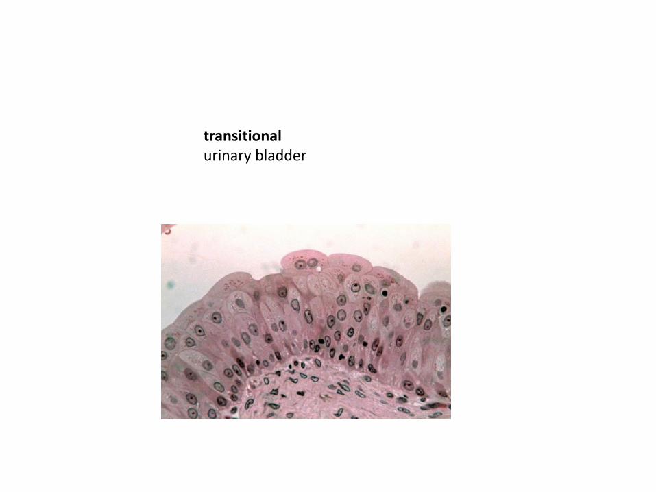
transitional
urinary bladder
glands – both excretory and secretory
endocrine – ductless
exocrine – possess ducts
shape: tubular, alveolar, simple, complex
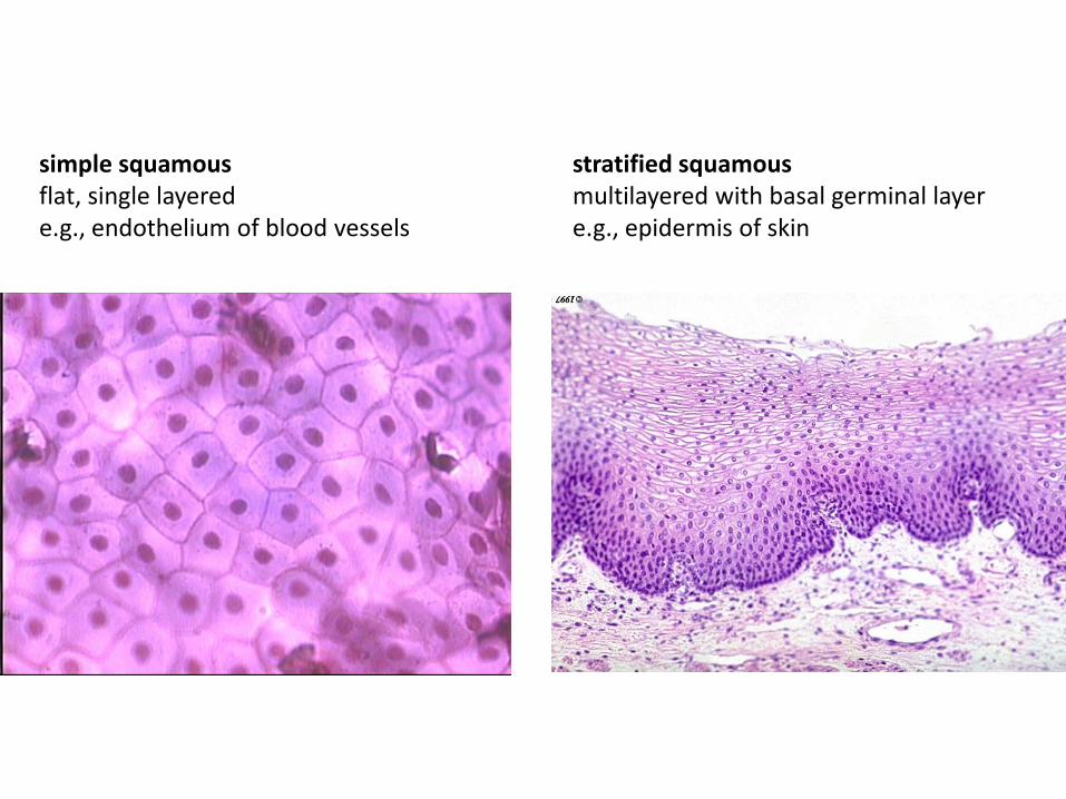
simple squamous stratified squamous flat, single layered multilayered with basal germinal layer e.g., endothelium of blood vessels e.g., epidermis of skin
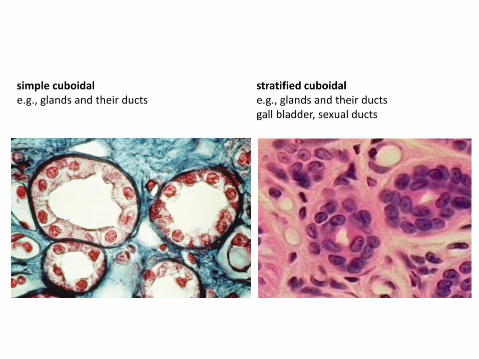
simple cuboidal stratified cuboidal e.g., glands and their ducts e.g., glands and their ducts gall bladder, sexual ducts
simple columnar pseudostratified columnar cells tall, parallel cells tall, not parallel brush border or microvilli ciliated e.g., endothelium of intestines e.g., endothelium of trachea
transitional urinary bladder
glands – both excretory and secretory endocrine – ductless exocrine – possess ducts shape: tubular, alveolar, simple, complex
Tissues – 4 basic types
epithelium –
connective – acellularity; extracellular matrix>>cells; provides
structure and/or substrate for blood vessels, nerves, lymphatics,
and glands; extracellular matrix or ground substance = water,
dissolved or precipitated salts, proteins, and carbohydrates
muscular –
nervous –
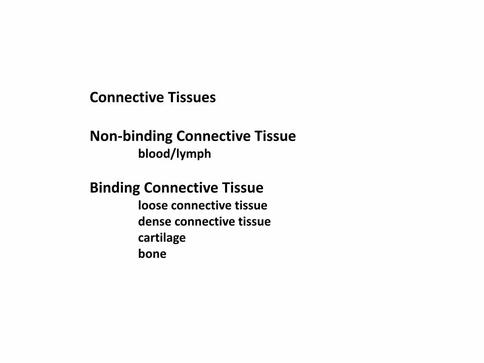
Connective Tissues Non-binding Connective Tissue blood/lymph
Binding Connective Tissue loose connective tissue dense connective tissue cartilage bone
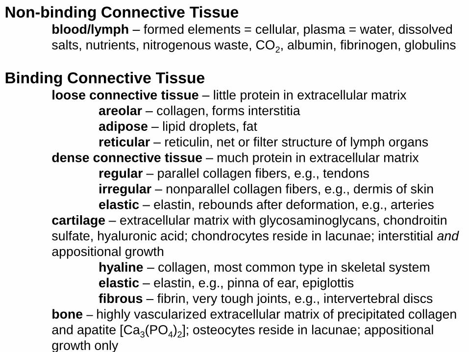
Non-binding Connective Tissue blood/lymph – formed elements = cellular, plasma = water, dissolved
salts, nutrients, nitrogenous waste, CO2, albumin, fibrinogen, globulins
Binding Connective Tissue loose connective tissue – little protein in extracellular matrix
areolar – collagen, forms interstitia
adipose – lipid droplets, fat
reticular – reticulin, net or filter structure of lymph organs
dense connective tissue – much protein in extracellular matrix
regular – parallel collagen fibers, e.g., tendons
irregular – nonparallel collagen fibers, e.g., dermis of skin
elastic – elastin, rebounds after deformation, e.g., arteries
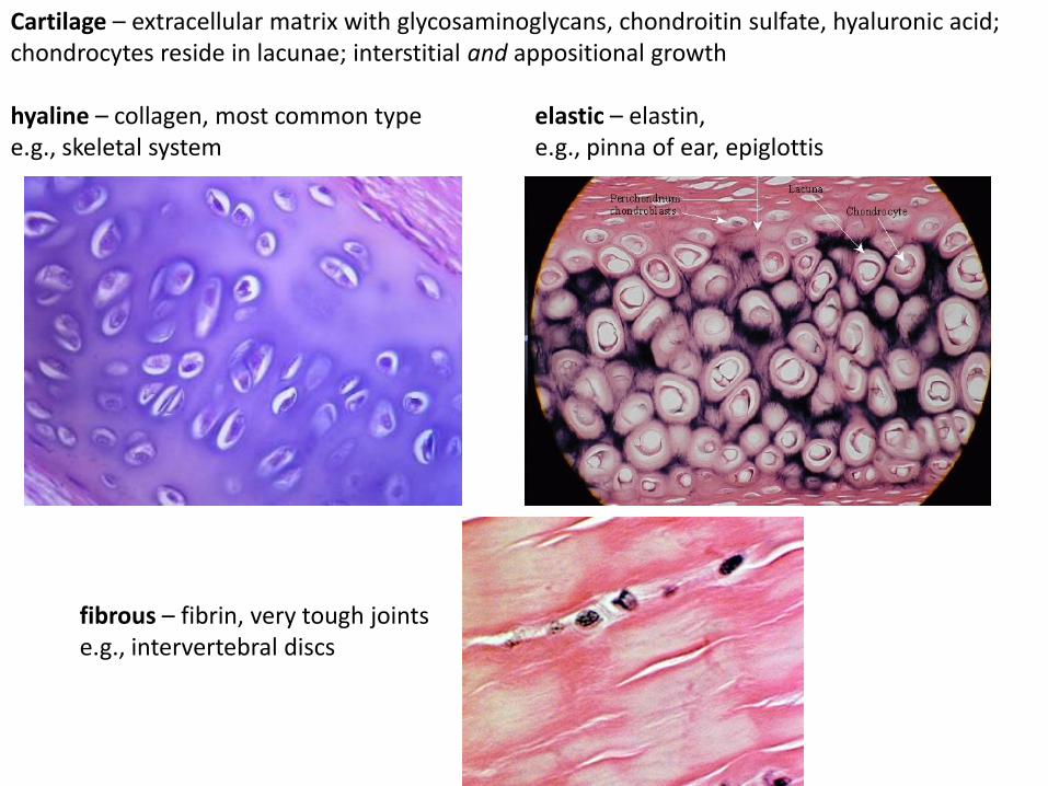
cartilage – extracellular matrix with glycosaminoglycans, chondroitin
sulfate, hyaluronic acid; chondrocytes reside in lacunae; interstitial and
appositional growth
hyaline – collagen, most common type in skeletal system
elastic – elastin, e.g., pinna of ear, epiglottis
fibrous – fibrin, very tough joints, e.g., intervertebral discs
bone – highly vascularized extracellular matrix of precipitated collagen
and apatite [Ca3(PO4)2]; osteocytes reside in lacunae; appositional
growth only
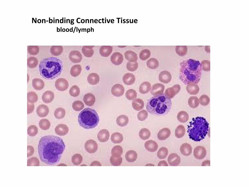
Non-binding Connective Tissue blood/lymph
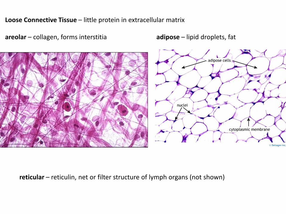
Loose Connective Tissue – little protein in extracellular matrix areolar – collagen, forms interstitia adipose – lipid droplets, fat
reticular – reticulin, net or filter structure of lymph organs (not shown)
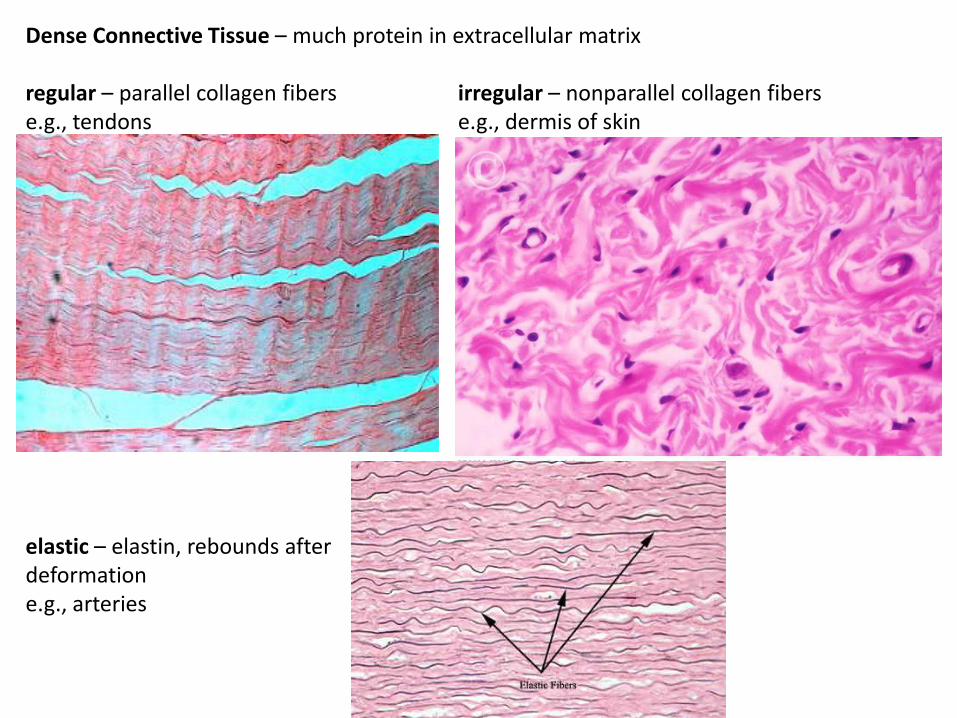
Dense Connective Tissue – much protein in extracellular matrix regular – parallel collagen fibers irregular – nonparallel collagen fibers e.g., tendons e.g., dermis of skin
elastic – elastin, rebounds after deformation e.g., arteries
fibrous – fibrin, very tough joints e.g., intervertebral discs
Cartilage – extracellular matrix with glycosaminoglycans, chondroitin sulfate, hyaluronic acid; chondrocytes reside in lacunae; interstitial and appositional growth hyaline – collagen, most common type elastic – elastin, e.g., skeletal system e.g., pinna of ear, epiglottis
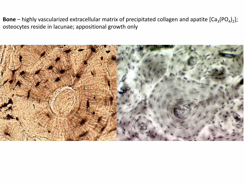
Bone – highly vascularized extracellular matrix of precipitated collagen and apatite [Ca3(PO4)2]; osteocytes reside in lacunae; appositional growth only
Tissues – 4 basic types
epithelium –
connective –
muscular – electrochemically excitatory and contractile
nervous –
Muscular Tissue – Ca++ and ATP dependent contraction; protein
myofilaments of two types: thin filaments – actin, troponin, meromyosin, others depending on muscle type; and thick filaments – myosin smooth muscle striated muscle skeletal striated muscle cardiac striated muscle
Muscular Tissue – Ca++ and ATP dependent contraction; thin filaments –
actin, troponin, meromyosin; thick filaments – myosin
smooth muscle – spindle shaped cells, mononucleate, in walls of organs (e.g.,
digestive tract, blood vessels, skin), respond to hormones, stretch, and
innervation by autonomic nervous system
striated muscle – myofilaments bundled into myofibrils forming ‘striated’
sarcomeres (alternating interdigitating bands of thick and thin filaments),
juxtaposed to sarcoplasmic reticulum (ER sequesters Ca++) and transverse
tubules (invaginations of sarcolemma); numerous mitochondria
skeletal striated muscle – giant multinucleate linear cells of voluntary skeletal
muscular system, syncytium, every cell innervated with motor endplate,
responds to neurotransmitter acetylcholine; denervation results in atrophy
cardiac striated muscle – myocardium of heart; branching cells joined at
intercalated discs; syncytium but few nuclei; intercalated discs possess gap
junctions (electrical connectivity) and desmosomes; cells depolarize
spontaneously and wave of contraction passes from cell to cell; rate modulated
by hormones and autonomic innervation
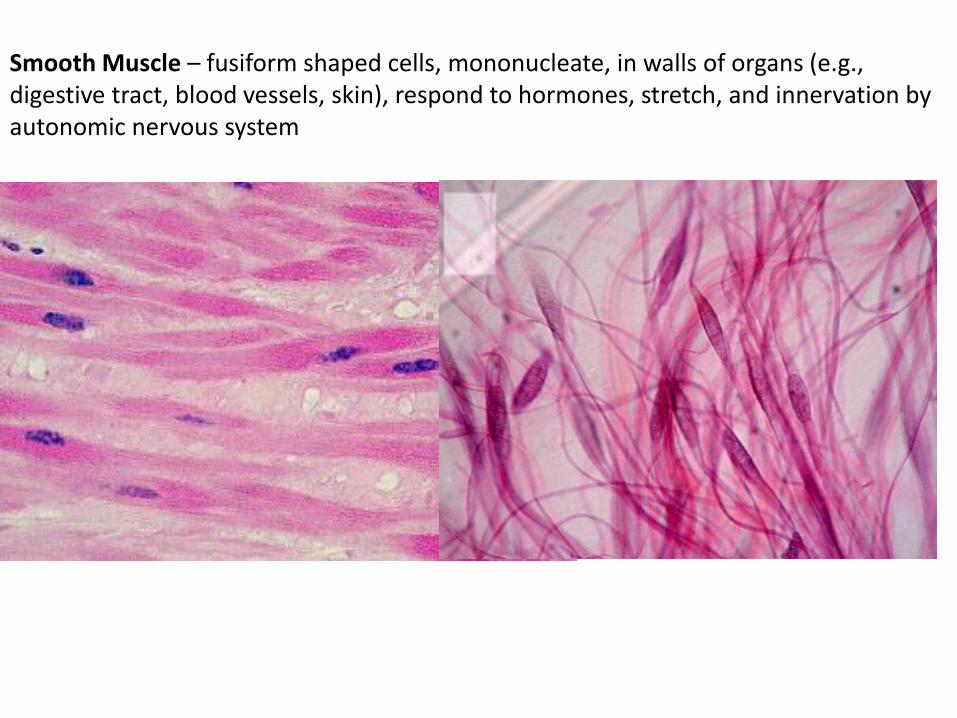
Smooth Muscle – fusiform shaped cells, mononucleate, in walls of organs (e.g., digestive tract, blood vessels, skin), respond to hormones, stretch, and innervation by autonomic nervous system
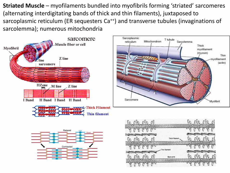
Striated Muscle – myofilaments bundled into myofibrils forming ‘striated’ sarcomeres (alternating interdigitating bands of thick and thin filaments), juxtaposed to sarcoplasmic reticulum (ER sequesters Ca++) and transverse tubules (invaginations of sarcolemma); numerous mitochondria
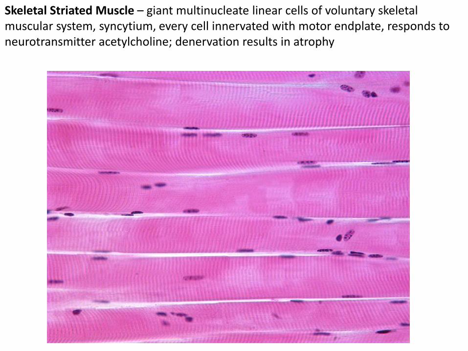
Skeletal Striated Muscle – giant multinucleate linear cells of voluntary skeletal muscular system, syncytium, every cell innervated with motor endplate, responds to neurotransmitter acetylcholine; denervation results in atrophy
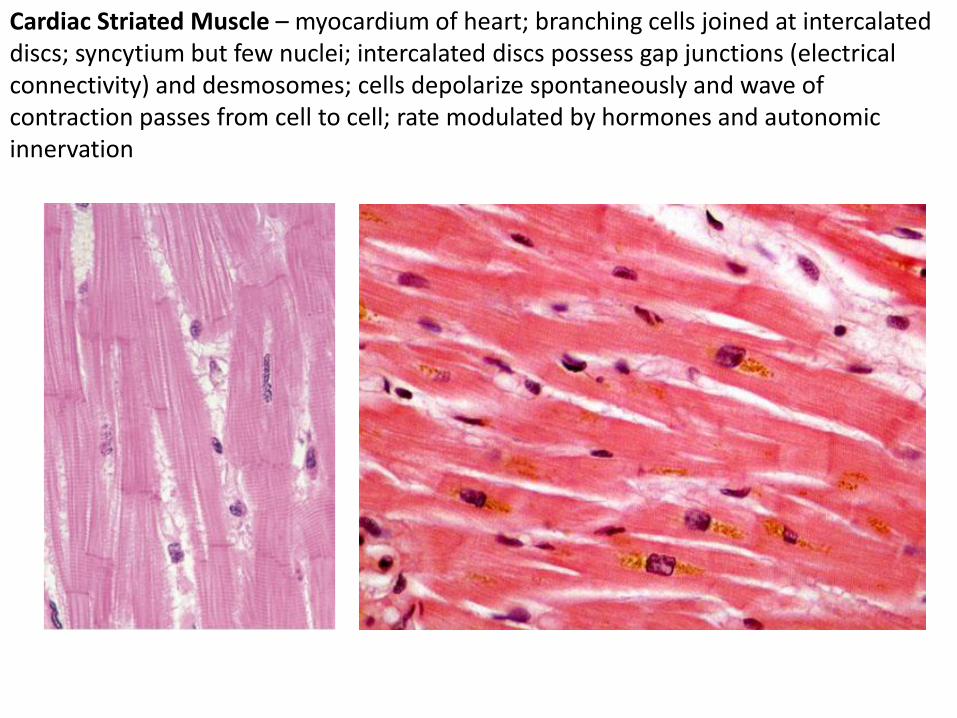
Cardiac Striated Muscle – myocardium of heart; branching cells joined at intercalated discs; syncytium but few nuclei; intercalated discs possess gap junctions (electrical connectivity) and desmosomes; cells depolarize spontaneously and wave of contraction passes from cell to cell; rate modulated by hormones and autonomic innervation
Tissues – 4 basic types
epithelium –
connective –
muscular –
nervous - electrochemically excitatory and conductive
Nervous Tissue
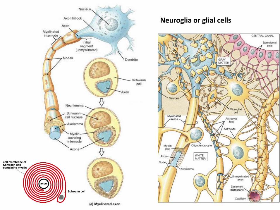
neuroglia or glial cells neurons
Nervous Tissue
neuroglia or glial cells – structural, supportive, insulating
neurons – excitatory; cell body or ‘neuron’; cell processes
are axons and dendrites; slow transport of neurotransmitters
from neuron to presynaptic vesicles of axon; membrane
depolarization causes release of neurotransmitters into
synapse which are bound by receptors of postsynaptic
dendrites; neurotransmitters may be excitatory, inhibitory, or
modifying to membrane depolarization of postsynaptic
neuron
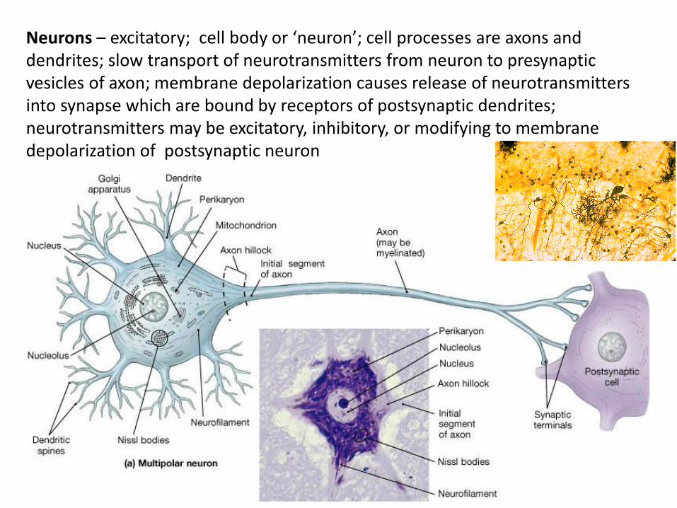
Neurons – excitatory; cell body or ‘neuron’; cell processes are axons and dendrites; slow transport of neurotransmitters from neuron to presynaptic vesicles of axon; membrane depolarization causes release of neurotransmitters into synapse which are bound by receptors of postsynaptic dendrites; neurotransmitters may be excitatory, inhibitory, or modifying to membrane depolarization of postsynaptic neuron
Neuroglia or glial cells