Embed Size (px)
Citation preview

Title
HISTOLOGICAL CHANGES IN THE BRAINS OFMALFORMED FETUSES FROM THE URETHANETREATED MOTHER MOUSE AND THEIR POSSIBLERELATION TO OCCURRENCE OF GLIOMAS INCHILDREN
Author(s) YAMAZAKI, NORIO
Citation 日本外科宝函 (1959), 28(5): 1530-1550
Issue Date 1959-06-01
URL http://hdl.handle.net/2433/206902
Right
Type Departmental Bulletin Paper
Textversion publisher
Kyoto University
1530 日本外科宝函第28巻第5号
HISTOLOGICAL CHANGES IN THE BRAINS OF MALFORMED FETUSES FROM THE URETHANE TREATED MOTHER
MOUSE AND THEIR POSSIBLE RELATION TO OCCURRENCE OF GLIOMAS IN CHILDREN
bv
N ORIO YAMAZAKI
From the !st Surgical Division, Kyoto University Medical School (Director : Prof. Dr Cm3ATO ARAKr) Received for publication May 14, 1959
INTRODUCTORY NOTES
It has been kmwn that the s'トcalledembryonβl cell rests are not infrequently
found in the immature brain of a new-born infant or fetus. A number of authors
(CusHING, 1930; RAFF and KERNοHAN, 1944; BRZusTow1cz and KERNOHAN, 1952;
SHIMADA, 1954; and OsTERTAG, 1956) assumed that gliomas of the brain, particularly
those in chi!出℃n,may pC>ssibly arise from such cell rests or clusters of persistent
immature cells. Seats of predilection for occurrence of the gliomas in children under
10 years of age are generall~’ believed to be the vermis of the cerebellum, especially
its posterior part,日oorof the 4th ventricle, pons and quadrigeminal body, while
those in persons around the adolescence the 3rd ventricle, pineal body, .optic nerve,
thalamus, basal ganglia and septum pellucidum. Some authors (0sTERTAG) believe
that gliomas in adult of the olfactory brain, edge of the lateral ventricle (in the
fetal period) and callosal i川l ~’ as well as di町useor multiple gliomas ma~・ have
something to do with developmental anomaly of the tissue.
It has recent!? become clear that variable exogenous factors can give rise to
tissue malformation, some of which ma~ -, at the same time, play a role in fostering
formation of tumor. Having injected methylcholanthrene to mice, for instance,
STRONG (1945) was able rather constant!~· to produce in ti日ir descendants certain
malformations, such as adenomatous changes in the stomach, situs inversus visceralis, dextrocardia, apigmentatio piliaris, etc.
It scc1m, therefore, rF1ite probable that the cell rests, which can be found even
in the brain under the process of normal development, are liable to increase in
number and sh;>w greater tendenc¥・ to formation of tumor, \\’hen that brain is
subjected to factors womoting tissue malformation.
In the p1℃淀川 stud ~-, the author for the first step examined histologically the
brain of normal fetus o! mouse, following the developmental sequence, in the aim
to see whether the same or similar cell rests as those in human fetus are pt℃sent
・and in what part .of the brain, if present, are thむγ111()日tlikely to hむ found. Then,
the brains of malformed fetal mice, de刈 C'Jl(lantsof a mother mouse having undergone
intraperitoneal injection of ethylurethane solution during the pregnancy after the
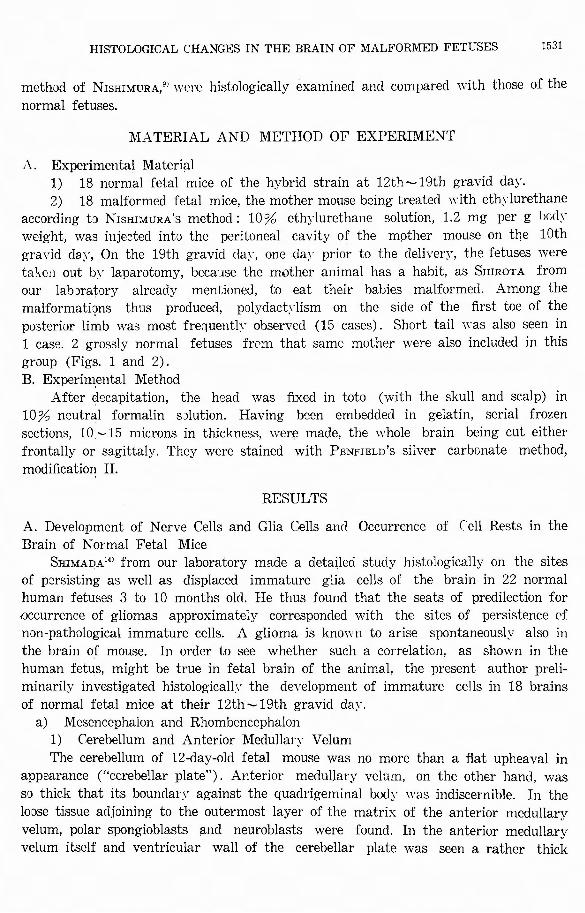
HISTOLOGICAL CHANGES IN THE BRAIN OF MALFORMED FETUSES 1531
method of N1sHIMURA/> were histologically examined and compared with those of the
normal fetuses.
MATERIAL AND METHOD OF EXPERIMENT
A. Experimental Material 1) 18 normal fetal mice of the hybrid strain at 12th~19th gravid day.
2) 18 malformed fetal mice, the mother mouse being treated with ethylurethane
according to N1sm~uRA’s method: 10% ethylurethane solution, 1.2 mg per g bod~·
weight, was injected into the peritoneal cavity of the mother mouse on the 10th
gravid day, On the 19th gravid cla~ · , one da~' prior to the deliver~', the fetuses were
ta1,cn out by laparotomy, because the mother animal has a habit, as SHIROTA from
our labJratory already mentioned, to eat their babies malformed. Among the
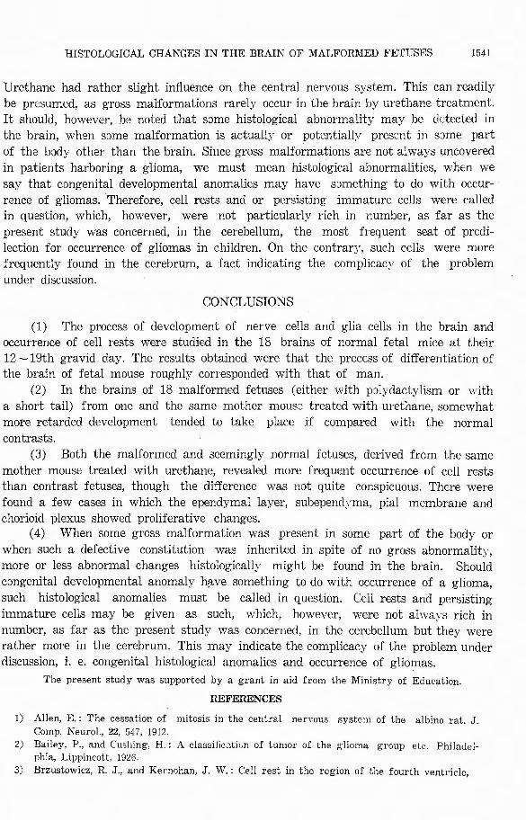
malformations thus pr吋 ucecl, polydactylism on the side of the first toe of the
posterior limb was most frequently observed (15 cases). Short tail was also seen in
1 case. 2 grossly normal fetuses from that same mother were also included in this
group (Figs. 1 and 2).
B. Experin~ental Method After decapitation, the head was 白xedin toto (with the skull and scalp) in
10% neutral formalin sJlution. Having been embedded in gelatin, serial frozen
sections, 10 -15 microns in thickness, ¥¥:ere made, the whole brain being cut either
frontally or sagittaly. They were stained with PENFIELD’S silver carbonate method,
modification II.
RESULTS
A. Development of Nerve Cells and Glia Cells and Occurrence of (冶11Rests in the
Brain of Normal Fetal Mice
SHIMAJ)A14' from our laboratory made a detailed study histologically on the sites
of persisting as well as displaced immature glia cells of the brain in 22 normal
human fetuses 3 to 10 months old. He thus found that the seats of predilection for
.occurrence of gliomas approximately corresponded with the sites of persistence 0f
non-pathological immature cells. A glioma is known to arise spontaneously’ also in
the brain of mouse. In order to see whether such a correlation, as shown in the
human fetus, might be true in fetal brain of the animal, the present author preli-
minarily investigated histologically the development of immature cells in 18 brains
of normal fetal mice at their 12th~19th gravid day.
a) Mesencephalon and Rhombencephalon
1) Cerebellum and Anterior Medullary Velum
The cerebellum of 12・day-oldfetal mouse was no more than a flat upheaval in
appearance (“cerebellar plate"). Anterior medullary velum, on the other hand, was
so thick that its boundary against the quadrigeminal body was indiscernible. In the
loose tissue adjoining to the outermost layer of the matrix of the anterior medullary
velum, polar spongioblasts and neuroblasts were found. In the anterior medullary
velum itself and ventricular wall of the cerebellar plate was seen a rather thick
1532 目本外科宝函第28巻第5号
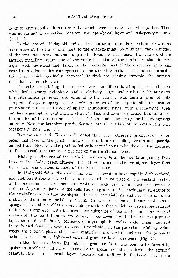
la~℃r of argentophilic immature cells which were densely packed together. There
was no distinct demarcation between the epend~'mal layer and subependymal zone
(matrix:). In the case of 13-daγ-ol〔l fetus, the anterior medullar}ァ velum showed an
indentation at the transitional part to the quadrigeminal l川 lyso that the distinction
of the tw0 structures became apparent. Even at this stage, the matrix of the
anterior medullary velum and of the ventral rnrtion of the cerebellar plate interm-
ingled with the epcndymal layer. In the posterior part of the cerebellar plate and
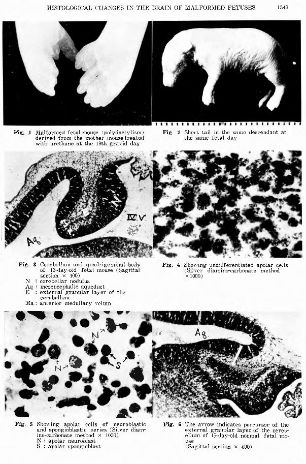
near the midline, which corresponded to the cerebellar no〔lulus,the matrix formed a
thick layer which gradually decreased its thickness coming towards the anterior
medullan・ velum (Fig. 3).
The cells constituting the matrix were undi汀erentiatedapolar cells (Fig. 4)
which had a scanty c::toplasm and a relatively large oval nucleus with numerous
fine intranuclear granule日. Just external to the matrix was seen a layer of cells
composed of ap:ilar sp.mgioblastic series possessed of an argentophilic and oval or
pear-shaped nucleus an〔lthose of ap0lar neuroblastic series with a somewhat larger
but less argentophilic oval nucleus (Fig. 5). This cell la~·er wa日 foundthinned around
the midline of the cerebellar plate but thicker and more irregular in arrangement
laterall?. X ear the brachium pontis, clensel~’ packed clusters of immature cells were occasionall? seen (Fig. 6).
BRzusTow1cz and KERNOHAN3i stated that thev observed proliferation of the
epencl~·mal layer at the junction between the anterior medullary velum and quadrig・
cminal bod~-. However, the proliferated cells seemed to us to be those of the precursor
of the external granular la~モr but not of the ependymal layer.
Histological findings of the brain in 14-day-old fetus did not di仔ergreatl:yア from
those in the 13-cla~· cases, although the di百erentiationof the epend~·mal layer from the matrix was obvious in most of the former cases.
In 15-da;,ア・oldfetus, the cerebellum was observed to have rapidl}ア di百erentiated,
and undi町erentiatedapolar cells were uncovered in no place on the ventral portion
of the cerebellum other than the posterior medullarv velum and the cerebellar
nodulus. A great majorit;,・ of the cells had emigrated in the meclullary substance of
the cerebellum where they d引でlopedinto polar spongioblasts and neuroblasts. In the
matrix of the .anterior medullary velum, on the other hand, innumerable apolar
spc:mgioblasts and neurobl,flsts were still pres・mt; a fact which indicates more retarded
maturity as comparecl with the medullary substance of the cerebellum. The external
叩rfaceof the cerebellum in its entirety was covered with the external granular
la~℃r, as a thin cell layer, composed of argentophilic apolar cells which here and
there formed dense!.¥・ packed clusters. In particular, in the posterior medullary velum
where the chorioid plexus of the 4th ventricle is attached to and near the cerebellar
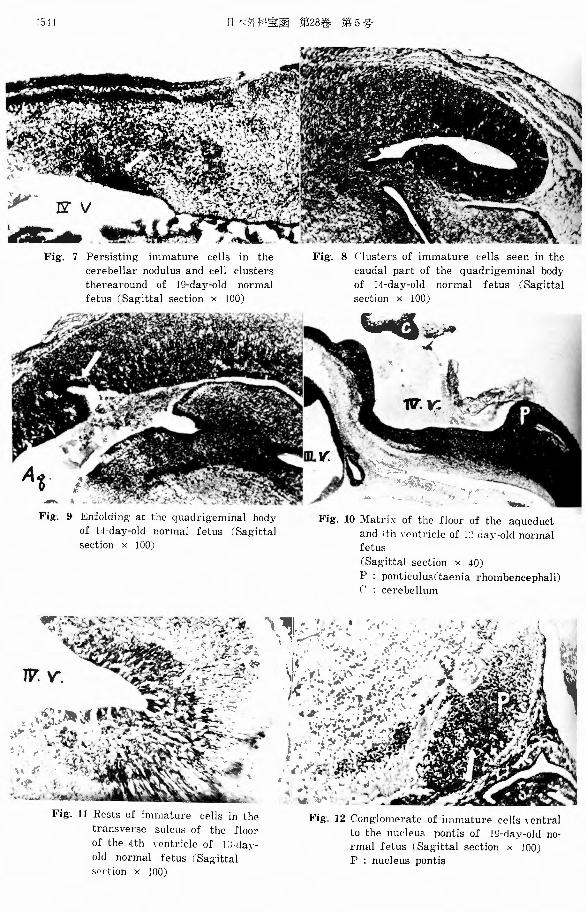
nodulus, a cつnsiderablythickened external granular l叫℃rwas seen (Fig. 7)・
In the 16-〔la>ザ-oldfetus, the internal granular la~ザer was seen to be formed l〕1. apolar spongiobl
granular layer. The intern叫 layer appeared not uniform in thickness, but in the
HISTOLOGICAL CHANGES IN THE BRAIN OF '.¥1ALFORMED FETUSES 1533
cerebellar vermis it was unusuall~· thick and in the cerebellar hemisphere rosary-like. In the 17-day-old cases, the differentiation of the cerebellum was almost the
same with that of the 16-day-group. At the 18th fetal da~· , polar neuroblasts were seen to have evidently aggregated
in the medullarv substance of the cerebellum, forming nuclei. The glia cells
surrounding these nuclei were rather mature, as contrasted with those in other
pe>rtions, as they already developed into astroblasts or even astrocytes. At the 19th fetal dav, the external granular layer bec..ame uniformly thick,
arranging itself parallel to the foliated outer surface of the cerebellum. Polar spong-
ioblasts were found to have arranged, toward the molecular laJァer,from the external
granular layer which consequently appeared thinner than that in the 18-day cases.
In the vermis, near the anterior medullary velum, posterior medullary velum and
around the cerebellar nodulus still persisted undi百erentiated apolar cells, the density
of which was most pronounced in the cerebellar no【lulus;the more the nearer to the medulla円ア sQbstance.Accordingly, in the meclullary substance were seen innumerable
cell clusters. which had indistinct delimitation from the surrounding tissue (Fig. 7).
Similar cell ,conglomerates were also visible in other portions of the brain.
2) Quadrigeminal Body
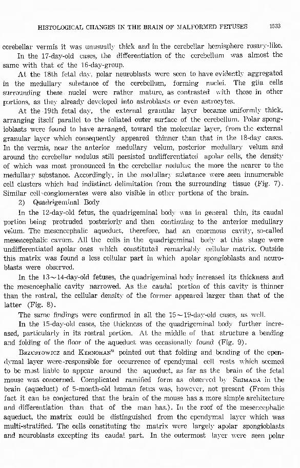
In the 12-day-old fetus, the quadrigeminal body was in general thin, its caudal
portion being protruded posteriorlJァ andthen continuing to the anterior medullary
velum. The mesencephalic aqueduct, therefore, had an enormous cavity, so-called
mesencephalic cavum. All the cells in the quadrigeminal bocly at this stage were undifferentiated apolar ones which constituted remarkably cellular matrix. Outside
this matrix・ was found a less cellular part in which apolar spongioblasts and neuro-
blasts were observed.
In the 13~14-day-old fetuses, the quadrigeminal bodv increased its thickness and
the mesencephalic cavity narrowed. As the caudal portion of this cavity is thinner
than the rostral, the cellular densit)γof the former appeared larger than that of the
latter (Fig. 8).
The same findings were confirmed in all the 15~19・daJ・-oldcases, as ¥vcll. In the 15-day-olcl cases, the thickness of the quadrigeminal bod)ァ furtherincre-
ased, particularly in its rostral portion. At the middle of that structure a bending
and folding of the floor of the aqueduct was occasionally found (Fig. 9).
BRzusTowrcz and KERNOHAN3> pointed out that folding and bending of the epen-
dymal layer were-responsible for occurrence of ependymal cell rests which seemed to be m:lst liable to appear around the aqueduct, as far as the brain of the fetal
mouse was concerned. Complicated ramified form as obsen℃cl by SHIMADA in the
brain (aqueduct) of 5・month-oldhuman fetus was, however, not present (From this
fact it can be conjectured that the brain of the mouse has a more simple architecture
and differentiation than that of the man has.). In the roof of the mesenccphalic
aqueduct, the matrix could be distinguished from the ependymal laJ℃r which was
multi-stratified. The cells constituting the matrix were largely apolar spongioblasts
and neuroblasts excepting its caudal part. In the outermost layer were seen polar
1534 日本外科宝函第28苦手第5号
spongioblasts, while apolar spongioblasts and neuroblasts reappeared in aml near the
subpial region. In the 16-day cases, the epend:-・mal layer became so thin ’that it was composed
of onlγone la>≪:r of rather mature cells of the Yentricular wall, although multi-
stratified parts could be seen in some places. Such an irregularity in the thickness
of the ep巴nd~ヤI
cell rests around the mesencephalic aqueduct. In the 17岨 and18-day cases, the cells of the quadrigeminal body were generally
matur_!?d. In the lateral portion, in particular, the cells developed into astroblasts or
even astrnc吋か.Near the midline and in the subpial zone 邸 wellas in the caudal
portion, immatm℃ cells m乙reseen still persisting.
In the 19-dav old fetus, the state remain仁d almost unchanged. That is to say,
the quadrigeminal bod>・ showed, among the brain structures distal to the midbrain,
a more retarded development. Such a tendency was in much resemblance with what
was found by SHIMADA in the human fetal brain.
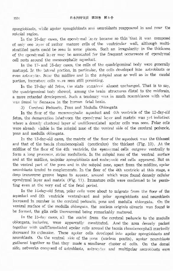
3) (、erebralPeduncle, Pons and :'.¥Iedulla Oblongata In the ftlnr of the mesencephalic aqueduct and 4th ventricle of the 12・・day-old
fetus, the demarcation inbetween the epencl~·mal la~℃r and matrix was yet indistinct
where a densely clustered layer of undi百erentiated apolar cells was seen. Polar cells
were already visible in the subpial zone of the ventral side of the cerebral peduncle,
pons and medulla oblongata.
In the 13・clajア・oldcases, the matrix of the floor of the aqueduct was the thinnest
and that of the taenia rhombencephali (ponticulus) the thickest (Fig. 10). At the
midline of the丹o:irof the 4th ventricle, the epernlymal cells outgrew ventrally to
form a long processu日, striaemedullaris. In the subpial zone of the ventral surface
and at the midline, unipolar spongioblasts and embryonic rod cells appeared. But on
the ventral part of the pons and in the subpial zone, apart from the midline, apolar
neuroblasts t~mded to conglomerate. In the floor of the 4th ventricle at this stage, a
deep transverse groove began to appear, around which were found densely cellular
ependymal layer and matrix (Fig. 11). Immature cells were confirmed to be persis-
ting even at the very end of the fetal period.
In the 14-day-old fetus, polar cells were about to migrate from the floor of the
aqueduct and 4th ventricle ventrahrnrcl and polar spongioblasts and neuroblasts
increased in number in the cerebral peduncle, pons and medulla oblongata. On the
ventral surface of the medulla oblongata, the nucleus originis olivaris was found to
be formed, the glia cells therearound being remarkably matured.
In the 15-day cases, all the nuclei from the cerebral peduncle to the medulla
oblongata, inclusive, were apparentl~ア constituted. And the area densely・ packed
together with undi庁erentiatedapolar cells around the taenia rhombencephali markedly
decreased its cx:tension. These apolar cells developed into apolar spongioblasts and
neuroblasts. On the ventral ぉideof the pons (nucleus pontis), apolar neuroblasts
gathered together so that they made a semilunar cluster of cells. On the dorsal
side, networks comp;Jsed of astroblasts, astrc淀川cs and multipolar neuroblasts were
HISTOLOGICAL CHANGES IN THE BRAIN OF MALFORMED FETUSES 1535
seen. In the 18・and19・day 伺 ses, all the cells inbetween the cerebral peduncle and
medulla oblongata further differentiated, and astroblasts and astrocytes with fine
processes were uncovered especially in the lateral parts and adjacent to the pia mater.
The nerve cells constituting various nuclei also progressively di百erentiated,although
apolar spongioblasts and neuroblasts were observed to have persisted in some part
of the taenia rhombencephali. Ventral to the p:mtine nucleus and subpially were also
seen apolar neuroblasts and spongioblasts (Fig. 12).
b) Prosencepha lon
1) Diencephalon
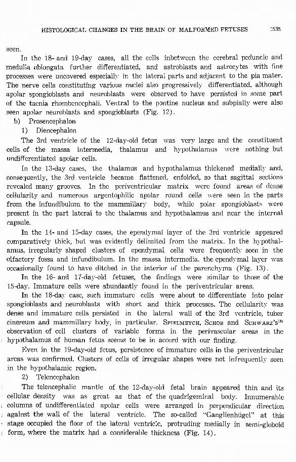
The 3rd ventricle of the 12-day-old fetus was very large and the constituent
cells of the massa intermedia, thalamus and hypothalamus "℃ re nothing but
undi仔erentiateclapolar cells.
In the 13・daycases, the thalamus and hypothalamus thickened medially and,
consequently, the 3rd ventricle became flattened, enfolded, so that sagittal sections revealed many grooves. In the periventricular matrix were found areas of dense cellularity and numerous argentophilic apolar round cells were seen in the parts from the infundibulum to the mammillary body, while polar spongioblasts were
present in the part lateral to the thalamus and hypothalamus and near the internal
capsule.
In the 14-and 15-day cases, the ependymal layer of the 3rd ventricle appeared
comparatively thick, but was evidently delimited from the matrix. In the hypothal-amus, irregularly shaped clusters of ependymal cells were frequently seen in the
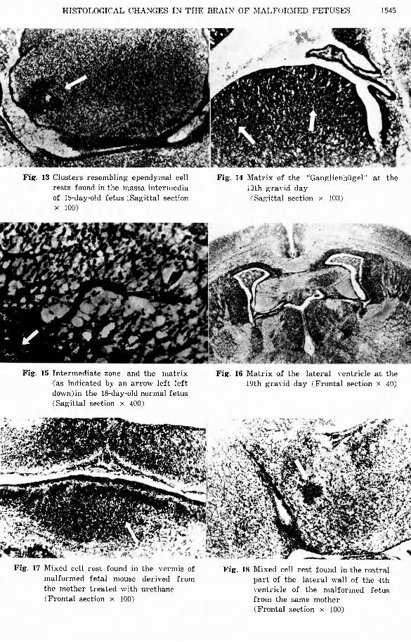
olfactory fossa and infundibulum. In the massa intermedia, the ependymal layer was
occasionally found to have ditched in the interior of the parenchyma (Fig. 13).
In the 16・and 17-day-old fetuses, the findings were similar to those of the
15-day. Immature cells were :'.lbundantlyア foundin the perかentricularareas.
In the 18・da)’ case,such immature cells were about to differentiate into polar
spongioblasts and neuroblasts with short and thick processes. The cellularity was
dense and immature cells persisted in the lateral wall of the 3rd ventricle, tuber
cinereum and mammillary body, in particular. SPIELMEYCH, ScHOB and ScHw ARz's13J observation of cell clusters of variable forms in the perivascular areas in the
hypothalamus of human fetus seems to be in accord with our finding.
Even in the 19・day-oldfetus, persistence of immature cells in the per討entricular
areas was confirmed. Clusters of cells of irregular shapes were not infrequently seen
in the hypothalamic region.
2) Telencephalon
The telencephalic mantle of the 12-day-old fetal brain appeared thin and its
cellular density was as great as that of the quadrigeminal body. Innumerable
, columns of undi町erentiated apolar cells were arranged in perpendicular direction
against the wall of the lateral ventricle. The so-called “Ganglienhi.igel'’ at this
, stage occupied the floor of the lateral ventricle, protruding medially in semi-globoid
form, where the matrix had a considerable thickness (Fig. 14).
1536 白木外科宝函第28巻第5号
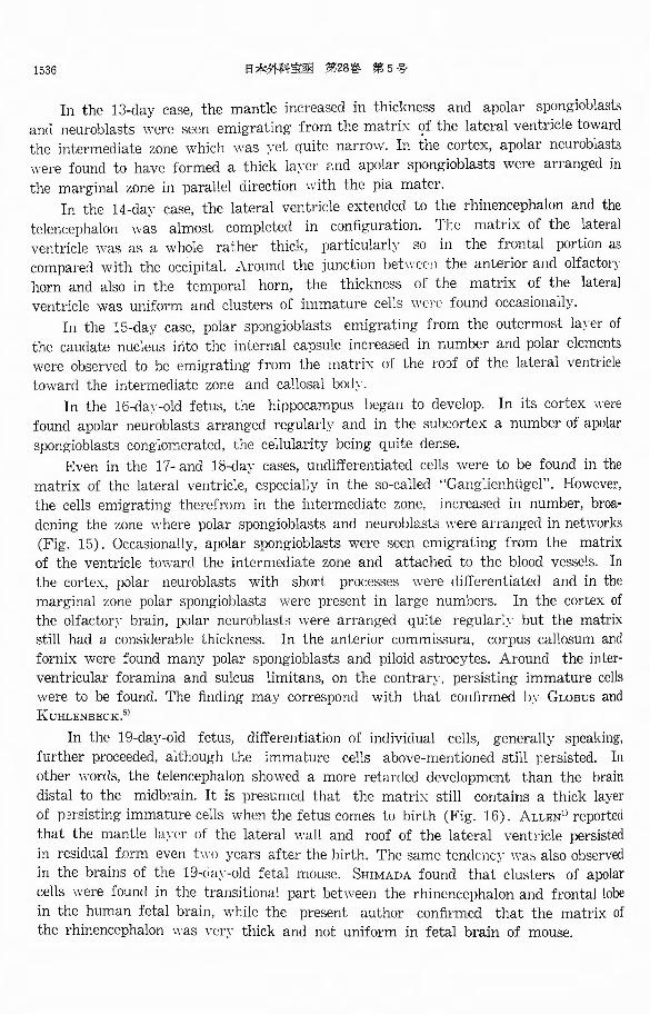
In the 13・daycase, the mantle increased in thickness and apolar spongioblasts
and neuroblasts were seen emigrating from the matrix of the lateral ventricle toward
the intermediate zone which was ~·et quite narmw. In the cortex, apolar neuroblasts
were found to have formed a thick layer ['.nd apolar spongioblasts were arranged in
the marginal zone in parallel direction with the pia mater.
In the 14・daycase, the lateral ventricle extended to the rhinencephalon and the
telencephalon was almost completed in configuration. The matrix of the lateral
ventricle was as a whole rather thick, particularly so in the frontal portion as
compared with the occipital.λround the junction bet\\℃むnthe anterior and olfactory
horn and also in the temporal horn, the thickness of the matrix of the lateral
ventricle was uniform and clusters of immature cells mでrefound occasionally.
In the 15・daycase, polar spongioblasts emigrating from the outermost layer of
the caudate nucleus irito the internal capsule increased in number and polar elements
were observed to be emigrating from the matrix of the ro'.)f of the lateral ventricle
toward the intermediate zone and callosal body.
In the 16・d旬、oldfetus, the hippocampus began to develop. In its cortex were
found apolar neuroblasts arranged regularly and in the subcortex a number of apolar
spongioblasts conglomerated, the cellularity being quite dense.
Even in the 17-and 18・daycases, undi百erentiatedcells were to be found in the
matrix of the lateral ventricle, especially in the so-called “Ganglienhiigel”. However,
the cells emigrating therefrom in the intermediate zone, increased in number, broa-
dening the zone where polar spongioblasts and neuroblasts were arranged in networks
(Fig. 15). Occasionally, apolar spongioblasts were seen emigrating from the matrix
of the ventricle toward the intermediate zone and attached to the blood vessels. In
the cortex, polar neuroblasts with short processes were di仔erentiated and in the
marginal zone polar spongioblasts were present in large numbers. In the cortex of
the olfacton・ brain,polar neuroblasts were arranged quite regular]~’ but the matrix
still had a considerable thickness. In the anterior commissura, corpus callosum and
fornix were found many polar spongioblasts and piloid astrocytes. Around the inter-
ventricular foramina and sulcus limitans, on the contrary, persisting immature cells
were to be found. The finding may correspond with that confirmed by Gwsus and
KUHLENBECK.5)
In the 19・day-old fetus, di百erentiationof individual cells, generally speaking,
further proceeded, although the immature cells above-mentioned still persisted. In
other words, the telencephalon showed a more retarded development than the brain
distal to the midbrain. It is presumed that the matrix still contains a thick layer
of p2rsisting immature cells when the fetus comes to birth (Fig. 16). ALLEN1> reported
that the mantle la~℃r of the lateral wall and roof of the lateral ventricle persisted
in residual form even two years after the birth. The same tendency w出 alsoobserved
in the brains of the 19-d町二oldfetal mouse. SHIMADA found that clusters of apolar
cells were found in the transitional part between the rhinencephalon and frontal lobe
in the human fetal brain, while the present author confirmed that the matrix of
the rhinencephalon was very thick and not uniform in fetal brain of mouse.
HISTOLOGICAL CHANGES IN THE BRAIN OF MALFORMED FETUSES 1537
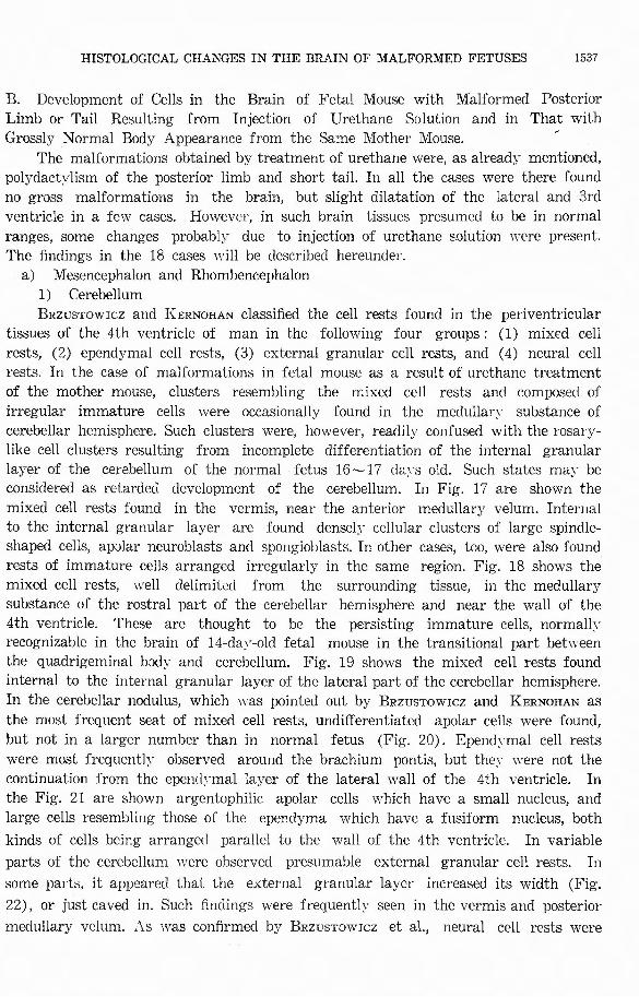
B. Development of Cells in the Brain of Fetal Mouse with Malformed Posterior Limb or Tail Resulting from Injection of Urethane Solution and in That with
Grossly Normal Body Appearance from the Same Mother Mouse. '
The malformations obtained by treatment of urethane were, as alread)ア mentioned,
polydact~'lism of the posterior limb and short tail. In all the cases were there found
no gross malformations in the brain, but slight dilatation of the lateral and 3rd
ventricle in a few cases. However, in such brain tissues presumed to be in normal
ranges, some changes probably due to injection of urethane s.olution were present.
The findings in the 18 cases will be described hereunder.
a) Mesencephalon and Rhombencephalon
1) Cerebellum
BRzusTow1cz and KERNOHAN classified the cell rests found in the periventricular tissues of the 4th ventricle of man in the following four groups: (1) mixed cell
rests, (2) ependymal cell rests, (3) external granular cell rests, and ( 4) neural cell
rests. In the case of malformations in fetal mouse as a result of urethane treatment
of the mother mouse, clusters resembling the mixed cell rests and composed of
irregular immature cells were occasionally found in the medullar・3’ substanceof cerebellar hemisphere. Such clusters were, however, readily confused with the rosary-
like cell clusters resulting from incomplete differentiation of the internal granular
layer of the cerebellum of the normal fetus 16~17 c1a}・s old. Such states ma}ァ beconsidered as retarded development of the cerebellum. In Fig. 17 are shown the
mixed cell rests found in the vermis, near the anterior medullary velum. Internal to the internal granular layer are found densely cellular clusters of large spindle-
shaped cells, apolar neuroblasts ancl spongioblasts. In other cases, too, were also found rests of immature cells arranged irregularly in the same region. Fig. 18 shows the
mixed cell rests, well delimited from the surrounding tissue, in the medullar:yア
substance of the rostral part of the cerebellar hemisphere and near the wall of the 4th ventricle. These are thought to be the persisting immature cells, normally recognizable in the brain of 14-day-old fetal mouse in the transitional part between
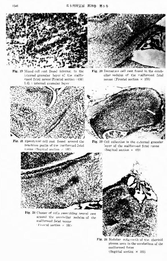
the quadrigeminal body and cerebellum. Fig. 19 shows the mixed cell rests found
internal to the internal granular layer of the lateral part of the cerebellar hemisphere.
In the cerebellar nodulus, which was pointed out by BRzusTow1cz and KERNOHAN as
the most frequent seat of mixed cell rests, undi町erentiatcd apolar cells were found,
but not in a larger number than in normal fetus (Fig. 20). Ependymal cell rests
were most frequently observed around the brachium pontis, but th匂’ werenot the
continuation from the epenc1γmal layer of the lateral wall of the 4th ventricle. In
the Fig. 21 are shown argentophilic apolar cells which have a small nucleus, and
large cells resembling those of the epend:yァma which have a fusiform nucleus, both
kinds of cells being arranged parallel to the wall of the 4th ventricle. In variable
parts of the cerebellum were observed presumable external granular cell rests. In
some parts, it appeared that the external granular layer increased its width (Fig.
22), or just caved in. Such findings were fr判 1淀川lyseen in the vermis and posterior
medullary velum. As was confirmed by BRzusTow1cz et al., neural cell rests were

1538 日本外科宝函第28苦手第5号
found quite rarely. In Fig. 23 were seen cell clusters rich in apolar spongioblasts,
but apolar neuroblasts were also seen intermingled. Beside the variable cell rests
aboveベvrittcn,the pial membrane of the cerebellum was occasionally seen to have
caved in the cつrtcx.Proliferation of cells took place also in some part of the chorioid
plexus of the 4th ventricle, forming nodules (Fig. 24).
2) Mesencephalon
The cell rests in the midbrain appeared largely around the mesencephalic aque-
duct. This can be reasoned from the fact that the wall of the aqueduct, which
was initially quite a large cavitγ,\ms gradual!~マ enfolded as the quadrigeminal body
and cerebral peduncles became thicker and that the thickness of the ependymal
layer was not uniform in some parts (at the 14th and 15th gravid day). In the
brain of the malformed mouse (malformation either of the posterior limb or tail),
such a tendenc~· w::i.s much pronounced and clusters of cells simulating ependymal
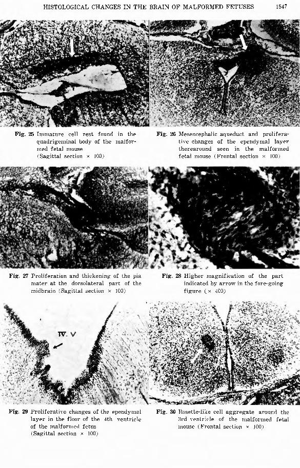
cell rests were uncovered around the aqueduct. The Fig. 25 shows densely packed
cell conglomerates arranged parallel to the aqueduct in the caudal portion of the
quadrigeminal body, some of which were in close contact with the ependymal l何百.
In the transitional part b:ctwc2n the aqueduct and 3rd ventricle, the ependymal layer
was seen proliferating (Fig. 26). In the cases with urethane treatment, proliferative
abnormality was confirmed not only around the aqueduct but also in other parts of
the brain. Changes also of the pia mater were noticed in the midbrain. As shown
in Figs. 27 and 28, cells of the pial membrane which had indistinct outline and a
large fusiform nucleus were seen proliferated, forming large clusters in the dorso-
lateral surface of the quadrigeminal bod~· , where this was in close contact with the
occipital lobe. These proliferative changes of the pia mater were in resemblance with
the outgrowth of the chorioid plexus of the cerebellum of the mouse treated with urethane (Fig. 24).
3) Pons and Medulla Oblongata
Cell rests were scarcely noticed in the pons and medulla oblongata. However, around the transverse sulcus of the 4th ventricle resulting from bending and folding
of the pons at the 13th gravid day, persisting immature cells could be observed even
in the normal cases. In the brain of the mouse treated with urethane, such a
tendency was much conspicuous and more marked and characteristic proliferative
changes could be seen. Fig. 29 is one of the most representative illustrations, in
which are shown multiple protrusions in irregular forms of the ependymal layer
toward the ventricular cavity. The cells which constituted these protrusions were
1mmature cells which had scanty cytoplasm and an argentophilic oval nucleus. The
:_pendymal la~-er in other parts was thin and the ependymal cells were quite matured. tlRzusTow1cz et al. and SHIMADA reported that epemlymal cell rests were frequently
observed in the taenia rhombencephali of the human fetal brain. In the brain of
~ouse treated with urethane, no such cell rests we1℃ present and protrusions of
1mmature ependymal cells similar to those described above were predominantly found.
b) Prosencephalon
HISTOLOGICAL CHANGES IN THE BRAIN OF MALFORMED FETUSES 1539
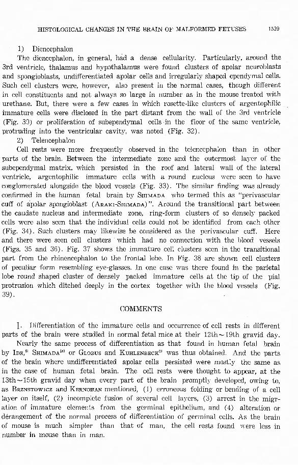
1) Diencephalon
The diencephalon, in general, had a dense cellularity. Particularly, around the
3rd ventricle, thalamus and hypothalamus were found clusters of apolar neuroblasts
and spongioblasts, undi百erentiatedapolar cells and irregularly shaped ependymal cells.
Such cell clusters were, however, also present in the normal cases, though different
in cell constituents and not always so large in number as in the mouse treated with
urethane. But, there were a few cases in which rosette-like clusters of argentophilic
immature cells were disclosed in the part distant from the wall of the 3rd ventricle
(Fig. 30) or proliferation of subependymal cells in the floor of the same ventricle,
protruding into the ventricular cavity, was noted (Fig. 32).
2) Telencephalon
Cell rests were more frequently observed in the telencephalon than in other
parts of the brain. Between the intermediate zone and the outermost layer of the
subependymal matrix, which persisted in the roof and lateral wall of the lateral
ventricle, argentophilic immature cells with a round nucleus were seen白 have
conglomerated alongside the blood vessels (Fig. 33). The similar finding was already
confirmed in the human fetal brain by SHIMADA who termed this as“perivascular cuff of apJlar spongioblast (ARAKI-SHIMADA)”. Around the transitional part between the caudate nucleus and intermediate zone, ring-form clusters of so densely packed
cells were also seen that the individual cells could not be identified from each other
(Fig. 34). Such clusters may likewise be considered as the perivascular cuff. Here and there were seen cell clusters which had no connection. with the blood vessels
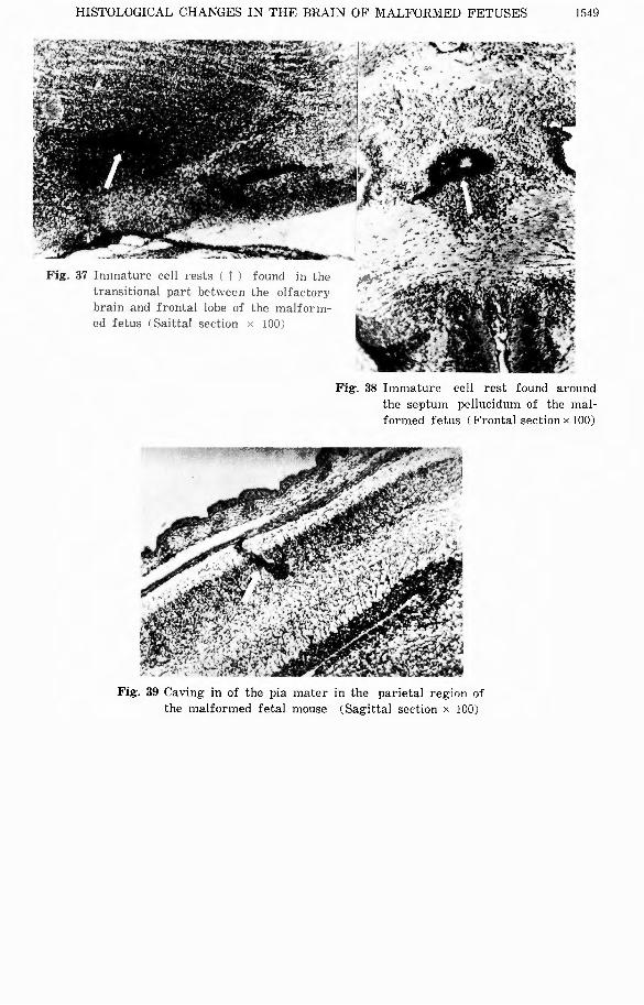
(Figs. 35 and 36). Fig. 37 shows the immature cell clusters seen in the transitional
part from the rhinencephalon to the frontal lobe. In Fig. 38 are shown cell clusters
of peculiar form resembling eye-glasses. In one case was there found in the parietal lobe round shaped cluster of densely packed immature cells at the tip of the pial
protrusion which ditched deeply in the cortex together with the blood vessels (Fig. 39).
COMMENTS
J. Di町erentiationof the immature cells and occurrence of cell rests in di百erentparts of the brain were studied in normal fetal mice at their 12th~19th gravid day.
Nearly the same process of differentiation as that found in human fetal brain by lDE,6) SHIMADA14J or GLoBus and KuHLENBECK5J was thus obtained. And the parts
of the brain where undi古erentiated apolar cells persisted were mostly the same as
in the case of human fetal brain. The cell rests were thought to appear, at the
13th~15th gravid day when every part of the brain promptly developed, owing to,
as BRzNsTowrcz and KERNOHAN mentioned, (1) erroneous folding or bending of a cell
layer on itself, (2) incomplete fusion of several cell layers, (3) arrest in the migr-
ation of immature elements from the germinal epithelium, and ( 4) alteration or
derangement of the normal process of di町erentiationof germinal cells. As the brain
of mouse is much simpler than that of man, the cell rests found were less in
number in mouse than in man.
1540 日本外科宝函第28巻 第5号
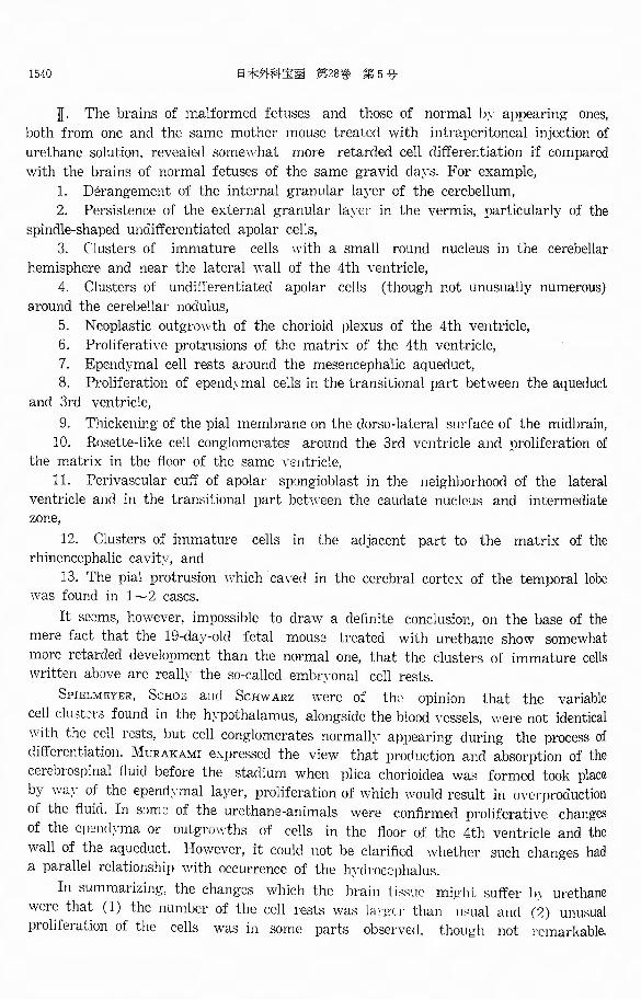
JI. The brains of malformed fetuses and those of normal by appearing ones,
both from one and the same mother mouse treated with intraperitoneal injection of
urethane solution, revealed somewhat more retarded cell di百erentiationif compared
with the brains of normal fetuses of the same gravid da~'S. For example,
1. Derangement of the internal granular layer of the cerebellum,
2. Persistence of the external granular layer in the vermis, particularly of the
spindle-shaped undi苛erentiatedapolar cells,
3. (、lustersof immature cells with a small round nucleus in the cerebellar
hemisphere and near the lateral wall of the 4th ventricle,
4. Clusters of undifferentiated apolar cells (though not unusually numerous)
around the cerebellar nodulus,
5. Neoplastic outgrowth of the chorioid plexus of the 4th ventricle,
6. Proliferative protrusions of the matrix of the 4th ventricle,
7. Ependymal cell rests around the mesencephalic aqueduct, 8. Proliferation of ependymal cells in the transitional part between the aqueduct
and 3rd ventricle,
9. Thickening of the pial membrane on the dorso-lateral smface of the midbrain,
10. Rosette-like cell conglomerates around the 3rd ventricle and proliferation of the matrix in the floor of the same vcntriclc,
11. Peri-Vascular cu古 ofapolar spongioblast in the neighborhood of the lateral
ventricle and in the transitional part between the caudate nucleus and intermediate zone,
12. Clusters of immature cells in the adjacent part to the matrix of the rhinencephalic cavit~-, and
13. The pial protrusion which caved in the cerebral cortex of the temporal lobe was found in l~2 cases.
It seems, however, impossible to draw a definite conclusion, on the base of the
mere fact that the 19・day-old fetal mouse treated with urethane show somewhat
more retarded development than the normal one, that the clusters of immature cells written above are really the so-called embryonal cell rests.
SPIELMEYER, Scttoz ancl Scttw ARZ were of the opinion that the variable
cell clu3t:;rs found in the hypothalamus, alongside the blood vessels, were not identical
with the cell rests, but cell conglomerates normally appearing during the process of
di庁erentiation.MURAKAMI expressed the view that production and absorption of the
cerebrospinal fluid before the stadium when plica chorioidea wa日 formedtook place
by w何- of the epend~·mal layer, proliferation of which would result in overproduction of the fluid. In srimc of the urethane-animals were confirmed proliferative changes
of the cpenclγma or outgrowths of cells in the floor of the 4th ventricle and the
wall of the aqueduct. However, it could not be clarified whether such changes had a parallel relationship with occurrence of the hγdroccphalus.
In summarizing, the changes ¥Vhich the brain 1 issue mii . .dit su汀erlハurethane
were that (1) the numl.Jer of the cell rests was lai-どι1・than usual and (2) unusual
proliferation of the cells was in some parts observed, though not I℃markable.
HISTOLOGICAL CHANGES IN THE BRAIN OF MALFORMED FETUSES 1541
Urethane had rather slight influence on the central nervous system. This can readily
be presumed, as gross malformations rarely occur in the brain by urεthane treatment.
It should, however, be noted that some histological abnormality may be detected in
the brain, when some malformation is actually or potentially presεnt in some part
of the body other than the brain. Since gross malformations are not always uncovered
in patients harboring a glioma, we must mean histological abnormalities, whrn we
say that congenital developmental anomalies may have something to do with occur-
rence of gliomas. Therefore, cell rests and or persisting immature cells were called in question, which, however, were not particularly rich in number, as far as the
present study was concerned, in the cerebellum, the most frequent seat of predi-
lection for occurrence of gliomas in children. On the contrary, such cells were more frequently found in the cerebrum, a fact indicating the complicacy of the problem
under discussion.
CONCLUSIONS
(1) The process of development of nerve cells and glia cells in the brain and occurr町田 ofcell rests were studied in the 18 brains of normal fetal mice at their 12~19th gravid day. The results obtained were that the precess of di百erentiationof the brain of fetal mouse roughly corresponded with that of man.
(2) In the brains of 18 malformed fetuses (either with pol:yァdactylismor with
a short tail) from one and the same mother mouse treated with urethane, somewhat
mote retarded development tended to take place if compared with the normal
contrasts.
(3) Both the malformed and seemingly normal fetuses, derived from the same
mother mouse treated with urethane, revealed more frequent occurrence of cell rests
than contrast fetuses, though the difference was not quite conspicuous. There were
found a few cases in which the ependymal layer, subependyma, pial membrane and chorioid plexus showed proliferative changes.
( 4) When some gross malformation was present in some part of the body or
when such a defective constitution was inherited in spite of no gross abnormality,
more or less abnormal changes histologically might be found in the brain. Should
congenital developmental anomaly 切vesomething to do with occurrence of a glioma,
such histological anomalies must be called in question. Cell rests and persisting
immature cells may be given as such, which, however, were not always rich in
number, as far as the present study was concerned, in the cerebellum but they were rather more in the cerebrum. This may indicate the complicacy of the problem under discussion, i. e. congenital histological anomalies and occurrence of gliomas.
The present study was supported by a grant in aid from the Ministry of Education.
REFERENCES
1) Allen, E.: The cessation of mitosis in the central nervous system of the albino rat. J. Comp. Neurol., 22, 547, 1912.
2) Bailey, P., and Cushing, H. : A classilica.ti(,n of tumor of the glioma group etc. Philadel-phia, Lippincott, 1926.
3) Brzustowicz, R. J., and Kernohan, J. W. : Cell rest in the region of the fourth ventricle,

1542 日本外科宝函第28巻第5号
(a) Their site and incidence according to age and sex. Arch. Neurol. and Psychiat., 67, 585,
1952.
(b) Histologic and embryologic condition. ibid, 67, 59士, 1952.
IC) Their relation to the development of gliomas. ibid, 67, 602, 1952.
4) Cushing, H.: Experiences with cerebellar astrocytomas. Critical review of 76 cases. Surg.
Gynec. and Obst., 52, 129, 1931.
5) Globus, J. H., and Kuhlenbeck, H. : Tumor of the striatothalamic and related region. Their
probable source of origin and more common forms. Arch. Path., 34, 674, 1942.
6) Ide, Z.: Embryologic studies on th2 cytogenesis and structure of glial cell. Japan Path.
s .. 38, 163. 1949.
7) Murakami, U.: Manifestation of some abnormalities in abnormal environments. Studi.2s on
phenocopies in mice (First report). Jap. J. Genet., 27, 176, 1953.
8) Nakahara, W.: Problem in cancer research. Sogoigaku, 10, 10, 517, 1953,
9) Nishimura, H. : How do environmental factors put influence on the congenital constitution,
as viewed from the experimental study of constitution in the human fetus. Saishinigaku,
11, 12, 1956.
10) Ostertag, B.: l¥Iissbildungen, in Lubarsch et al.: Handbuch d. sp2z. path. Arat. u. Histol.,
Bd. 13, N. Tei!, Kap. X. 577, 1956.
11) Pfleger, L.: B2obachtungen iiber het.crctopic grauer Substanz im Mark des Kleinhirns.
Zentralbl. f. med、 Wissensch.,18, 468, 1880.
12) Raff, J. and Kernohan, J‘ W. : Relation of abnornnl coll巴ctionof cells in posterior medull-
ary刊 lumof cerebellum to origin of medulloblastoma. Arch. Neurol. & Psychiat., 52, 163, 1944.
13) Schwarz, H., Goolker, P. & Globus, J. H.・Thenorm:il histology of infant's brains. Am. J. Dig. Chile, 43, 889, 1932.
14) Shimada, 1¥1.: Persistence and misplacement of immature glial cells in various parts of
the human fetal brain and their possible relation to the devεlopment of gliomas. Folia Psychiat. et x’eurol. Japon., 8, 238, 1954.
15) Shirota, G.: On the possible relationship of the predilection site for the glial abnormalities
in the malformed fetal mouse brain to that for the human infant’s gliomas. Arch. f. Jap. Chir., 37, 579, 1958.
HISTOLOGICAL C'HA:-.JGES I:-..1 THE BRAIN OF MALFOR:'IIED FETUSES 1543
Fig. 1 Malformed fetal mouse lPOlydactylism; derived from the mother mouse treated with urethane at the 19th gra¥'id day
Fig. 3 Cerebellum and quadrigeminal body of 13-day-old f巴talmouse (Sagittal section × 400)
N : cerebellar nodulus Aq : mesencephalic aqueduct E : external granular layer of the
cerebellum Ma : anterior medullary ,-clum
Fig. 5 Showin邑・ apolar cells of neuroblastic and spongioblastic series (Silver diam-ino・carbonatemethod x 1000) N : apolar neuroblast S : apolar spongioblast
Fig・. 2 Short tail in the same <le白cen<lantat the same fetal day
Fig. 4 Showing undifferentiated apolar cells (Sih・er <liamino-carbonate method ×1000)
l<'ig. 6 The arrow indicates percursor of the external granular layer of the cereb-ellum of 15-day-old normal fetal mo・use (Sagittal section x 400)
154-1 日本外科宝函第28巻第5号
、r
Fig・. 11 Hests of immature cells in the
transverse sulcus of the floor
of the 4th、cntricleof 1 :J-day-
old normal fetus <Sagittal section × 100)
匂,....ぶ
Fig-. 10 Matrix of the floor of the aqueduct
and Ith、cntricleof 1 ~寸lay-old normal fetus
(Sagittal section× 40)
P : ponticulus(taenia rhombencephali) 「: cerebellum
HISTOLOGICAL CHANGES IN THE BRAIN OF :VIALFOI-mED FETUSES 1545
Fig・. 15 Intermediate zone and the matrix (as indicated by an arrow left left down)in the 18-day-old normal fetus (Sagittal section × 400)
ー _ ....._,.,..._...,.._一-- ·~現~曙;,_;--
Fig. 14 Matrix of the “Ganglienhiigel'’ at the
13th gra円dtlay (Sag・ittal section × 100)
Fig・. 16 Matrix of the lateral 、entricleat the 19th gravid day (Frontal section × 40)
1546 白木外科宝函第28巻第5号
Fig-. 19 '.¥fixed cell rest found internal to the Fig・. 20 Immature cell rest found in the cereb・
internal granular layer of the malfo- ellar nodulus of the malformed fetal
rme<l fetal mouse(Frontal sectionメ400) mouse (Frontal section× 100)
Fig-. :.Uβpendymal cell rest found around the Fig-. 22 Cell collection in the external granular
brachium pontis of the malformed fetal layer of the malformed fetal mouse
γ11ouse 'Sag・ittal section •' 100) (Sa宵ittalsection x JOO)
Fig-. 23 Cluster of cells resembling neural rest
around the cercl》〈、liar noclulus of the
malformed fetal mouse
'Frnnl乱lsection × 100)
・”ヨζ
Fig-. 24 Nodular outιrnwth of the chorioid
plexus seen in the cerebellum of the
malformed fetus
(Sag・ittal section x 100)
HISTOLOGICAL CHANGES IN THE BRAIN OF MALFORMED FETUSES 1547
認・・・・F -ー・...,,..-・'""τ.Fig・. 27 Proliferation and thickenin宮 ofthe pia
mater at the dorsolateral part of the
midbrain (Sagittal section x 100)

1548 日本外科宝函第ヨ8巻第5号
主,A.
HISTOLOGICAL CHANGES IN THE BRAIN OF MALFORMED FETUSES 1549
Fig._ 37 Im111atu1・c cell rests < i I found in lhc trnnsitional part bet、,・ccnthe olfac:Lor:-' brnin and frontal lobe of the malfo1・Ill-ed fetus < Sai ttal sec:Liun× 100) ~
./'!/ 、-
2・4「シ’~
Fig-. 38 Immature cell rest found around
the septum pellucidum of the mal-formed fetus (Frontal section×100)
1550 日本外科宝函第2B~会第 5号
和文抄録
ウレタン処置崎形マウス脳の組織学的変化
ー一 人間小児脳グリオームの発生より見fこるー
京都大学医学部外科学教室第 1講座 (指導:荒木千里教授)
山崎徳雄
最近実験的に,種々な外因で崎形を発生せしめ得る
ことが分ったがp 筒形を発生させる外因の中のあるも
のは腰湯舟も惹起せしめ得るのでは無いかと考えられ
ている(Strong1945!.それで脳組織の様に,その正
常な発育過程中に於てもp 一種の組織崎形と見倣すべ
きCellrests舟生ずる臓器に' {.催時形因子手作用させ
れば,更に多くの Cell rests を発生するのではない
か,そしてそれ;土より大なZ腫疹発生素因を意味する
ものではないかということが考えられる.それでp 私
は正常マウス胎仔脳の発育過程を組織学的に検索しp
マウス胎仔脳に於ける Cellrestsの存在及び未成熟細
胞残存好発部位を追求した後p 西村氏法に従って妊娠
10日目の母マウス腹腔内にエチールウレタンを注入す
第ることによって生じた崎形胎仔の脳(妊娠第19日目
に関l復して取出した)を同様組織学的に検索しF i欠の
結果を得た.
A. 正常マウス胎仔脳に於ける神経細胞及びゲリア
細胞の分化過程及び Cellrestsの発生について.
在常マウス胎仔月以に於ける神経細胞並び、にグリア細
胞の成熟過程はp 先に島田3 井出y Globus及びKuhl-
enheekがノ問!同厄に就いて検索したと略々同様であ
りp 未成熟細胞残存部も人間胎児のものに似ており,
小脳外新粒層p 小脳結節p 菱脳紐,四丘休特に下丘,
間脳腹側,側脳室周辺 Ganglienhiig巴l及び喫脳室周
辺であった.Cell restsの発現に関してはp 胎生13日
目頃から15日間頃にかけてp 脳各部が急速に発達す
る時期に生ずると考えられP Brzustowicz及び
Kernohanの見解に一致する様である.併しマウス脳
は人間の脳と比べると単純である為Cellrestsの発現
数は人間胎仔脳に比べて透かに少数である.
B. ウレタン処置を行った崎形胎仔及び同腹の外見
上正常胎仔朕iに就いて.
この場合同日数の正常胎仔脳に比べp 多少成熟が遅
延する傾向が認められP Cell restsの発現も若干対照
例よりも多い.併し小児グリオームの最大好発部位で
ある小脳よりもむしろ大脳に多く認められた事:;,
Cell 1estsの発現と脳腫蕩の関連性という問題の複雑
さを示唆するものと思われる.又,第四脳室底部上1¥
下層y 中脳水道周辺Matrix,第三脳室底部:\fatrixに
於ける増殖性変化p 第四脳室脈絡叢の増生p 中脳背外
側軟脳膜肥厚等の変化をも認めたが,ウ Lタンの中枢
神経系に与えた作用は一般bこいつて軽微であった.