Embed Size (px)
Citation preview
1309
Int. J. Morphol.,28(4):1309-1314, 2010.
Histological Changes in Ovaries of Mice Exposedto Butea monosperma: Preliminary Study
Cambios Histológicos en Ovarios de Ratón Expuestos a Butea monosperma: Estudio Preliminar
*Neelam Gupta; **Gajendra Singh; ***S. M. Singh & ****K. R. C. Reddy
GUPTA, N.; SINGH; G.; SINGH, S. M. & REDDY, K. R. C. Histological changes in ovaries of mice exposed to Butea monosperma:preliminary study. Int. J. Morphol., 28(4):1309-1314, 2010.
SUMMARY: In Ayurvedic practice Butea monosperma (Palash) is in clinical use for hundreds of years as a contraceptive. Seedsof Butea monosperma are also used as an anthelmitic (Ansani et al., 1979) and antimicrobial (Avirutnant & Pongpan, 1983). Buteamonosperma (Fabaceae family) locally known as Palash (Dhak) if given for 3 consecutive days acts as an antifertility agent for which ithas been is traditionally used since time immemorial. The objective of the present study was to search the effect of Butea monospermaseeds on the ovary of mice. Observations in the present study were massive degeneration of ova in almost all the follicles, irrespective ofthe stage of their development. The ova from treated animals showed different stages of necrotic process. Moreover, the arrangement offollicular cells was also disturbed. The Palash seeds in the form of powder when administered orally with distilled water, according to thebody weight i.e.2g/Kg, of female mice, for three consecutive days showed notable changes in ovaries. The animals were sacrificed onday next to the last day of treatment and ovaries were extirpated. Ovaries studied histologically after Haematoxylin & Eosin stainingshowed most of the follicle in immature state with undefined nucleus and nucleoli in the ovum. Others showed degenerative changes inthe ovum. Follicles had lost their normal shape and arrangement and organization of granulosa cells. It was conspicuous to find thatalmost all follicles including graafian follicles of treated ovaries were undergoing degenerative changes simultaneously. The rate ofapoptosis in the granulosa cells when studied was found increased in treated cases as compared with control. The study suggests that thedisintegration of ova in the ovaries is a specific effect of Butea monosperma seed administration.
KEY WORDS: Palash; Butea monosperma; Apoptosis; Disintegration; Follicles.
INTRODUCTION
Various Ayurvedic preparations are known to interferewith the reproductive ability of women (Bhattachaya et al.,1980; Chaundry, 1993; Takase et al., 1995; Tandon et al.,2003; Prasad et al., 2006; Ganguly, 2007). These preparationsare in clinical use in Ayurveda for thousands of years toprevent pregnancy. However, the mechanism of action ofthese preparations in interfering with the process ofconception remains largely unclear.
The ancient literature of Ayurveda, Yogachintamani:Mishradhikar-1, mentions that a recipe for producing sterilityin the prostitutes is by using seeds of Palash (Buteamonosperma) taken with water in the form of a fine powderfor three consecutive days following menstruation. In another
ancient Ayurvedic literature designated as the Garudapuran,198:27, it is stated that Palash when made to a paste by addinghoney and then taken by the women during the menstrualperiod leads to sterility, preventing both menstruation andconception. In Yogratna Samuchaya, Anant Kumar, YogratnaSamuchchaya – Part III Bandhya – Rogadhikar, 187,Travancore University, Trivandrum (1940) there is nomention of the prevention of conception by the intake ofthis recipe but only the blockage of the menstruation isdescribed as one of its primary effect. In order to understandthe mechanism of the antireproductive action of Palash(Butea monosperma) seeds on ovary, the present study wasundertaken. To study the effect of oral administration ofPalash (Butea monosperma) seeds on the ovarian histology.
* PhD Scholar, Department of Anatomy, Institute of Medical Sciences Banaras Hindu University, India.** Professor, Department of Anatomy, Director, Institute of Medical Sciences Banaras Hindu University, India.*** Professor School of Biotechnology, Banaras Hindu University, Varanasi-221 005, U.P. India.**** Reader, Department of Rasashatra, Faculty of Ayurveda, Institute of Medical Sciences, Banaras Hindu University, Varanasi-221 005, U.P. India.
1310
Mice were used as an animal model. Hence a need was feltto study scientifically if such claims about Palash (Buteamonosperma) are true, and this can be used effectively as adrug to prevent conception.
MATERIAL AND METHOD
Twenty seven Swiss white female mice weighing 18-34 g and age of about 50 days were used. These were handledwith utmost human care in an animal laboratory having airconditioned room with temperature maintained at 75º F, dailydark and light cycle of 12 hours each with humidity at 40%.The mice were fed on commercially obtained diet pellets(Hindustan Lever, Mumbai, India) and tap water wasprovided ad libitum.
Mature seeds of Palash (Butea monosperma) werecollected from a tree in the botanical garden of departmentof Dravyaguna, Faculty of Ayurveda. These were made dryand powdered in a grinder in the Ayurvedic Pharmacy of theInstitute. This powder was then sieved to isolate a finepowder. The seed powder of Butea monosperma wasadministered orally in the dose of 2g/kg body weight, mixedwith 0.5ml of distilled water through a cannula fitted to asyringe, for three consecutive days to adult female Swissalbino mice of reproductive age group (weighing 20-34g,nulliparous). The mice were sacrificed by overdose of etheranaesthesia on the day next to last day of treatment. In thecontrol mice no drug was administered only vehicle (distilledwater) was given orally. After opening the abdomen bymidline incision, both uterine horns and ovaries wereinspected and removed from mice and collected in 10%formalin after these were examined under dissectingmicroscope for external morphology. Ovaries were fixed,and cut in sections of 8 micron thickness and stained withHaematoxylin & Eosin and observed under a lightmicroscope.
The ovaries were weighed after dissecting them outfrom the mice and wiping them dry with blotting paper. TheSartorius make (MC 210 P) balance was used for this purpose
which had a minimum count of 0.0l mg. Only those ovarieswhich could not be dissected out in toto and damaged werediscarded.
RESULTS AND DISCUSSION
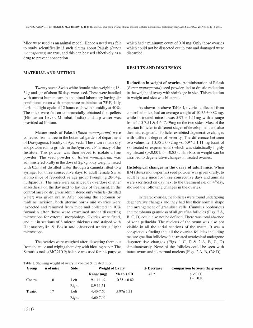
Reduction in weight of ovaries. Administration of Palash(Butea monosperma) seed powder, led to drastic reductionin the weight of ovary with shrinkage in size. This reductionin weight and size was bilateral.
As shown in above Table I, ovaries collected fromcontrolled mice, had an average weight of 10.35 ± 0.82 mg.while in treated mice it was 5.97 ± 1.11mg with a rangefrom 4.40-7.51 & 4.6- 7.49mg on the two sides. Most of theovarian follicles in different stages of development and alsothe matured graafian follicles exhibited degenerative changeswith different degree of severity. The difference betweentwo values i.e. 10.35 ± 0.82mg vs. 5.97 ± 1.11 mg (controlvs. treated or experimental) which was statistically highlysignificant (p<0.001, t= 10.83) . This loss in weight can beascribed to degenerative changes in treated ovaries.
Histological changes in the ovary of adult mice. WhenBM (Butea monosperma) seed powder was given orally, toadult female mice for three consecutive days and animalswere sacrificed on day next to the treatment i.e. on 4th day,showed the following changes in the ovaries.
In treated ovaries, the follicles were found undergoingdegenerative changes and they had lost their normal shapeand arrangement of granulosa cells. Cumulus oophoricusand membrana granulosa of all graafian follicles (Figs. 2 A,B, C, D) could also not be defined. There was total absenceof zona pellucida. The nucleus of the ovum was also notvisible in all the serial sections of the ovum. It was aconspicuous finding that all the ovarian follicles includingmature graafian follicles of the treated ovaries had undergonedegenerative changes (Figs. 1 C, D & 2 A, B, C, D)simultaneously. None of the follicles could be seen withintact ovum and its normal nucleus (Figs. 2 A, B, C& D).
Weight of Ovary % Decrease Comparison between the groupsGroup n of mice Side
Range (mg) Mean ± SD
Left 9.1-11.49Control 10
Right 8.9-11.51
10.35 ± 0.82
Left 4.40-7.60Treated 17
Right 4.60-7.40
5.97± 1.11
42.21 p < 0.001t = 10.83
Table I. Showing weight of ovary in control & treated mice.
GUPTA, N.; SINGH; G.; SINGH, S. M. & REDDY, K. R. C. Histological changes in ovaries of mice exposed to Butea monosperma: preliminary study. Int. J. Morphol., 28(4):1309-1314, 2010.

1311
The granulosa cells were well arranged in Graafian follicle of con-trol ovaries, with a demarcated area between the ovum and the granulosacells on one hand and between granulosa cell and theca interna, theca exter-na and stroma of the ovary on the other hand (Fig. 1A). However, in thetreated ovaries the granulosa cells had lost there typical arrangement andwere randomly scattered except a few of them poorly aligned around thesite of ovum (Figs. 3A, B, C).
In histological study of treated ovaries the principal observation wasdegeneration of ova in all the follicles simultaneously, which were in differentstages of their development (Figs. 2A, B, C, D & 3A, B, C).
The first sign noticed during the atresia in a follicle was pyknosis
A. Photomicrograph of a section of control miceovary showing a mature Graafian follicle withovum and its nucleus and nucleolus (Black arrow)which is surrounded by the follicular cells the co-rona radiata (Red Arrow). Zone of granulosa cellsare well defined with a demarcating basementmembrane (Green arrow). The Graafian follicle islocated towards the surface of the ovary as if readyto deliver ovum. The follicle is surrounded by thecaexterna (Yellow arrow) and theca interna cells(Blue arrow). The follicular cells surrounding thefollicular cavity the membrana granulosa (ThinBlack arrow) is well defined. The ovum is attachedto membrana granulosa by cumulus oophoricus(Star) (X 400.)B. The follicle from a treated ovary showing aprominent ovum with well demarcated nucleus(Red arrow) and well defined nucleolus (ThinBlack arrow) the zona pellucida is present only ina limited zone(Blue arrow) and is not present inthe lower part of the photograph (Green arrow).The cells surrounding the ovum can be well definedcorona radiata (Thick Black arrow), however, thegranulosa cells in the region of cavity appearingin its territory, show increased incidence ofapoptosis (Yellow arrows). Such apoptosis is notvisible in the follicular antrum, in the periphery, inFig. 1 A (X1000).C. In this photomicrograph of ovary collected fromtreated mice with BM, the region of the ovum andfollicular antrum are well defined. However, theooplasm shows marked degenerative changeswithout any indication of residue of nucleus ornucleolus. The changes of the section not goingthrough nucleus cannot be ruled out. However, theentire region of corona radiata (Black arrow) andfollicular antrum show massive apoptosis (Redarrows) which can not be seen in Fig. 1 A (X 400).
D. Like the fig. B & C this section also show degenerative changes in the ovum. There is shrinkage and shift of ooplasm to one side(Black arrow) with associated non visibility of nucleus and nucleolus and absence of zona pellucida (Red arrow). The area surroundingthe ovum shows certain cavities (Green arrows). In the stroma of the ovary many vacant areas (Blue arrows) were seen, some of these arefull of fluid (Yellow arrows). The typical appearance of a follicle has been totally lost (X400.)
and fragmentation of the inner granulosacells (Fig. 1C). This was followed by thecloudy changes in the follicles (Valsala &Karpagaganapathy, 2002) (Fig. 3C). Theresulting eosinophilic granular cell residualmaterial passes in to liquor folliculi. Therefollows thinning of the cumulus, freeing ofthe oocyte into the liquor and finallysloughing of the entire follicle leaving avacant space (Figs. 2A, B, C& D).
In treated group ovaries, it wasobserved that the sizes of almost all follicles
GUPTA, N.; SINGH; G.; SINGH, S. M. & REDDY, K. R. C. Histological changes in ovaries of mice exposed to Butea monosperma: preliminary study. Int. J. Morphol., 28(4):1309-1314, 2010.
1312
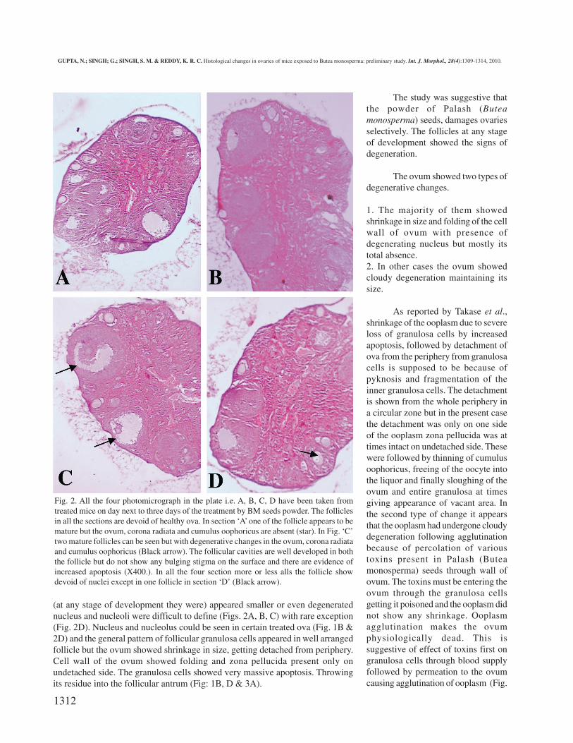
(at any stage of development they were) appeared smaller or even degeneratednucleus and nucleoli were difficult to define (Figs. 2A, B, C) with rare exception(Fig. 2D). Nucleus and nucleolus could be seen in certain treated ova (Fig. 1B &2D) and the general pattern of follicular granulosa cells appeared in well arrangedfollicle but the ovum showed shrinkage in size, getting detached from periphery.Cell wall of the ovum showed folding and zona pellucida present only onundetached side. The granulosa cells showed very massive apoptosis. Throwingits residue into the follicular antrum (Fig: 1B, D & 3A).
Fig. 2. All the four photomicrograph in the plate i.e. A, B, C, D have been taken fromtreated mice on day next to three days of the treatment by BM seeds powder. The folliclesin all the sections are devoid of healthy ova. In section ‘A’ one of the follicle appears to bemature but the ovum, corona radiata and cumulus oophoricus are absent (star). In Fig. ‘C’two mature follicles can be seen but with degenerative changes in the ovum, corona radiataand cumulus oophoricus (Black arrow). The follicular cavities are well developed in boththe follicle but do not show any bulging stigma on the surface and there are evidence ofincreased apoptosis (X400.). In all the four section more or less alls the follicle showdevoid of nuclei except in one follicle in section ‘D’ (Black arrow).
The study was suggestive thatthe powder of Palash (Buteamonosperma) seeds, damages ovariesselectively. The follicles at any stageof development showed the signs ofdegeneration.
The ovum showed two types ofdegenerative changes.
1. The majority of them showedshrinkage in size and folding of the cellwall of ovum with presence ofdegenerating nucleus but mostly itstotal absence.2. In other cases the ovum showedcloudy degeneration maintaining itssize.
As reported by Takase et al.,shrinkage of the ooplasm due to severeloss of granulosa cells by increasedapoptosis, followed by detachment ofova from the periphery from granulosacells is supposed to be because ofpyknosis and fragmentation of theinner granulosa cells. The detachmentis shown from the whole periphery ina circular zone but in the present casethe detachment was only on one sideof the ooplasm zona pellucida was attimes intact on undetached side. Thesewere followed by thinning of cumulusoophoricus, freeing of the oocyte intothe liquor and finally sloughing of theovum and entire granulosa at timesgiving appearance of vacant area. Inthe second type of change it appearsthat the ooplasm had undergone cloudydegeneration following agglutinationbecause of percolation of varioustoxins present in Palash (Buteamonosperma) seeds through wall ofovum. The toxins must be entering theovum through the granulosa cellsgetting it poisoned and the ooplasm didnot show any shrinkage. Ooplasmagglutination makes the ovumphysiologically dead. This issuggestive of effect of toxins first ongranulosa cells through blood supplyfollowed by permeation to the ovumcausing agglutination of ooplasm (Fig.
GUPTA, N.; SINGH; G.; SINGH, S. M. & REDDY, K. R. C. Histological changes in ovaries of mice exposed to Butea monosperma: preliminary study. Int. J. Morphol., 28(4):1309-1314, 2010.
1313
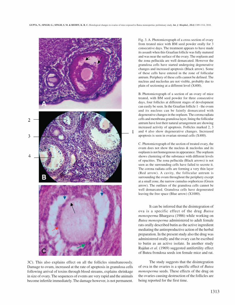
3C). This also explains effect on all the follicles simultaneously.Damage to ovum, increased at the rate of apoptosis in granulosa cellsfollowing arrival of toxins through blood streams, explains shrinkagein size of ovary. The sequences of events are very rapid and the animalsbecome infertile immediately. The damage however, is not permanent.
Fig. 3. A. Photomicrograph of a cross section of ovaryfrom treated mice with BM seed powder orally for 3consecutive days. The treatment appears to have madeits assault when this Graafian follicle was fully maturedand was near the surface of the ovary. The ooplasm andthe zona pellucida are well demarcated. However thegranulosa cells have started undergoing degenerativechanges and increased apoptosis (Black arrow). Someof these cells have entered in the zone of follicularantrum. Periphery of these cells cannot be defined. Thenucleus and nucleolus are not visible, probably due toplain of sectioning at a different level (X400).
B. Photomicrograph of a section of an ovary of micetreated, with BM seed powder for three consecutivedays, four follicles at different stages of developmentcan easily be seen. In the Graafian follicle 1 - the ovumand its nucleus can be faintly demarcated withdegenerative changes in the ooplasm. The corona radiatacells and membrana granulosa layer, lining the follicularantrum have lost their natural arrangement are showingincreased activity of apoptosis. Follicles marked 2, 3and 4 also show degenerative changes. Increasedapoptosis is seen in ovarian stromal cells (X400).
C. Photomicrograph of the section of treated ovary, theovum does not show the nucleus & nucleolus and itsooplasm is not homogenous in appearance. The ooplasmshows clustering of the substance with different levelsof opacities. The zona pellucida (Black arrows) is notseen as the surrounding cells have failed to secrete it.The corona radiata cells are forming a very thin layer(Red arrow). A cavity, the follicular antrum issurrounding the ovum throughout the periphery exceptat a small zone, the narrow cumulus oophoricus (Greenarrow). The outlines of the granulosa cells cannot bewell demarcated. Granulosa cells have degeneratedleaving the free space (Blue arrow) (X1000).
It can be inferred that the disintegration ofova is a specific effect of the drug Buteamonosperma Bhargava (1986) while working onButea monosperma administered to adult femalerats orally described butin as the active ingredientmediating the antireproductive action of the herbalpreparation. In the present study also the drug wasadministered orally and the ovary can be escribedto butin as an active isolate. In another studyRajdan et al. (1969) suggested antifertility effectof Butea frondosa seeds ion female mice and rat.
The study suggests that the disintegrationof ova in the ovaries is a specific effect of Buteamonosperma seeds. These effects of the drug onthe ovaries causing destruction of the follicles arebeing reported for the first time.
GUPTA, N.; SINGH; G.; SINGH, S. M. & REDDY, K. R. C. Histological changes in ovaries of mice exposed to Butea monosperma: preliminary study. Int. J. Morphol., 28(4):1309-1314, 2010.
1314
ACKNOWLEDGEMENT. This article is a part of the Ph.D. thesis of Neelam Gupta. We are grateful for funding fromDepartment of Anatomy, Institute of Medical Sciences,
Banaras Hindu University, Varanasi, U. P. as a researchfellow is gratefully acknowledged.
GUPTA, N.; SINGH; G.; SINGH, S. M. & REDDY, K. R. C. Cambios histológicos en ovarios de ratón expuestos a Butea monosperma:estudio preliminar. Int. J. Morphol., 28(4):1309-1314, 2010.
RESUMEN: En la práctica Ayurvédica Butea monosperma (Palash) se encuentra en uso clínico durante cientos de años comométodo anticonceptivo. Semillas de Butea monosperma también se utilizan como un antihemético y antimicrobiano. Butea monosperma(familia Fabaceae) conocida localmente como Palash (Dhak) si se administra durante 3 días consecutivos actúa como un agente anticoncep-tivo que se utiliza tradicionalmente desde tiempos inmemoriales. El objetivo del presente estudio fue buscar el efecto de las semillas deButea monosperma en ovarios de ratones.Se obsevó degeneración masiva de los óvulos en casi todos los folículos, independientemente dela fase de su desarrollo. Los óvulos de los animales tratados mostraron las diferentes etapas del proceso necrótico. Por otra parte, ladisposición de las células foliculares se mostró alterada. El polvo de semillas de Palash, cuando se administra a los ratones, por vía oral enagua destilada, i.e. 2g/Kg peso corporal, durante tres días produce cambios en los ovarios. Los animales se sacrificaron al día siguienteterminado el tratamiento y fueron extirpados los ovarios. Los ovarios se estudiaron histológicamente con HE mostrándose la mayoría de losfolículos en estado inmaduro, con núcleo definido y nucléolos en el óvulo. Otros mostraron cambios degenerativos en los óvulos. Losfolículos habían perdido su forma normal y la disposición y organización de células de la granulosa. Se encontró que casi todos los folículosincluyendo los folículos mostraban cambios degenerativos de manera simultánea. En los casos tratados, la tasa de apoptosis en las célulasde la granulosa estaba aumentada, en comparación con el grupo control. El estudio sugiere que la desintegración de los óvulos en los ovarioses un efecto específico de la administración de las semillas de Butea monosperma.
PALABRAS CLAVE: Palash; Butea monosperma; Apoptosis; Desintegración; Folículos.
REFERENCES
Ansari, N. A.; Rastogi, S. K.; Tewari, J. P. & Srivastava, M.C. Anthelmintic activity of Butea monosperma (Palash)seed. Indian J. Pharmacy, 11:65, 1979.
Avirutnant, W. & Pongpan, A. The antimicrobial activity ofsome Thai flowers and plants. Mahidol Univ. J. Pharm.Sci., 10(3):81-6, 1983.
Bhargava, S. K. Estrogenic and Postcoital anticonceptiveactivity in rats of butin isolated from Butea monospermaseeds. J. Ethnopharmacol., 18(1):95-101, 1986.
Bhattacharya, S.; Tiwari, K.; Majumdar, R. & Misra, A. K.Folklore medicine from district Kamrup (Assam). Bull.Med. Ethnobot. Res., 1:447-60, 1980.
Chaudhury, R. R. The quest for an herbal contraceptive. Natl.Med. J. India, 6(5):199-201, 1993.
Ganguly, M.; Kr Borthakur, M.; Devi, N. & Mahanta, R.Antifertility activity of the methanolic leaf extract ofCissampelos pareira in female albino mice. J.Ethnopharmacol., 111:688-91, 2007.
Takase, K.; Ishikawa, M. & Hoshiai, H. Apoptosis in thedegeneration process of unfertilized mouse ova. TohokuJ. Exp. Med., 175(1):69-76, 1995.
Prasad, P. V.; Subhaktha, P. K.; Narayana, A. & Rao, M. M.Pala˘s´a (Butea monosperma (Lamk.) Taub.) and itsmedico-historical study. Bull. Indian Inst. Hist. Med.Hyderabad, 36(2):117-28, 2006.
Rajdan, M. K.; Kapila, K. & Bhide, N. K, Antifertility effectof some pharmacological action of Butea frondosa seedsextracts. Indian J. Physiol. Pharmacol., 13(4):239-49,1969.
Tandon, R.; Shivanna, K. R. & Mohan Ram, H. Y.Reproductive biology of Butea monosperma (Fabaceae).Ann. Bot., 92(5):715-23, 2003.
Valsala, S. & Karpagaganapathy, P. R. Effect of Mimosapudica root powder on oestrous cycle and ovulation incycling female albino rat Rattus norvegicus. Photother.Res., 16:190-2, 2002.
Correspondence to:Dr. Neelam GuptaDepartment of AnatomyInstitute of Medical SciencesBanaras Hindu UniversityVaranasi -221005, U.P.INDIA Email: [email protected] [email protected]
Received: 11-05-2009Accepted: 18-02-2010
GUPTA, N.; SINGH; G.; SINGH, S. M. & REDDY, K. R. C. Histological changes in ovaries of mice exposed to Butea monosperma: preliminary study. Int. J. Morphol., 28(4):1309-1314, 2010.