Embed Size (px)
Citation preview

ISSN 2349-7823
International Journal of Recent Research in Life Sciences (IJRRLS) Vol. 1, Issue 3, pp: (32-49), Month: October - December 2014, Available at: www.paperpublications.org
Page | 32 Paper Publications
Histochemical Studies of Enzymes in the
Adrenal Gland of Rat & Rabbit during
Pregnancy 1Dr. Shobha Chaturvedi,
2Dr. Vibha Dave
1,2 Department of Zoology, PMB Gujarati Science College, Indore (M.P), India
Abstract: Administration of ACTH stimulates adrenal secretion of progesterone as well as corticosterone (Resko,
19691; Feder et al., 1969; Feder et al., 1971; Piva et al., 1973). Progesterone is both an obligatory intra-adrenal
substrate for corticosterone production and a steroid essential for maintenance of pregnancy. Thus, the regulation
of adrenal steroidogenesis during pregnancy has two potentially important aspects: i.) Maintenance of optimal
blood levels of corticosterone and ii.)Contributing significant amounts of progesterone to the total maternal pool.
Since the extended luteotrophic function of ovary in rat & mice during pregnancy is related to the Peroxidase-
Ascorbate system (Agrawal, P. & Laloraya, M.M. 1979). It appears likely that synthesis of progesterone under the
action of ACTH during pregnancy may be controlled by a similar mechanism as reported for LH in the ovary,
thus causing increased synthesis and secretion of the Progesterone and corticosteroids from the adrenal gland.
1. INTRODUCTION
The various in vivo and in vitro studies have demonstrated that the ovary and adrenal possess the side-chain cleaving
system to convert C27 cholesterol to pregnenolone which are mainly a C22-C20 lyase and hydroxylases (Simmer,
1968). Steroidogenic enzymes such as 3B-OH-steroid dehydrogenase and 20 -hydroxysteroid dehydrogenase have been
reported (Beyer et al., 1956; Burstein et al., 1963 and Weist et al., (1963) which are involved in the biosynthesis of
progesterone and androgens.
The sex hormones produced by the adrenal cortex of both males and females are progesterone, testosterone and estrogens.
The adrenal gland is the source of sex hormones until the testis and ovaries mature at puberty. The secretion of these
hormones is controlled by ACTH and not by gonadotrophins which stimulate the testes and the ovaries.
Since ,adrenals are known to secrete large quantities of progesterone, which is an oxidation product of pregnenolone, it
appears probable that conversion of pregnenolone to progesterone may be brought about peroxidatively by the operation
of peroxidase as suggested in the ovary (Agrawal and Laloraya, 1977 ).
The role of Peroxidase in the endocrine regulation of hormone action in the adrenal which is closely interlinked in
reproductive functioning of different groups of animals remains largely unknown. Also the hormone regulation and the
enzymic mechanism which lead to the rapid formation and secretion of hormone namely progesterone and corticosteroids
in the adrenal gland is largely unknown.
ACTH has a major role in the synthesis of progesterone which is known to be a precursor of several steroid hormones
including androgens, estrogens and corticoids (Gorbman and Bern, 1974). A preovulatory surge of progesterone in the
systemic plasma of adrenal origin preceeding the display of behavioural estrous in rodents has been reported (Feder et al.,
1968).ACTH is also known to cause depletion of adrenal ascorbate and cholesterol in the hypophysectomized rat
(Tyslowitz, 1943; Sayer et al., 1946) which is shown to occur within minutes of ACTH injection and to exhibit a
characteristic time sequence.

ISSN 2349-7823
International Journal of Recent Research in Life Sciences (IJRRLS) Vol. 1, Issue 3, pp: (32-49), Month: October - December 2014, Available at: www.paperpublications.org
Page | 33 Paper Publications
The role of Peroxidase in the endocrine regulation of hormone action in the adrenal which is closely interlinked in
reproductive functioning of different groups of animals remains largely unknown. Also the hormone regulation and the
enzymic mechanism which lead to the rapid formation and secretion of hormone namely progesterone and corticosteroids
in the adrenal gland is largely unknown.
2. MATERIAL & METHODS
Colony-bred albino rats (Wistar Strain) of our departmental colony maintained on a regimenof 12hrs.light/12 hrs.dark in
a temperature controlled room (25 0
- +
10
C) were used in this study. They received food & water ad libitum. Vaginal
smears were taken daily and at least two complete cycles were observed in each rat prior to its use in an experiment. The
mature female rats used for the study showed a regular 4-5 days estrous cycle. The adrenal of sexually mature rat were
used. Pregnancy, which lasted 21 days in rats, was induced by mating functional females with proven males on the day of
vaginal cornification, which was designated day 0. The day of conception was ascertained by examining vaginal smears
daily; the observed presence of sperms in a smear being taken to indicate the first day of pregnancy. The pregnant females
were sacrificed at various stages of pregnancy by cervical dislocation and dissected. The adrenal were stored at -200C and
were later subjected to various histochemical analyses.
Colony bred mature female white rabbits of our departmental colony were caged individuallyIn a controlled environment
with light-dark cycle of 14:10 hours. Water and food were supplied ad libitum. They were mated twice with different
bucks of proven fertility followed by i.v. injection of 100 i.u. of human chorionic gonadotropin (CG-5, Sigma Chemicals
co. USA) to induce ovulation and were designated as pregnant rabbits. The pregnant females were anesthesized by i.v.
injection of Sodium pentabarbitone at various stages of pregnancy. The dissected tissues were stored at -200C. The
adrenals were subjected to histochemical studies.
Histochemical Procedure: Gelatin fixed frozen sections (4µ) of the adrenal gland were cut in an American Optical
Cryocut and were then used for the localization of various enzymes.
1. Peroxidase : This enzyme was localized by the modified method of Van Duija (1951) using Benzidine as donor.
Another method Graham &Karnovsky (1966) using diaminobenzidine as a donor was also applied. Similar pattern was
obtained with this donor. Therefore, benzidine as donor was used in the histochemical tests of peroxidase. The activity
was also tested with the other donor namely Guaiacol.
2. Cytochrome Oxidase: The method followed for the localization of Cytochrome oxidase was that of Burstone, 1959)).
3. ∆5
3β- Hydroxysteroid dehydrogenase: This enzyme was localized by the method of Wattenberg(1958).
4. Acid Phosphatase: This enzyme was localized after the method of Gomori,(1950).
5. Alkaline Phosphatase :The Calcium-Cobalt method for Alkaline phosphatase , Gomori, (1952) was followed .
6. Lipids: Lipids were stained in frozen sections (4µ) by Herxheimer’s fat stain method (1903).
7. Ascorbic Acid:Histochemical localization of ascorbic acid brought about by the method of Chenoy (1969b).
3. RESULTS
Peroxidase:
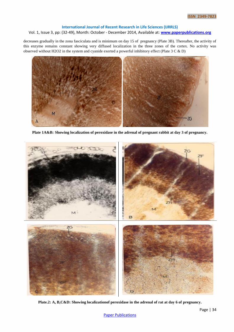
Peroxidase is observed to be detectable during early gestation period in rat & rabbit. Low activity of peroxidase is
observed in zona glomerulosa, zona fasciculate and zona Reticularis (Plate 1A& B). A diffuse activity of this enzyme is
seen in the blood vessels of zona reticularis during the first day of pregnancy in rabbit (Plate 2A & B). The activity of
peroxidase increases on days 4-7 of pregnancy in the zona fasciculate of rat & rabbit while low activity is seen in the
zona reticularis (Plate 2A,B,C & D and Plate 3A).During the later periods of gestation, the activity of peroxidase
ISSN 2349-7823
International Journal of Recent Research in Life Sciences (IJRRLS) Vol. 1, Issue 3, pp: (32-49), Month: October - December 2014, Available at: www.paperpublications.org
Page | 34 Paper Publications
decreases gradually in the zona fasciculata and is minimum on day 15 of pregnancy (Plate 3B). Thereafter, the activity of
this enzyme remains constant showing very diffused localization in the three zones of the cortex. No activity was
observed without H2O2 in the system and cyanide exerted a powerful inhibitory effect (Plate 3 C & D)
Plate 1A&B: Showing localization of peroxidase in the adrenal of pregnant rabbit at day 3 of pregnancy.
Plate.2: A, B,C&D: Showing localizationof peroxidase in the adrenal of rat at day 6 of pregnancy.

ISSN 2349-7823
International Journal of Recent Research in Life Sciences (IJRRLS) Vol. 1, Issue 3, pp: (32-49), Month: October - December 2014, Available at: www.paperpublications.org
Page | 35 Paper Publications
Plate.3: A&B: Showing localization of peroxidase in the adrenal of pregnant rat & rabbit at different days of pregnancy. C&D:
Cyanide treated Control sections of adrenal of rat & rabbit showing no activity of peroxidase.
Hydoxysteroid dehydrogenase
The enzyme activity is , in general, very strong in the mammals and shows significant Species differences. Under similar
conditions it has been shown to be absent in the mouse Adrenal, weak in rabbit & very strong in pregnant mouse,cow
sheep and dog ( Rubin et al. , 1963). In rat and rabbit steroid dehydrogenase is mainly localized in the outer part of the
zona fasciculate and average activity is seen in the zona reticularis during early days of pregnancy (Plate 4A,B & C).
Higher activity of ∆5-3β-Hydroxysteroid dehydrogenase is maintained in the zona fasciculate throughout the early
gestation period in rat & rabbit (Plate 5A, B & C).The activity of ∆5-3β-Hydroxysteroid dehydrogenase decreases in the
zona fasciculate & zona reticularis of rat & rabbit during later days of pregnancy (Plate6A & B)

ISSN 2349-7823
International Journal of Recent Research in Life Sciences (IJRRLS) Vol. 1, Issue 3, pp: (32-49), Month: October - December 2014, Available at: www.paperpublications.org
Page | 36 Paper Publications
Plate.4: A, B&C: Showing localization of ∆5-3β-Hydroxy Steroid dehydrogenase in the pregnant rat & rabbit adrenal gland at
day 5 of pregnancy
Plate 5A, B&C: Showing localization of ∆5-3β-Hydroxysteroid dehydrogenase in the pregnant rat & rabbit adrenal gland at
day 12 of pregnancy.

ISSN 2349-7823
International Journal of Recent Research in Life Sciences (IJRRLS) Vol. 1, Issue 3, pp: (32-49), Month: October - December 2014, Available at: www.paperpublications.org
Page | 37 Paper Publications
Plate6A&B: Showinglocalizationof∆5-3β-Hydroxysteroid dehydrogenase in the pregnant rat & rabbit adrenal gland at day 18
of pregnancy.
Cytochrome oxidase
A positive Cytochrome oxidase activity is seen in the adrenals of rat and rabbit during pregnancy. The Enzymes activity is
low in the zona fasciculate and reticularis layers at days 1-4 of pregnancy (Plate 7A & B). A high activity of Cytochrome
oxidase is seen in the fascicular zone from days 6-14 of pregnancy In rat and rabbit (Plate 8A,B & C) and decreases
gradually during the later period of gestation (Plate 9A,B & C). The control sections without the substrate i.e. α- naphthol
show no activity thereby confirming the presence of the enzyme in these regions (Plate 9D).
Plate.7: A&B: Showing localization of Cytochrome oxidase inthe pregnant rat & rabbit at day 3 of pregnancy.

ISSN 2349-7823
International Journal of Recent Research in Life Sciences (IJRRLS) Vol. 1, Issue 3, pp: (32-49), Month: October - December 2014, Available at: www.paperpublications.org
Page | 38 Paper Publications
Plate.8: A,B&C: Showing localization of Cytochrome oxidase in the adrenal of rat pregnant rat at day 10 & rabbit at day 14
of pregnancy.

ISSN 2349-7823
International Journal of Recent Research in Life Sciences (IJRRLS) Vol. 1, Issue 3, pp: (32-49), Month: October - December 2014, Available at: www.paperpublications.org
Page | 39 Paper Publications
Plate 9: A,B,C&D: Showing localization of Cytochrome oxidase in the adrenal of rat & rabbit at day 18 of pregnancy.

ISSN 2349-7823
International Journal of Recent Research in Life Sciences (IJRRLS) Vol. 1, Issue 3, pp: (32-49), Month: October - December 2014, Available at: www.paperpublications.org
Page | 40 Paper Publications
Alkaline phosphatase
A number of authors have notedthe difference in localization of the adrenal alkaline phosphatase Activity depending on
the species examined (Nicander, 1952). In our studies the entire adrenal cortex
of rat and rabbit has a histochemically detectable alkaline phosphatase activity during pregnancy, which predominates in
the zona glomerulosa, outer zona fasciculate and zona reticularis (Plate 10A,B,C & D).A high activity of alkaline
phosphatase is seen in the zona reticularis on day 16 ofpregnancy while it is low in the zona glomerulosa in rat (Plate 11A
& B) and rabbit (Plate 11 C). Elftman (1947) has shownThat female adrenal cortex is rich in alkaline phosphatase activity
mainly in the reticular zone which correlates very well with our histochemical observations. No activity is seen in the
control sections i.e., minus the substrate, Sodium-β- glycerophosphate, thereby confirming the presence of this enzyme to
these regions (Plate12 A & B).
Plate.1: 0A,B,C&D: Showing localization of alkaline phosphatase in the adrenal ofpregnant rat and rabbit at day 6 of
pregnancy.

ISSN 2349-7823
International Journal of Recent Research in Life Sciences (IJRRLS) Vol. 1, Issue 3, pp: (32-49), Month: October - December 2014, Available at: www.paperpublications.org
Page | 41 Paper Publications
Plate 11 A,B&C: Showing localization of alkaline phosphatase in the adrenal of pregnant rat and rabbit day 16 of pregnancy.
Plate 12A&B: Control sections of rat & rabbit adrenal minus the substrate showing no acitivtyof Alkaline phosphatase at day
6 of pregnancy.

ISSN 2349-7823
International Journal of Recent Research in Life Sciences (IJRRLS) Vol. 1, Issue 3, pp: (32-49), Month: October - December 2014, Available at: www.paperpublications.org
Page | 42 Paper Publications
Acid phosphates:
In mammals, the histochemically detectable acid phosphatase activity is more uniformly distributed than the alkaline
phosphatase. The acid phosphatase activity is high in the fascicular zone and in the reticular zone on day 4 of pregnancy
in rat and rabbit (Plate 13A, B & C). A uniformly distributed activity of acid phosphatase is seen in the three layers of the
cortex on day 10-12 of pregnancy (Plate 14A,B & C) The glomerular zone is richer than the rest of the cortex in rabbit
(Plate 14C). In rat it is clearly less rich than the rest of the cortex (Plate 14A). In rabbit, the enzyme activity predominates
in the internal zone (Plate 14 B). In rat the entire cortex is poor in acid phosphatase and contrasts with the adrenal medulla
which has an extremely strong activity(Plate14A). No activity is seen in the control sections i.e., minus substrate Sodium-
β-glycerophosphate (Plate 15A & B), thereby confirming the presence of this enzyme in these regions
Plate 13 A,B&C: Showinglocalizationofacid phosphatase In the pregnant rat & rabbit adrenal at day 4 of pregnancy.
Plate 14 A,B&C: Showing localization of acid phosphatase in the pregnant rat & rabbit adrenal at day 10 of pregnancy.

ISSN 2349-7823
International Journal of Recent Research in Life Sciences (IJRRLS) Vol. 1, Issue 3, pp: (32-49), Month: October - December 2014, Available at: www.paperpublications.org
Page | 43 Paper Publications
Plate 15 A&B: control sections of rat& rabbit adrenaminus the substrate showing no activity of acid phosphatase in the
cortex (C )and medulla (M).
Lipids
The fat laden fasciculate cells are filled with sudanophilic lipids on days 4 and 5 of pregnancy, whilesparsely scattered
lipid bodies are seen in the zona reticularis of the rat and rabbit adrenals (Plate 16A,B & C). The dense localization of
lipids is seen in the zona fasciculata in rabbit on days 8-16 of pregnancy(Plate 17A & B) while on days 10 and 16 in rat
(Plate 17C). In the later days of pregnancy the fasciculata and reticularis layers show diffuse lipid droplets in rabbit (Plate
17D), while no change is seen in rat. Cholesterol has been shown to be the precursor in the steroid biosynthesis. The
presenceof lipids demonstrated histochemically in the cells of zona fasciculata provide sufficient amounts of cholesterol
for use as a substrate for steroid synthesis.

ISSN 2349-7823
International Journal of Recent Research in Life Sciences (IJRRLS) Vol. 1, Issue 3, pp: (32-49), Month: October - December 2014, Available at: www.paperpublications.org
Page | 44 Paper Publications
Plate 16 A,B&C: Showing loczlization of lipids in the adrenal of pregnant rat at day 4 of pregnancy.
Plate 17A,B,C&D: Showing localization of lipids in the adrenal of pregnant rat & rabbit at day 11 of pregnancy.

ISSN 2349-7823
International Journal of Recent Research in Life Sciences (IJRRLS) Vol. 1, Issue 3, pp: (32-49), Month: October - December 2014, Available at: www.paperpublications.org
Page | 45 Paper Publications
Ascorbic acid:
The inner region of zona fasciculata and zona reticularis of the adrenal cortex in rat and rabbit is darkly stained during the
first 4 days of pregnancy (Plate 18A, B, C & D and Plate 19A, B, C & D ) indicating the presence of ascorbic acid. The
polyhedral columnar cells contain a fine deposition of silver granules. The reticular cells of the hypertrophied cortex show
deposition of silver granules in the outer region while the zona glomerulosa cells show black crystals indicating high
concentration of ascorbic acid(Plate 20A&B). The chromaffin cells show brown diffuse localization which is not a
characteristic of ascorbic acid. From days 5-11 of pregnancy the ascorbate content is very low. The zona fasciculata and
reticularis layers show diffused & scattered deposits of silver granules (Plate 21A, B & C and Plate 22A, B & C) in rat
and rabbit suggesting very low levels of ascorbate in the adrenal cortex. The high staining reaction in zona fasciculata is
observed from day 13 onwards while moderate reaction is seen in zona reticularis (Plate 22A, B, C & D) which is not seen
in the later days of pregnancy that is before parturition in rat and rabbit. Thus the histochemical studies clearly show that
the zona fasciculata is characterized by the presence of high peroxidase , ∆5
- 3β –Hydroxysteroid dehydrogenase , acid
phosphatase and alkaline phosphatase activity while moderate to low activity is seen in the reticular zone of the adrenals
of rat and rabbit.
Plate 18 A,B,C&D: Showing localization of AA in the adrenal of rat during early pregnancy.
Plate 19A,B,C&D: Showing localization of AA in the adrenal of rabbit during early pregnancy.

ISSN 2349-7823
International Journal of Recent Research in Life Sciences (IJRRLS) Vol. 1, Issue 3, pp: (32-49), Month: October - December 2014, Available at: www.paperpublications.org
Page | 46 Paper Publications
Plate 20 A, B&C: Showing localization of AA in the adrenal of rat at day 6 of pregnancy.
Plate 21 A,B&C: Showing localization of AA in the adrenal of rabbit at day 7 of pregnancy.
Plate 22: Showing localization of AA in the adrenal of rat & rabbit during later days of pregnancy (at day 12 & 14).

ISSN 2349-7823
International Journal of Recent Research in Life Sciences (IJRRLS) Vol. 1, Issue 3, pp: (32-49), Month: October - December 2014, Available at: www.paperpublications.org
Page | 47 Paper Publications
4. DISCUSSION
The adrenal cortical complexes in different classes of vertebrates present enormous morphological Differenc es. The
hormones elaborated by these complexes differ not only quantitatively but also Qualitatively. The adrenal cortex in man,
ape and dog produces mainly cortisol, that of rabbit and rat has a massive biosynthesis of corticosterone(Bush,1955),
while that of aves, amphibians and fish elaborates exclusively aldosterone and corticosterone (Carstensen et al., 1961;
Sander et al., 1976).
peroxidase is present in the inner layer of adrenocortical Cells but not in chromaffin cells of ovulatory animals of different
groups of vertebrates which are associated with the functioning of ACTH hormone and progesterone, corticosteroid
secretion. The ∆ 5 -
3β – Hydroxysteroid dehydrogenase and Cytochrome oxidase are present in the adrenocortical cells
during the entire period of sexual cycle. Thus the characteristic function of adrenocortical cells regulating large secretion
of progesterone during increased sexual activity appears to be related to the presence of peroxidase in these
compartments. The adrenal cortex of many non-mammalian speciesare recognized as the chief site of conversion of 14
C
acetate to progesterone (Vinson and Whitehouse, 1973a). Also the biochemical studies have shown that adrenal cortex is
the chief site for the synthesis of steroid hormones namely Progesterone, cortisol and corticosterone ( Hayano et al.,
1956; Resko, 1969; Holzbauer, 1969) .
Furthermore, since peroxidase mediated reactions are many fold faster than dehydrogenase reactions, the association of
high peroxidase activity in these regions, and lack of activity in adrenocortical cells at follicular phase, in growing follicle
of the ovary and IGT of the ovary would suggest that the high rate of progesterone formation may be associated with the
functioning of this enzyme at specific sites. Peroxidase thus appears to be involved in the biosynthetic machinery
controlling corticosteroidogenesis.
At the same time, presence of high peroxidase activity and the following depletion of ascorbate during days 5-11 of
pregnancy in the adrenal correlates well with stimulated steroid biogenesis in adrenal cortex during this period.
The histochemical changes in acid and alkaline phosphatases and lipids in the adrenocortical cells and ovary at various
reproductive phases have been shown by a number of workers ( Galli Mainini, 1951; Botte, 1964) .High acid phosphatase
activity is shown to be present at ovulatory Furthermore phase in the adrenocortical cells and ovary of fish, amphibians
and reptiles, while alkaline phosphatase attains zenith during the secretory phase ( spawning phase ) . Sudanophilic
granules have been shown to increase markedly in the adrenocortical cells,TI and IGT of the ovary At the follicular
phase and disappears during the spawning period. Under the hormonal stimuli ( Guraya ,1974) the marked decrease in the
lipids in ovary with increase in acid and alkaline phosphatase activity of spawning period confirm these reports.
The presence of active cytochrome oxidase in the adrenocortical cells and TI,CL,IGT of the ovary is suggestive of high
metabolic activity in these tissues. The operation of active Cytochrome oxidase suggest that the necessary respiratory
energy in the form of ATP molecules for the biosynthesis of lipids would be available at the site.The hypertrophied TI and
CL of non-mammalian vertebrates are characterized by high vascularization and increased blood flow is also visible in
adrenocortical cells of all vertebrates. The oxidative sites thus provided with adequate Oxygen supply with the activated
blood flow thus converting these sites into intense oxidative sites and thus the intense Cytochrome oxidase activity in
these sites becomes meaningful. The Peroxidase and Cytochrome oxidase would also seem to transform adrenocortical
cells and hypertrophied TI into highly oxidative compartments of the adrenal and ovary which attributes to the oxidation
of pregnenolone to progesterone and corticosteroids towards maturation and ovulation of the oocyte from the ovary
REFERENCES:
[1] Agrawal,P. and Laloraya, M.M. (1977):Ascorbate and peroxidase changes during pregnancy in
albino rat and Swiss mouse.Histochemical studies on the peroxidase localization in the rat ovary and uterus during
various reproductive stages Biochem.J.166:205-208.
[2] P Agrawal,& M.M. Laloraya (1979): Ascorbate and peroxidase changes during pregnancy in albino rat and Swiss
mouse J. Biol. Chem. 219: 66-76.
ISSN 2349-7823
International Journal of Recent Research in Life Sciences (IJRRLS) Vol. 1, Issue 3, pp: (32-49), Month: October - December 2014, Available at: www.paperpublications.org
Page | 48 Paper Publications
[3] Beyer,K.F. and Samuels,L.T. (1956): Distribution of steroid-3ß-ol-dehydrogenase in cellular structures of the
adrenal gland. J. Biol. Chem. 219: 66-76.
[4] Botte,V. (1964): Plasma sex hormones and post-reproductive period in the green frog, Rana esculenta complex
Atti Soc.Peloritana Sci. Fis.Mat.Natur 10:521-528.
[5] Burstein,S. and Dorfman, R.I. (1963): Determination of mammalian steroid sulfatase with 7 a-H3-3, fl,-
hydroxyandrost-5-en-17-one sulfate. J.Biol. Chem.238:1656-1660.
[6] Burstone,M.S. (1959): New histochemical techniques for the demonstration of tissue oxid-ases (cytochrome
oxidase).J.Histochem.Cytochem. 7: 112–122 – The relationship between... Histochem. Cytochem. 7: 112-122
Chicago.
[7] BUSH, I.E.(1955): Ciba Foundation Colloquia on Endocrinology London, J. and A. Churchill Ltd. 7: 210. 1953. ...
1959.
[8] Carstensen, H., A. C. J. Burgers, and C.H. Li(1961): Demonstration of aldosterone and corticosterone as the
principal steroids formed in incubates of adrenals of the american bullfrog (Rana catesbeiana) and stimulation of
their production by mammalian adrenocorticotropin. Gen. comp. Endocr. 1, 37–50 . 9.Chenoy,N.J. (1969b):
Histochemie 20, 105-107 On the specificity of alcoholic acidic silver nitratereagent for the histochemical
localization of ascorbic acid.
[9] F. Piva, P. Gagliano, M. Motta, L. Martini(1973):Endocrinology, 93 pp. 1178–1184Adrenal progesterone:Factors
controlling its secretion.
[10] C. Galli-Mainini(1951): Sécrétion de l'oviducte due crapaud par l'ovaire en ovulation. CR Soc. Biol, 145 pp. 131–
133.
[11] Gomori, G. (1952): Microscopic Histochemistry: Principles & Practice, University of Chicago Press, G. Chicago,
Illinois p. 121.
[12] Gomori,G (1950): An improved histochemical technic for acid phosphatase. Stain. Tech. 25-81 Temporal Effects
of Ovine Luteinizing Hormone and Desoxycorticosterone Acetate on Maturation and Ovulation of Oocytes of the
Catfish, Heteropneustes fossilis (Bloch): An in Vivo and in Vitro Study, J.
[13] Gorbman,A and Bern,H.A.(1974): Adrenal cortex & Interrenal gland.In: “ A textbook of comparative
endocrinology”. Wiley Eastern University Edition 1974 pp.297-339.
[14] GP Vinson, JBG Bell, BJ Whitehouse (1976): J. steroid Biochem. 7 407-411. Productin of of testosterone and
corticosteroids by the rat adrenal gland incubated in vitro and the effects of stimulation with ACTH, LH and FSH.
[15] Graham, R.C. & Karnowsky, M.J. (1966): The early stages of absorption of injected horse radish peroxidase in the
proximal tubules of mouse kidney: ultrastructural cytochemistry by a new tech- nique. J.Histochem. Cytochem..
14: 291-302.
[16] Guraya, S.S. (1974): Comparativemorphological and histochemical observations on the ovarian stromal
compartment in mammalswith special reference to steroidogenesis Acta Anat. 90: 250-284.
[17] Hayano, M.,Saba, N., Dorfman, R.I. and Hechter, O. (1956): Some aspects of the biogenesis of adrenal steroid
hormones. Recent Prog. Horm. Res. 12 pp. 79–123.
[18] Herxheimer, G. (1901): Ueber Fettfarbstoffe Deutsche Med. Wschr. 36,607.
[19] HH Feder, JA Resko, RW Goy(1968): Progesterone levels in the arterial plasma of
pre-ovulatory and ovariectomized rats. J. Endocrinol., 41 pp. 563–569.
[20] HH Feder, K Brown-Grant, CS Corker (1971): - Journal of endocrinology- Soc Endocrinology Pre-ovulatory
progesterone, the adrenal cortex and the'critical period'for luteinizing hormone release in rats.
[21] HH Feder, KB Ruf(1969): - Endocrinology- press.endocrine.org Stimulation of progesterone release and estrous
behavior by ACTH in ovariectomized rodents.

ISSN 2349-7823
International Journal of Recent Research in Life Sciences (IJRRLS) Vol. 1, Issue 3, pp: (32-49), Month: October - December 2014, Available at: www.paperpublications.org
Page | 49 Paper Publications
[22] Holzbauer, M., Newport, H.M. Birmingham, M.K. and Traikov, H. (1969): Secretion of pregn-4-ene-3,20- dione
(progesterone) in vivo by the adrenal gland of the rat Nature 221: 572-573
[23] P Agrawal, MM Laloraya(1979): - Am. J. Physiol, Anim .Physiological Soc.
[24] Resko(1969):Science 164:70-71The concentration of progesterone in systemic plasma of ovariectomized rats can
be aug- mented by injection of ACTH but not by injectionof LH
[25] Resko, J. (1969): Endocrine control of adrenal progesterone secretion in the ovariectomized rat Science 164:70-71.
[26] Sandor, T., Fazekas, A.G. and Robinson, B.H. (1976):In:“General Comparative and Clinical Endocrinology of the
Adrenal Cortex “. (I. Chester Jones and I.W. Henderson, eds.), Vol I,pp.25-125.Academic Press, London and New
York.
[27] Sayers, G., Sayers, M.A., Liang, T.Y. and Long, C.N.H. (1946): Endocrinology 38: 1-9 The effect of pituitary
adrenotrophic hormone on the cholesterol and ascorbic acid content of the adrenal of the rat and the guinea pig .
[28] .Sayers, M.A. Sayers, G. and Woodbury, L.A. (1948): endocrinology 42: 379 The assay of adrenocorticotrophic
hormone by the adrenal ascorbic acid-depletion method 1
[29] Simmer, H.H. (1968): In: Placental Hormones In: Biology of Gestation Vol. I ed. N.S. Assali, A.P. New York.
29:347.
[30] Tyslowitz,R. (1943): Effect of hypophysectomy on the concentration of ascorbic acid in the adrenals of the rat
endocrinology 32:103.
[31] Van Duija, P. (1951): An improved histochemical benzidine‐blue peroxidase method and a note on the composition
of the blue reaction product Recueil 74: 771-777
[32] Vinson, G.P.and Whitehouse, B.J.(1973a): Compartmental arrangement of steroids Formed from [I-14C] acetate
by rat adrenal zona glomerulosa and the effect of corticotropin. Acta endocr. Copenh.72 pp. 737–745. Acta
Endocr., Copnh. 737-745.
[33] Wattenberg, L.W. (1958): Microscopic histochemical demonstration of 3fl-ol-dehydrogenase in tissue sections. J.
Histochem. Cytochem. 6: 225- 232
[34] Weist, W.G., Kidwell, W.R. and Kirschbaum, T.H. (1963):In vitro metabolism of progesterone and 20α-
hydroxypregn-4-en-3-one by tissues of thefemaleratSteroids.617-630. XII: 79-123.