Embed Size (px)
Citation preview

Advances in Experimental Medicine and Biology 1042
Hisao MasaiMarco Foiani Editors
DNA ReplicationFrom Old Principles to New Discoveries

Advances in Experimental Medicine and Biology
Volume 1042
Editorial BoardIRUN R. COHEN, The Weizmann Institute of Science, Rehovot, IsraelABEL LAJTHA, N.S. Kline Institute for Psychiatric Research, Orangeburg, NY, USAJOHN D. LAMBRIS, University of Pennsylvania, Philadelphia, PA, USARODOLFO PAOLETTI, University of Milan, Milan, ItalyNIMA REZAEI, Tehran University of Medical Sciences, Children’s Medical Center Hospital, Tehran, Iran

More information about this series at http://www.springer.com/series/5584

Hisao Masai • Marco FoianiEditors
DNA ReplicationFrom Old Principles to New Discoveries

ISSN 0065-2598 ISSN 2214-8019 (electronic)Advances in Experimental Medicine and BiologyISBN 978-981-10-6954-3 ISBN 978-981-10-6955-0 (eBook)https://doi.org/10.1007/978-981-10-6955-0
Library of Congress Control Number: 2017960795
© Springer Nature Singapore Pte Ltd. 2017Chapter 2 was created within the capacity of an US governmental employment. US copyright protection does not apply.This work is subject to copyright. All rights are reserved by the Publisher, whether the whole or part of the material is concerned, specifically the rights of translation, reprinting, reuse of illustrations, recitation, broadcasting, reproduction on microfilms or in any other physical way, and transmission or information storage and retrieval, electronic adaptation, computer software, or by similar or dissimilar methodology now known or hereafter developed.The use of general descriptive names, registered names, trademarks, service marks, etc. in this publication does not imply, even in the absence of a specific statement, that such names are exempt from the relevant protective laws and regulations and therefore free for general use.The publisher, the authors and the editors are safe to assume that the advice and information in this book are believed to be true and accurate at the date of publication. Neither the publisher nor the authors or the editors give a warranty, express or implied, with respect to the material contained herein or for any errors or omissions that may have been made. The publisher remains neutral with regard to jurisdictional claims in published maps and institutional affiliations.
Printed on acid-free paper
This Springer imprint is published by Springer NatureThe registered company is Springer Nature Singapore Pte Ltd.The registered company address is: 152 Beach Road, #21-01/04 Gateway East, Singapore 189721, Singapore
EditorsHisao MasaiDepartment of Genome MedicineTokyo Metropolitan Institute of Medical ScienceTokyo, Japan
Marco FoianiIstituto FIRC di Oncologia MolecolareMilan, Italy

v
Preface
The discovery of the double-helix structure in 1953 provided the basic concept of how genetic materials are duplicated. However, this also triggered the quest for understanding the whole picture of DNA replication. A semi-conservative replica-tion was demonstrated in a historical experiment by Messelson and Stahl, and Arthur Kornberg discovered an enzyme responsible for synthesizing DNA. At that time, no one envisioned such complicated systems required to make copies of DNA.
The proposal of the replicon hypothesis by Jacob made a major impact in point-ing the direction in which research of DNA replication in the following years would be led. In fact, the results of genetic studies in bacteria provided evidence for the presence of the factors (initiator and replicator) hypothesized in the model. This was striking and revealing, and the research of DNA replication in the following 50 years tried to recapitulate this finding in different organisms. The elegant single-molecule analyses of replicating DNA in mammalian cells done by Huberman and Riggs in 1966 were interpreted in the framework of the replicon model, becoming the basis for the “multiple replicon” hypothesis for the eukaryotic genomes.
Molecular genetic studies in Escherichia coli, in combination with development of recombinant DNA technology, clarified the structures of the replication origin (oriC), and “resolution and reconstitution” studies of the single-stranded DNA phage and eventually those of oriC replication elucidated the mechanisms of DNA chain elongation and initiation at the bacterial chromosome replicator.
A similar approach was taken for eukaryotes, and the studies using viruses as a model significantly contributed to the elucidation of eukaryotic DNA replication machinery. Genetic studies, in conjunction with newly developed methods for phys-ical mapping of origins, in a unicellular eukaryote, yeast, led to identification of specific sequences that could serve as replicators. In contrast, the “initiator” remained elusive until Bell and Stillman discovered ORC (Origin Recognition Complex), which appears to fulfill all the requirement to be qualified as the “initia-tor”. All the data pointed to the adherence to the old “replicon” principle even in eukaryotes.
Fifty-four years after the replicon hypothesis, almost the entire process for eukaryotic DNA replication was reconstituted with purified proteins, and the

vi
detailed mechanisms are bound to be discovered in the ensuing years. Given the large extent of conservation of most of the core replication factors between yeast and higher eukaryotes, the basic mechanisms of origin activation and assembly of a replisome would be conserved through evolution.
Compared to the replication machinery, the sequences that define the “replica-tor” appear to have great divergence between species. A genome-wide approach with NGS (Next Generation Sequencing) generated an enormous amount of new information on the profiles of replication origins in higher eukaryotes, but consen-sus sequences, similar to those found in the yeast replication origins, have not yet been discovered. An additional complication is the absence of sequence-specific DNA binding activity in mammalian ORC. These results suggest the existence of a determinant other than nucleotide sequences that dictate the assembly of a replica-tion complex.
This book compiles various timely topics in DNA replication. The volume starts with a historical description on studies of eukaryotic DNA replication by Professor Thomas Kelly. Professor Kelly has made major contributions in this field for the past over 40 years through his studies on viral DNA replication, human genome replication, and yeast replication. The history of Professor Kelly’s research itself represents how this field evolved and materialized into the current understanding of eukaryotic DNA replication. We are very honored and excited to have this chapter at the beginning of the book. This chapter is followed by two chapters describing replication origins in higher eukaryotes. Dr. Mirit Aladjem describes various fea-tures that define metazoan replication origins; those include sequence bias, open chromatin, and histone modifications. Drs. Nozomi Sugimoto and Masatoshi Fujita focus on chromatin remodeling factors that determine origin activity though promo-tion of replication licensing.
Studies in E. coli, starting from those on phage replicons to those of oriC plas-mid, have led the field of DNA replication. In spite of evolutional distance from eukaryotes, the expertise from the bacterial systems have been proven to play lead-ing roles in elucidating the mechanisms of eukaryotic DNA replication. Dr. Tsutomu Katayama describes the most recent discovery on the detailed mechanism of E. coli oriC replication and various modes of its regulation. Studies in Archaea have filled unique roles in shedding new light on regulation of eukaryotic DNA replication. Dr. Stephen Bell has contributed a chapter describing initiation and its regulation in Archaea replicons.
In the following two chapters, replication machinery for DNA chain elongation is discussed. Drs. Joseph Stodola and Peter Burgers deal with the mechanism of lagging strand DNA replication in eukaryotes, which is a critical step for DNA chain elongation and is also crucial for stable maintenance of genome, while Drs. Eiji Ohashi and Toshiki Tsurimoto discuss the multiple clamp and its loaders, which are now known to play major roles in coordinating the process of DNA replication with various other chromosome maintenance systems, including checkpoint/DNA damage repair systems, epigenetic transmission, and chromosome partition.
Replication termination is a recent hot topic. In bacteria, replication termination occurs when two replication forks collide 180° from the origin, and their progres-
Preface

vii
sion is arrested at the ter signals bound with tus protein. On the eukaryotic chromosomes, combined actions of topoisomerase and a ubiquitin ligase are required to dislodge the replisome at the termination site.
The recent technical revolution in cryoelectron microscopy has enabled struc-tural analyses of huge protein complexes at a resolution close to that achieved by X-ray crystallography. Application of this new technology has revealed the complex structures and operation mechanism of MCM (minichromosome maintenance) and CMG helicase as well as more complex replisome assembly. Drs. Yuanliang Zhai and Bik-Kwoon Tye; Drs. Lin Bai, Zuanning Yuan, Jingchuan Sun, Roxana Georgescu, Michael O’Donnell, and Huilin Li describe their state-of-the-art analy-ses of yeast MCM2–7 double hexameter and the replisome complex, respectively.
Long-standing questions about DNA replication timing regulation are now being addressed in detail at a genome-wide level and detailed landscape of replication domains has been presented in various cell types. Drs. Peiyao Zhao, Juan Carlos Rivera-Mulia, and David Gilbert, a leading group in this area, discuss their current model on how replication domains are related to genome compartmentalization and chromatin architecture. The conserved Rif1 protein, originally identified as a telo-mere binding factor in yeast, was rediscovered as a critical regulator of replication timing in fission yeast. Rif1 also plays a major role in organizing the replication domains in mammalian cells. Dr. Sara Buonomo describes how chromatin architec-ture is regulated by Rif1 in mammalian cells to define the replication domains. Drs. Kenji Moriyama, Mong Sing Lai, and Hisao Masai describe functions of Rif1 in both fission yeast and mammalian cells, with particular emphasis on its ability to specifically recognize the G-quadruplex structure (G4), and also the potential roles of G4 in the regulation of chromosome functions. The presence of G4 is one of the most prevalent features associated with replication origins from higher eukaryotes. Dr. Marie-Noëlle Prioleau describes the genetic experiments that show the require-ment for G4 in origin activity and discusses the potential roles of G4 in origin regulation.
Chromatin structures play central roles in almost all the metabolism of DNA. Drs. Constance Alabert, Zuzana Jasencakova, and Anja Groth discuss how chromatin is inherited during the course of DNA replication.
DNA replication is a part of cell cycle events. Therefore, how it is integrated in the global cell cycle regulation is an important issue, especially from the point of view that cell proliferation is regulated by the extracellular stimuli, and that it is this pathway that is often deregulated in cancer cells. Drs. Gavin Grant and Jeanette Cook discuss cell cycle regulation of the S phase, with particular emphasis on the events in G1 that are crucial for regulated execution of the S phase.
Modification of proteins with small polypeptides such as ubiquitin or SUMO permits rapid and reversible regulation of various biological reactions, and DNA replication is no exception. Drs. Tarek Abbas and Anindya Dutta describe how unperturbed DNA replication is regulated by protein ubiquitination and its relation to diseases. Drs. Sara Villa-Hernández, Avelino Bueno, and Rodrigo Bermejo describe protein ubiquitylation in cellular responses to perturbed DNA replication
Preface

viii
or DNA damages. SUMOylation also recently has been shown to play important roles during DNA replication, which is covered by Drs. Lei Wei and Xiaolan Zhao.
DNA replication and transcription take place on the same template DNA, and how they are coordinated has been an intriguing issue. It is usually assumed that collision of replication and transcription can cause genomic instability and needs to be avoided if possible. However, especially on the eukaryotic genome where there are many origins, this is inevitable. Drs. Yathish Achar and Marco Foiani describe how cells coordinate replication with transcription.
The processes of DNA replication and its regulation are directly linked to main-tenance of genomic stability. Overexpression of Cyclin E has been known to induce aberrant DNA replication that leads to genome instability. Drs. Leonardo Teixeira and Steven Reed describe how cyclin E overexpression induces genome instability. Repetitive DNA sequences and sequences capable of forming unusual DNA struc-tures are ubiquitous on the human genome, and increasing evidence points to their pathogenic nature for various diseases. Chromosomal fragile sites, often composed of unusual repetitive sequences, have long been known and implicated in genome instability. Drs. Wenyi Feng and Arijita Chakraborty provide a comprehensive his-torical account of chromosomal fragile sites and provide a detailed discussion of their disease association and then how their expression is related to DNA replica-tion. Advaitha Madireddy and Jeannine Gerhardt discuss the consequence of repli-cation through repetitive DNA elements and how they could lead to specific diseases.
With the astounding amount of new information on replication origins and pro-tein associations on the chromatin and protein–protein/inter-chromatin networks and with the long-awaited reconstitution system of eukaryotic DNA replication in hand, we are at one of the most exciting moments in the field of DNA replication. While we will gain detailed mechanistic insight into the molecular basis of eukary-otic DNA replication, the basic principle of initiation of eukaryotic chromosomal replication remains elusive. It is becoming clear that initiation of replication in higher eukaryotes is quite distinct from that in bacteria in that it can be initiated almost anywhere, albeit with varied efficiency, and the initiation event may be quite stochastic, pointing to the possibility that the bacterial replicator–initiator concept may not apply to regulation in higher eukaryotes.
We hope that this book will help readers to get a taste of the newest trends in this exciting field and will trigger a new wave of research in search of a new principle of DNA replication.
Tokyo, Japan Hisao MasaiMilan, Italy Marco Foiani
Preface

ix
Contents
1 Historical Perspective of Eukaryotic DNA Replication ....................... 1Thomas Kelly
2 Regulation of Replication Origins ......................................................... 43Anna B. Marks, Haiqing Fu, and Mirit I. Aladjem
3 Molecular Mechanism for Chromatin Regulation During MCM Loading in Mammalian Cells ........................................ 61Nozomi Sugimoto and Masatoshi Fujita
4 Initiation of DNA Replication at the Chromosomal Origin of E. coli, oriC .............................................................................. 79Tsutomu Katayama
5 Initiation of DNA Replication in the Archaea ...................................... 99Stephen D. Bell
6 Mechanism of Lagging-Strand DNA Replication in Eukaryotes ........................................................................................... 117Joseph L. Stodola and Peter M. Burgers
7 Functions of Multiple Clamp and Clamp- Loader Complexes in Eukaryotic DNA Replication ......................................... 135Eiji Ohashi and Toshiki Tsurimoto
8 Termination of Eukaryotic Replication Forks ...................................... 163Agnieszka Gambus
9 Structure of the MCM2-7 Double Hexamer and Its Implications for the Mechanistic Functions of the Mcm2-7 Complex ......................................................................... 189Yuanliang Zhai and Bik-Kwoon Tye

x
10 Architecture of the Saccharomyces cerevisiae Replisome .................... 207Lin Bai, Zuanning Yuan, Jingchuan Sun, Roxana Georgescu, Michael E. O’Donnell, and Huilin Li
11 Replication Domains: Genome Compartmentalization into Functional Replication Units .......................................................... 229Peiyao A. Zhao, Juan Carlos Rivera-Mulia, and David M. Gilbert
12 Rif1-Dependent Regulation of Genome Replication in Mammals ............................................................................................. 259Sara B.C. Buonomo
13 G-Quadruplexes and DNA Replication Origins ................................... 273Marie-Noëlle Prioleau
14 Interaction of Rif1 Protein with G-Quadruplex in Control of Chromosome Transactions .............................................. 287Kenji Moriyama, Mong Sing Lai, and Hisao Masai
15 Chromatin Replication and Histone Dynamics .................................... 311Constance Alabert, Zuzana Jasencakova, and Anja Groth
16 The Temporal Regulation of S Phase Proteins During G1 ................... 335Gavin D. Grant and Jeanette G. Cook
17 Roles of SUMO in Replication Initiation, Progression, and Termination ................................................................ 371Lei Wei and Xiaolan Zhao
18 The Multiple Roles of Ubiquitylation in Regulating Challenged DNA Replication ................................................................. 395Sara Villa-Hernández, Avelino Bueno, and Rodrigo Bermejo
19 Regulation of Mammalian DNA Replication via the Ubiquitin- Proteasome System ......................................................... 421Tarek Abbas and Anindya Dutta
20 Coordinating Replication with Transcription ...................................... 455Yathish Jagadheesh Achar and Marco Foiani
21 Fragility Extraordinaire: Unsolved Mysteries of Chromosome Fragile Sites ................................................................. 489Wenyi Feng and Arijita Chakraborty
22 Cyclin E Deregulation and Genomic Instability .................................. 527Leonardo K. Teixeira and Steven I. Reed
23 Replication Through Repetitive DNA Elements and Their Role in Human Diseases ....................................................... 549Advaitha Madireddy and Jeannine Gerhardt
Contents

xi
About the Editors
Hisao Masai Hisao Masai graduated from the University of Tokyo in 1981 and received his Ph.D. from the same university in 1986 for his work on DNA replica-tion in E. coli, which he conducted at the DNAX Research Institute (Palo Alto, California) under the supervision of Dr. Ken-ichi Arai. He did postdoctoral studies at the same institute and became an assistant professor (1990) and then associate professor (1995) of the Institute of Medical Science, the University of Tokyo. In 2000, he moved to the Tokyo Metropolitan Institute of Medical Science to be the head of the Department of Cell Biology. He is currently a vice director of the insti-tute, leading a laboratory of genome dynamics. He continues to work on the mecha-nisms of DNA replication using E. coli, fission yeast, mammalian cells, and animal models. One of his current interests is to elucidate the biological significance of G (guanine)-quadruplex in various chromosome transactions including DNA replica-tion. Dr. Masai served on the editorial board of The Journal of Biological Chemistry (2009–2012). He is currently an editor for Biochemical and Biophysical Research Communications and an associate editor for Genes to Cells. The awards he has received include the Tokyo Metropolitan Governor’s Award (2006), A-IMBN (Asian International Molecular Biology Network) Arthur Kornberg Memorial Award (2012), the Prize for Science and Technology (Research Category), and the Commendation for Science and Technology from the Minister of Education, Culture, Sports, Science and Technology (2017).
Marco Foiani Marco Foiani obtained his Ph.D. in molecular and cell biology from the University of Milan in 1985. After conducting postdoctoral research at the University of Milan and at NIH-NICHD, USA (laboratory of Dr. Alan Hinnebusch), he became a faculty member at the University of Milan in 1990, and in 2000, he became the head of the Genome Integrity Laboratory at IFOM, Milan. He also became a full professor of molecular biology at the University of Milan in 2002. Since 2009 he has been the scientific director of IFOM, an international cancer center that hosts 20 groups and 300 scientists. At IFOM, Dr. Foiani is responsible for strategic research planning, the development of programs aimed at results reduc-tion into practice, and the establishment of national and international cooperation

xii
programs and joint ventures. He is the founder of the European Nanomedicine Foundation (CEN) that aims to support multidisciplinary projects/teams in biomed-icine. He is cofounder of the IFOM-IEO Campus, hosting IFOM, the European Institute of Oncology, the European School of Molecular Medicine, the Italian Institute of Technology, and Cogentech. Since 2012 he has directed a cancer genet-ics diagnostic laboratory within Cogentech. In 2004 he was elected an EMBO mem-ber and in 2010 a member of the Academia Europaea. He is currently a member of the editorial board of Cell.
About the Editors

1© Springer Nature Singapore Pte Ltd. 2017 H. Masai, M. Foiani (eds.), DNA Replication, Advances in Experimental Medicine and Biology 1042, https://doi.org/10.1007/978-981-10-6955-0_1
Chapter 1Historical Perspective of Eukaryotic DNA Replication
Thomas Kelly
Abstract The replication of the genome of a eukaryotic cell is a complex process requiring the ordered assembly of multiprotein replisomes at many chromosomal sites. The process is strictly controlled during the cell cycle to ensure the complete and faithful transmission of genetic information to progeny cells. Our current under-standing of the mechanisms of eukaryotic DNA replication has evolved over a period of more than 30 years through the efforts of many investigators. The aim of this perspective is to provide a brief history of the major advances during this period.
Keywords DNA replication • Eukaryotes • Viral models • Origin of DNA replica-tion • Prereplicative complex • Helicase • Initiator • Replisome
1.1 Introduction
The quest to understand how our genomes are duplicated began in earnest with the description of the double helix in 1953. The self-complementary structure of DNA immediately suggested how the information in the sequence of nucleotides could be copied during DNA replication and repaired after DNA damage. The first enzyme activities capable of synthesizing DNA were described within 5 years of the publi-cation of the DNA structure (Bessman et al. 1956), but it would take many more years to achieve even a basic understanding of the complex machinery required to replicate genomes and to elucidate some of the mechanisms that control it. As one of the fundamental processes of life, DNA replication has been a central focus of molecular biology from the very beginning and remains so today.
Early studies of DNA replication focused on prokaryotic systems because of their relative simplicity. The work of many investigators established most of the basic principles that govern DNA replication in all organisms from prokaryotes to archaea and eukaryotes. In 1958 Meselson and Stahl demonstrated that the
T. Kelly (*) Sloan Kettering Institute, Memorial Sloan Kettering Cancer Center, New York, NY, USAe-mail: [email protected]
2
replication process in bacteria is semiconservative (Meselson and Stahl 1958). A few years later, Cairns showed that replication in E. coli begins at a single site in the circular chromosome and that DNA synthesis occurs on two arms of a forked struc-ture (Cairns 1963). Taken together, these observations implied the existence of a replication machine that is loaded on the genomic DNA at an “origin” and subse-quently moves along the DNA, unwinding the two parental strands and synthesizing new complementary strands. One of the key features of replication machines was discovered in 1968 when Okazaki showed by pulse-labeling experiments that newly synthesized DNA in E. coli cells consists of short fragments (Okazaki et al. 1968). Further analysis in vitro suggested that DNA replication is semi-discontinuous at each growing point (Olivera and Bonhoeffer 1972; Herrmann et al. 1972). One strand (the leading strand) is synthesized continuously 5′–3′ in the same direction as the fork moves, while the other strand (the lagging strand) is synthesized discon-tinuously 5′–3′ in the direction opposite to fork movement, producing short DNA fragments that are subsequently joined together. In the 30 years following the pub-lication of the DNA structure, many of the components of prokaryotic replication machines were identified and characterized, including origin recognition proteins, DNA polymerases, processivity factors, primases, single-stranded DNA-binding proteins, topoisomerases, etc. This saga is the main subject of the superb mono-graphs on DNA replication by Kornberg and more recent reviews (e.g., Kornberg 1981; Kornberg and Baker 1992; Johnson and O’Donnell 2005; Lewis et al. 2016). It was (correctly) surmised that many of the components of the eukaryotic replica-tion machinery would have similar functions to their prokaryotic counterparts.
The identification and characterization of essential prokaryotic replication fac-tors were largely accomplished by classical biochemical approaches – developing assays specific for putative replication functions and then purifying the active proteins(s) to near homogeneity from crude cell extracts. Initial work focused on viruses of E. coli, such as T4, T7, and φX174, because their genomes are relatively small and readily obtained. Some of these viruses (e.g., T4, T7) encode most of the factors required for the replication of their genomes, while others (e.g., ϕX174) rely largely on E. coli replication proteins. The isolation of many bacterial and phage mutants with conditional defects in DNA replication accelerated the identification and purification of replication proteins by in vitro complementation assays. Other proteins were purified by straightforward fractionation and reconstitution approaches. By 1980 most of the proteins required for propagation of a bacterial replication fork had been identified, and their functions characterized through stud-ies of viruses and plasmids (Kornberg 1981). Understanding of the mechanisms involved in initiation of bacterial DNA replication during the subsequent decade required the development and analysis of cell-free systems capable of replicating plasmids containing oriC, the E. coli origin of DNA replication (Kornberg and Baker 1992). These studies established that initiation of bacterial DNA replication is largely controlled at the level of occupancy of the oriC origin by the DnaA initia-tor protein, consistent with the replicon model of Jacob et al. (1964).
It was clear early on that eukaryotic DNA replication was likely to be much more complex than prokaryotic DNA replication because eukaryotic genomes can be orders of magnitude larger than their prokaryotic counterparts. It follows that to
T. Kelly

3
complete chromosome duplication in a timely fashion, DNA replication must start at many sites along the chromosomal DNA. Using fiber autoradiographic methods similar to those of Cairns, two graduate students at Caltech, Joel Huberman and Arthur Riggs, demonstrated in 1968 that a mammalian cell utilizes tens of thou-sands of origins and that replication is bidirectional from each origin (Huberman and Riggs 1968). It was also known that eukaryotic chromosomes are duplicated precisely once each cell cycle, so the requirement for multiple origins raised a num-ber of regulatory issues that were not apparent in bacterial systems with single ori-gins per chromosome. When E. coli cells are growing rapidly in rich media, they initiate DNA replication at oriC prior to the complete duplication of the chromo-some. As a result, DNA synthesis is continuous throughout the cell cycle, and seg-ments of the bacterial chromosome proximal to oriC are present in more than two copies (Skarstad and Katayama 2013). This scenario does not happen in eukaryotes where each segment of DNA is duplicated once and only once during a defined period of each cell cycle. These considerations strongly implied that the logic of replication control in eukaryotes would prove to be substantially different from that of prokaryotes and that the biochemical mechanism of initiation of eukaryotic DNA replication would be correspondingly complex. Because of this complexity, the pathway of initiation of eukaryotic DNA replication was not worked out in detail and recapitulated in cell-free systems until comparatively recently.
As described in the succeeding chapters of this volume, the complexity of eukaryotic DNA replication extends to the organization of the machinery at the replication fork. A number of the core components of the eukaryotic (and archaeal) replisomes are unrelated, or only distantly related, to their bacterial counterparts, suggesting that the prokaryotic and eukaryotic/archaeal replication machineries may have evolved largely independently (Edgell and Doolittle 1997; Leipe et al. 1999; Makarova and Koonin 2013). The eukaryotic replisome contains multiple DNA polymerases and a replicative helicase with a considerably more complex subunit composition than that of E. coli. In addition, several factors associated with the replisome have no clear prokaryotic counterparts. The reasons for this extra complexity are not yet completely clear, but may be related to the need for greater regulation of DNA chain elongation in large genomes. For example, the packaging of the nuclear DNA into nucleosomes and higher-order structures likely creates unique problems for the elongation machinery. In addition, the replication machin-ery must deal with multiple sources of endogenous and exogenous DNA damage and potentially other barriers to DNA synthesis, such as large transcriptional units, DNA-binding proteins, etc. While these obstacles are not unique to eukaryotes, the dimension of the problem may be magnified by the large number of replicons involved in the replication of eukaryotic genomes.
In this perspective, I describe some of the major milestones in the study of eukaryotic DNA replication over the past 35 years. There were numerous contribu-tors to this story, and, generally speaking, each of the major advances described here was built on discoveries made in many different laboratories. Given the span of time covered and the great progress that has been made, this perspective can’t be comprehensive and of necessity must be somewhat idiosyncratic. While many pri-mary references are provided, they represent a tiny fraction of the important publi-
1 Historical Perspective of Eukaryotic DNA Replication

4
cations of the last three or four decades, so I have also cited review articles that contain more comprehensive reference lists in particular areas of investigation. In writing this historical review, I was struck by the number of different lines of inves-tigation that have converged to give us our current picture of how genomes are duplicated in eukaryotic cells. The field has grown enormously over the years and is now relatively mature, but the excitement remains.
1.2 The Beginning: Viral Models for Eukaryotic DNA Replication
The study of eukaryotic DNA replication was hampered for many years by the primitive state of genetic approaches in most eukaryotes and the lack of simple systems for biochemical analysis. The impasse was overcome by the development and characterization of cell-free systems capable of replicating the genomes of ani-mal viruses. This advance, inspired by the success of previous studies of bacterial virus DNA replication, opened a viable pathway for identifying and purifying cel-lular replication proteins and characterizing their mechanisms of action. The result was a rapid acceleration of the study of eukaryotic DNA replication.
The replication of the human adenoviruses (Ad) was the first to be established in a completely soluble cell-free system (Challberg and Kelly 1979). The genome of these viruses consists of a linear duplex DNA molecule of about 35 kb with two features that are significant for viral DNA replication: the ends of the genome have identical sequences, and the 5′ terminus of each DNA strand is covalently attached to a virus-encoded protein of 55 kDa (the terminal protein or TP). Extracts of adenovirus- infected human cell nuclei are capable of carrying out the complete rep-lication of exogenously added viral DNA molecules by a mechanism that closely resembles viral DNA synthesis in vivo. Analysis of replication in vitro led to the identification of the minimal essential requirements for the reaction and defined the basic mechanisms involved in initiation and chain elongation (Challberg and Kelly 1982; Stillman 1983; Nagata et al. 1983; Sussenbach and van der Vliet 1984; de Jong et al. 2003). Three viral proteins are required for adenovirus DNA replication: an 80 kDa precursor to the adenovirus 55 kDa terminal protein (pTP), a DNA poly-merase (Ad Pol), and a single-stranded DNA-binding protein (Ad DBP). DNA rep-lication is initiated by a novel protein-priming mechanism in which the first nucleotide in the adenovirus genome is covalently linked to a serine residue in the pTP (Rekosh et al. 1977; Challberg et al. 1980, 1982; Desiderio and Kelly 1981; Enomoto et al. 1981; Tamanoi and Stillman 1982; King and van der Vliet 1994). The biochemical mechanism of the initiation reaction is quite interesting although not directly germane to cellular DNA replication. One novel feature of the reaction is the requirement for two cellular transcription factors, nuclear factor I (NF-1/CTF) and Oct-1 (NF-III/Otf-1/Oct-1), for efficient initiation (Nagata et al. 1982; Tamanoi and Stillman 1983; Pruijn et al. 1986; Rosenfeld and Kelly 1986; O’Neill et al. 1988). These two factors bind to sequences at the ends of the viral genome and act to stabilize the binding of a complex of the pTP and Ad Pol and to facilitate the
T. Kelly

5
initial unwinding of the DNA. Together these cellular factors increase the efficiency of initiation by more than 100-fold. After initiation, daughter strand synthesis pro-ceeds in the 5′–3′ direction by a strand displacement mechanism mediated by the adenovirus DNA polymerase and the Ad DBP (Challberg and Kelly 1982; Stillman 1983; Sussenbach and van der Vliet 1984). No separate helicase is required for duplex unwinding during chain elongation. The energy provided by hydrolysis of the nucleotide precursors and by the cooperative binding of the Ad DBP to single- stranded DNA is sufficient to drive strand displacement, which is further facilitated by a cellular topoisomerase. The products of this first round of DNA replication are a daughter duplex and a displaced parental single strand. Annealing of the self- complementary ends of the displaced single strand generates a duplex segment iden-tical to the ends of the original viral genome. Following a second initiation event by the same protein-priming mechanism, complementary strand synthesis proceeds from one end of the genome to the other, generating the second daughter duplex.
Many features of adenovirus DNA replication differ from those of cellular DNA replication. The protein-priming mechanism represents an efficient solution to the end replication problem of linear DNA molecules but is not utilized for the ends of eukaryotic chromosomes. The adenovirus replisome is a remarkably efficient machine, requiring only a DNA polymerase and a single-stranded DNA binding protein, plus topoisomerase activity. At each growing point, only one of the two strands is synthesized, so there is no need for the complexities inherent in discon-tinuous DNA synthesis. On the other hand, the mechanism is unsuitable for cellular DNA replication because it exposes long regions of single-stranded DNA which is more sensitive to many DNA-damaging agents. Thus, the study of adenovirus DNA replication did not provide much insight into the normal mechanism of cellular DNA replication. However, the extremely rapid progress in defining the adenovirus replication mechanism after the initial development of the in vitro system clearly demonstrated the potential of the fractionation-reconstitution approach for studying DNA replication in eukaryotic cells and provided strong motivation to develop more informative models.
1.3 The SV40 DNA Replication System
A major turning point in the study of eukaryotic DNA replication was the discovery that extracts of primate cells infected with the polyomavirus SV40 could support the complete replication of added viral genomes (Li and Kelly 1984). Previous studies had indicated that SV40 offered many advantages as a model system. The viral genome is only about 5 kb and exists in infected cells as a minichromosome with a nucleoprotein structure like that of cellular chromatin. DNA replication initiates at a single origin and proceeds bidirectionally, similar to a single cellular replicon (Danna and Nathans 1972). Importantly, most of the proteins required for SV40 DNA replication are provided by the host cell, so biochemical dissection of the cell- free system provided a powerful approach for identifying cellular replication pro-teins and characterizing their mechanisms of action (Fig. 1.1a).
1 Historical Perspective of Eukaryotic DNA Replication

6
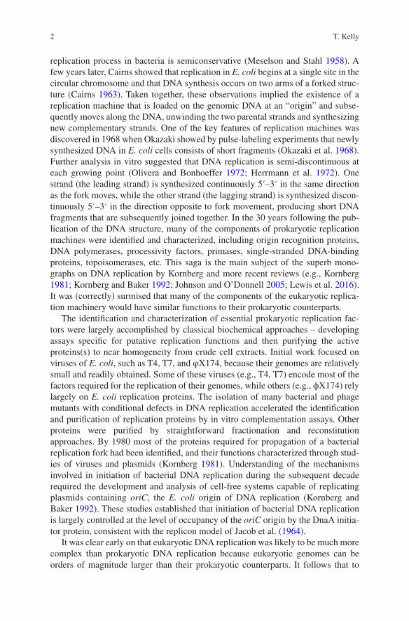
Fig. 1.1 SV40 and cellular DNA replication. A diagrammatic representation of the major steps in the viral and cellular replication pathways and the protein requirements for each step. See the text for details. (The steps involved in maturation of Okazaki fragments, relaxation of supercoiling, replication termination, and decatenation are left out for clarity.) (a) SV40 DNA replication. T, T antigen; α, DNA polymerase alpha-primase; R, RPA; δ, DNA polymerase delta; P, PCNA. (b) Cellular DNA replication. M, MCM2-7; 45, Cdc45; G, GINS, ε, DNA polymerase epsilon; α, DNA polymerase alpha-primase; R, RPA; δ, DNA polymerase delta; P, PCNA
T. Kelly

7
The only SV40-encoded protein required for DNA replication in vivo is the viral T antigen, which binds to the origin of DNA replication and serves both as the ini-tiator protein and as the replicative helicase (Tegtmeyer 1972; Tjian 1978; Delucia et al. 1983). In the initial description of the SV40 cell-free DNA replication system, it was demonstrated that extracts from uninfected primate cells, supplemented with purified T antigen, were sufficient for replication of DNA molecules containing the
M45G
α
α
α
α
δ
δε
ε
ε
ε
ε
ε
P
P
P
P
M45G
M45
G
M
M
45G
45G
M45
G
MM
{ORC, Cdc6, Cdt1, MCM2-7, ATP}
{Cdc45, GINS, Sld2, Sld3, Dpb11, Pol ε, RPA, Mcm10, DDK, CDK, ATP}
Core Helicase Loading
Helicase Activation
Initiation
Chain Elongation
B. Cellular DNA Replication
{Pol α, NTPs, dNTPs}
{Pol δ, RFC, PCNA}
Fig. 1.1 (continued)
1 Historical Perspective of Eukaryotic DNA Replication

8
SV40 origin of DNA replication (Li and Kelly 1984). Detailed studies demonstrated that T antigen monomers assemble into a double hexamer around the origin DNA in a reaction dependent upon ATP (Dean et al. 1992; Valle et al. 2000). The hexamers interact head-to-head via the N-terminal origin binding domains of T antigen and alter the structure of the origin DNA (Borowiec and Hurwitz 1988). In the presence of ATP and a single-stranded DNA-binding protein, each T antigen hexamer func-tions as a helicase to unwind the template DNA (Stahl et al. 1986; Dean et al. 1987; Wold et al. 1987). Helicase activity is dependent upon a C-terminal AAA+ module, which binds and hydrolyzes ATP to drive translocation of the hexamer in the 3′–5′ direction along the leading strand template at each replication fork (Fig. 1.1a). Structural studies of the T antigen helicase revealed a double-ring with one tier containing the AAA+ motor domains (Li et al. 2003a). The central channel contains basic residues that can interact with the DNA. The precise mechanism of T antigen helicase translocation on DNA is not yet understood, although structural studies have inspired some interesting models. In addition to serving as the replicative heli-case, T antigen interacts with other replication proteins to organize the replisome and coordinate its activities (see more below).
Biochemical analysis of the cell-free system derived from human cells demon-strated that a number of cellular proteins were required for SV40 DNA replication in vitro. All of these proteins proved to be involved in cellular DNA replication and were subjects of extensive biochemical and structural studies in the ensuing years (Reviewed in Kelly 1988; Challberg and Kelly 1989; Stillman 1989; Hurwitz et al. 1990; Brush et al. 1995; Waga and Stillman 1998).
The first cellular protein identified by fractionation of the SV40 system was RPA, the eukaryotic single-stranded DNA-binding protein (Wobbe et al. 1987; Wold and Kelly 1988; Fairman and Stillman 1988; Wold 1997; Chen and Wold 2014). The three nonidentical subunits of RPA contain multiple OB folds that bind single-stranded DNA. RPA was subsequently found to be subject to multiple post-translational modifications and to interact with many cellular proteins involved in a wide range of transactions involving DNA (Chen and Wold 2014). The contrast of the multi-subunit structure of RPA with the simpler bacterial single-stranded DNA- binding protein (SSB) was an early hint that the eukaryotic replisome would prove to be much more complex than that of prokaryotes.
An unexpected discovery that emerged from analysis of the SV40 system was that viral DNA synthesis in vitro is dependent on more than one cellular DNA poly-merase. The eukaryotic DNA polymerase α had been discovered many years prior to the development of the SV40 system and was thought to be the major, perhaps the only, replicative DNA polymerase in eukaryotic cells on the basis of many indirect lines of evidence (Campbell 1986; Lehman and Kaguni 1989). After initial difficul-ties with biochemical characterization, the enzyme was eventually shown to contain four subunits, one of which harbors the polymerase activity. The two smallest sub-units comprise a primase enzyme (Tseng and Ahlem 1982; Kaguni et al. 1983; Plevani et al. 1985). The primase catalyzes de novo synthesis of RNA primers on single-stranded DNA templates that can be further extended into nascent DNA chains by the DNA polymerase activity of the enzyme. By antibody depletion and
T. Kelly
9
fractionation/reconstitution experiments, it was demonstrated that DNA polymerase α is absolutely required for SV40 DNA replication in vitro. In the presence of T antigen and RPA, which are sufficient to drive extensive DNA unwinding, DNA polymerase α can initiate DNA synthesis on DNA molecules containing the SV40 origin and catalyze DNA synthesis on both the leading and lagging strand templates (Li and Kelly 1984; Murakami et al. 1986, 1992; Wold et al. 1988; Fig. 1.1a).
DNA polymerase δ, a second eukaryotic DNA polymerase, was identified in the 1970s but had been largely ignored (Byrnes et al. 1976). The activity of DNA poly-merase δ was initially distinguished from that of DNA polymerase α because it contained an associated 3′–5′ proofreading exonuclease activity, which was lacking in DNA polymerase α. The first clue that this polymerase might play a role in SV40 DNA replication was the discovery that PCNA, a 37 kDa protein essential for SV40 DNA replication in vitro, was identical to a previously identified factor that increased the processivity of DNA polymerase δ (Tan et al. 1986; Prelich et al. 1987). The requirement for DNA polymerase δ was subsequently confirmed by direct reconsti-tution of the SV40 replication reaction with purified proteins (Lee et al. 1989; Weinberg and Kelly 1989; Tsurimoto et al. 1990). As described in more detail below, DNA polymerase δ catalyzes the bulk of DNA synthesis on both the leading and lagging strands of SV40 (Fig. 1.1a).
The PCNA processivity factor required for efficient DNA synthesis by DNA polymerase δ was reminiscent of the sliding clamps previously described in the prokaryotic T4 and E. coli systems (Tan et al. 1986; Prelich et al. 1987; Tsurimoto and Stillman 1990). It was ultimately shown by elegant biochemical and structural studies that PCNA, the E. coli β-clamp, the T4 gp45 sliding clamp, and archaeal sliding clamps are ring-shaped proteins with pseudo sixfold symmetry that accom-modate duplex DNA in a topological linkage (Jeruzalmi et al. 2002). The general structure of the rings and the process by which they are loaded onto DNA have been highly conserved in evolution. In addition to its role in mediating processive DNA synthesis, PCNA plays major roles in other processes, such as Okazaki fragment maturation, DNA repair, recombination, chromatin assembly, cell cycle control, etc. (Moldovan et al. 2007).
The eukaryotic clamp loader RF-C, identified initially as a fraction required for SV40 DNA replication, consists of five subunits, each of which contains an AAA+ domain (Tsurimoto and Stillman 1989; Lee et al. 1991; Cai et al. 1996; Bowman et al. 2005). RF-C, like prokaryotic and archaeal clamp loaders, functions as a machine that couples the energy of ATP hydrolysis to open PCNA and load it at a primer terminus (Bowman et al. 2005; Yao et al. 2006). After dissociation of the RF-C loader, DNA polymerase δ associates with the loaded PCNA to form a highly processive complex. Much has been learned about the structural basis of the specific-ity of RF-C for primer termini and about the biochemical mechanism of clamp load-ing (Yao and O’Donnell 2012). On the lagging strand, RF-C and other clamp loaders can also function to unload PCNA rings from double-stranded DNA after comple-tion of Okazaki fragment synthesis and ligation so that they can be recycled.
Studies of SV40 DNA replication in vitro also allowed the detailed analysis of the roles of DNA topoisomerases (Yang et al. 1987). It was shown that topoisomer-
1 Historical Perspective of Eukaryotic DNA Replication

10
ase activity is required during DNA chain elongation to remove supercoils gener-ated by DNA unwinding and that this function can be mediated by either type I or type II topoisomerase. Human topoisomerase I binds to T antigen and may play the predominant role in relieving topological stress at replication forks (Simmons et al. 1996). Interestingly, yeast topoisomerase I co-purifies with the cellular helicase, suggesting that it may also travel with the replication fork (Gambus et al. 2006). Topoisomerase activity is additionally required for the decatenation and segregation of completed daughter DNA molecules synthesized in vitro, but this activity can only be provided by type II topoisomerase. These observations are entirely consis-tent with studies of SV40 DNA replication in vivo (Sundin and Varshavsky 1980, 1981).
Analysis of the interactions among the proteins required for SV40 DNA replica-tion provided insights into the functional organization of the replication fork, many of which are relevant to understanding the cellular replication fork (Reviewed in Kelly 1988; Challberg and Kelly 1989; Stillman 1989; Hurwitz et al. 1990; Waga and Stillman 1998; Fig. 1.1a). Movement of the replication fork, driven by the T antigen helicase motor, generates single-stranded DNA bound by RPA. During this process, the complex of DNA polymerase δ with PCNA advances, synthesizing the leading strand, while a region of the lagging strand template accumulates in single- stranded form prior to synthesis of a primer by DNA polymerase α. Interestingly, it was observed that DNA polymerase α, in the absence of other factors, is completely unable to initiate primer synthesis on DNA templates that are coated with bound RPA (Collins and Kelly 1991; Melendy and Stillman 1993). This observation led to the discovery that T antigen promotes primer synthesis via specific interactions with DNA polymerase α and RPA that presumably destabilize bound RPA and allow access of primase to the template (Collins and Kelly 1991; Dornreiter et al. 1992; Collins et al. 1993; Melendy and Stillman 1993; Zhou et al. 2012). Thus, the T anti-gen helicase-DNA polymerase α complex constitutes a mobile primosome that is active in primer synthesis and likely limits priming to the vicinity of replication forks. Presumably, functionally similar interactions involving the cellular helicase or associated factors are required for priming by DNA polymerase α at cellular replication forks.
Another important phenomenon discovered in the SV40 system is switching among DNA polymerases (Lee et al. 1989; Weinberg and Kelly 1989; Tsurimoto et al. 1990; Waga et al. 1994; Waga and Stillman 1994, 1998; Fig. 1.1a). Polymerase switching is unique to eukaryotic DNA replication and occurs in both viral and cel-lular systems. DNA synthesis is initiated on both the leading and lagging strand templates by the primase activity of DNA polymerase α. After synthesis of a short RNA primer, DNA polymerase α extends it into an “initiator DNA” chain. In the case of SV40, DNA polymerase α is subsequently replaced by the complex of PCNA and DNA polymerase δ, which carries out the bulk of DNA synthesis. The mechanism of this polymerase switch involves a competition between DNA poly-merase α and RF-C for the primer terminus (Tsurimoto et al. 1990; Tsurimoto and Stillman 1991a, b; Waga et al. 1994). Since DNA polymerase α is not highly proces-sive, RF-C will gain access to the primer terminus at some point during synthesis of
T. Kelly

11
the initiator DNA. When this happens, RF-C loads PCNA and then dissociates, allowing the binding of DNA polymerase δ to PCNA. The PCNA-DNA polymerase δ complex is stable, creating a highly processive polymerase engine capable of syn-thesizing long nascent DNA strands. On the leading strand, the polymerase switch is only required once, while on the lagging strand, one switching event occurs for each Okazaki fragment. DNA polymerase δ efficiently completes the synthesis of Okazaki fragments, after which they are joined together by DNA ligase in a matura-tion process that involves removal of the RNA primer and much of the initiator DNA (Waga and Stillman 1994; Balakrishnan and Bambara 2013). DNA poly-merase switching plays an important role in maintaining the integrity of the genome. DNA polymerase δ has a proofreading exonuclease, but DNA polymerase α does not, so the polymerase switch, together with the removal of the primer and much of the initiator DNA during completion of Okazaki fragment synthesis, helps ensure the fidelity of DNA replication. Subsequent genetic and biochemical studies in yeast revealed the existence of a third eukaryotic DNA polymerase, DNA poly-merase ε (Pol ε), that functions in cellular DNA replication but apparently not in SV40 DNA replication (Budd et al. 1989; Morrison et al. 1990; Araki et al. 1992; Zlotkin et al. 1996; Pospiech et al. 1999). Multiple recent studies indicate that DNA polymerase ε plays the major role in the synthesis of the leading strand during cel-lular DNA replication, while DNA polymerase δ synthesizes most of the DNA on the lagging strand (Burgers et al. 2016). It follows that a switch from DNA poly-merase α to DNA polymerase ε occurs at the terminus of the leading strand shortly after synthesis of the first primer. The mechanism of this switch is not yet known. However, yeast DNA polymerase ε is quite processive, even in the absence of RF-C and PCNA, so it may simply replace the less processive DNA polymerase α after synthesis of a short segment of initiator DNA. In the yeasts S. cerevisiae and S. pombe, the catalytic activity of DNA polymerase ε is not essential, although DNA replication is somewhat abnormal in its absence (Dua et al. 1999; Kesti et al. 1999; Feng and D’Urso 2001). In this circumstance DNA polymerase δ is presumably responsible for most DNA synthesis on both the leading and lagging strand tem-plates, as is the case with SV40.
The SV40 system also focused attention on the potential for regulation of DNA replication by protein phosphorylation. Viral DNA replication in vivo and in vitro was found to be completely dependent upon phosphorylation of T antigen on Thr- 124 by cyclin-dependent kinase (McVey et al. 1989, 1993; Moarefi et al. 1993). The unphosphorylated protein is competent to bind to the origin and to induce structural distortions in origin DNA and is also competent to catalyze unidirectional unwind-ing in helicase assays with artificial DNA substrates containing single- and double- stranded DNA. However, the unphosphorylated protein is completely inactive in bidirectional helicase activity from the SV40 origin, indicating that phosphorylation controls a step subsequent to assembly of the T antigen double hexamer on the DNA, possibly an interaction between T antigen hexamers that is required for the initial unwinding of the origin. Interestingly, it was observed that phosphorylation of other residues in T antigen, notably Ser-120 and Ser-123, is inhibitory to initia-tion of DNA replication and, again, the modifications appear to regulate a step sub-
1 Historical Perspective of Eukaryotic DNA Replication
12
sequent to assembly of the double hexamer (Virshup et al. 1989, 1992; Scheidtmann et al. 1991; Cegielska and Virshup 1993; Cegielska et al. 1994). Thus, T antigen helicase function is regulated both positively and negatively by phosphorylation. The biological role of this regulation is still unclear, but it may function in part to coordinate SV40 replication with the cell cycle, to enhance the efficiency of produc-tion of viral genomes. Consistent with this idea, human CDK can promote SV40 DNA replication in G1 extracts that are normally deficient in replication activity (D’Urso et al. 1990). These discoveries presaged the description of the (much more complex) control of the activation of the cellular helicase by protein phosphoryla-tion (see below).
As the foregoing indicates, analysis of SV40 DNA replication in vitro by many investigators resulted in the identification of key cellular replication proteins and generated many insights into their mechanisms of action. Naturally, these advances kindled great interest in identifying the cellular counterparts of T antigen – the mol-ecules that are required for origin recognition in cellular chromosomes and the mol-ecules that comprise the cellular helicase. A related issue of great interest was how initiation of replication in cells is controlled to ensure that each segment of the genome is faithfully duplicated once each cell cycle. These are the main issues dis-cussed in the remainder of this perspective. To attack these problems, attention shifted from SV40 to other model systems. The analysis of DNA replication in bud-ding yeast proved particularly fruitful because of the availability of highly devel-oped genetic approaches and the existence of defined origins of replication. Fission yeast provided many novel insights as well and, in many cases, complemented the work in budding yeast. Many advances also came from studying replication in extracts of eggs of Xenopus laevis. The Xenopus system lacks genetics and is not particularly amenable to large-scale fractionation/reconstitution approaches. However, it can be used effectively to test specific requirements for cellular replica-tion by antibody depletion-reconstitution experiments. It is also especially useful for analyzing the regulation of cellular DNA replication.
Because of methodological advances, there was also a gradual shift away from classical approaches for identifying cellular replication proteins by fractionation of crude extracts toward more efficient methods. The products of putative replication genes identified in genetic screens or homology searches were expressed as tagged variants that could be quickly purified and biochemically characterized. But recon-stitution with purified proteins remained the gold standard for defining the minimal constellation of proteins required for eukaryotic DNA replication.
1.4 Cellular Origins and Initiators
The first cellular origins of DNA replication to be characterized were those of the budding yeast S. cerevisiae (Hsiao and Carbon 1979; Stinchcomb et al. 1979; Newlon 1988; Fangman and Brewer 1991; Campbell and Newlon 1991). In early genetic studies, it was discovered that certain chromosomal DNA segments, called
T. Kelly

13
autonomously replicating sequences or ARSs, could increase the transformation efficiency and replication stability of yeast plasmids. It was subsequently shown by direct physical assays that ARS elements represent the start sites for initiation of bidirectional replication in budding yeast (Brewer and Fangman 1987; Huberman et al. 1987). Detailed analysis revealed that ARS elements share a common sequence of 11 bp called the ARS consensus sequence, as well as adjacent, less highly con-served sequence elements (Broach et al. 1983; Celniker et al. 1984; Van Houten and Newlon 1990; Marahrens and Stillman 1992; Breier et al. 2004). The strong sequence specificity and other properties of S. cerevisiae origins of DNA replication were similar to the familiar prokaryotic origins of DNA replication and the origins of eukaryotic viruses like SV40. Importantly, this sequence specificity would prove crucial for identifying the cellular proteins that assemble at origins of replication prior to the onset of DNA synthesis. Central among these is the origin recognition complex (ORC), which was detected as a protein that binds specifically to ARS ele-ments and is essential for initiation of DNA replication (Bell and Stillman 1992; Bell et al. 1993). The discovery of ORC was a major advance that opened the way to study the early events in cellular DNA replication.
ORC was found to have six subunits, five of which are related to the AAA+ pro-tein family of ATPases (Bell et al. 1995; Bell and Stillman 1992; Bell 1995; Bell and Dutta 2002; Speck et al. 2005; Clarey et al. 2006). High-affinity binding of ORC to ARS elements is dependent upon ATP binding to the largest subunit, Orc1. In the years following the identification of ORC, a general picture of the first steps in cel-lular replication emerged from studies in yeast cells and the Xenopus egg extract system. These steps result in the loading of the core replicative helicase onto the origin DNA. An early finding that opened a path toward defining the helicase load-ing reaction was the identification of a protein assembly at yeast origins of DNA replication that is referred to as the prereplicative complex or pre-RC (Diffley and Cocker 1992; Diffley et al. 1994). The pre-RC was originally characterized as a distinct pattern of nuclease protection in genomic footprinting experiments that was observed at yeast origins during the G1 phase of the cell cycle. The requirements for pre-RC formation and the protein composition of the complex were not immediately apparent but were rapidly determined by a variety of experimental approaches (Cocker et al. 1996; Rowles et al. 1996; Coleman et al. 1996; Romanowski et al. 1996b; Donovan et al. 1997; Tanaka et al. 1997; Aparicio et al. 1997; Nishitani et al. 2000; Maiorano et al. 2000; Labib et al. 2001; Devault et al. 2002; Tanaka and Diffley 2002). These experiments demonstrated that in the initial step of pre-RC formation, ORC recruits the Cdc6 protein to the origin. Cdc6 is a AAA+ protein in the same clade as the Orc1-5 subunits. The complex of ORC and Cdc6 then recruits the Cdt1 protein and the MCM2-7 complex. Cdt1, a factor first identified in fission yeast, binds to MCM2-7 and facilitates its interaction with ORC-Cdc6 bound at the origin. The MCM2-7 complex is the core of the eukaryotic replicative helicase. It is loaded onto the origin DNA by ORC-Cdc6-Cdt1 in a reaction requiring ATP hydro-lysis (Fig. 1.1b). The nature of the association of MCM2-7 with DNA was not clear from these early experiments but was eventually determined by elegant biochemical studies with purified components (Evrin et al. 2009; Remus et al. 2009; Kawasaki
1 Historical Perspective of Eukaryotic DNA Replication

14
et al. 2006). Two hexameric rings of MCM2-7 are loaded in a head-to-head configu-ration with duplex DNA passing through a central channel. In the loading reaction, ORC and Cdc6 function as a molecular machine to close a gate between the Mcm2 and Mcm5 subunits, leaving the MCM2-7 rings topologically linked to the DNA. The current view is that the two hexamers are loaded in separate steps which are dependent on distinct Cdc6 and Cdt1 molecules (Bell and Kaguni 2013; Sun et al. 2014; Ticau et al. 2015). Once the MCM double hexamer is loaded on the DNA, subsequent events in initiation of DNA replication do not require Cdc6, Cdt1, or ORC, and in vitro studies suggest that these factors may dissociate from the DNA (Rowles et al. 1999; Hua and Newport 1998; Gros et al. 2014; On et al. 2014; Ticau et al. 2015). Thus, loaded double hexamers of MCM2-7 mark the potential sites of initiation of DNA replication in chromosomal DNA at the end of the G1 phase of the cell cycle. The transformation of MCM2-7 into an active helicase and the assem-bly of the other replisome components begin at the onset of S phase and require the CDK and DDK protein kinases (more about this below).
It was quickly established that the early events in DNA replication just described are largely conserved in all eukaryotes. ORC molecules were identified in eukary-otic species other than S. cerevisiae, including humans and other metazoa, and the general requirements for pre-RC assembly proved to be universal (Bell and Dutta 2002). However, studies in other systems did not reveal the strong sequence speci-ficity of budding yeast ARS elements. It gradually became apparent that DNA rep-lication origins in most eukaryotes conform to a paradigm that is somewhat different from that of S. cerevisiae. A well-characterized example is the fission yeast S. pombe. It was possible to isolate segments from S. pombe chromosomal DNA that, like ARS elements in budding yeast, confer stable plasmid replication in fission yeast cells (Dubey et al. 1994, 1996; Clyne and Kelly 1995; Dai et al. 2005). However, the properties of the active segments are different from those of budding yeast: they are much larger than budding yeast ARS elements, and they exhibit little conservation of nucleotide sequence. The average AT content of the sequences active as origins is greater than that of the fission yeast genome (Segurado et al. 2003; Dai et al. 2005), an observation that was explained, at least in part, by the discovery that S. pombe ORC binds to chromosomal DNA via multiple copies of a DNA-binding motif called the AT hook (Chuang and Kelly 1999; Kong and DePamphilis 2001). But fission yeast origins do not share a common consensus sequence. In fact, the best predictors of the ability of a segment of the fission yeast genome to function as a plasmid origin are AT content and length (Segurado et al. 2003; Dai et al. 2005). At least half the intergenic regions in the fission yeast genome can exhibit detectable origin activity in plasmid assays. It was suggested that the properties of fission yeast origins are best explained by a stochastic model in which ORC can bind and drive initiation of DNA replication at many potential sites in the genome with little intrinsic sequence specificity, and during each cell cycle these potential sites are chosen largely at random with perhaps some preference for AT-rich intergenic DNA (Dai et al. 2005). Subsequent DNA combing experiments demonstrated that the distribution of distances between start sites in a fission yeast genome is exponential, which is completely consistent with a stochastic model for
T. Kelly

15
the spatial distribution of potential origins (Patel et al. 2006; Kaykov and Nurse 2015). The firing of potential origins appears to be stochastic in time as well as space, although the rates of firing per unit time may vary from origin to origin (Heichinger et al. 2006; Kaykov and Nurse 2015).
The nature of origins of DNA replication in metazoan organisms is still not well understood, but the available evidence suggests that there are many potential origin sites and that such sites lack strong sequence determinants (Leonard and Mechali 2013). Consistent with this impression is the observation that purified metazoan ORC molecules, such as those of Drosophila or human cells, do not exhibit a strong preference for any particular DNA sequence (Vashee et al. 2003; Remus et al. 2004). Human ORC can drive replication of essentially any DNA molecule in a replication system derived from Xenopus eggs (Vashee et al. 2003).
It is important to recognize that even in the case of S. cerevisiae, the highly spe-cific interactions between ORC and the ARS consensus sequences are not essential for the fundamental mechanism by which ORC promotes initiation of DNA replica-tion. Specific binding by ORC as observed in budding yeast simply affects the dis-tribution of ORC molecules over the genome. In cell-free systems with S. cerevisiae proteins, ORC can efficiently initiate DNA replication on DNA molecules lacking ARS elements (Gros et al. 2014; On et al. 2014). Moreover, deleting chromosomal ARS elements from an S. cerevisiae chromosome has surprisingly little effect on the efficiency of DNA replication (Dershowitz et al. 2007). Thus, it seems likely that initiation of DNA replication in most, if not all, eukaryotes may occur on essentially any DNA molecule with some efficiency. S. cerevisiae may have acquired the capac-ity to initiate DNA replication at more specific sites because its chromosomes are relatively small. If replication initiation were purely random, there would be some probability that a small chromosome would not be duplicated in a significant frac-tion of cell cycles.
Why have eukaryotes largely departed from the prokaryotic paradigm of highly specific origin sequences enshrined in the replicon model? One possibility is that the distribution of chromosomal sites that are available for ORC binding and initia-tion may vary considerably in eukaryotic cells in different physiological states (e.g., different states of development, different states of differentiation, different tissue environments, etc.) because the composition and organization of chromatin and the pattern of gene expression are different in such states. Thus, the ability to initiate DNA replication in almost any accessible region may be advantageous in the face of the increased complexity of eukaryotic biology.
Because of the many potential origins of replication, the dynamics of DNA rep-lication in an individual eukaryotic cell is extremely complex and is different from one cell cycle to the next. The factors that determine the sites where replication is initiated and the timing of the replication of each genomic sequence are not yet understood in detail in any organism. The overall pattern of DNA replication is established by events that occur at two stages of initiation: the loading of the core helicase in G1 and the activation of the helicase at the onset of S phase. The distribu-tion of loaded MCMs obviously depends upon the relative affinity of ORC for par-ticular DNA sequences, which may be affected to some degree by other components
1 Historical Perspective of Eukaryotic DNA Replication
16
of the pre-RC (e.g., Cdc6) or by interactions with other cellular proteins (e.g., Beall et al. 2002; Speck and Stillman 2007). The distribution of loaded MCMs is also greatly influenced by competition with the multitude of other chromatin factors that bind to chromosomal DNA. This competition affects the accessibility of chromo-somal loci to the helicase loading factors and probably explains why many studies have correlated initiation of DNA replication with so-called open chromatin domains (Berbenetz et al. 2010; Eaton et al. 2011; Leonard and Mechali 2013). Once S phase begins, the loading of MCMs is precluded (see below), so their num-ber and their approximate locations are fixed (although there is some recent evi-dence that they may have limited mobility) (Gros et al. 2015). Therefore, subsequent events are largely determined by the pattern of activation of loaded MCMs by the CDK and DDK protein kinases to generate functional replisomes. This pattern is also extremely complex. Not all loaded MCMs are activated in a given cell cycle – many are functionally inactivated when they are replicated passively by a replisome originating at another site (Friedman et al. 1997; Santocanale et al. 1999; Vujcic et al. 1999). Thus, the determination of which loaded MCMs are activated and which are not is highly dependent upon their order of activation, and this is likely to be largely stochastic in a given cell cycle. There is no convincing evidence that the ordering of helicase activation in the cell cycle is deterministic, but, as discussed later in this perspective, the relative rates of activation of loaded MCMs at different sites can differ, so that on average activation at some sites occurs earlier than at oth-ers. In the end, the timing of replication of a particular sequence in the genome during a given cell cycle is a function of (1) its distance from the flanking initiation sites, (2) the times of initiation at those sites, and (3) the rates at which the forks move after initiation. These factors, particularly the first and second, are subject to variation from one cell cycle to the next. This is an active area of research, and it is expected that further insights will come from increasing the spatial and temporal resolution of single molecule and ensemble approaches to studying replication tim-ing and from improving the methods of analysis in silico.
1.5 CMG the Cellular Helicase
Following the discovery of ORC, the cellular initiator protein, another 15 years were required to definitively identify the active form of the cellular helicase. This advance was driven by two separate lines of investigation that eventually came together. Genetic studies in budding yeast defined a group of genes required for the stable maintenance of plasmids as autonomously replicating units in cells (Moir et al. 1982; Maine et al. 1984). Although isolated by several different assays, it was eventually appreciated that the corresponding proteins form a complex, which was named the MCM2-7 complex (for minichromosome maintenance 2-7). The six sub-units of the MCM2-7 complex were shown to be related members of the AAA+ superfamily of ATPases (Chong et al. 1996), and a number of studies suggested that they are required for initiation of DNA replication (Yan et al. 1991; Hennessy et al.
T. Kelly

17
1990; Chong et al. 1995; Madine et al. 1995; Romanowski et al. 1996a). The first clue regarding their potential function was the discovery that a sub-complex of MCM4-6-7 purified from human cells possessed ATP-dependent helicase activity in vitro (Ishimi 1997). This insight was followed by the demonstration that the sin-gle archaeal MCM protein forms multimeric complexes with robust helicase activ-ity (Kelman et al. 1999; Chong et al. 2000). Analysis of the distribution of MCM proteins in yeast chromosomes during S phase strongly suggested that the MCM complex travels with replication forks, and it was subsequently shown that deple-tion of MCM subunits interrupts replication fork progression (Aparicio et al. 1997; Labib et al. 2000). Experiments in Xenopus egg extracts confirmed that inhibition of MCM activity prevents efficient DNA unwinding (Pacek and Walter 2004). In the course of these studies, it was observed that Cdc45, another protein thought to be involved in initiation of DNA replication, interacts with the MCM complex and is also required for both DNA unwinding and fork progression (Hopwood and Dalton 1996; Aparicio et al. 1997; Pacek and Walter 2004). While these studies, taken together, established the likelihood that the MCM complex was a required compo-nent of the cellular helicase, it did not seem to be sufficient for helicase activity, since purified yeast MCM2-7, containing all six subunits, did not have detectable helicase activity under the usual assay conditions.
A second line of investigation that proved critical for the identification of the cellular helicase was a product of the discovery that DNA polymerase ε is required for DNA replication in yeast (Morrison et al. 1990; Araki et al. 1992). A network of proteins that exhibited genetic and physical interactions with each other and with DNA polymerase ε was discovered and characterized. The key players in this net-work were Dpb11, Sld2, Sld3, and a complex of four proteins called the GINS complex (Araki et al. 1995; Kamimura et al. 1998, 2001; Takayama et al. 2003). It was observed that GINS colocalizes with Cdc45 and MCM2-7 at sites of DNA unwinding during DNA replication and is required to maintain the association of Cdc45 with MCM2-7 during DNA chain elongation (Aparicio et al. 1997; Gambus et al. 2006; Pacek et al. 2006). The definitive biochemical definition of the cellular helicase was a result of experiments aimed at purifying the Cdc45 protein from extracts of Drosophila embryos (Moyer et al. 2006; Ilves et al. 2010). The purifica-tion yielded a complex of 11 polypeptides that included MCM2-7 and GINS, as well as Cdc45, and is now referred to as CMG. Strikingly, the purified CMG com-plex possessed robust 3′–5′ helicase activity on partially duplex DNA molecules. Reconstitution of the complex with recombinant proteins revealed that the presence of GINS and Cdc45 dramatically changed the properties of the MCM2-7 complex, increasing its affinity for DNA and stimulating its ATPase activity by two orders of magnitude (Ilves et al. 2010; Costa et al. 2011, 2014). Since GINS and Cdc45 do not have ATPase active sites, the observed stimulation is due to allosteric remodeling of the core MCM2-7 helicase engine. More recent studies have examined the structure of the CMG in the presence of single-stranded DNA and non-hydrolysable ATP (Costa et al. 2014). In this complex the AAA+ motor domains of the MCM2-7 hex-amer form a cracked-ring, right-handed spiral with the crack at the MCM2-5 inter-face as predicted from biochemical studies (Bochman and Schwacha 2008). GINS
1 Historical Perspective of Eukaryotic DNA Replication