Embed Size (px)
Citation preview
Hip DysplasiaChapter 83
Wayne H. Riser, W. Harker Rhodes, and Charles D. Newton
Section One: Theories of Pathogenesis
Section Two: Growth and Development of the Normal Canine Pelvis, Hip Joints, and Femur from Birth toMaturity
Section Three: The Dysplastic Hip Joint: Radiologic and Histologic Development
Section Four: Clinical Manifestations
References
Section One: Theories of PathogenesisCanine hip dysplasia is a complex disease. It is a concentration of factors from a pool of genetic weaknessesand environmental stresses that fall into a programmed pattern of progressive remodeling and degenerativejoint disease. The degree of involvement varies from minute changes in bone structure to total destruction ofthe hip joint. Investigators have searched intensively for genetic, chemical, and metabolic defects, but thecause has remained obscure.
Hip dysplasia affects humans and all other domestic mammals. In humans, 1.3 children in 1000 are affected.In dogs the prevalence may run over 50% in large dogs if control measures have not been practiced. Few dataare available on the prevalence of hip dysplasia in other mammals, but it is thought to be low. The disease isundoubtedly rare in undomesticated animals.
No specific genetic pattern of inheritance has been demonstrated in this variable disease. It has beendemonstrated that both genetic and environmental influences contribute to development, regardless of thespecies affected.(15,31, 32,40,74,76) Consequently, the disease has been designated as polygenic ormultigenic.(28) As in most polygenic diseases, there are both major and minor causative factors. There is noevidence that a primary defect of bone exists but rather the disease is a failure of the muscles and other softtissues to hold the hip joint in full congruity.(31,32) This is further supported by the fact that bony dysplasiacan be increased, decreased, or prevented by controlling the degree of joint instability and incongruity.(53)No other malformations are associated with the disease.(79) A causal relationship between muscles and softtissue defects or pathologic changes other than lack of muscle mass or strength has not been established.(40,41)
Experimentally, hip dysplasia may be produced in many ways.(43,56,74,76,87,88) These include anycircumstances that contribute to an unstable hip joint, namely, adductor forces, lack of muscle strength,
chemical relaxation of the pelvic soft tissues, traumatic injury to the hip joint, and overloading of the joint byweight. Hip dysplasia is a concentration of factors from a pool of genetic weaknesses and environmentalstresses that fall into a programmed pattern of progressive remodeling and degenerative joint disease.
The general cause of hip dysplasia, when defined, must be broad enough to explain its development, not onlyin dogs, but also in all other affected animals. Many genetic and environmental factors can trigger events thatbring about the condition secondarily.(74,77,79,88) Hip dysplasia, therefore, is not one disease but manydiseases that result in common degenerative lesions of the hip joints.(77)
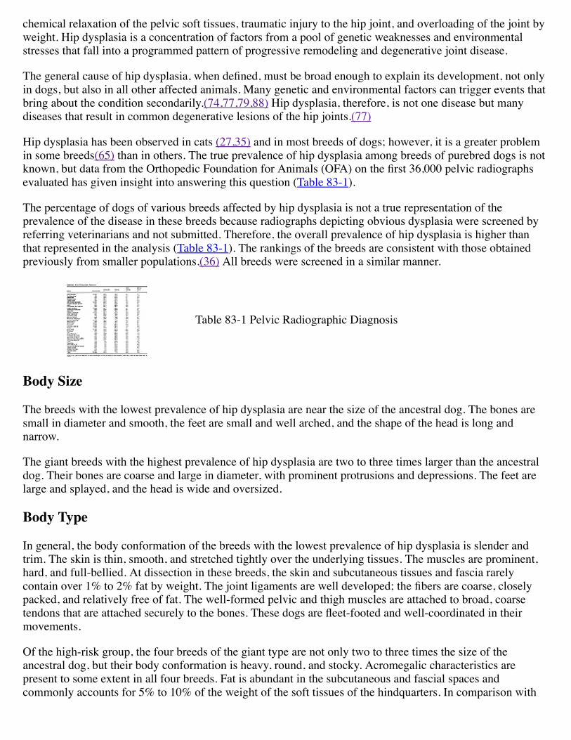
Hip dysplasia has been observed in cats (27,35) and in most breeds of dogs; however, it is a greater problemin some breeds(65) than in others. The true prevalence of hip dysplasia among breeds of purebred dogs is notknown, but data from the Orthopedic Foundation for Animals (OFA) on the first 36,000 pelvic radiographsevaluated has given insight into answering this question (Table 83-1).
The percentage of dogs of various breeds affected by hip dysplasia is not a true representation of theprevalence of the disease in these breeds because radiographs depicting obvious dysplasia were screened byreferring veterinarians and not submitted. Therefore, the overall prevalence of hip dysplasia is higher thanthat represented in the analysis (Table 83-1). The rankings of the breeds are consistent with those obtainedpreviously from smaller populations.(36) All breeds were screened in a similar manner.
Table 83-1 Pelvic Radiographic Diagnosis
Body Size
The breeds with the lowest prevalence of hip dysplasia are near the size of the ancestral dog. The bones aresmall in diameter and smooth, the feet are small and well arched, and the shape of the head is long andnarrow.
The giant breeds with the highest prevalence of hip dysplasia are two to three times larger than the ancestraldog. Their bones are coarse and large in diameter, with prominent protrusions and depressions. The feet arelarge and splayed, and the head is wide and oversized.
Body Type
In general, the body conformation of the breeds with the lowest prevalence of hip dysplasia is slender andtrim. The skin is thin, smooth, and stretched tightly over the underlying tissues. The muscles are prominent,hard, and full-bellied. At dissection in these breeds, the skin and subcutaneous tissues and fascia rarelycontain over 1% to 2% fat by weight. The joint ligaments are well developed; the fibers are coarse, closelypacked, and relatively free of fat. The well-formed pelvic and thigh muscles are attached to broad, coarsetendons that are attached securely to the bones. These dogs are fleet-footed and well-coordinated in theirmovements.
Of the high-risk group, the four breeds of the giant type are not only two to three times the size of theancestral dog, but their body conformation is heavy, round, and stocky. Acromegalic characteristics arepresent to some extent in all four breeds. Fat is abundant in the subcutaneous and fascial spaces andcommonly accounts for 5% to 10% of the weight of the soft tissues of the hindquarters. In comparison with
the low-dysplasia group, the muscles are less prominent and less developed. Fat is infiltrated into the tendonsand ligaments. The fibers of these two structures are smaller in diameter than those of the low-risk group. Thegait of the giant breeds is less graceful and slower than that of the smaller breeds.
Growth Pattern
Breeds with the highest prevalence of hip dysplasia grow and mature more rapidly than those in the low-riskgroup. Starting at birth, this group gains rapidly. The pups of these breeds are aggressive eaters, both as theynurse and as they take supplemental food. In a study involving 222 German shepherds, 63% of the dogs thatweighed more than the mean of this group at 60 days of age were dysplastic at 1 year of age, whereas only37% of those less than the mean became dysplastic. The same rapid rise in weight in other breeds of thegroup at high-risk for dysplasia has been observed.(63)
Hip dysplasia has not been reported in the wild undomesticated carnivorous animals, such as wolves andfoxes. A study of their pattern of growth found that the pups were slow-growing and late maturing. Theyoung pups were whelped in dens. As newborns, they received their nourishment by nursing during the firstfew weeks. When more food was required, the mother killed rodents and either brought them to the den or atethe animal where it was killed and then returned to the den where the ingested rodents were regurgitated forthe young to eat.(61) Young carnivores were quite mature and 6 to 10 months old before they began to hunt.The amount of food available for the growing members of a litter was limited. This caused the young tomature slowly and remain thin and light for their body size. Such an environment favored the completion ofossification and developmental maturity of the joint before the hips could be subjected to possible injury,incongruity, or subluxation from excessive extrinsic forces (e.g., excessive body weight) (65,69)
Genetic Influences and Heritability
Few genes analyzed thus far directly affect osseous structures.(17) The shape of bones reflects changes bybiomechanical stresses.(15)
In the dog no clear-cut pattern of inheritance has been recognized.(23,28,30) This means that many genes areaffected, and polygenic traits are subject to environmental modifications. New data have substantiated thesefindings.(29)
The spread of hip dysplasia centers around the genetic transmission and heritability of a particular body size,type, conformation, movement, growth pattern, and temperament. This conclusion is based on the facts thatthe prevalence of hip dysplasia is approximately the same in a number of breeds with similar bodycharacteristics and there is no gene flow between these purebred breeds. Since these facts must be respected,biomechanical and environmental factors associated with certain body conformation and size must beconsidered as causes.(69)
Critical evaluation of the heritability of hip dysplasia has been made in the German shepherd in 244 offspringfrom 54 full subfamilies. In one report, "heritability was defined as a property not only of the character (trait)but also the population and the environmental circumstances to which individuals are subjected. Heritability,because it represents the proportion of the total phenotypical variance, receives the attributes of a positivenumber which may range from 0 to 1.0 in magnitude".(29) On this scale and based on evaluations ofradiographs from 2 year-old dogs, the heritability was given an average estimate of 0.25. The conclusionswere that canine hip dysplasia be termed a moderately heritable diseased.(30)
In a study involving 236 German shepherds, it was demonstrated that the most reliable way to eliminatecanine hip dysplasia was through the establishment of "pedigree depth," that is, by the use of ancestral linesof dogs radiographically free of hip dysplasia.(33)
Results of controlled breeding programs in Sweden further indicated that the prevalence of hip dysplasia inthe German shepherd was substantially reduced by mating only dogs with radiographically normal hips.(7,50) Similar decreases in prevalence have occurred in another controlled breeding program in a colony ofguide dogs (Seeing Eye, Inc. Morristown, NJ).
In another account, with 584 progeny in a closed colony of German shepherds, it was shown that theprevalence of hip dysplasia was noticeably reduced by selectively breeding dogs proved radiographically tohave normal hips at 1 year of age or older. In 3-1/2 years the incidence of hip dysplasia was lowered from39% to less than 17%.(64) The male dogs in this colony had a wide variation in their ability to transmitnormal hips to their progeny. For example, only 8.7% of the progeny of one dog with radiographically normalhips at 2 years of age developed hip dysplasia, whereas 37.8% of the pups of another dog with similarradiologic evaluation mated to the same bitches developed hip dysplasia.(20)
Environmental and Man-Made Influences
Embryologically, articular joints are differentiated as units in situ from a mass of skeletal mesenchyme.(90)Development progresses normally in each joint as long as there is full congruity between the parts. Thecongruity remains as long as the supporting tissues are strong enough to withstand the mechanical orphysiological factors that tend to pull them apart.(77)
In humans, intrauterine stress has been cited as contributing to hip dysplasia, particularly if the fetus ispositioned with the legs in adduction and extension.
Hip dysplasia in humans is rarely associated with teratology abnormalities. Other hip abnormalitiesdistinctive from dysplasia, however, are frequently associated with such deformities as clubfoot,hyperextension of the knees, spinal deformities, arthrogryposis multiplex, and chondro-osteodystrophy.(22)
In the young child, the position of the legs during infant care is found to be very important to normal hipdevelopment.(71,73,75) Abduction and flexion of the legs has a stabilizing effect on the hip joints. Thesquare diaper favors greater abduction of the legs than does the three cornered diaper. The Bantu baby, who iscarried with its front side bound to the mother's back with its legs in acute abduction and flexion, seldom hasabnormal hip joints.(71,75) In contrast, the Navajo Indian baby, who spends its first years of life strapped to acradleboard with the legs in abduction and extension, has a high rate of hip joint instability.(70)
Other factors such as femoral anteversion and spastic shortening of the psoas muscle have been shown tofavor acetabular dislocation when the leg was extended.(44) These observations indicate that bothenvironmental and hereditary influences are important.(28,42)
In the dog, the hip joints are normal at birth.(43,68) The long bones of the pup are short during prenatal life,and mechanical stresses that bring about dislocation of the femoral heads are minimal. Teratologicabnormalities of the joints are rare in the dog, except for congenitally dislocated elbows and an occasionalclubfoot deformity. Congenital malformation of the hips is also rare.
Extrauterine Influences
EARLY WEIGHT GAIN
In 222 German shepherds born consecutively, 100 were dysplastic, and the prevalence of hip dysplasia at 1year had a direct correlation with their weight at 60 days of age. The heavier dogs, that is, the heaviest malesand heaviest females at 60 days of age, had the highest incidence of hip dysplasia at maturity.(63) (See Fig.83-2.)
These data suggested a number of indirect genetic factors influencing the rate of hip dysplasia. Theaggressiveness in nursing may be inherited, as may be the quality and quantity of the supporting tissuesaround the hip joint. It was concluded that when growth, gain in weight, and nursing aggressiveness exceededthe strength of the supporting tissues, subluxation and hip dysplasia occurred.(63)
The first subluxating stress on the hips occurs when the pup supports itself while nursing, and the hindlegsare in forceful adduction and extension. The heaviest pups were the more aggressive, worked the hardestwhile nursing, and spent the most time feeding.(63)
PELVIC MUSCLE MASS
Data indicate that here is a positive correlation between the amount of pelvic muscle mass and the prevalenceof hip dysplasia. Of three large breeds of dogs, the greyhound is relatively free of hip dysplasia; over half ofthe German shepherds are affected with hip dysplasia, and nearly all the July foxhounds are dysplastic.(69)
These data further emphasize that hip dysplasia encompasses biologic height, weight, and muscle bracing.The builder, before architecture was a science, learned that when the height of a structure was doubled, thebracing had to be tripled or the structure would fall of its own weight.(82) This basic rule, learned many yearsago, illustrates clearly why a low foot stool fits solidly on the floor and the tall stool of the same area wobbleswhen supporting weight.(82) Similarly, it has been found that dogs less than 30.5 cm in height and less than11.3 kg in weight (dachshund) are relatively free of hip dysplasia. On the other hand, at least half the largedogs, those 34 kg or more in weight and more than 50.8 cm in height, are affected with dysplasia.(66)
MUSCLE MYOPATHIES
All newborn mammals, including human infants, undergo many metabolic changes during their transitionfrom intrauterine to extrauterine life. The muscle tissues are relatively immature both anatomically andbiochemically at birth. Lack of muscular maturation in the newborn influences the manner in which thenewborn responds to function. This immaturity accounts for the failure of many mammals, including thehuman, dog, and cat, to walk at birth.(88)
There is evidence that the wide range of acetabular and femoral changes occurring in hip dysplasia is theconsequence of joint laxity. The possibility that this may be associated with or influenced by the rate ofmuscle maturation has not been explored. The rate of muscle maturation may be an inherited factor.(12,43)Consequently, the degree of subluxation in the young may be influenced by subnormal muscular function. Inhumans, the possibility of iliopsoas muscle spasm in the infant has been explored. (41,44)
In the adult dog, the light microscope was used to examine histologically the individual pelvic musclesassociated with hip joint motion. Evidence of muscle disease was not recognized. In dogs with advanced hipdysplasia and associated osteoarthritis, atrophy of the pelvic muscles was present but changes such asmuscular necrosis, inflammation, and extensive fibrosis were not found.(66,69)
One observer suggested that in young dogs with developing dysplasia, the pectineus muscles were in spasmand contained a degenerative lesion.(4) The pectineus muscle (an adductor), when in spasm, was thought tofavor forcing the femoral heads out of the acetabula. This observer further suggested that if the pectineuswere cut in the dog at an early age, the occurrence of hip dysplasia would be drastically lowered.(4)
A causal relationship between the pectineus muscles and hip dysplasia was not established in an experimentusing the pelvic muscles from Labrador retrievers, German shepherds, Alaskan malamutes, and beagles.(40)Pectineus muscles in these dogs with both normal and dysplastic hips were examined and compared. Therelationship between pectineus muscle abnormality and hip dysplasia remains undefined. The pectineusmuscles from some young pups showed both hypotrophic and hypertrophic changes. It was suggested that the
alterations seen in the pectineus muscles of dysplastic dogs probably represented secondary manifestationsassociated with a disease of developing hip joints (hip dysplasia).(4,12) The available evidence does notsupport the concept that abnormal pectineus muscle behavior is a cause of hip dysplasia. (39)
Developmental myopathy with type II fiber hypotrophy has been described in the pectineus muscles of veryyoung dysplastic German Shepherds These investigators failed to establish a relationship between this musclechange, joint laxity, and dysplasia but have suggested the possibility of such a relationship. In theirexperiments using an enzyme stain, the small fibers stained as type I (white) and the large fibers as type II(dark). They considered the differentiation between small and large fibers in young dogs to be a myopathy.No myopathies were present in either the normal or dysplastic adult dogs in their study.(12) This change inthe young dog resembles muscle fiber hypotrophy, which follows the cutting of the nerve to a muscle. Thesehypotrophied muscles become functional again and the fibers become normal in size when the nerve unitesand use is restored.(34) Atrophied muscle due to a severed nerve and immature muscle are similar inappearance. (34)
Metabolic Influences
SEX
In humans, the female is affected with hip dysplasia four to eight times more often than the male.(22) In thedog an equal number of females and males are affected. The reasons for this difference have not beenexplained. Of 100 dysplastic German shepherds at the Armens Hund Skula (Sweden), 49 were males and 51were females.(63)
CHEMICAL AND HORMONAL INFLUENCES
Pelvic tissue relaxation is a well-known physiological phenomenon that occurs during the terminal phase ofpregnancy in mammals. This reaction has been associated with the female hormone, estrogen.Experimentally, this reaction has been studied by injecting ovarian extracts into dogs to produce pelvic tissuerelaxation resembling that seen at the termination of pregnancy. The specific polypeptide hormone that iscommonly used is called relaxin. Male and spayed and virgin females when "primed" with estrogen beforerelaxin was administered responded sufficiently to relax pelvic tissues around the hip joints.(43,55)
The urine of newborns was examined to see if there was a correlation between high estrogen levels and theunstable hip. From the first tests, it appeared that such a correlation existed, but the use of more refined testsfailed to verify these findings. (1,3,81) The conclusion is that hormonal influence is not associated with thedevelopment of congenital hip dysplasia in humans or animals.(1,3,71,81)
In the dog it has been possible to increase the incidence of hip dysplasia by giving relaxin to newborn pupsand to produce hip dysplasia in the greyhound. (18,43,51,55) "It does not prove, however, that estrogens haveanything to do with etiology and pathogenesis of spontaneously occurring hip dysplasia."(19) There is noevidence that estrogen levels within the biologic range have a relationship to the incidence of hip dysplasia indogs.(19,52,55,81)
Defective protein biosynthesis of collagen was suggested as a cause for increasing articular cartilagedegradation in osteoarthritic joints. Soluble collagen was reported to be found in the acetabular cartilage ofdysplastic dogs, while predominantly insoluble collagen was present in dogs with normal hip joints. It wasnot possible to relate these changes to hip dysplasia or to osteoarthritis.(39,40)
Inborn metabolic errors of chemical or hormonal origin have not been found in human or canine hipdysplasia.(39,40,52,87)
DIET
A variety of nutritional and mineral supplements have been used in attempts to alter or prevent the course ofhip dysplasia in the dog. Diet has not affected the occurrence or course of the disease other than themechanical effect of increased or decreased weight upon the hip joint.(66)
Prevention
In the child the development of hip dysplasia can be stopped and the condition can be reversed to a stablenormal hip if it is discovered early before remodeling has begun. The key to treatment is the restoration offull congruity between the femoral head and acetabulum by placing the legs in an abductor-flexed position.(76,88)
In the young dog genetically conditioned to develop hip dysplasia, confinement to a small cage (1 m3) wherethe dog spends most of his time sitting on his haunches (abductor-flexed position) will prevent thedevelopment of hip dysplasia.(66,68) Surgical improvement of joint congruity can also be very beneficial.
Section Two: Growth and Development of the Normal Canine Pelvis,Hip Joints, and Femur from Birth to MaturityA clear understanding of the normal development of the hip joints, pelvis, and femur is imperative as a basisfor comparison in evaluating change associated with disease and/or injury of the hip. The American racinggreyhound is the model used to obtain normal growth patterns because of the low incidence of hip disease,particularly hip dysplasia, in this breed. Furthermore, the shape, size, and rate of growth and development ofthe bones of the pelvis, hindlegs, and caudal half of the spine are similar to other medium-large dogs, some ofwhich have a high prevalence of hip dysplasia.
Longitudinal Growth of the Femur
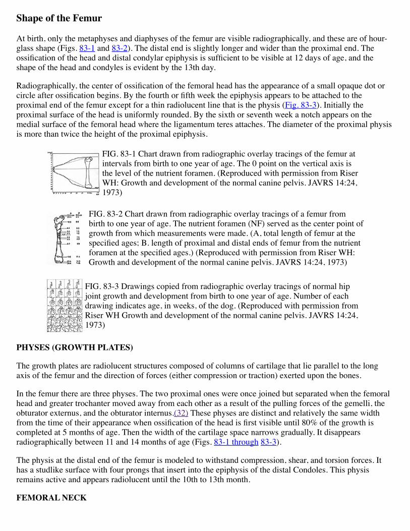
The nutrient artery around which the bony foramen later forms is at the center of a long bone when thecartilage mold is laid down in the skeleton of the fetus.(45) By identifying the nutrient artery, it is possible tomeasure the growth at the two ends of a long bone. At birth, it is difficult to measure the length of a longbone radiographically because only the diaphysis between the proximal and distal growth plates isradiopaque. The cartilaginous epiphyses are radiolucent.
At birth, the estimated total length of the femur is 3.5 cm, with 1.5 cm on the proximal end of the femur and 2cm on the distal end. The femur maintains a uniform weekly length increase (Fig. 83-1). When measurementsare charted, the weekly growth as indicated on the chart for the proximal part maintains an increase at theslope of approximately 20° until growth slows at the 30th week. Between the 10th and 30th weeks, the slopeincreases to approximately 25° and 30°. The distal part of the femur grows more quickly. Its growth rate is ata slope of approximately 50°. A 1: 1.5 ratio is maintained between the proximal and distal parts of the femurfrom birth to adulthood.
At 30 weeks (7 months) of age the overall length of the femur is 23.5 cm, with 9.5 cm at the proximal partand 14 cm at the distal end. This length represented 95% of the total adult femoral length and is within 0.9cm of the length of the femur of the 1-year-old, 26-kg (56-lb), male greyhound, whose femur measured 24.4cm, with 9.9 cm in the proximal and 14.5 cm in the distal parts (a ratio of 1:1.5 between the proximal anddistal ends).
Shape of the Femur
At birth, only the metaphyses and diaphyses of the femur are visible radiographically, and these are of hour-glass shape (Figs. 83-1 and 83-2). The distal end is slightly longer and wider than the proximal end. Theossification of the head and distal condylar epiphysis is sufficient to be visible at 12 days of age, and theshape of the head and condyles is evident by the 13th day.
Radiographically, the center of ossification of the femoral head has the appearance of a small opaque dot orcircle after ossification begins. By the fourth or fifth week the epiphysis appears to be attached to theproximal end of the femur except for a thin radiolucent line that is the physis (Fig. 83-3). Initially theproximal surface of the head is uniformly rounded. By the sixth or seventh week a notch appears on themedial surface of the femoral head where the ligamentum teres attaches. The diameter of the proximal physisis more than twice the height of the proximal epiphysis.
FIG. 83-1 Chart drawn from radiographic overlay tracings of the femur atintervals from birth to one year of age. The 0 point on the vertical axis isthe level of the nutrient foramen. (Reproduced with permission from RiserWH: Growth and development of the normal canine pelvis. JAVRS 14:24,1973)
FIG. 83-2 Chart drawn from radiographic overlay tracings of a femur frombirth to one year of age. The nutrient foramen (NF) served as the center point ofgrowth from which measurements were made. (A, total length of femur at thespecified ages; B. length of proximal and distal ends of femur from the nutrientforamen at the specified ages.) (Reproduced with permission from Riser WH:Growth and development of the normal canine pelvis. JAVRS 14:24, 1973)
FIG. 83-3 Drawings copied from radiographic overlay tracings of normal hipjoint growth and development from birth to one year of age. Number of eachdrawing indicates age, in weeks, of the dog. (Reproduced with permission fromRiser WH Growth and development of the normal canine pelvis. JAVRS 14:24,1973)
PHYSES (GROWTH PLATES)
The growth plates are radiolucent structures composed of columns of cartilage that lie parallel to the longaxis of the femur and the direction of forces (either compression or traction) exerted upon the bones.
In the femur there are three physes. The two proximal ones were once joined but separated when the femoralhead and greater trochanter moved away from each other as a result of the pulling forces of the gemelli, theobturator externus, and the obturator internus.(32) These physes are distinct and relatively the same widthfrom the time of their appearance when ossification of the head is first visible until 80% of the growth iscompleted at 5 months of age. Then the width of the cartilage space narrows gradually. It disappearsradiographically between 11 and 14 months of age (Figs. 83-1 through 83-3).
The physis at the distal end of the femur is modeled to withstand compression, shear, and torsion forces. Ithas a studlike surface with four prongs that insert into the epiphysis of the distal Condoles. This physisremains active and appears radiolucent until the 10th to 13th month.
FEMORAL NECK
The diameter of the femoral neck narrows slightly directly below the head. The joint capsule attaches aroundthe neck immediately distal to the area where the femoral head unites with the neck. The trochanteric fossaappears as a deep cavity. The angle of the neck with the diaphysis as viewed from the ventrodorsalradiograph is 135°. This neck angle is constant from birth through all stages of development (Figs. 83-1through 83-3).
The anteversion angle of the femoral neck is 0° to 20°, with a mean of 10°. It has been obtained from themeasurements of 32 macerated femora of 16 mature greyhounds.
In the embryologic state the trochanter and femoral head are composed of a single piece of cartilaginoustissue. With the development and contraction of the muscles of the pelvis, the trochanteric fossa is formed toallow for the insertion of the obturator internus, the obturator externus, and the gemelli muscles.(21,45) Thegreater trochanter is extended by the dorsal and medial pull of the three pairs of gluteal muscles. Thetrochanter becomes visible radiographically between the eighth and ninth weeks of life. The lesser trochanteron the medial side of the femur appears as an apophysis and is composed of cartilage during the early weeksof life but is identifiable radiographically at 10 to 12 weeks. When the physis closes (11-13 months), thisprocess attaches as a single piece to the diaphysis of the femur (Fig. 83-3).
FEMORAL SHAFT
The metaphyses develop and extend proximally and distally as the columns of cartilage cells of the physesmature and disintegrate, and new trabeculae ossify and the bones lengthen. Both the length and diameter ofthe femoral shaft increase to six to seven times their size (Fig. 83-4). There is a direct correlation betweengrowth in length and increase in diameter.
FIG. 83-4 Chart drawn from overlay tracings of radiographs of one half of thepelvis of a dog at birth, 8 weeks, 16 weeks, and one year. The bipolar growth plate(B) between the ilium and ischium served as the center point of growth.(Reproduced with permission from Riser WH: Growth and development of thenormal canine pelvis JAVRS 14:24, 1973)
FEMORAL CONDYLES
The shape of the condyles at the distal end of the femur varies very little during growth. The increase in sizeis in proportion to that of the rest of the bone.
Growth of the Os Coxae (Innominate Bone)
The innominate bone is united with its contralateral fellow to form the pelvis, which comprises the ilium,ischium, pubis, and the acetabular bone. Only parts of the ilium and ischium are visible radiographically atbirth. (See Fig. 83-3.)
At birth all the bones are present, and the hip joint with the acetabulum and femur is functional and stable.The parts making up the hip joint are cartilaginous and radiolucent, except for parts of the ilium and ischium(Figs. 83-3 and 83-4).
Growth of the Pelvis
Cartilaginous physes unite the ilium, ischium, and pubis. These physes are functionally bipolar; for example,enchondral ossification takes place on both sides of the cartilage strip in the area that unites the ilium,
ischium, and pubis. By the 28th week practically all the enchondral growth at the pelvis has been completed.An additional increase of 2 cm in total length (1 cm at each end) occurs as the tuber-ilium and tuber-ischiumare stimulated by traction of the attached muscles.(57) Growth in these two areas is completed by the 30thweek.
At birth the ilium is approximately one third longer than the ischium. These two parts maintain a 3:2 growthratio during the growth period and throughout life. The total weekly length increase of the os coxae isuniform during the growth period. When plotted on a graph, the growth slope of the ilium is 30° and that ofthe ischium is 20° (Fig. 83-5). Between the 10th and 30th weeks both slopes increase somewhat until the 29thweek, when skeletal growth slows abruptly. The upper and lower curves of the growth patterns of the oscoxae and the femora are almost identical.
FIG. 83-5 Chart drawn from radiographic overlay tracings of one-half ofthe pelvis of a dog at intervals from birth to one year of age. The bipolargrowth plate between the ilium and ischium served as the center point ofgrowth. (B. size at birth.) (Reproduced with permission from Riser WHGrowth and development of the normal canine pelvis JAVRS 14:24, 1973)
ILIUM
The ilium is a scapula-shaped bone that is divided into a wide cranial part known as the wing and a caudalcompressed part that forms the cranial half of the acetabulum. Midway on the medial surface is a powerfulsynarthrosis that unites the pelvis and hindquarters with the sacrum. The growth lines, the shape of the ilium,and the angle (slope) at which the ilium is secured to the sacrum change relatively little from birth tomaturity.
ISCHIUM
The ischium consists of the body, the sciatic tuberosity with its cartilaginous caudal border, and the curvedramus. These parts are all present at birth, but only a small rectangular piece of the ischial body is visibleradiographically. The shape of the ischial body and ramus becomes visible rapidly within the next 20 days.Even by the first week the body lengthens and the caudal end becomes paddle-shaped and hooked as thetuberosity and ramus continue to ossify. The obturator foramen becomes evident when the pubis and ischiumossify at the seventh week.
PUBIS
The pubis appears radiographically by the fourth week and is identified with its contralateral mate by theninth week. The pubic symphysis and obturator foramen are visible by the 11th week. (See Fig. 83-3.)
ACETABULUM
The acetabulum is a cotyloid lunate cavity created by the fusion of the ends of three bones: the ilium, theischium, and the pubis. These encircle a fourth bone, the acetabular bone. The articular surface of theacetabular cavity is horseshoe-shaped and open ventrally. There is a central acetabular fossa. It is estimatedthat the ilium and ischium each contribute two fifths of the acetabulum and the pubis and acetabular bonetogether contribute one fifth.
Radiographically the union of these bones is masked by the head of the femur and cannot be distinguisheduntil the 12th week when the acetabular cavity has ossified sufficiently to give a good radiographic image.(See Fig. 83-3.) A Y-strip endochondral cartilage unites the ilium and ischium at the dorsal rim of the
acetabulum. This is the last area to ossify. A secondary center of ossification located on the craniodorsalacetabular rim is sometimes visible on ventrodorsal pelvic radiographs of dogs between the 11th and 14thweeks of life. (See Fig. 83-3. )
Relationship of the Femoral head and Acetabulum
When the femoral head is first recognized, as a radiopaque dot, it is positioned well within the visibleboundaries of the acetabular cavity. As the bones of the cavity and head mineralize sufficiently to berecognized, approximately two thirds of the globe of the femoral head lies within the acetabular cavity. (SeeFig. 83-3.) This relationship does not change as the area ossifies. As ossification progresses, the radiolucent,nonossified cartilaginous spaces narrow.
Gross and Histologic Development of the Normal Hip
The hip joints of all dogs are normal at birth.(43,62) The joints continue to develop normally as long as fullcongruity is maintained between the acetabulum and the femoral head.(80)
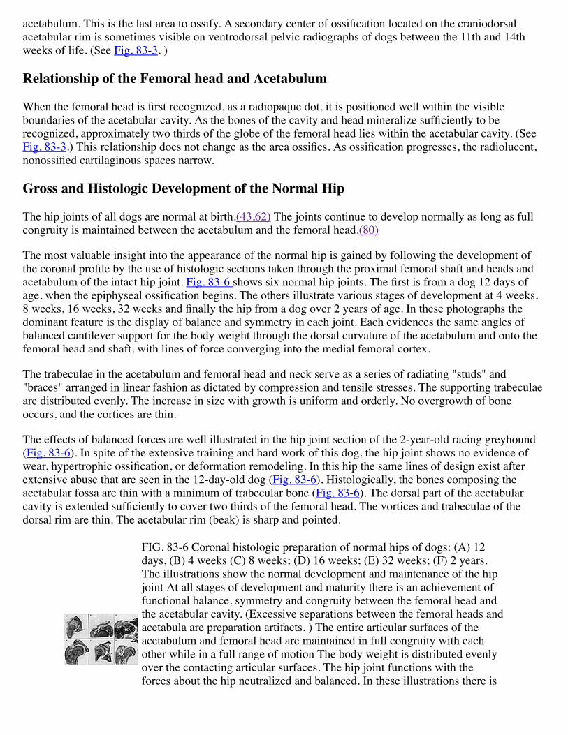
The most valuable insight into the appearance of the normal hip is gained by following the development ofthe coronal profile by the use of histologic sections taken through the proximal femoral shaft and heads andacetabulum of the intact hip joint. Fig. 83-6 shows six normal hip joints. The first is from a dog 12 days ofage, when the epiphyseal ossification begins. The others illustrate various stages of development at 4 weeks,8 weeks, 16 weeks, 32 weeks and finally the hip from a dog over 2 years of age. In these photographs thedominant feature is the display of balance and symmetry in each joint. Each evidences the same angles ofbalanced cantilever support for the body weight through the dorsal curvature of the acetabulum and onto thefemoral head and shaft, with lines of force converging into the medial femoral cortex.
The trabeculae in the acetabulum and femoral head and neck serve as a series of radiating "studs" and"braces" arranged in linear fashion as dictated by compression and tensile stresses. The supporting trabeculaeare distributed evenly. The increase in size with growth is uniform and orderly. No overgrowth of boneoccurs, and the cortices are thin.
The effects of balanced forces are well illustrated in the hip joint section of the 2-year-old racing greyhound(Fig. 83-6). In spite of the extensive training and hard work of this dog, the hip joint shows no evidence ofwear, hypertrophic ossification, or deformation remodeling. In this hip the same lines of design exist afterextensive abuse that are seen in the 12-day-old dog (Fig. 83-6). Histologically, the bones composing theacetabular fossa are thin with a minimum of trabecular bone (Fig. 83-6). The dorsal part of the acetabularcavity is extended sufficiently to cover two thirds of the femoral head. The vortices and trabeculae of thedorsal rim are thin. The acetabular rim (beak) is sharp and pointed.
FIG. 83-6 Coronal histologic preparation of normal hips of dogs: (A) 12days, (B) 4 weeks (C) 8 weeks; (D) 16 weeks; (E) 32 weeks; (F) 2 years.The illustrations show the normal development and maintenance of the hipjoint At all stages of development and maturity there is an achievement offunctional balance, symmetry and congruity between the femoral head andthe acetabular cavity. (Excessive separations between the femoral heads andacetabula are preparation artifacts. ) The entire articular surfaces of theacetabulum and femoral head are maintained in full congruity with eachother while in a full range of motion The body weight is distributed evenlyover the contacting articular surfaces. The hip joint functions with theforces about the hip neutralized and balanced. In these illustrations there is
no evidence of abnormal development, remodeling, or wear. (Reproducedwith permission from Riser WH Growth and development of the normalcanine pelvis. JAVRS 14:24, 1973)
Histologically, the proximal ends of the femur and trochanter appear almost osteoporotic. There is very littlebone at the subchondral plate beneath the articulating surface of the femoral head and an absence of extrabone beneath the attachment of the teres ligament. The trabeculae supporting the femoral head are few andthin in diameter, but they form a triangle converging into the medial cortex (Fig. 83-6). The medial cortex isdense and thin, indicating that stress forces are minimal. In the trochanteric fossa, the site where the gemelli,obturator externus, and obturator internus muscle insert, the trabecular response is minimal, indicating thatthe hip has balanced sufficiently that there are no unusual forces on these attachments. In the trochanter majorthere is also a scarcity of both cortical and trabecular bone. The cortex is slightly thicker at the dorsal roundedtip of the trochanter and at the point where the trochanter joins the femur. These are the sites of attachmentfor the three gluteal muscles.
The long axes of the diaphysis and the neck form an angle of nearly 135°. The structure of the joint andadjacent bones reflects neutral stresses (compression, tension, and torsion), sufficient lubrication, andbalanced muscle pull upon all of the bony components in the joint.
The acetabular rims are stimulated to grow by mild traction applied by the joint capsule and gluteal musclesattached along their dorsal borders, and from pressure by the femoral heads upon the articular surfaces. Thediameters of the acetabular cavities are only slightly larger than the femoral heads.
Discussion
A normal hip must be an example of symmetry and balance as it develops and maintains itself from birththrough adulthood. The hip joint is composed of specialized tissues, all of which participate in a programmedchain of development.(33) Except in the racing and toy dog types, there is a great chance that the hip willdevelop abnormally.(60)
The morphologic characteristics of the complex hip structure show that biomechanical behavior is the primeinfluence in the growth of this joint.(32) The symmetry in the femoral head and neck, the trochanter and theacetabulum of the normal hip, is impressive when coronal histologic sections are studied. The photographs ofthis joint from dogs of all ages-from shortly after birth to late maturity-illustrate how these qualities aremaintained. The photograph of a highly trained racing dog retired after more than 2 years of grueling workdisplays no evidence of joint instability, wear, remodeling, or degeneration. (See Fig. 83-6.)
The laws that control bone and soft tissue dynamics control the development of the hip. Newton's law ofneutral forces when applied to biologic tissues means that a joint is in functional equilibrium when all forcesupon that joint mutually neutralize one another both in intensity and direction. Wolffs law introduces theconcept of bone transformation in that changes in function of a bone are attended by alterations in its internalstructure. This law applies to cancellous and cortical bone.(l4)
Section Three: The Dysplastic Hip Joint: Radiologic and HistologicDevelopment
Chronologic Changes Seen in Hip Dysplasia
Birth to 30 Days of Age
Eighty-seven dogs from birth to 30 days of age were dissected. Their appearance was compared with normalgreyhounds of the same age.(51) Eighty-four of these appeared normal. In three dogs that were 30 days ofage, the teres ligaments of the hip joints were edematous, a few ligament fibers were torn, and capillaryhemorrhage dotted the surface of the ligaments at the point of the tears. These changes were considered thefirst findings that might be linked to hip dysplasia.
From the dissection of the hips of these young dogs, it appears that the teres ligament is largely responsiblefor holding the femoral head in place for the first month. For the first 2 weeks the teres ligament is so shortthat the femoral head attachment fractures at the fovea when luxation of the femoral head is forced. After thefirst 2 weeks, the teres ligament begins to lengthen very slowly. After the first 4 weeks the femoral head maybe subluxated laterally 1 mm to 2 mm. In the normal adult dog the ligament is lengthened sufficiently topermit femoral head subluxation to the edge of the acetabular rim after the muscles are removed.
THIRTY TO SIXTY DAYS OF AGE
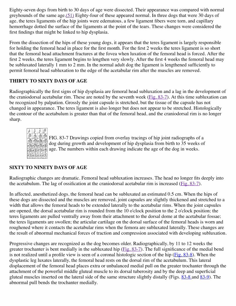
Radiographically the first signs of hip dysplasia are femoral head subluxation and a lag in the development ofthe craniodorsal acetabular rim. These are noted by the seventh week (Fig. 83-7). At this time subluxation canbe recognized by palpation. Grossly the joint capsule is stretched, but the tissue of the capsule has notchanged in appearance. The teres ligament is also longer but does not appear to be stretched. Histologicallythe contour of the acetabulum is greater than that of the femoral head, and the craniodorsal rim is no longersharp.
FIG. 83-7 Drawings copied from overlay tracings of hip joint radiographs of adog during growth and development of hip dysplasia from birth to 35 weeks ofage. The numbers within each drawing indicate the age of the dog in weeks.
SIXTY TO NINETY DAYS OF AGE
Radiographic changes are dramatic. Femoral head subluxation increases. The head no longer fits deeply intothe acetabulum. The lag of ossification at the craniodorsal acetabular rim is increased (Fig. 83-7).
In affected, anesthetized dogs, the femoral head can be subluxated an estimated 0.5 cm. When the hips ofthese dogs are dissected and the muscles are removed, joint capsules are slightly thickened and stretched to awidth that allows the femoral heads to be extended laterally to the acetabular rims. When the joint capsulesare opened, the dorsal acetabular rim is rounded from the 10 o'clock position to the 2 o'clock position; theteres ligaments are pulled ventrally away from their attachment to the dorsal dome at the acetabular fossae;the teres ligaments are swollen; the articular cartilage on the dorsal surface of the femoral heads is worn androughened where it contacts the acetabular rims when the femora are subluxated laterally. These changes arethe result of abnormal mechanical forces of traction and compression associated with developing subluxation.
Progressive changes are recognized as the dog becomes older. Radiographically, by 11 to 12 weeks thegreater trochanter is bent medially in the subluxated hip (Fig. 83-7). The full significance of the medial bendis not realized until a profile view is seen of a coronal histologic section of the hip (Fig. 83-8). When thedysplastic leg luxates laterally, the femoral head rests on the dorsal rim of the acetabulum. This lateraldisplacement of the femoral head places extra or unbalanced medial pull on the greater trochanter through theattachment of the powerful middle gluteal muscle to its dorsal tuberosity and by the deep and superficialgluteal muscles inserted on the lateral side of the same structure slightly distally (Figs. 83-8 and 83-9). Theabnormal pull bends the trochanter medially.
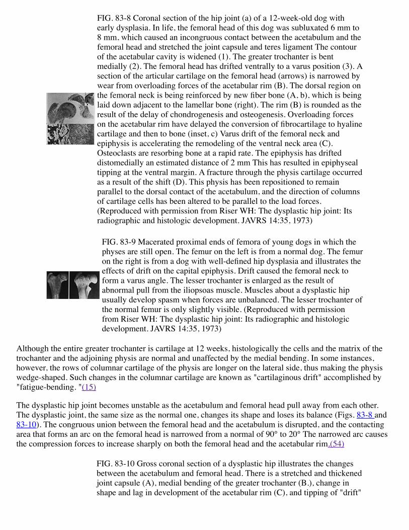
FIG. 83-8 Coronal section of the hip joint (a) of a 12-week-old dog withearly dysplasia. In life, the femoral head of this dog was subluxated 6 mm to8 mm, which caused an incongruous contact between the acetabulum and thefemoral head and stretched the joint capsule and teres ligament The contourof the acetabular cavity is widened (1). The greater trochanter is bentmedially (2). The femoral head has drifted ventrally to a varus position (3). Asection of the articular cartilage on the femoral head (arrows) is narrowed bywear from overloading forces of the acetabular rim (B). The dorsal region onthe femoral neck is being reinforced by new fiber bone (A, b), which is beinglaid down adjacent to the lamellar bone (right). The rim (B) is rounded as theresult of the delay of chondrogenesis and osteogenesis. Overloading forceson the acetabular rim have delayed the conversion of fibrocartilage to hyalinecartilage and then to bone (inset, c) Varus drift of the femoral neck andepiphysis is accelerating the remodeling of the ventral neck area (C).Osteoclasts are resorbing bone at a rapid rate. The epiphysis has drifteddistomedially an estimated distance of 2 mm This has resulted in epiphysealtipping at the ventral margin. A fracture through the physis cartilage occurredas a result of the shift (D). This physis has been repositioned to remainparallel to the dorsal contact of the acetabulum, and the direction of columnsof cartilage cells has been altered to be parallel to the load forces.(Reproduced with permission from Riser WH: The dysplastic hip joint: Itsradiographic and histologic development. JAVRS 14:35, 1973)
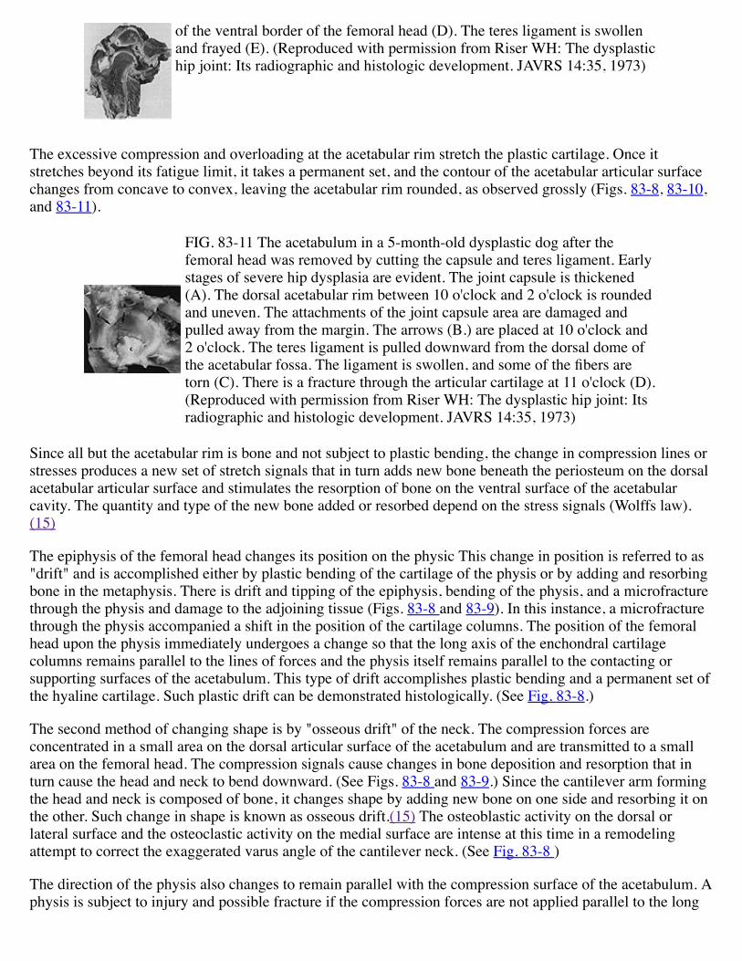
FIG. 83-9 Macerated proximal ends of femora of young dogs in which thephyses are still open. The femur on the left is from a normal dog. The femuron the right is from a dog with well-defined hip dysplasia and illustrates theeffects of drift on the capital epiphysis. Drift caused the femoral neck toform a varus angle. The lesser trochanter is enlarged as the result ofabnormal pull from the iliopsoas muscle. Muscles about a dysplastic hipusually develop spasm when forces are unbalanced. The lesser trochanter ofthe normal femur is only slightly visible. (Reproduced with permissionfrom Riser WH: The dysplastic hip joint: Its radiographic and histologicdevelopment. JAVRS 14:35, 1973)
Although the entire greater trochanter is cartilage at 12 weeks, histologically the cells and the matrix of thetrochanter and the adjoining physis are normal and unaffected by the medial bending. In some instances,however, the rows of columnar cartilage of the physis are longer on the lateral side, thus making the physiswedge-shaped. Such changes in the columnar cartilage are known as "cartilaginous drift" accomplished by"fatigue-bending. "(15)
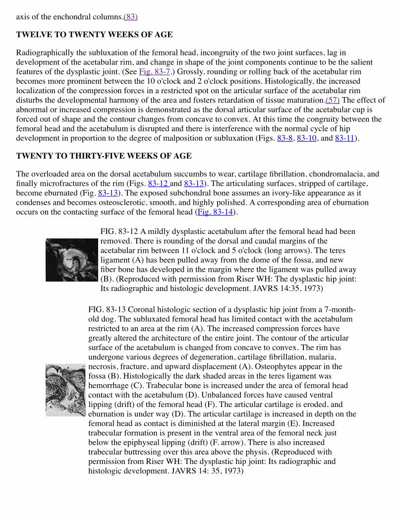
The dysplastic hip joint becomes unstable as the acetabulum and femoral head pull away from each other.The dysplastic joint, the same size as the normal one, changes its shape and loses its balance (Figs. 83-8 and83-10). The congruous union between the femoral head and the acetabulum is disrupted, and the contactingarea that forms an arc on the femoral head is narrowed from a normal of 90° to 20° The narrowed arc causesthe compression forces to increase sharply on both the femoral head and the acetabular rim.(54)
FIG. 83-10 Gross coronal section of a dysplastic hip illustrates the changesbetween the acetabulum and femoral head. There is a stretched and thickenedjoint capsule (A), medial bending of the greater trochanter (B.), change inshape and lag in development of the acetabular rim (C), and tipping of "drift"
of the ventral border of the femoral head (D). The teres ligament is swollenand frayed (E). (Reproduced with permission from Riser WH: The dysplastichip joint: Its radiographic and histologic development. JAVRS 14:35, 1973)
The excessive compression and overloading at the acetabular rim stretch the plastic cartilage. Once itstretches beyond its fatigue limit, it takes a permanent set, and the contour of the acetabular articular surfacechanges from concave to convex, leaving the acetabular rim rounded, as observed grossly (Figs. 83-8, 83-10,and 83-11).
FIG. 83-11 The acetabulum in a 5-month-old dysplastic dog after thefemoral head was removed by cutting the capsule and teres ligament. Earlystages of severe hip dysplasia are evident. The joint capsule is thickened(A). The dorsal acetabular rim between 10 o'clock and 2 o'clock is roundedand uneven. The attachments of the joint capsule area are damaged andpulled away from the margin. The arrows (B.) are placed at 10 o'clock and2 o'clock. The teres ligament is pulled downward from the dorsal dome ofthe acetabular fossa. The ligament is swollen, and some of the fibers aretorn (C). There is a fracture through the articular cartilage at 11 o'clock (D).(Reproduced with permission from Riser WH: The dysplastic hip joint: Itsradiographic and histologic development. JAVRS 14:35, 1973)
Since all but the acetabular rim is bone and not subject to plastic bending, the change in compression lines orstresses produces a new set of stretch signals that in turn adds new bone beneath the periosteum on the dorsalacetabular articular surface and stimulates the resorption of bone on the ventral surface of the acetabularcavity. The quantity and type of the new bone added or resorbed depend on the stress signals (Wolffs law).(15)
The epiphysis of the femoral head changes its position on the physic This change in position is referred to as"drift" and is accomplished either by plastic bending of the cartilage of the physis or by adding and resorbingbone in the metaphysis. There is drift and tipping of the epiphysis, bending of the physis, and a microfracturethrough the physis and damage to the adjoining tissue (Figs. 83-8 and 83-9). In this instance, a microfracturethrough the physis accompanied a shift in the position of the cartilage columns. The position of the femoralhead upon the physis immediately undergoes a change so that the long axis of the enchondral cartilagecolumns remains parallel to the lines of forces and the physis itself remains parallel to the contacting orsupporting surfaces of the acetabulum. This type of drift accomplishes plastic bending and a permanent set ofthe hyaline cartilage. Such plastic drift can be demonstrated histologically. (See Fig. 83-8.)
The second method of changing shape is by "osseous drift" of the neck. The compression forces areconcentrated in a small area on the dorsal articular surface of the acetabulum and are transmitted to a smallarea on the femoral head. The compression signals cause changes in bone deposition and resorption that inturn cause the head and neck to bend downward. (See Figs. 83-8 and 83-9.) Since the cantilever arm formingthe head and neck is composed of bone, it changes shape by adding new bone on one side and resorbing it onthe other. Such change in shape is known as osseous drift.(15) The osteoblastic activity on the dorsal orlateral surface and the osteoclastic activity on the medial surface are intense at this time in a remodelingattempt to correct the exaggerated varus angle of the cantilever neck. (See Fig. 83-8 )
The direction of the physis also changes to remain parallel with the compression surface of the acetabulum. Aphysis is subject to injury and possible fracture if the compression forces are not applied parallel to the long
axis of the enchondral columns.(83)
TWELVE TO TWENTY WEEKS OF AGE
Radiographically the subluxation of the femoral head, incongruity of the two joint surfaces, lag indevelopment of the acetabular rim, and change in shape of the joint components continue to be the salientfeatures of the dysplastic joint. (See Fig. 83-7.) Grossly, rounding or rolling back of the acetabular rimbecomes more prominent between the 10 o'clock and 2 o'clock positions. Histologically, the increasedlocalization of the compression forces in a restricted spot on the articular surface of the acetabular rimdisturbs the developmental harmony of the area and fosters retardation of tissue maturation.(57) The effect ofabnormal or increased compression is demonstrated as the dorsal articular surface of the acetabular cup isforced out of shape and the contour changes from concave to convex. At this time the congruity between thefemoral head and the acetabulum is disrupted and there is interference with the normal cycle of hipdevelopment in proportion to the degree of malposition or subluxation (Figs. 83-8, 83-10, and 83-11).
TWENTY TO THIRTY-FIVE WEEKS OF AGE
The overloaded area on the dorsal acetabulum succumbs to wear, cartilage fibrillation, chondromalacia, andfinally microfractures of the rim (Figs. 83-12 and 83-13). The articulating surfaces, stripped of cartilage,become eburnated (Fig. 83-13). The exposed subchondral bone assumes an ivory-like appearance as itcondenses and becomes osteosclerotic, smooth, and highly polished. A corresponding area of eburnationoccurs on the contacting surface of the femoral head (Fig. 83-14).
FIG. 83-12 A mildly dysplastic acetabulum after the femoral head had beenremoved. There is rounding of the dorsal and caudal margins of theacetabular rim between 11 o'clock and 5 o'clock (long arrows). The teresligament (A) has been pulled away from the dome of the fossa, and newfiber bone has developed in the margin where the ligament was pulled away(B). (Reproduced with permission from Riser WH: The dysplastic hip joint:Its radiographic and histologic development. JAVRS 14:35, 1973)
FIG. 83-13 Coronal histologic section of a dysplastic hip joint from a 7-month-old dog. The subluxated femoral head has limited contact with the acetabulumrestricted to an area at the rim (A). The increased compression forces havegreatly altered the architecture of the entire joint. The contour of the articularsurface of the acetabulum is changed from concave to convex. The rim hasundergone various degrees of degeneration, cartilage fibrillation, malaria,necrosis, fracture, and upward displacement (A). Osteophytes appear in thefossa (B). Histologically the dark shaded areas in the teres ligament washemorrhage (C). Trabecular bone is increased under the area of femoral headcontact with the acetabulum (D). Unbalanced forces have caused ventrallipping (drift) of the femoral head (F). The articular cartilage is eroded, andeburnation is under way (D). The articular cartilage is increased in depth on thefemoral head as contact is diminished at the lateral margin (E). Increasedtrabecular formation is present in the ventral area of the femoral neck justbelow the epiphyseal lipping (drift) (F. arrow). There is also increasedtrabecular buttressing over this area above the physis. (Reproduced withpermission from Riser WH: The dysplastic hip joint: Its radiographic andhistologic development. JAVRS 14: 35, 1973)
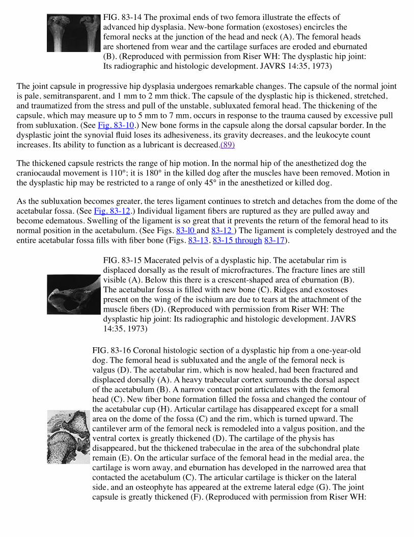
FIG. 83-14 The proximal ends of two femora illustrate the effects ofadvanced hip dysplasia. New-bone formation (exostoses) encircles thefemoral necks at the junction of the head and neck (A). The femoral headsare shortened from wear and the cartilage surfaces are eroded and eburnated(B). (Reproduced with permission from Riser WH: The dysplastic hip joint:Its radiographic and histologic development. JAVRS 14:35, 1973)
The joint capsule in progressive hip dysplasia undergoes remarkable changes. The capsule of the normal jointis pale, semitransparent, and 1 mm to 2 mm thick. The capsule of the dysplastic hip is thickened, stretched,and traumatized from the stress and pull of the unstable, subluxated femoral head. The thickening of thecapsule, which may measure up to 5 mm to 7 mm, occurs in response to the trauma caused by excessive pullfrom subluxation. (See Fig. 83-10.) New bone forms in the capsule along the dorsal capsular border. In thedysplastic joint the synovial fluid loses its adhesiveness, its gravity decreases, and the leukocyte countincreases. Its ability to function as a lubricant is decreased.(89)
The thickened capsule restricts the range of hip motion. In the normal hip of the anesthetized dog thecraniocaudal movement is 110°; it is 180° in the killed dog after the muscles have been removed. Motion inthe dysplastic hip may be restricted to a range of only 45° in the anesthetized or killed dog.
As the subluxation becomes greater, the teres ligament continues to stretch and detaches from the dome of theacetabular fossa. (See Fig. 83-12.) Individual ligament fibers are ruptured as they are pulled away andbecome edematous. Swelling of the ligament is so great that it prevents the return of the femoral head to itsnormal position in the acetabulum. (See Figs. 83-l0 and 83-12 ) The ligament is completely destroyed and theentire acetabular fossa fills with fiber bone (Figs. 83-13, 83-15 through 83-17).
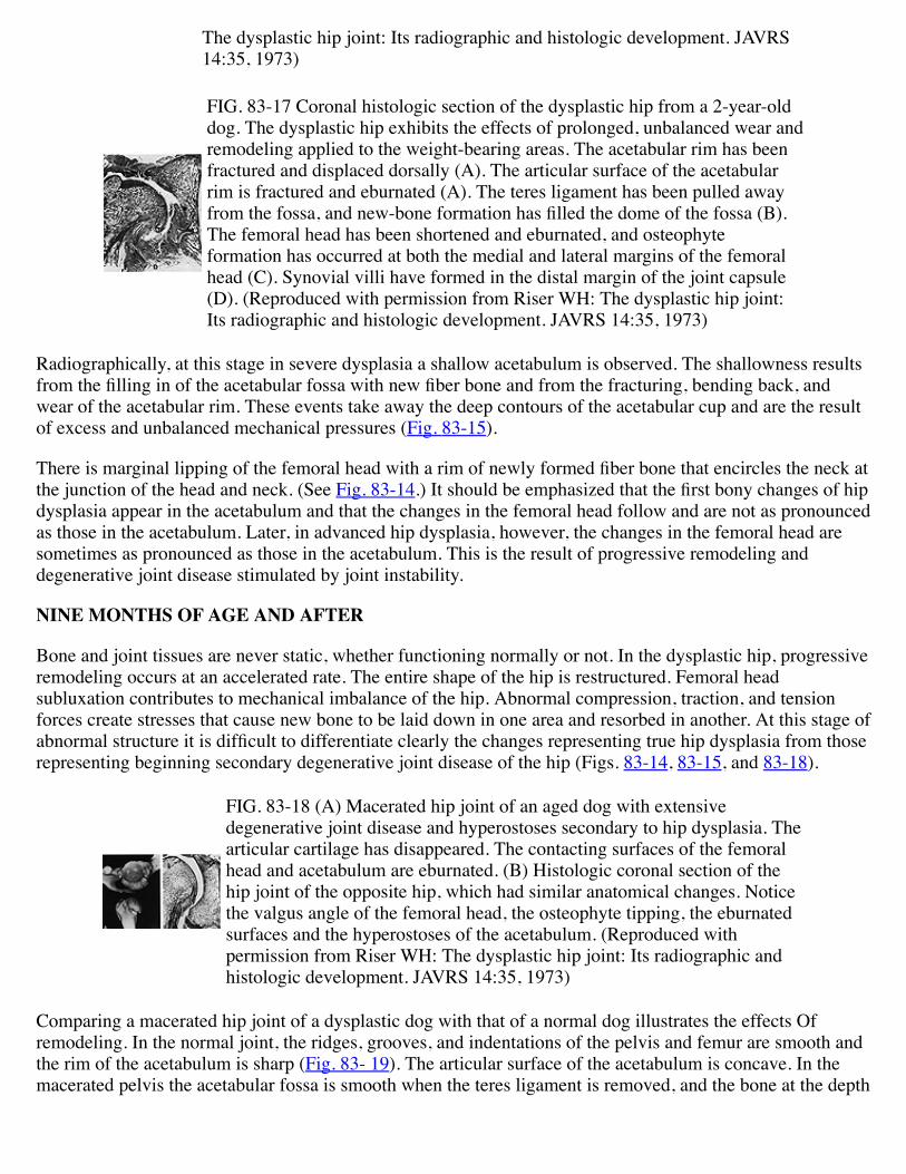
FIG. 83-15 Macerated pelvis of a dysplastic hip. The acetabular rim isdisplaced dorsally as the result of microfractures. The fracture lines are stillvisible (A). Below this there is a crescent-shaped area of eburnation (B).The acetabular fossa is filled with new bone (C). Ridges and exostosespresent on the wing of the ischium are due to tears at the attachment of themuscle fibers (D). (Reproduced with permission from Riser WH: Thedysplastic hip joint: Its radiographic and histologic development. JAVRS14:35, 1973)
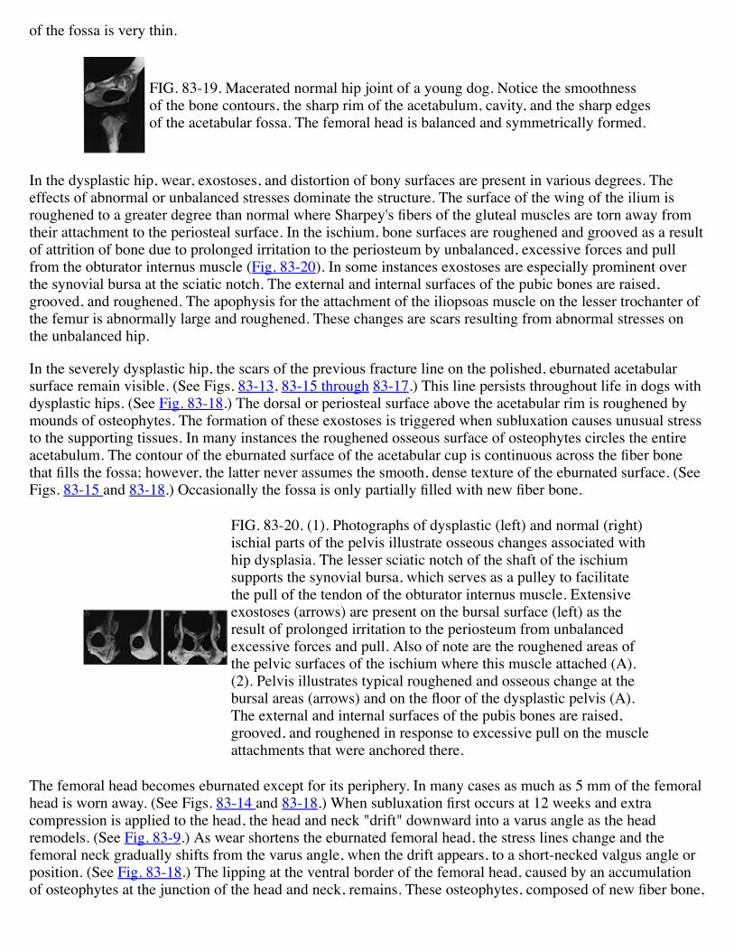
FIG. 83-16 Coronal histologic section of a dysplastic hip from a one-year-olddog. The femoral head is subluxated and the angle of the femoral neck isvalgus (D). The acetabular rim, which is now healed, had been fractured anddisplaced dorsally (A). A heavy trabecular cortex surrounds the dorsal aspectof the acetabulum (B). A narrow contact point articulates with the femoralhead (C). New fiber bone formation filled the fossa and changed the contour ofthe acetabular cup (H). Articular cartilage has disappeared except for a smallarea on the dome of the fossa (C) and the rim, which is turned upward. Thecantilever arm of the femoral neck is remodeled into a valgus position, and theventral cortex is greatly thickened (D). The cartilage of the physis hasdisappeared, but the thickened trabeculae in the area of the subchondral plateremain (E). On the articular surface of the femoral head in the medial area, thecartilage is worn away, and eburnation has developed in the narrowed area thatcontacted the acetabulum (C). The articular cartilage is thicker on the lateralside, and an osteophyte has appeared at the extreme lateral edge (G). The jointcapsule is greatly thickened (F). (Reproduced with permission from Riser WH:
The dysplastic hip joint: Its radiographic and histologic development. JAVRS14:35, 1973)
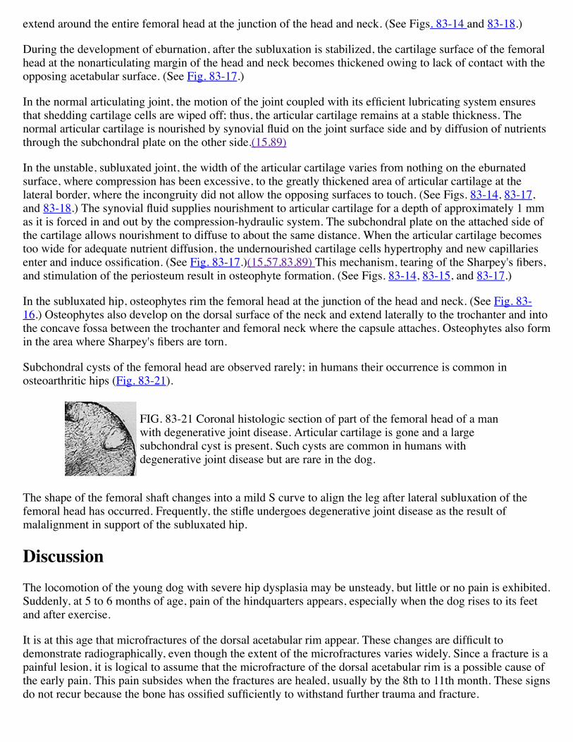
FIG. 83-17 Coronal histologic section of the dysplastic hip from a 2-year-olddog. The dysplastic hip exhibits the effects of prolonged, unbalanced wear andremodeling applied to the weight-bearing areas. The acetabular rim has beenfractured and displaced dorsally (A). The articular surface of the acetabularrim is fractured and eburnated (A). The teres ligament has been pulled awayfrom the fossa, and new-bone formation has filled the dome of the fossa (B).The femoral head has been shortened and eburnated, and osteophyteformation has occurred at both the medial and lateral margins of the femoralhead (C). Synovial villi have formed in the distal margin of the joint capsule(D). (Reproduced with permission from Riser WH: The dysplastic hip joint:Its radiographic and histologic development. JAVRS 14:35, 1973)
Radiographically, at this stage in severe dysplasia a shallow acetabulum is observed. The shallowness resultsfrom the filling in of the acetabular fossa with new fiber bone and from the fracturing, bending back, andwear of the acetabular rim. These events take away the deep contours of the acetabular cup and are the resultof excess and unbalanced mechanical pressures (Fig. 83-15).
There is marginal lipping of the femoral head with a rim of newly formed fiber bone that encircles the neck atthe junction of the head and neck. (See Fig. 83-14.) It should be emphasized that the first bony changes of hipdysplasia appear in the acetabulum and that the changes in the femoral head follow and are not as pronouncedas those in the acetabulum. Later, in advanced hip dysplasia, however, the changes in the femoral head aresometimes as pronounced as those in the acetabulum. This is the result of progressive remodeling anddegenerative joint disease stimulated by joint instability.
NINE MONTHS OF AGE AND AFTER
Bone and joint tissues are never static, whether functioning normally or not. In the dysplastic hip, progressiveremodeling occurs at an accelerated rate. The entire shape of the hip is restructured. Femoral headsubluxation contributes to mechanical imbalance of the hip. Abnormal compression, traction, and tensionforces create stresses that cause new bone to be laid down in one area and resorbed in another. At this stage ofabnormal structure it is difficult to differentiate clearly the changes representing true hip dysplasia from thoserepresenting beginning secondary degenerative joint disease of the hip (Figs. 83-14, 83-15, and 83-18).
FIG. 83-18 (A) Macerated hip joint of an aged dog with extensivedegenerative joint disease and hyperostoses secondary to hip dysplasia. Thearticular cartilage has disappeared. The contacting surfaces of the femoralhead and acetabulum are eburnated. (B) Histologic coronal section of thehip joint of the opposite hip, which had similar anatomical changes. Noticethe valgus angle of the femoral head, the osteophyte tipping, the eburnatedsurfaces and the hyperostoses of the acetabulum. (Reproduced withpermission from Riser WH: The dysplastic hip joint: Its radiographic andhistologic development. JAVRS 14:35, 1973)
Comparing a macerated hip joint of a dysplastic dog with that of a normal dog illustrates the effects Ofremodeling. In the normal joint, the ridges, grooves, and indentations of the pelvis and femur are smooth andthe rim of the acetabulum is sharp (Fig. 83- 19). The articular surface of the acetabulum is concave. In themacerated pelvis the acetabular fossa is smooth when the teres ligament is removed, and the bone at the depth
of the fossa is very thin.
FIG. 83-19. Macerated normal hip joint of a young dog. Notice the smoothnessof the bone contours, the sharp rim of the acetabulum, cavity, and the sharp edgesof the acetabular fossa. The femoral head is balanced and symmetrically formed.
In the dysplastic hip, wear, exostoses, and distortion of bony surfaces are present in various degrees. Theeffects of abnormal or unbalanced stresses dominate the structure. The surface of the wing of the ilium isroughened to a greater degree than normal where Sharpey's fibers of the gluteal muscles are torn away fromtheir attachment to the periosteal surface. In the ischium, bone surfaces are roughened and grooved as a resultof attrition of bone due to prolonged irritation to the periosteum by unbalanced, excessive forces and pullfrom the obturator internus muscle (Fig. 83-20). In some instances exostoses are especially prominent overthe synovial bursa at the sciatic notch. The external and internal surfaces of the pubic bones are raised,grooved, and roughened. The apophysis for the attachment of the iliopsoas muscle on the lesser trochanter ofthe femur is abnormally large and roughened. These changes are scars resulting from abnormal stresses onthe unbalanced hip.
In the severely dysplastic hip, the scars of the previous fracture line on the polished, eburnated acetabularsurface remain visible. (See Figs. 83-13, 83-15 through 83-17.) This line persists throughout life in dogs withdysplastic hips. (See Fig. 83-18.) The dorsal or periosteal surface above the acetabular rim is roughened bymounds of osteophytes. The formation of these exostoses is triggered when subluxation causes unusual stressto the supporting tissues. In many instances the roughened osseous surface of osteophytes circles the entireacetabulum. The contour of the eburnated surface of the acetabular cup is continuous across the fiber bonethat fills the fossa; however, the latter never assumes the smooth, dense texture of the eburnated surface. (SeeFigs. 83-15 and 83-18.) Occasionally the fossa is only partially filled with new fiber bone.
FIG. 83-20. (1). Photographs of dysplastic (left) and normal (right)ischial parts of the pelvis illustrate osseous changes associated withhip dysplasia. The lesser sciatic notch of the shaft of the ischiumsupports the synovial bursa, which serves as a pulley to facilitatethe pull of the tendon of the obturator internus muscle. Extensiveexostoses (arrows) are present on the bursal surface (left) as theresult of prolonged irritation to the periosteum from unbalancedexcessive forces and pull. Also of note are the roughened areas ofthe pelvic surfaces of the ischium where this muscle attached (A).(2). Pelvis illustrates typical roughened and osseous change at thebursal areas (arrows) and on the floor of the dysplastic pelvis (A).The external and internal surfaces of the pubis bones are raised,grooved, and roughened in response to excessive pull on the muscleattachments that were anchored there.
The femoral head becomes eburnated except for its periphery. In many cases as much as 5 mm of the femoralhead is worn away. (See Figs. 83-14 and 83-18.) When subluxation first occurs at 12 weeks and extracompression is applied to the head, the head and neck "drift" downward into a varus angle as the headremodels. (See Fig. 83-9.) As wear shortens the eburnated femoral head, the stress lines change and thefemoral neck gradually shifts from the varus angle, when the drift appears, to a short-necked valgus angle orposition. (See Fig. 83-18.) The lipping at the ventral border of the femoral head, caused by an accumulationof osteophytes at the junction of the head and neck, remains. These osteophytes, composed of new fiber bone,
extend around the entire femoral head at the junction of the head and neck. (See Figs. 83-14 and 83-18.)
During the development of eburnation, after the subluxation is stabilized, the cartilage surface of the femoralhead at the nonarticulating margin of the head and neck becomes thickened owing to lack of contact with theopposing acetabular surface. (See Fig. 83-17.)
In the normal articulating joint, the motion of the joint coupled with its efficient lubricating system ensuresthat shedding cartilage cells are wiped off; thus, the articular cartilage remains at a stable thickness. Thenormal articular cartilage is nourished by synovial fluid on the joint surface side and by diffusion of nutrientsthrough the subchondral plate on the other side.(15,89)
In the unstable, subluxated joint, the width of the articular cartilage varies from nothing on the eburnatedsurface, where compression has been excessive, to the greatly thickened area of articular cartilage at thelateral border, where the incongruity did not allow the opposing surfaces to touch. (See Figs. 83-14, 83-17,and 83-18.) The synovial fluid supplies nourishment to articular cartilage for a depth of approximately 1 mmas it is forced in and out by the compression-hydraulic system. The subchondral plate on the attached side ofthe cartilage allows nourishment to diffuse to about the same distance. When the articular cartilage becomestoo wide for adequate nutrient diffusion, the undernourished cartilage cells hypertrophy and new capillariesenter and induce ossification. (See Fig. 83-17.)(15,57,83,89) This mechanism, tearing of the Sharpey's fibers,and stimulation of the periosteum result in osteophyte formation. (See Figs. 83-14, 83-15, and 83-17.)
In the subluxated hip, osteophytes rim the femoral head at the junction of the head and neck. (See Fig. 83-16.) Osteophytes also develop on the dorsal surface of the neck and extend laterally to the trochanter and intothe concave fossa between the trochanter and femoral neck where the capsule attaches. Osteophytes also formin the area where Sharpey's fibers are torn.
Subchondral cysts of the femoral head are observed rarely; in humans their occurrence is common inosteoarthritic hips (Fig. 83-21).
FIG. 83-21 Coronal histologic section of part of the femoral head of a manwith degenerative joint disease. Articular cartilage is gone and a largesubchondral cyst is present. Such cysts are common in humans withdegenerative joint disease but are rare in the dog.
The shape of the femoral shaft changes into a mild S curve to align the leg after lateral subluxation of thefemoral head has occurred. Frequently, the stifle undergoes degenerative joint disease as the result ofmalalignment in support of the subluxated hip.
DiscussionThe locomotion of the young dog with severe hip dysplasia may be unsteady, but little or no pain is exhibited.Suddenly, at 5 to 6 months of age, pain of the hindquarters appears, especially when the dog rises to its feetand after exercise.
It is at this age that microfractures of the dorsal acetabular rim appear. These changes are difficult todemonstrate radiographically, even though the extent of the microfractures varies widely. Since a fracture is apainful lesion, it is logical to assume that the microfracture of the dorsal acetabular rim is a possible cause ofthe early pain. This pain subsides when the fractures are healed, usually by the 8th to 11th month. These signsdo not recur because the bone has ossified sufficiently to withstand further trauma and fracture.
By the 9th to 11th month, the pain gradually subsides until the dog is seemingly pain-free in rising and inlocomotion. The range of motion of the hindlegs is restricted, but this is usually not detectable from the dog'snormal gait because the dog does not swing the legs more than 45° in normal walking. Occasionally thebalance of the hindquarters is unstable, but frequently, even when the radiographic signs are severe, the gait isnormal, except for a low threshold of fatiguability.
Abnormal remodeling and hypertrophic osseous development are continually present in and around thedysplastic hip. Even though these mechanisms are functional during the disease, frequently there is poorcorrelation between the clinical signs of the patient and the radiographic signs of the involvement unless thedog is worked hard. Often the radiographic involvement is extensive, yet there are no physical signs ofinvolvement. In a few instances radiographic lesions are mild but signs of pain are severe.
The significant changes described above are recorded for comparison with the rate and pattern of normaldevelopment of the hip joint from birth to maturity.(60) These findings support the theory that hip dysplasiaoccurs only if hip joint instability and joint incongruity are present in the young dog. It is also believed thatthe disease can be prevented if hip joint congruity can be maintained until ossification makes the acetabulumless plastic and the abductor muscles and supporting soft tissues become sufficiently strong and functional toprevent femoral head subluxation.(74)
Section Four: Clinical Manifestations
History
Hip dysplasia is a disease of young animals. Most dogs will be between the ages of 5 months and 12 monthsat first presentation. A much smaller population will present with first signs as late as 36 months. Young dogspresent with joint instability, which may result in abnormal hindlimb gait, thigh muscle atrophy, pain, lowexercise tolerance, inability to climb stairs, an audible "click" when walking, or walking with an arched back.Any one (or several) of the above signs may be present. Past pedigree history is of little relevance, sincemany generations may be normal and still produce dysplastic progeny.
Physical Examination
Physical examination may or may not produce definitive information. Palpation of the hips may result in painon extremes of range of motion. Pressure on the hip may cause discomfort. Attempts to subluxate the hipshould be made either by lifting the hip laterally or by adducting with axial pressure. The latter may force thehip to subluxate. When the limb is then abducted, the femoral head will drop again into the acetabulum andmake a "popping" sound referred to as the Ortolani sign.
Many severely dysplastic dogs will subluxate their hips on each step. This can be monitored by walkingbehind the dog with a hand over each hip. Radiography is needed to produce definitive diagnosis. Old dogsdo not present with hip dysplasia. They present with degenerative joint disease of the hips, which issecondary to hip dysplasia as a young animal. Any subsequent workup or treatment is directed towardtreatment of the resultant arthritis rather than the dysplasia.
Radiography
Radiographs are the definitive visible diagnostic evidence of hip dysplasia. The diagnostic value of hipradiographs depends upon their quality, that is, proper patient positioning, proper centering of the x-ray beam,proper exposure technique, and proper darkroom technique. Positioning faults and technical deficiencies may
result in nondiagnostic radiographs or the illusion of hip disease where it does not exist. It is unlikely that allof the possible abnormalities associated with hip dysplasia will appear in any single radiograph. Anawareness of these changes will, however, aid in perceiving their presence and understanding theirsignificance.
POSITION FOR RADIOGRAPHY
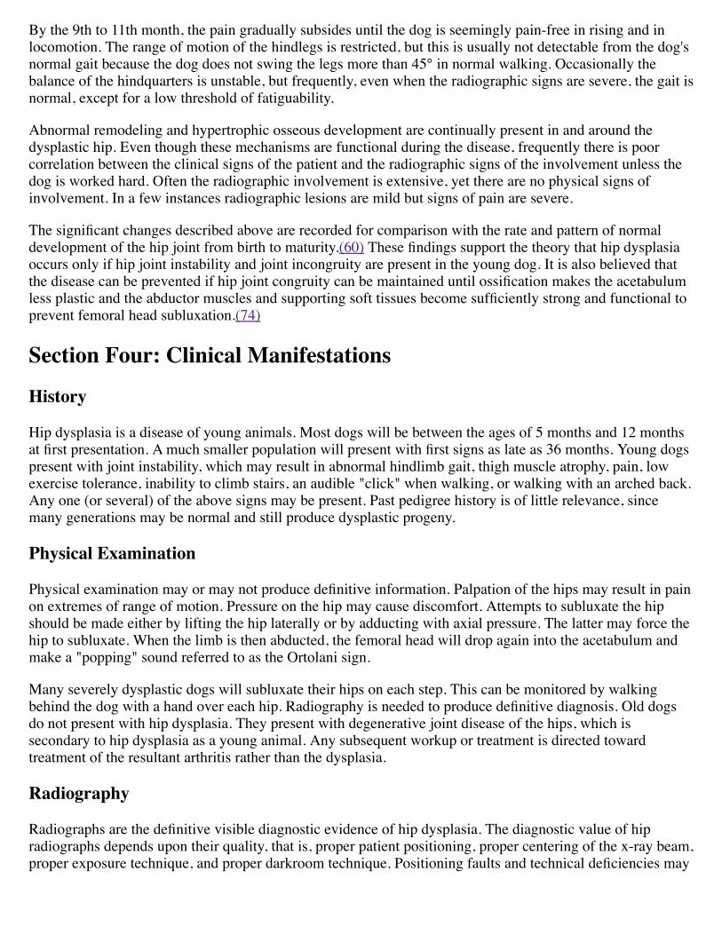
The ventrodorsal position is recommended for hip radiographs (Fig. 83-22). The dog is placed on its back.The hindlegs are extended fully in a caudal direction, held parallel, and rotated fully in a medial direction.When this position has been achieved, the cranial midline of the femora will face directly up toward the x-raytube.
Symmetric pelvic positioning may be achieved by determining that major palpable anatomical sites, such asthe iliac crest and greater trochanter, are equidistant from the cassette surface. Sedation or anesthesia may berequired to achieve this position in all but the most cooperative dogs.
An exposure technique should be selected that will produce clearly visible structural detail of the hipbone inthe finished radiograph. Radiographic evaluation of the hip may be severely limited by exposure techniqueinadequacies.
THE NORMAL HIP
The hip is a ball-and-socket joint formed by the convex, hemispherical femoral head and a deep, concaveacetabulum. The normal femoral head has a localized surface irregularity where the femoral head ligament isattached. The acetabular articular surface is a peripheral rim that is incomplete ventrally. The acetabularfossa, the second site of femoral head ligament attachment, extends medially and caudally from the articularsurface.(45) Hip bone detail should be clearly visible in good quality radiographs.
FIG. 83-22 Ventrodorsal hip radiographs of a normal canine pelvis andfemora positioned symmetrically. (A) All parts of the pelvis appearbilaterally symmetric. The legs are fully extended and parallel with thepatellae appearing over the femoral midline. (B) An enlargement of A.The hemispherical femoral heads fit snugly into the deep, concaveacetabulae. The flattened central area of the femoral head is the site offemoral-head ligament attachment. Cranial to this the femoral headcontours parallel that of the cranial acetabular walls. This is the normalhip component positional relationship. (cranial acetabular wall -; dorsalacetabular wall ....; joint space J)
The femoral head and acetabular articular cartilage is not discernible as a discrete radiodensity. Articularcartilage thickness and condition can, however, be inferred from hip joint space width and shape andsubchondral bone structure.
The hip joint capsule attaches on the pelvis adjacent to the acetabulum and on the femoral neck.(45) The jointcapsule is similar in density to the soft tissues surrounding it and does not appear as a discrete radiodensity.
The growth and development of the normal canine pelvis, hip joints, and femora from birth to maturity aredescribed elsewhere in this chapter. A thorough understanding of this description is imperative for assessingradiographically the many variations within the limits of normal hip conformation and differentiating thesevariations from hip disease. The coronal histologic preparations of normal dog hips (See Fig. 83-6) are ofspecial value in gaining this understanding, since much of what appears in these coronal preparations can be
perceived radiographically. A macerated normal hip joint is shown in Figure 83- 19.
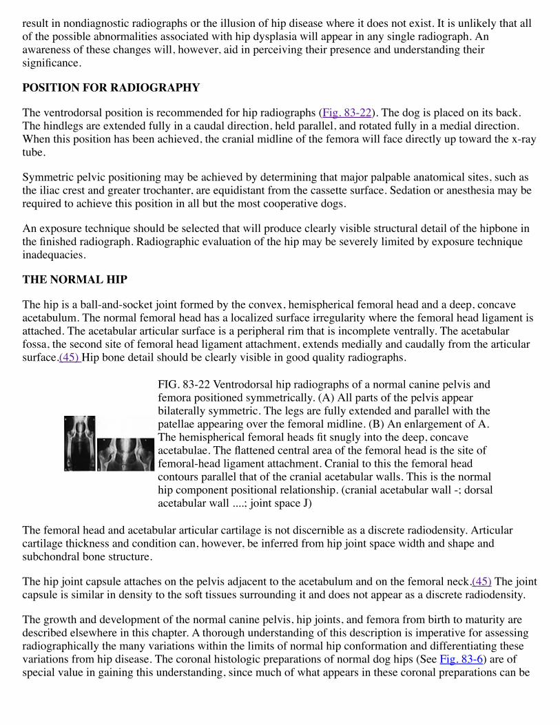
The correct position for hip radiography has been described. Strict adherence to this position is a prerequisitefor accurate radiographic assessment of hip structures. Major problems caused by variations from it includedistortions of acetabular depth and contours, the illusion of femoral head subluxation, and distortion of thefemoral neck-diaphyseal angle. Each of these variations can mimic signs of disease and lead to diagnosticerrors (Figs. 83-22 and 83-23).
The normal hips shown in Figures 83-24 through 83-26 should be examined in detail after reviewing Figures83-22 and 83-23. The reader should correlate the anatomical landmarks and contours of the macerated bonespecimens and the coronal histologic preparations with each of the radiographs.
FIG. 83-23 The same dog as shown in Figure 83-22 but positionedasymmetrically. The pelvis and femora are rotated about their long axes,and the femora are pulled laterally. Pelvic rotation in this figure ischaracterized by the wing of the ilium appearing wider and theobturator foramen less rounded on the right side than the left side. Theright acetabulum appears less deep and the left acetabulum more deepthan in the same pelvis positioned symmetrically (Fig. 83-22). Lateralfemoral rotation causes the femoral necks to appear shorter and thefemoral neck-diaphseal angles more obtuse than normal. The distalfemora appear bowed laterally, the patellae appear lateral to the femoralmidlines, and the patellae appear more medial than normal. In both Aand B. lateral femoral rotation has caused the joint space between thecranial acetabular wall and the femoral head to appear wedge shaped(subluxated) rather than of even width as in Figure 83-22 The apparentabnormalities seen in this figure mimic hip dysplasia but are illusionscreated by faulty positioning of a normal dog. (cranial acetabular wall;dorsal acetabular wall ....; joint space J)
FIG. 83-24 Excellent hip conformation in a 27-month-old female Germanshepherd. (Courtesy of the Orthopedic Foundation for Animals)
FIG. 83-25 Good hip conformation in a 32-month-old female German shepherd.(Courtesy of the Orthopedic Foundation for Animals)
The coronal histologic preparations and specimen photographs are of specialvalue, since much of what appears in these figures can be perceivedradiographically.
FIG. 83-26 Fair hip conformation in a 24-month-old female German shepherd.(Courtesy of the Orthopedic Foundation for Animals)
THE DYSPLASTIC HIP
The primary radiographic signs of hip dysplasia are a shallow acetabulum and a small, flattened femoralhead. The secondary radiographic signs of hip dysplasia are femoral head subluxation and degenerative jointdisease of the hip.
The cranial acetabular border becomes less concave; its dorsal border recedes medially. The femoral headbecomes less hemispherical than normal; and its size diminishes. The anatomical abnormalities are those ofan incompetent ball-and-socket joint, namely a structure inadequate for weight bearing and predisposing tocraniolateral and craniodorsal femoral head subluxation, the direction of maximum weight-bearing stress.
The earliest radiographic sign of femoral head subluxation is a wedge-shaped joint space between the femoralhead and the cranial acetabular wall. As the femoral head shifts laterally, the joint space widens medially andnarrows laterally at the point of maximum head weight-bearing stress against the acetabular rim. (See Fig.83-29,B.)
Femoral head subluxation is considered a secondary change of hip dysplasia because subluxation may haveother causes, including muscle atrophy, traumatic injuries, and leg axial deformities. Indeed, if femoral headsubluxation from any cause persists, the acetabulum and femoral head may remodel to resemble the primaryabnormalities of hip dysplasia.
Degenerative joint disease is secondary to femoral head subluxation and the associated joint tissueabnormalities. Frequently degenerative joint disease is first recognizable radiographically as a crescent-shaped line of hypertrophic bone at the site of joint capsule attachment to the femoral neck (See Fig. 83-30,B.) Hypertrophic bone spurs may also be seen at the femoral head border, where the joint capsule attachesto the pelvis, and at the craniolateral acetabular border. An increase in width and density of the craniolateralacetabular subchondral bone is a sign of degenerative joint disease that reflects excessive stress placed uponthat region by a subluxated femoral head. (See Figs. 83-16 and 83-29, B.) The gross bony proliferations anddeformities associated with hip dysplasia and secondary degenerative joint disease are seen in Figures 83-15,83-18 through 83-20.
Degenerative disease may result from any stresses or diseases that adversely affect joint biomechanicalbehavior or tissue. The presence of degenerative joint disease of the hip does not by itself constitute hipdysplasia and must not be so construed.
The radiographic and histologic development of the dysplastic hip is described elsewhere in this chapter. Athorough understanding of this description is imperative for assessing the early radiographic signs of hipdysplasia.
THE ORTHOPAEDIC FOUNDATION FOR ANIMALS
Consensus reports on pelvic radiographs from the OFA contain information for serious breeders andconcerned owners. The consensus is the result of independent review by three veterinary radiologists and isreported as one of the following categories. The first three are considered within normal radiographic limitsfor age and breed and are eligible for assignment of an OFA breed number if the dog is 24 months of age orolder at the time of radiography. The last four (Nos. 4-7) are not eligible for an OFA breed number.
1. Excellent hip joint conformation (Fig. 83-24): superior hip joint conformation as compared with otheranimals of the same breed and age.
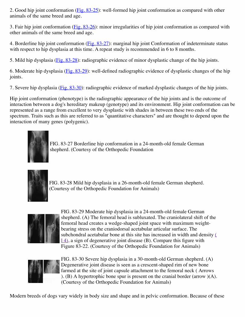
2. Good hip joint conformation (Fig. 83-25): well-formed hip joint conformation as compared with otheranimals of the same breed and age.
3. Fair hip joint conformation (Fig. 83-26): minor irregularities of hip joint conformation as compared withother animals of the same breed and age.
4. Borderline hip joint conformation (Fig. 83-27): marginal hip joint Conformation of indeterminate statuswith respect to hip dysplasia at this time. A repeat study is recommended in 6 to 8 months.
5. Mild hip dysplasia (Fig. 83-28): radiographic evidence of minor dysplastic change of the hip joints.
6. Moderate hip dysplasia (Fig. 83-29): well-defined radiographic evidence of dysplastic changes of the hipjoints.
7. Severe hip dysplasia (Fig. 83-30): radiographic evidence of marked dysplastic changes of the hip joints.
Hip joint conformation (phenotype) is the radiographic appearance of the hip joints and is the outcome ofinteraction between a dog's hereditary makeup (genotype) and its environment. Hip joint conformation can berepresented as a range from excellent to very dysplastic with shades in between these two ends of thespectrum. Traits such as this are referred to as "quantitative characters" and are thought to depend upon theinteraction of many genes (polygenic).
FIG. 83-27 Borderline hip conformation in a 24-month-old female Germanshepherd. (Courtesy of the Orthopedic Foundation
FIG. 83-28 Mild hip dysplasia in a 26-month-old female German shepherd.(Courtesy of the Orthopedic Foundation for Animals)
FIG. 83-29 Moderate hip dysplasia in a 24-month-old female Germanshepherd. (A) The femoral head is subluxated. The craniolateral shift of thefemoral head creates a wedge-shaped joint space with maximum weight-bearing stress on the craniodorsal acetabular articular surface. Thesubchondral acetabular bone at this site has increased in width and density (l 4), a sign of degenerative joint disease (B). Compare this figure withFigure 83-22. (Courtesy of the Orthopedic Foundation for Animals)
FIG. 83-30 Severe hip dysplasia in a 30-month-old German shepherd. (A)Degenerative joint disease is seen as a crescent-shaped rim of new bonefarmed at the site of joint capsule attachment to the femoral neck ( Arrows). (B) A hypertrophic bone spur is present on the cranial border (arrow )(A).(Courtesy of the Orthopedic Foundation for Animals)
Modern breeds of dogs vary widely in body size and shape and in pelvic conformation. Because of these
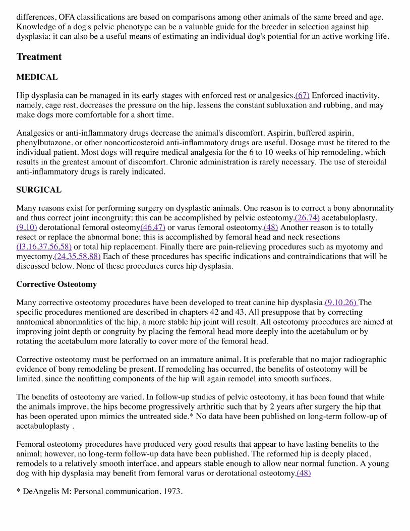
differences, OFA classifications are based on comparisons among other animals of the same breed and age.Knowledge of a dog's pelvic phenotype can be a valuable guide for the breeder in selection against hipdysplasia; it can also be a useful means of estimating an individual dog's potential for an active working life.
Treatment
MEDICAL
Hip dysplasia can be managed in its early stages with enforced rest or analgesics.(67) Enforced inactivity,namely, cage rest, decreases the pressure on the hip, lessens the constant subluxation and rubbing, and maymake dogs more comfortable for a short time.
Analgesics or anti-inflammatory drugs decrease the animal's discomfort. Aspirin, buffered aspirin,phenylbutazone, or other noncorticosteroid anti-inflammatory drugs are useful. Dosage must be titered to theindividual patient. Most dogs will require medical analgesia for the 6 to 10 weeks of hip remodeling, whichresults in the greatest amount of discomfort. Chronic administration is rarely necessary. The use of steroidalanti-inflammatory drugs is rarely indicated.
SURGICAL
Many reasons exist for performing surgery on dysplastic animals. One reason is to correct a bony abnormalityand thus correct joint incongruity; this can be accomplished by pelvic osteotomy,(26,74) acetabuloplasty,(9,10) derotational femoral osteomy(46,47) or varus femoral osteotomy.(48) Another reason is to totallyresect or replace the abnormal bone; this is accomplished by femoral head and neck resections(l3,16,37,56,58) or total hip replacement. Finally there are pain-relieving procedures such as myotomy andmyectomy.(24,35,58,88) Each of these procedures has specific indications and contraindications that will bediscussed below. None of these procedures cures hip dysplasia.
Corrective Osteotomy
Many corrective osteotomy procedures have been developed to treat canine hip dysplasia.(9,10,26) Thespecific procedures mentioned are described in chapters 42 and 43. All presuppose that by correctinganatomical abnormalities of the hip, a more stable hip joint will result. All osteotomy procedures are aimed atimproving joint depth or congruity by placing the femoral head more deeply into the acetabulum or byrotating the acetabulum more laterally to cover more of the femoral head.
Corrective osteotomy must be performed on an immature animal. It is preferable that no major radiographicevidence of bony remodeling be present. If remodeling has occurred, the benefits of osteotomy will belimited, since the nonfitting components of the hip will again remodel into smooth surfaces.
The benefits of osteotomy are varied. In follow-up studies of pelvic osteotomy, it has been found that whilethe animals improve, the hips become progressively arthritic such that by 2 years after surgery the hip thathas been operated upon mimics the untreated side.* No data have been published on long-term follow-up ofacetabuloplasty .
Femoral osteotomy procedures have produced very good results that appear to have lasting benefits to theanimal; however, no long-term follow-up data have been published. The reformed hip is deeply placed,remodels to a relatively smooth interface, and appears stable enough to allow near normal function. A youngdog with hip dysplasia may benefit from femoral varus or derotational osteotomy.(48)
* DeAngelis M: Personal communication, 1973.
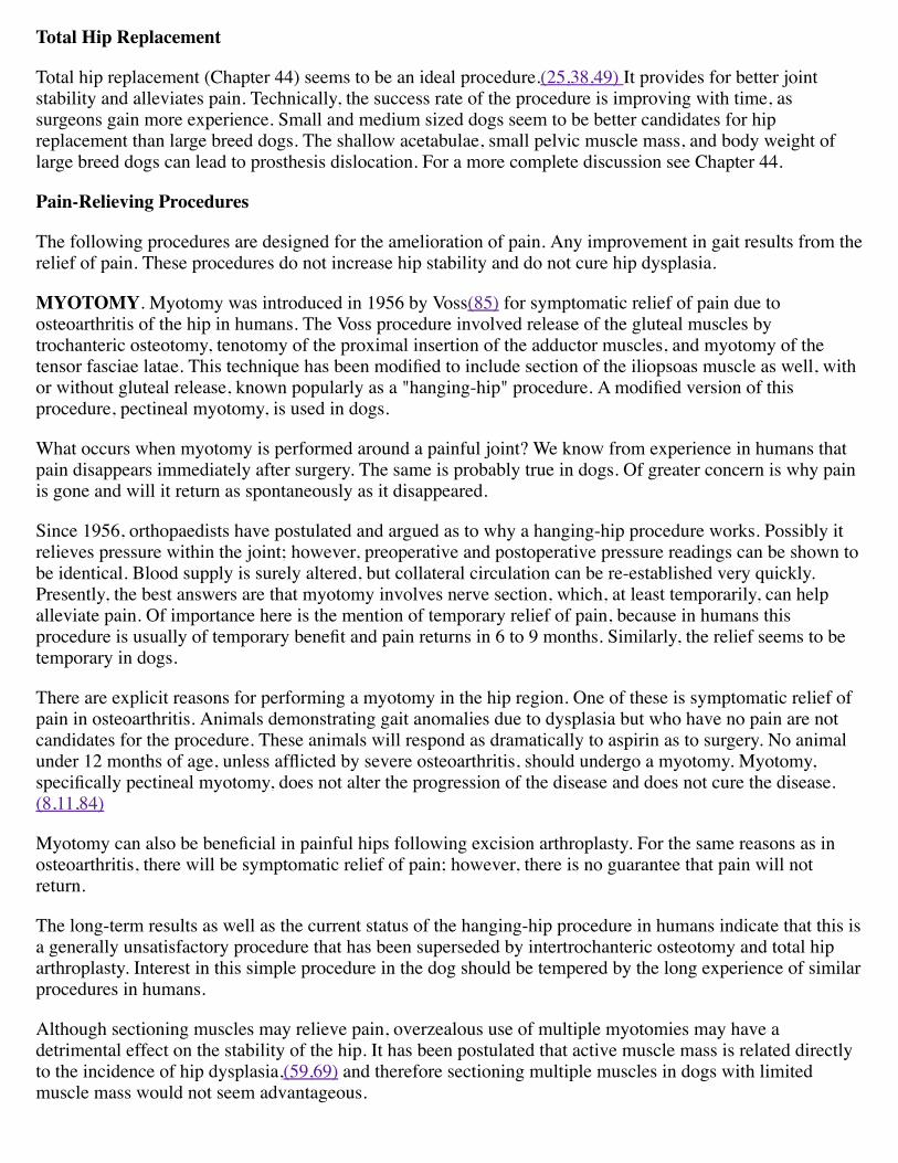
Total Hip Replacement
Total hip replacement (Chapter 44) seems to be an ideal procedure.(25,38,49) It provides for better jointstability and alleviates pain. Technically, the success rate of the procedure is improving with time, assurgeons gain more experience. Small and medium sized dogs seem to be better candidates for hipreplacement than large breed dogs. The shallow acetabulae, small pelvic muscle mass, and body weight oflarge breed dogs can lead to prosthesis dislocation. For a more complete discussion see Chapter 44.
Pain-Relieving Procedures
The following procedures are designed for the amelioration of pain. Any improvement in gait results from therelief of pain. These procedures do not increase hip stability and do not cure hip dysplasia.
MYOTOMY. Myotomy was introduced in 1956 by Voss(85) for symptomatic relief of pain due toosteoarthritis of the hip in humans. The Voss procedure involved release of the gluteal muscles bytrochanteric osteotomy, tenotomy of the proximal insertion of the adductor muscles, and myotomy of thetensor fasciae latae. This technique has been modified to include section of the iliopsoas muscle as well, withor without gluteal release, known popularly as a "hanging-hip" procedure. A modified version of thisprocedure, pectineal myotomy, is used in dogs.
What occurs when myotomy is performed around a painful joint? We know from experience in humans thatpain disappears immediately after surgery. The same is probably true in dogs. Of greater concern is why painis gone and will it return as spontaneously as it disappeared.
Since 1956, orthopaedists have postulated and argued as to why a hanging-hip procedure works. Possibly itrelieves pressure within the joint; however, preoperative and postoperative pressure readings can be shown tobe identical. Blood supply is surely altered, but collateral circulation can be re-established very quickly.Presently, the best answers are that myotomy involves nerve section, which, at least temporarily, can helpalleviate pain. Of importance here is the mention of temporary relief of pain, because in humans thisprocedure is usually of temporary benefit and pain returns in 6 to 9 months. Similarly, the relief seems to betemporary in dogs.
There are explicit reasons for performing a myotomy in the hip region. One of these is symptomatic relief ofpain in osteoarthritis. Animals demonstrating gait anomalies due to dysplasia but who have no pain are notcandidates for the procedure. These animals will respond as dramatically to aspirin as to surgery. No animalunder 12 months of age, unless afflicted by severe osteoarthritis, should undergo a myotomy. Myotomy,specifically pectineal myotomy, does not alter the progression of the disease and does not cure the disease.(8,11,84)
Myotomy can also be beneficial in painful hips following excision arthroplasty. For the same reasons as inosteoarthritis, there will be symptomatic relief of pain; however, there is no guarantee that pain will notreturn.
The long-term results as well as the current status of the hanging-hip procedure in humans indicate that this isa generally unsatisfactory procedure that has been superseded by intertrochanteric osteotomy and total hiparthroplasty. Interest in this simple procedure in the dog should be tempered by the long experience of similarprocedures in humans.
Although sectioning muscles may relieve pain, overzealous use of multiple myotomies may have adetrimental effect on the stability of the hip. It has been postulated that active muscle mass is related directlyto the incidence of hip dysplasia,(59,69) and therefore sectioning multiple muscles in dogs with limitedmuscle mass would not seem advantageous.
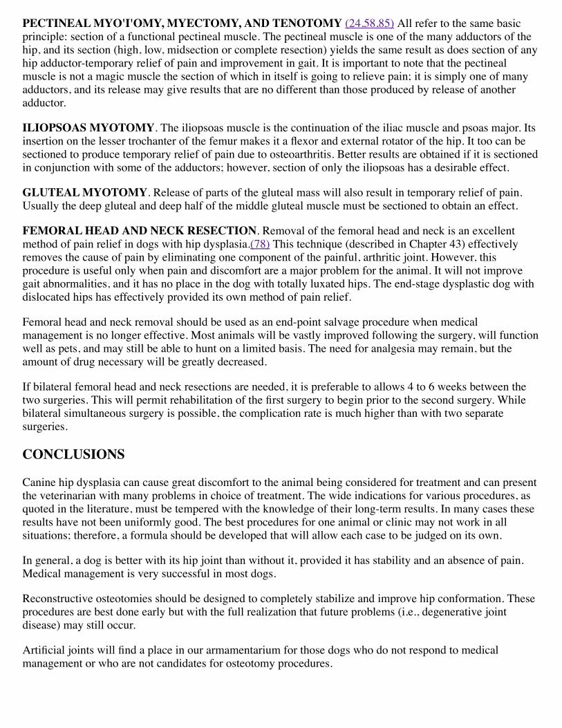
PECTINEAL MYO'I'OMY, MYECTOMY, AND TENOTOMY (24,58,85) All refer to the same basicprinciple: section of a functional pectineal muscle. The pectineal muscle is one of the many adductors of thehip, and its section (high, low, midsection or complete resection) yields the same result as does section of anyhip adductor-temporary relief of pain and improvement in gait. It is important to note that the pectinealmuscle is not a magic muscle the section of which in itself is going to relieve pain; it is simply one of manyadductors, and its release may give results that are no different than those produced by release of anotheradductor.
ILIOPSOAS MYOTOMY. The iliopsoas muscle is the continuation of the iliac muscle and psoas major. Itsinsertion on the lesser trochanter of the femur makes it a flexor and external rotator of the hip. It too can besectioned to produce temporary relief of pain due to osteoarthritis. Better results are obtained if it is sectionedin conjunction with some of the adductors; however, section of only the iliopsoas has a desirable effect.
GLUTEAL MYOTOMY. Release of parts of the gluteal mass will also result in temporary relief of pain.Usually the deep gluteal and deep half of the middle gluteal muscle must be sectioned to obtain an effect.
FEMORAL HEAD AND NECK RESECTION. Removal of the femoral head and neck is an excellentmethod of pain relief in dogs with hip dysplasia.(78) This technique (described in Chapter 43) effectivelyremoves the cause of pain by eliminating one component of the painful, arthritic joint. However, thisprocedure is useful only when pain and discomfort are a major problem for the animal. It will not improvegait abnormalities, and it has no place in the dog with totally luxated hips. The end-stage dysplastic dog withdislocated hips has effectively provided its own method of pain relief.
Femoral head and neck removal should be used as an end-point salvage procedure when medicalmanagement is no longer effective. Most animals will be vastly improved following the surgery, will functionwell as pets, and may still be able to hunt on a limited basis. The need for analgesia may remain, but theamount of drug necessary will be greatly decreased.
If bilateral femoral head and neck resections are needed, it is preferable to allows 4 to 6 weeks between thetwo surgeries. This will permit rehabilitation of the first surgery to begin prior to the second surgery. Whilebilateral simultaneous surgery is possible, the complication rate is much higher than with two separatesurgeries.
CONCLUSIONS
Canine hip dysplasia can cause great discomfort to the animal being considered for treatment and can presentthe veterinarian with many problems in choice of treatment. The wide indications for various procedures, asquoted in the literature, must be tempered with the knowledge of their long-term results. In many cases theseresults have not been uniformly good. The best procedures for one animal or clinic may not work in allsituations; therefore, a formula should be developed that will allow each case to be judged on its own.
In general, a dog is better with its hip joint than without it, provided it has stability and an absence of pain.Medical management is very successful in most dogs.
Reconstructive osteotomies should be designed to completely stabilize and improve hip conformation. Theseprocedures are best done early but with the full realization that future problems (i.e., degenerative jointdisease) may still occur.
Artificial joints will find a place in our armamentarium for those dogs who do not respond to medicalmanagement or who are not candidates for osteotomy procedures.
Myotomy procedures should be used only as pain-relieving procedures in dogs with proven degenerativejoint disease of the hips. Myotomy is inappropriate in immature dogs or as a proposed "cure" for hipdysplasia.
Femoral head and neck resection, a good tool for relief of pain, should be regarded only as a salvageprocedure.
References
1. Andren L: Pelvic instability in newborns with special reference to congenital dislocation of the hip andhormonal factors. Acta Radiol (suppl) 212, 1962
2. Andren L, Borglin NE: Disturbed urinary excretion patterns of oestrogens in newborns with congenitaldislocation of the hip: I. The excretion of oestrogen during the first few days of life. Acta Endocrinol 37:422,1961
3. Asrskog D, Stoa KR, Thorsen T: Urinary oestrogen excretion in newborn infants with congenital dysplasiaof the hip joint. Acta Paediatr 55:394, 1966
4. Bardens JW, Hardwick H: New observations on the diagnosis and cause of hip dysplasia. Vet Med/SmallAnim Clin 63: 238, 1968
5. Barlow TG: Early diagnosis and treatment of congenital dislocation of the hip. J Bone Joint Surg 44B:292,1962
6. Berzon JL, Howard PE, Covell SJ et al: A retrospective study of the efficacy of femoral head and neckexcisions in 94 dogs and cats. vet Surg 9:88, 1980
7. Bornfors S. Palsson K, Skude G: Hereditary aspects of hip dysplasia in German shepherd dogs. J Am VetMed Assoc 145: 15, 1964
8. Bowen JM, Lewis RE, Kneller SK et al: Progression of hip dysplasia in German shepherd dogs afterunilateral pectineal myotomy. J Am Vet Med Assoc 161:899, 1972
9. Brinker WO: Corrective osteotomy procedures for treatment of canine hip dysplasia. Vet Clin North Am1:467, 1971
10. Brinker WO, Flo G. Cross JF: Acetabuloplasty as a corrective surgical procedure in selected cases ofcongenital hip dysplasia in the dog. Scientific Proceedings of the 35th Annual Meeting of the AmericanAnimal Hospital Association 1968, pp 260-262
11. Cardinet GH, Guffy MM, Wallace LJ: Canine hip dysplasia: Effects of pectineal tenotomy in thecoxofemoral joints of German shepherd dogs. J Am Vet Med Assoc 164:591, 1974
12. Cardinet GH, Wallace LJ, Fedde MR et al: Developmental myopathy in the canine. Arch Neurol 21:620,1969
13. Duff R. Campbell JR: Radiographic appearance and clinical progress after excision arthroplasty of thecanine hip. J Small Anim Pract 19:439, 1978
14. Enlow DH: Principles of Bone Remodeling. Springfield, IL, Charles C Thomas, 1963
15. Frost HM: An Introduction to Biomechanics. Springfield, IL, Charles C Thomas, 1967
16. Gendreau CG, Cawley AJ: Excision of the femoral head and neck: The long term results of 35 operations.J Am Anim Hosp Assoc 13:605, 1977
17. Gruneberg H: The Pathology of Development: A Study of Inherited Skeletal Disorders in Animals. NewYork, John Wiley & Sons, 1963
18. Gustafsson PO, Beling CG: Estradiol-induced changes in beagle pups: Effects of prenatal and postnataladministration. Endocrinology 85:481, 1969
19. Gustafsson PO, Olsson SE, Kasstrom H et al: Skeletal development of greyhounds, German shepherds,and their crossbred offspring. Acta Radiol (suppl) 1-27, 1971
20. Hardisty JF, Scalera SE, Castleberry MW et al: Genetic influence of the male on the incidence of caninehip dysplasia. Proceedings of the Orthopedic Foundation of America Symposium on Hip Dysplasia, St Louis,1972
21. Hare WC: Canine pelvic limb. J Am Vet Med Assoc 136:603, 1960
22. Hass J: Congenital Dislocation of the Hip. Springfield, IL, Charles C Thomas, 1951
23. Henricson B. Norberg J. Olsson SE: On the etiology and pathogenesis of hip dysplasia. A comparativereview. J Small Anim Pract 7:673, 1966
24. Henry JD: A modified technique for pectineal tendonectomy in the dog. J Am Vet Med Assoc 163:465,1973
25. Hoefle WD: A surgical procedure for prosthetic total hip replacement in the dog. J Am Anim Hosp Assoc10:269, 1974
26. Hohn RB, Janes JM: Pelvic osteotomy in the treatment of canine hip dysplasia. Clin Orthop 62:70, 1969
27. Holt PE: Hip dysplasia in a cat. J Small Anim Pract 19:273, 1978
28. Hutt FB: Genetic selection to reduce the incidence of hip dysplasia in dogs. J Am Vet Med Assoc151:1041, 1967
29. Jessen CR, Spurrell FA: The heritability of canine hip dysplasia. Proceedings of the OrthopedicFoundation of America Symposium on Hip Dysplasia, St Louis, 1972
30. Jessen CD, Spurred FA: Canine hip dysplasia: 1. Radiographic detection in known age groups.Proceedings of the Orthopedic Foundation of America Symposium on Hip Dysplasia, St Louis, 1972
31. Johnson LC: Kinetics of osteoarthritis. Lab Invest 8:1223, 1959
32. Johnson LC: Morphologic analysis in pathology. In Johnson LC fed): Bone Biodynamics, p 559. Boston,Little, Brown & co, 1964
33. Kaman CH, Gossling HR: A breeding program to reduce hip dysplasia in German shepherd dogs. J AmVet Med Assoc 151 :562, 1967
34. Kelly AM, Zacks Sl: The histogenesis of rat intercostal muscle. J Cell Biol 42: 135, 1969
35. Kolde DL: Pectineus tenectomy for treatment of hip dysplasia in a domestic cat: A case report. J AmAnim Hosp Assoc 10:564, 1974
36. Larsen JS: The distribution of dysplastic radiographic evaluation among 36 breeds of dogs. Proceedingsof the Orthopedic Foundation of America Symposium on Hip Dysplasia, St Louis, 1972
37. Lee R. Fry PD: Some observations on the occurrence of Legg-Calve-Perthes disease (coxa plana) in thedog and an evaluation of excision arthroplasty as method of treatment. J Small Anim Pract 10:309, 1969
38. Lewis RH, Jones JP: A clinical study of canine total hip arthroplasty. Vet Surg 9:20, 1980
39. Lust G. Craig PH, Geary JC et al: Changes in pelvic muscle tissue associated with hip dysplasia in dogs.Am J Vet Res 33: 1097, 1972
40. Lust G. Craig PH, Ross GE et al: Studies on pectineus muscle in canine hip dysplasia. Cornell Vet62:628, 1972
41. McCarroll HR: Congenital subluxation and dislocation of the hip in infancy. J Bone Joint Surg 47A:589,1965
42. McKibbin B: Anatomical factors in the stability of the hip joint in the newborn. J Bone Joint Surg52B:148, 1970
43. Mansson J. Norberg J: Dysplasia of the hip in dogs. Medlemsbl Sver Vet Forb 13:330, 1961
44. Michele AA: iliopsoas. Springfield, IL Charles C Thomas, 1962
45. Miller Ml, Christensen GC, Evans HE: Anatomy of the Dog. Philadelphia, WB Saunders, 1964
46. Nunamaker DM: Surgical correction of large femoral anteversion angles in the dog. J Am Vet Med Assoc165:1061, 1974
47. Nunamaker DM, Biery DN, Newton CD: Femoral neck anteversion in the dog: Its radiographicmeasurement. J Am Vet Radiol Soc 14:45, 1973
48. Nunamaker DM, Newton CD: Canine hip disorders. In Bojrab MJ (ed): Current Techniques in SmallAnimal Surgery, pp 436-442. Philadelphia, Lea & Febiger, 1975
49. Olmstead ML, Hohn RB, Turner TM: Technique for canine total hip replacement. Vet Surg 10:44, 1981
50. Olsson SE: The control of canine hip dysplasia in Scandinavian countries: Advances in small animalpractice. Proc Br Small Anim Vet Assoc 3:112, 1961
51. Olsson SE: Hoftledsdysplasin pa tillbakagang. Hundsport. Svenska Kennelklubbens Tidskr 11:16, 1963
52. Owen R: Editorials and annotations: Early diagnosis of congenital unstable hip. J Bone Joint Surg50B:453, 1968
53. Palmen K: Preluxation of the hip joint: Diagnosis and treatment in the newborn and the diagnosis ofcongenital dislocation of the hip joint in Sweden during the years 1948-1960. Acta Paediatr 50 (suppl): 129,1961
54. Pauwel F: Neue Richtlinien fur die operative Behandlung der Koxarthrose. Verh. 48 Kongr Dtsch OrthopGes, 33 2 (Enke, Stuttgart 1961)
55. Pierce KR, Bridges CH: The role of estrogens in the pathogenesis of canine hip dysplasia: Metabolism ofexogenous estrogens. J Small Anim Pract 8:383, 1967
56. Piermattei DL: Femoral head ostectomy in the dog: Indications, technique, and results in ten cases. AnimHosp 1: 180, 1965
57. Ponseti IV, Pedrini-Mille A, Pedrini V: Histological analysis of human iliac crest cartilage: 1.Observations on trunk growth. Calcif Tissue Res 2:197,1968
58. Rickards DA, Hinko PJ, Morse EM: Pecanectomy vs. pectinotomy in the treatment of hip dysplasia. VetMed/Small Anim Clin 67:976, 1972
59. Riser H: An analysis of the current status of hip dysplasia in the dog. J Am Vet Med Assoc 144:709, 1964
60. Riser WH: The dog as a model for hip dysplasia. Libr Vet Med. Fak University of Bern, 1973
61. Riser WH: Canine hip dysplasia: Cause and control. J Am Vet Med Assoc 164:360, 1974
62. Riser WH: The dog as a model for the study of hip dysplasia. Vet Pathol 12:235, 1975
63. Riser WH, Cohen D, Lindqvist S et al: Influence of early rapid growth and weight gain on hip dysplasiain the German shepherd dog. J Am Vet Med Assoc 145:661, 1964
64. Riser WH, Hardisty JF, Castleberry MW: Reduction of frequency of hip dysplasia in a colony of dogs.Proceedings of the Orthopedic Foundation of America Symposium on Hip Dysplasia, St Louis, 1972
65. Riser WH, Larsen J S: Influence of breed somatotypes on the prevalence of hip dysplasia in the dog. JAm Vet Med Assoc 165:79, 1974
66. Riser WH, Miller H: Canine hip dysplasia and how to control it. Orthopedic Foundation for Animals,Columbia, MO, 1966
67. Riser WH, Newton CD: Canine hip dysplasia as a disease. In Bojrab MJ (ed):Pathophysiology of SmallAnimal Surgery, pp 618-624. Philadelphia, Lea & Febiger, 1981
68. Riser WH, Shirer JF: Hip dysplasia: Coxofemoral abnormalities in neonatal German shepherd dogs. JSmall Anim Pract 7:7, 1966
69. Riser WH, Shirer JF: Correlation between canine hip dysplasia and pelvic muscle mass: A study of 95dogs. Am J Vet Res 124:769, 1967
70. Robin DL, Barnett CR, Arnold WD et al: Untreated congenital hip disease. A study of the epidemiology,natural history, and social aspects of the disease in a Navajo population. Am J Public Health 55:(suppl) 2,1965
71. Rosen S von: Diagnosis and treatment of congenital dislocation of the hip joint in the newborn. J BoneJoint Surg 44B:284, 1962
72. Salter RB: Innominate osteotomy in the treatment of congenital dislocation and subluxation of the hip. JBone Joint Surg 43B:518, 1961
73. Salter RB: Experimental dysplasia of the hip and its reversibility in newborn pigs: Abstracts of theAnnual Meeting of the American Orthopedic Association, Hot Springs, VA, 1963, pp 24-27
74. Salter RB: Etiology, pathogenesis and possible prevention of congenital dislocation of the hip. Can MedAssoc J 98:933, 1968
75. Sheldon WH: The Varieties of Temperament. New York, Harper & Row, 1942
76. Smith WS, Coleman CR, Alex ML et al: Etiology of congenital dislocation of the hip. J Bone Joint Surg45A:491, 1963
77. Sokoloff L: The Biology of Degenerative Joint Disease. Chicago, University of Chicago Press, 1969
78. Spreull JSA: Excision arthoplasty as a method of treatment of hip joint diseases in the dog. Vet Rec73:573, 1961
79. Stanisavlievic S: Diagnosis and Treatment of Congenital Hip Pathology in the Newborn. Baltimore,Williams & Wilkins, 1964
80. Strayer LM: The embryology of the human hip joint. Clin Orthop Rel Res 74:221, 1971
81. Thieme WT, Wynne-Davies R. Blair HAF et al: Clinical examination and urinary oestrogen assays innewborn children with congenital dislocation of the hip. J Bone Joint Surg 50B:546, 1968
82. Thompson DAW: In Bonner (ed): On Growth and Form. New York, Cambridge University Press, 1961
83. Trueta J: Studies of the Development and Decay of the Human Frame, p 360. Philadelphia, WB Saunders1968
84. Vaughan LC, Clayton Jones DG, Lane JG: Pectineus muscle resection as a treatment for hip dysplasia indogs. Vet Rec 145, 1975
85. Voss C: Koxarthrose-die "temporare Hangehufte" ein neues Verfahren zur operativen Behandlung derschmerzhaften Altershufte und anderer chronisch deformierender Huftgelenkserkrankungen . M unch MedWochenschr 98:954, 1956
86. Wallace LK: Pectineus tendonectomy or tenotomy for treating clinical canine hip dysplasia. Vet ClinNorth Am 1:455, 1971
87. Wilkinson JA: Femoral anteversion in the rabbit. J Bone Joint Surg 44B:386, 1962
88. Wilkinson JA: Prime factors in the etiology of congenital dislocation of the hip. J Bone Joint Surg45B:269, 1963
89. Wright V: Lubrication and Wear in Joints. Philadelphia, JB Lippincott, 1972


![The Demographics of Canine Hip Dysplasia in the United ...downloads.hindawi.com/archive/2017/5723476.pdf · Canine hip dysplasia (CHD) is a well-known disorder in veterinarymedicine[1–4],especiallyamongstcertainbreeds](https://img.pdfslide.us/doc/110x75/5edab6c4272674784f04f1ad/the-demographics-of-canine-hip-dysplasia-in-the-united-canine-hip-dysplasia.jpg)