Embed Size (px)
Citation preview
REVIEW
Hinge/floating craniotomy as an alternative technique for cerebraldecompression: a scoping review
Hugo Layard Horsfall1,2 & Midhun Mohan1,2& B. Indira Devi2,3 & Amos O. Adeleye2,4,5
& Dhaval P. Shukla2,3 &
Dhananjaya Bhat2,3 & Mukhtar Khan2,6& David J. Clark1,2 & Aswin Chari7,8 & Franco Servadei9 & Tariq Khan2,6
&
Andres M. Rubiano2,10,11& Peter J. Hutchinson1,2
& Angelos G. Kolias1,2
Received: 24 June 2019 /Revised: 20 August 2019 /Accepted: 12 September 2019# The Author(s) 2019
AbstractHinge craniotomy (HC) is a technique that allows for a degree of decompression whilst retaining the bone flap in situ, in a‘floating’ or ‘hinged’ fashion. This provides expansion potential for ensuing cerebral oedema whilst obviating the need forcranioplasty in the future. The exact indications, technique and outcomes of this procedure have yet to be determined, but it islikely that HC provides an alternative technique to decompressive craniectomy (DC) in certain contexts. The primary objectivewas to collate and describe the current evidence base for HC, including perioperative parameters, functional outcomes andcomplications. The secondary objective was to identify current nomenclature, operative technique and operative decision-making. A scoping review was performed in accordance with the PRISMA-ScR Checklist. Fifteen studies totalling 283 patients(mean age 45.1 andM:F 199:46) were included. There were 12 different terms for HC. The survival rate of the cohort was 74.6%(n = 211). Nine patients (3.2%) required subsequent formal DC. Six studies compared HC to DC following traumatic brain injury(TBI) and stroke, finding at least equivalent control of intracranial pressure (ICP). These studies also reported reduced rates ofcomplications, including infection, in HC compared to DC. We have described the current evidence base of HC. There is noevidence of substantially worse outcomes compared to DC, although no randomised trials were identified. Eventually, arandomised trial will be useful to determine if HC should be offered as first-line treatment when indicated.
Keywords Neurosurgery . Decompressive craniectomy . Traumatic brain injury . Stroke
Introduction
A recent study estimated 60% of global neurosurgical case-load is traumatic brain injury (TBI) and stroke (6.2 and 2.8
million, respectively)—the majority in low-to-middle-incomesettings [1]. There are significant societal costs associatedwithTBI due to high levels of mortality and morbidity. A rigorous
Hugo Layard Horsfall and Midhun Mohan are joint first authors.
* Angelos G. [email protected]
1 Division of Neurosurgery, Department of Clinical Neurosciences,Addenbrooke’s Hospital and University of Cambridge,Cambridge, UK
2 NIHR Global Health Research Group on Neurotrauma, University ofCambridge, Cambridge, UK
3 Department of Neurosurgery, National Institute for Mental Healthand Neurosciences, Bangalore, India
4 Division of Neurological Surgery, Department of Surgery, College ofMedicine, University of Ibadan, Ibadan, Nigeria
5 Department of Neurological Surgery, University College Hospital,Ibadan, Nigeria
6 Department of Neurosurgery, North West General Hospital andResearch Center, Peshawar, Pakistan
7 Department of Neurosurgery, Great Ormond Street Hospital,London, UK
8 Institute of Child Health, University College London, London, UK9 Department of Neurosurgery, Humanitas University and Research
Hospital, Milan, Italy10 INUB/MEDITECH Research Group, El Bosque University,
Bogota, Colombia11 MEDITECH Foundation, Clinical Research, Cali, Colombia
Neurosurgical Reviewhttps://doi.org/10.1007/s10143-019-01180-7
evidence base to guide treatment strategies remains an inter-national public health priority.
The literature describing decompressive craniectomy (DC)is varied [2]. Recent and ongoing randomised controlled trials(RCTs) for the use of DC in TBI (DECRA [3]; RESCUEicp[4]; RESCUE-ASDH) and stroke (DECIMAL [5]; DESTINY[6]; HAMLET [7]) though have demonstrated its potentialutility and efficacy, they raise ongoing concerns, in some ofthe studies, regarding the higher rates of disability observed insurvivors following DC. Regarding TBI, the DECRA trialshowed that neuroprotective bifrontal DC for moderate intra-cranial hypertension (ICP) is not helpful, whereas theRESCUEicp trial found that last-tier DC for severe and refrac-tory ICP can significantly reduce the mortality rate but is as-sociated with a higher rate of disability [2–4]. In relation toischaemic stroke, a Cochrane review [8] including data fromall the three extant randomised controlled trials (DECIMAL[5]; DESTINY [6]; HAMLET [7]) suggested that DC im-proves survival compared with best medical management,but that an increased proportion of individuals treated withDC survive with moderately severe or severe disability [8, 9].
A relatively novel and less well-utilised technique toachieve cerebral decompression in patients with brain swellingand/or raised ICP is the ‘hinge craniotomy’ (HC), also knownas hinged decompressive craniectomy. The technique was firstdescribed by three independent groups in 2007 [10–12] specif-ically for surgical modulation of post-traumatic medically in-tractable raised ICP, although it has been used by neurosur-geons for several years for sundry other indications.Adoption of HC into neurosurgical practice can potentiallyyield benefits over traditional DC in specific situations, suchas the potential to control at least moderate cerebral oedemawhilst simultaneously obviating the need for a subsequentcostly operative cranioplasty [2]. This is a particularly impor-tant consideration in resource-limited settings. Furthermore,unlike for the traditional surgical technique of DC, followingHC, there are reports of potential reduction in axonal stretchingand there are supposedly fewer complications such as syn-drome of the trephined, problems with CSF hydrodynamics,infection and resorption of the autologous bone flap [13].
However, HC has possible limitations; these revolve mainlyaround whether sufficient extracranial brain expansion volumewill be achieved and whether the patient will require the moretraditional DC later on. Central to theHCvsDCdebate is not justabout post-operative patient survival, but the subsequent func-tional outcome and associated morbidity that may be incurred.There is a paucity of rigorous data evaluating HC, and contem-porary evidence is based upon experience from small series insingle centres from disparate regions of the world.
Our primary objective in this study was to collate, assessand describe the current evidence base for the use of HC. Weassess the current indications, differing techniques, functionaloutcomes and complications of the procedure. To this end, we
performed a scoping review; a relatively novel study designthat determines the scope or coverage of a body of literatureon a given topic and gives a broad overview [14, 15]. Thisreview process is particularly useful for examining the emerg-ing evidence relating to HC whilst it still remains relativelyunclear what other, more specific questions can be posed andvaluably addressed by a more precise systematic review andmeta-analysis [14, 16]. This is important as there is variationin the definition and technique of HC as currently describedby diverse workers interested in it from different regions of theworld. Moreover, the exact indication for HC is unclear, for,although it may have a role between medical management andDC in both TBI and stroke, little robust evidence presentlyexists for it. A scoping review provides the perfect medium toreport on the current evidence surrounding HC, using system-atic methodology provided by the recently publishedPRISMA-ScR framework [17].
Methods
Protocol and registration
This scoping review has been reported in accordance with thePreferred Reporting Items for Systematic Reviews and Meta-Analysis extension for Scoping Reviews (PRISMA-ScR)[17]. Unlike systematic reviews, the protocol does not needto be registered with PROSPERO [15].
Eligibility criteria
The following advanced search strategy was used to search allPubMed on 22 June 2019:
((((((((craniotomy[Title/Abstract]) OR craniectomy[Title/Abstract]) OR decompress*[Title/Abstract])) AND((((((hinge*[Title/Abstract]) OR float*[Title/Abstract]) OR insitu[Title/Abstract]) OR riding[Title/Abstract]) ORosteoplastic[Title/Abstract]) OR anchored[Title/Abstract])))NOT “case reports”[Publication Type]))
Titles and abstracts were screened for relevance. Full-textarticles were then assessed for eligibility according to thePICOS criteria below. The reference lists of eligible studiesand relevant articles were searched for further studies notidentified by the initial search strategy. Manuscripts were ex-cluded if data was not available separately for the HC cohort;they were case reports; or were paediatric series.
PICOS criteria
& Population: Diagnosis of TBI or stroke and exposure toHC
& Intervention: Hinge craniotomy; in situ hingecraniectomy; the Tucci flap; in situ resin floating
Neurosurg Rev
cranioplasty; in situ free floating craniectomy; osteoplasticdecompression; hinge decompressive craniotomytemporalis; riding craniotomy; modified temporal musclehinge decompressive craniotomy; floating anchoredcraniotomy
& Comparison: Studies with and without controls were in-cluded due to nature of scoping review
& Outcomes and other data collected: Demographics, de-scription of indications and surgical techniques, intracra-nial pressure monitoring, mean length of stay, functionaloutcome, mortality
& Study design: All prospective and retrospective case se-ries, cohort studies, case-control and randomised con-trolled trials with n > 1 written in English were included
Selection of sources of evidence
The resulting titles and abstracts were screened independentlyby two authors (HLH and MM) using the PICOS criteriaabove. If disagreements occurred, a third author (AK) wasconsulted. Data extraction was performed independently bythe same two authors with disagreements resolved via furtherreview and discussion. Due to the heterogeneity of terminol-ogy used for hinge craniotomy in the global literature, when-ever a new term was identified from the references, it wasincorporated into the search strategy.
Data charting process
The data extraction process in a scoping review is known as‘data charting’. Key variables were screened and extractedfrom the papers. These data were inputted into a MicrosoftExcel Document, which was the basis of the data chartingform. This was continuously updated in an iterative process,as heterogeneity of data and reported outcomes meant oftennon-contiguous data points. However, if the data was impor-tant and remained an essential component to report on in thispaper, it was added to the charting process. Two reviewersindependently (HLH & MM) charted data from each eligiblearticle. Any disagreements were resolved through discussionbetween the two reviewers or further adjudication by a thirdreviewer (AK).
Data items
Indication for hinge craniotomy: age, sex and indication forhinge craniotomy. If there were multiple pathologies, for ex-ample subdural haematoma and intracerebral haemorrhagefollowing TBI, then the first pathology was listed as the indi-cation. For some of the studies, separate male:female ratio ofHC cohort was not stated; therefore, the male:female ratioreported in Results is less than the total number of patients.
Surgical technique and nomenclature: the named procedurewas recorded, with size of craniotomy, dural manipulation,hinge craniotomy practice and the placement of wound drain.
Perioperative parameters: pre- and post-operative variables(ICP, GCS, midline shift and CT-Rotterdam criteria) wererecorded.
Clinical outcomes: survival, Glasgow Outcome Scale(GOS) and modified Rankin Score (mRS) and follow-up wererecorded. The GOS is reported as favourable (GOS 4–5) andunfavourable (GOS 1–3). The mRS is reported as: ‘Good’ =0–2; ‘Moderate’ = 3–4; and ‘Poor’ = 5–6.
Complications: inadequate cerebral decompression was re-ported if the bone flap needed to be removed subsequently dueto refractory intracranial hypertension. Infection, need for re-operation and other complications were also recorded.
Comparison between HC and DC: studies that comparedthe two techniques were summarised.
Level of evidence: data was also extracted on article demo-graphics (journal, institution, level of evidence, summary ofarticle) and the income status of the country of origin.
Synthesis of results
The results in this manuscript are presented as a scoping re-view, including summary tables, and follow the followingformat: (1) indication for hinge craniotomy; (2) surgical tech-nique and nomenclature; (3) perioperative parameters; (4)functional outcomes; (5) complications; (6) summary of com-parison studies and (7) level of evidence.
Results
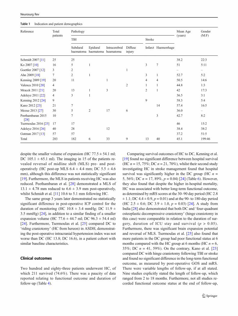
Indication and patient demographics
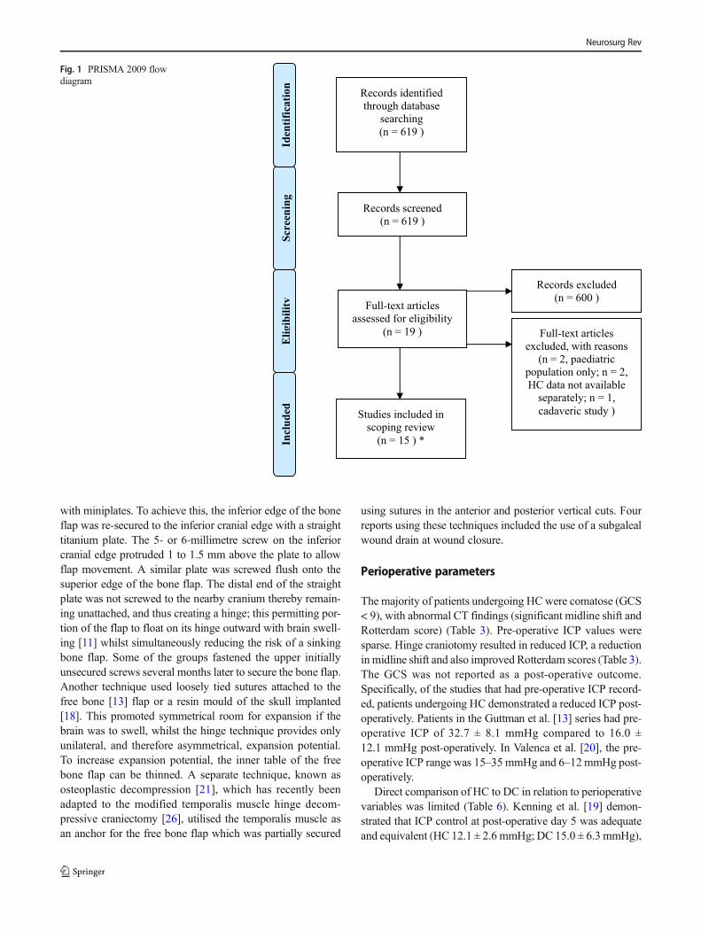
A total of 15 studies [10–13, 18–28] were eligible for inclu-sion (Fig. 1), comprising 283 patients with a mean age 45.1years and a male:female of 199:46 (Table 1). The majority ofpatients (n = 230, 81.3%) underwent HC following TBI. Ofthe patients who suffered TBI, the most common pathologywas acute subdural haematoma (n = 182, 79.1%), followed byintracerebral haemorrhage (n = 33, 14.3%) and epiduralhaematoma (n = 7, 3.0%). A number of patients (n = 53,18.7%) underwent HC following stroke: haemorrhagic (n =40, 75.5%) and ischaemic (n = 13, n = 24.5%).
Surgical technique and nomenclature
There were 12 separate terms used to describe hinge craniot-omy, of which hinge craniotomywas the most common (n = 4,33.3%). Every other term was used once (Table 2).
The ‘hinge’ was achieved via a variety of techniques(Table 2). Most commonly, the free bone flap was secured
Neurosurg Rev
with miniplates. To achieve this, the inferior edge of the boneflap was re-secured to the inferior cranial edge with a straighttitanium plate. The 5- or 6-millimetre screw on the inferiorcranial edge protruded 1 to 1.5 mm above the plate to allowflap movement. A similar plate was screwed flush onto thesuperior edge of the bone flap. The distal end of the straightplate was not screwed to the nearby cranium thereby remain-ing unattached, and thus creating a hinge; this permitting por-tion of the flap to float on its hinge outward with brain swell-ing [11] whilst simultaneously reducing the risk of a sinkingbone flap. Some of the groups fastened the upper initiallyunsecured screws several months later to secure the bone flap.Another technique used loosely tied sutures attached to thefree bone [13] flap or a resin mould of the skull implanted[18]. This promoted symmetrical room for expansion if thebrain was to swell, whilst the hinge technique provides onlyunilateral, and therefore asymmetrical, expansion potential.To increase expansion potential, the inner table of the freebone flap can be thinned. A separate technique, known asosteoplastic decompression [21], which has recently beenadapted to the modified temporalis muscle hinge decom-pressive craniectomy [26], utilised the temporalis muscle asan anchor for the free bone flap which was partially secured
using sutures in the anterior and posterior vertical cuts. Fourreports using these techniques included the use of a subgalealwound drain at wound closure.
Perioperative parameters
The majority of patients undergoing HCwere comatose (GCS< 9), with abnormal CT findings (significant midline shift andRotterdam score) (Table 3). Pre-operative ICP values weresparse. Hinge craniotomy resulted in reduced ICP, a reductionin midline shift and also improved Rotterdam scores (Table 3).The GCS was not reported as a post-operative outcome.Specifically, of the studies that had pre-operative ICP record-ed, patients undergoing HC demonstrated a reduced ICP post-operatively. Patients in the Guttman et al. [13] series had pre-operative ICP of 32.7 ± 8.1 mmHg compared to 16.0 ±12.1 mmHg post-operatively. In Valenca et al. [20], the pre-operative ICP range was 15–35 mmHg and 6–12mmHg post-operatively.
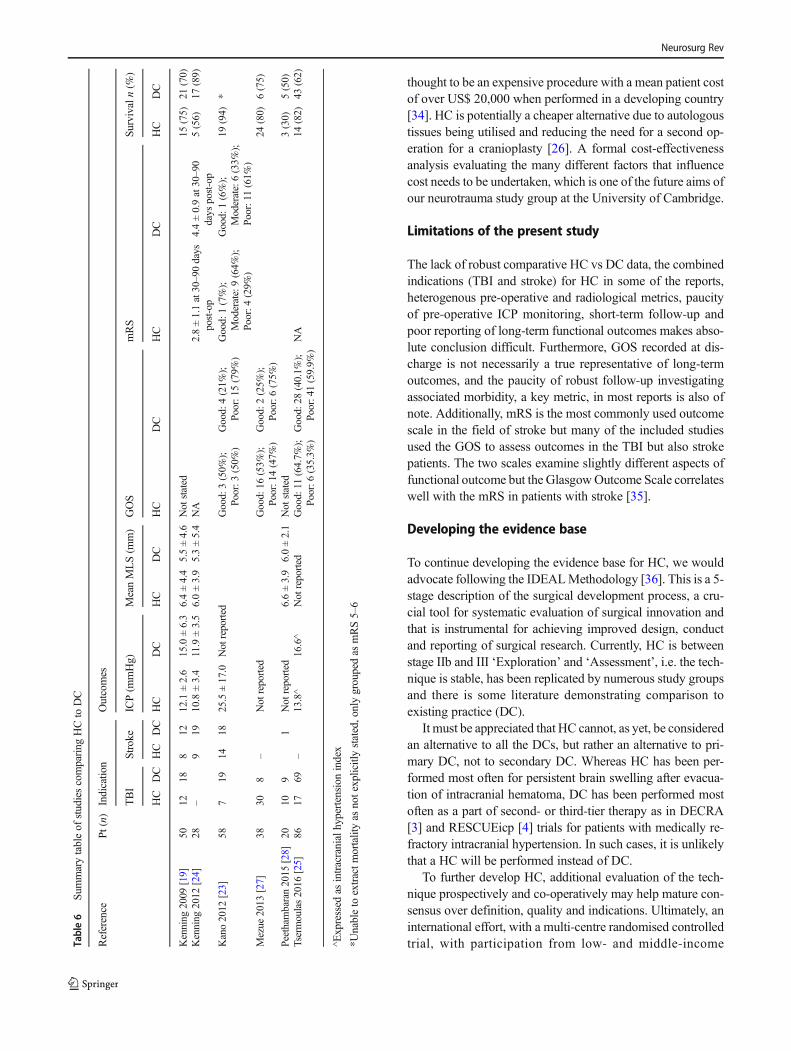
Direct comparison of HC to DC in relation to perioperativevariables was limited (Table 6). Kenning et al. [19] demon-strated that ICP control at post-operative day 5 was adequateand equivalent (HC 12.1 ± 2.6 mmHg; DC 15.0 ± 6.3 mmHg),
Records identified
through database
searching
(n = 619 )
Incl
uded
Elig
ibili
tyId
entif
icat
ion
Records screened
(n = 619 )
Records excluded
(n = 600 )Full-text articles
assessed for eligibility
(n = 19 ) Full-text articles
excluded, with reasons
(n = 2, paediatric
population only; n = 2,
HC data not available
separately; n = 1,
cadaveric study )Studies included in
scoping review
(n = 15 ) *
Scre
enin
g
Fig. 1 PRISMA 2009 flowdiagram
Neurosurg Rev
despite the smaller volume of expansion (HC 77.5 ± 54.1 ml;DC 105.1 ± 65.1 ml). The imaging in 15 of the patients re-vealed reversal of midline shift (MLS) pre- and post-operatively (HC post-op MLS 6.4 ± 4.4 mm; DC 5.5 ± 4.6mm), although this difference was not statistically significant[19]. Furthermore, the MLS in patients receiving HC was alsoreduced. Peethambaran et al. [28] demonstrated a MLS of13.1 ± 4.78 mm reduced to 6.6 ± 3.9 mm post-operatively,whilst Schmidt et al. [11] 10.6 to 5.1 mm following HC.
The same group 3 years later demonstrated no statisticallysignificant difference in post-operative ICP control for theduration of monitoring (HC 10.8 ± 3.4 mmHg; DC 11.9 ±3.5 mmHg) [24], in addition to a similar finding of a smallerexpansion volume (HC 77.6 ± 44.7 ml; DC 96.3 ± 54.4 ml)[24]. Furthermore, Tsermoulas et al. [25] compared DC to‘riding craniotomy’ (HC from hereon) in ASDH, demonstrat-ing the post-operative intracranial hypertension index was notworse than DC (HC 13.8; DC 16.6), in a patient cohort withsimilar baseline characteristics.
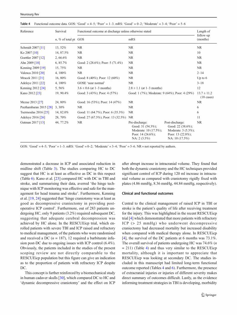
Clinical outcomes
Two hundred and eighty-three patients underwent HC, ofwhich 211 survived (74.6%). There was a paucity of datareported relating to functional outcome and duration offollow-up (Table 4).
Comparing survival outcomes of HC to DC, Kenning et al.[19] found no significant difference between hospital survival(HC n = 15, 75%; DC n = 21, 70%), whilst their second studyinvestigating HC in stroke management found that hospitalsurvival was significantly higher in the DC group (HC n =5, 56%; DC n = 17, 89%, p = 0.04) [24] (Table 6). However,they also found that despite the higher in-hospital mortality,HC was associated with better long-term functional outcome,as determined bymRS scores at the 30–90 day period (HC 2.8± 1.1; DC 4.4 ± 0.9, p = 0.01) and at the 90- to 180-day period(HC 2.5 ± 0.6; DC 3.9 ± 1.0, p = 0.03) [24]. A study fromIndia [28] also demonstrated that both DC and ‘four-quadrantosteoplastic decompressive craniotomy’ (hinge craniotomy inthis case) were comparable in relation to the duration of sur-gery, duration of ICU stay and survival (p > 0.05).Furthermore, there was significant brain expansion potentialand reversal of MLS. Tsermoulas et al. [25] also found thatmore patients in the DC group had poor functional status at 6months compared with the HC group at 6 months (HC n = 6,35%; DC n = 41, 59%). On the contrary, Kano et al. [23]compared DC with hinge craniotomy following TBI or strokeand found no significant difference in the long-term functionaloutcome, as measured by post-operative GOS and mRS.There were variable lengths of follow-up, if at all stated.Nine studies explicitly stated the length of follow-up, whichranged from 2 to 18 months. Furthermore, not all studies re-corded functional outcome status at the end of follow-up,
Table 1 Indication and patient demographics
Reference Totalpatients
Pathology Mean Age(years)
Gender(M:F)
TBI Stroke
Subduralhaematoma
Epiduralhaematoma
Intracerebralheamatoma
Diffuseinjury
Infarct Haemorrhage
Schmidt 2007 [11] 25 25 38.2 22:3
Ko 2007 [10] 16 5 1 3 7 51 5:11
Goettler 2007 [12] 3 2 1 – –
Ahn 2009 [18] 7 2 1 3 1 52.7 5:2
Kenning 2009 [19] 20 11 1 4 4 50.5 14:6
Valenca 2010 [20] 4 2 1 1 44.8 1:3
Mracek 2011 [21] 20 13 1 3 2 1 42 17:3
Adeleye 2011 [22] 4 3 1 36.5 3:1
Kenning 2012 [24] 9 9 58.3 5:4
Kano 2012 [23] 21 7 14 57.4 16:5
Mezue 2013 [27] 30 5 2 17 6 36.0 –
Peethambaran 2015[28]
10 7 3 42.7 8:2
Tsermoulas 2016 [25] 17 17 46 15:2
Adeleye 2016 [26] 40 28 12 38.4 38:2
Gutman 2017 [13] 57 57 37.2 51:5
Total 283 182 6 33 9 13 40 45.1 199:46
Neurosurg Rev
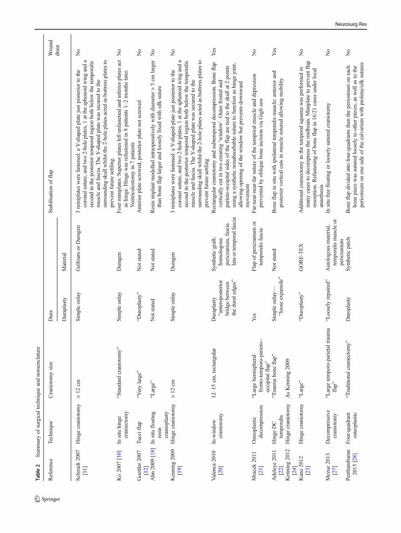
Table2
Sum
maryof
surgicaltechniqueandnomenclature
Reference
Technique
Craniotom
ysize
Dura
Stabilisatio
nof
flap
Wound
drain
Duraplasty
Material
Schm
idt2
007
[11]
Hinge
craniotomy
>12
cmSimpleonlay
Gelfoam
orDuragen
3miniplateswerefastened:a
Y-shapedplatejustposteriorto
the
coronalsuture,andtwo2-holeplates,1
atthesphenoidwinganda
second
intheposteriortemporalregionboth
belowthetemporalis
muscleandfascia.T
heY-shapedplatewas
securedto
the
surroundingskullw
hilstthe
2-holeplates
actedas
buttressplates
topreventfuturesettling.
No
Ko2007
[10]
Insitu
hinge
craniectom
y“Standardcraniotomy”
Simpleonlay
Duragen
Fourminiplates.Superiorplates
leftunfastened
andinferior
plates
act
ashinge.Hinge
was
refastened
in8patients1–2monthslater.
Ventriculostomyin
7patients
No
Goettler
2007
[12]
Tucciflap
“Verylarge”
“Duroplasty”
Not
stated
Anteriorplatescrewed,posterior
platenotscrew
edNo
Ahn
2009
[18]
Insitu
floatin
gresin
cranioplasty
“Large”
Not
stated
Not
stated
Resin
implantm
odelledintraoperativ
elywith
diam
eter
>5cm
larger
than
bone
flap
larger
andlooselyfixedwith
silk
suture
No
Kenning
2009
[19]
Hinge
craniotomy
>12
cmSimpleonlay
Duragen
3miniplateswerefastened:a
Y-shapedplatejustposteriorto
the
coronalsuture,andtwo2-holeplates,1
atthesphenoidwinganda
second
intheposteriortemporalregionboth
belowthetemporalis
muscleandfascia.T
heY-shapedplatewas
securedto
the
surroundingskullw
hilstthe
2-holeplates
actedas
buttressplates
topreventfuturesettling
No
Valenca
2010
[20]
In-w
indow
craniotomy
12–15cm
,rectangular
Duraplasty
“anteroposterior
bridge
between
theduraledges”
Synthetic
graft;
homologous
pericranium,fascia
lataor
temporalfascia
Rectangular
craniotomyandsubtem
porald
ecom
pression.B
oneflap
vertically
cutintwocreatin
g‘w
indow’.Outer
frontaland
parieto-occipitalsides
oftheflap
aretiedto
theskullat2
points
usingasynthetic
nonabsorbablesuture
tofunctio
nas
hingejoint,
allowingopeningof
thewindowbutp
reventsdownw
ard
movem
ent
Yes
Mracek2011
[21]
Osteoplastic
decompression
“Large
hemispheral
fronto-tem
poro-parieto--
occipitalflap”
Yes
Flapof
pericranium
ortemporalis
fascia
Far-near-near-farsuture
ofthetemporalm
uscleanddepression
preventedby
obliq
uebone
incision
viaGiglisaw
No
Adeleye
2011
[22]
Hinge
DC
temporalis
“Traum
abone
flap”
Simpleonlay—
“loose
expansile”
Not
stated
Boneflap
insitu
with
ipsilateraltem
poralis
muscle;anterior
and
posteriorverticalcutsin
musclesuturedallowingmobility
Yes
Kenning
2012
[24]
Hinge
craniotomy
AsKenning
2009
Kano2012
[23]
Hinge
craniotomy
“Large”
“Duroplasty”
GORE-TEX
Additionalcraniectom
yin
thetemporalsquam
awas
performed
inmanycasesto
decompressthemidbrain.Miniplateto
preventflap
resorptio
n.Refastening
ofbone
flap
in16/21casesunderlocal
anaethesia
No
Mezue
2013
[27]
Decom
pressive
craniotomy
“Large
temporo-parietaltraum
aflap”
“Loosely
repaired”
Autologousmaterial,
temporalis
muscleor
pericranium
Insitu
free
floatin
gor
looselysuturedcraniotomy
No
Peetham
baran
2015
[28]
Four-quadrant
osteoplastic
“Traditio
nalcraniectomy”
Duroplasty
Synthetic
patch
Boneflap
dividedinto
four-quadrantsthen
theperiosteum
oneach
bone
piecewas
suturedlooselyto
otherpieces,aswellasto
the
periosteum
ononeside
ofthecalvarium
with
prolene/silk
sutures
No
Neurosurg Rev
merely just that the patient was seen by healthcare practi-tioners in the time period. Six studies did not state durationof follow-up.
Complications
There were 54 reported complications in the HC cohort(Table 5). Nine patients (n = 9/283, 3.2%) required subsequentdecompressive craniectomy due to uncontrollable ICP, or inother words, ‘failure’ of hinge craniotomy. In one study [23],the bone flap was removed in 2 cases due to acute hydroceph-alus or brain herniation causing low cerebral perfusion pres-sure and in the other 2 cases, ICP was elevated immediatelysecondary to CT-confirmed epidural haemotoma requiringprogression to DC. In another study [24], one patient wasplaced in a barbiturate-induced coma at the request of thetreating neurologist, although the paper states that ICP wasin normal range.
Regarding the replaced free bone flap specifically, therewere 2 incidences of malfunctioning technique requiring re-moval of the bone flap [22] and two incidences of bone flapdepression [26] utilising the temporalis HC technique. Onepatient required reoperation to secure the bone flap due toincreased mobility [19]. There was no reported syndrome ofthe trephined or other complications uniquely associated withdecompressive craniectomy. In the reported studies, one pa-tient, who underwent HC with a resin implant, requested sub-sequent cranioplasty for cosmetic reasons [18]. The remainingpatients receiving HC group had satisfactory cosmetic out-comes. Ko et al. [10] refastened the hinge in 8 patients duringthe proceeding post-operative months, using local anaesthetic.Two patients refused this and subsequently died within the 8months of follow-up.
Kenning et al. [19, 24] found that there was no significantdifference between HC and DC in terms of operative time,need for reoperation, duration of mechanical ventilation orICU stay. Their analysis revealed a greater degree (not statis-tically significant) of post-operative parenchymal contusionenlargement with DC, which may reflect blossoming of thecontusions secondary to unconstrained brain expansion [19].Furthermore, Kenning et al. reported only one patient under-going HC requiring subsequent cranioplasty compared to 17patients who received DC (HC 1/5, 20%; DC 17/17, 100%)[24].
Kano et al. [23] report no bone flap infections in HC whilstthere were 6 of such in DC after autologous cranioplasty (p =0.02). The earliest of the six cases of bone flap infection in DCoccurred 1 week after the cranioplasty, and the latest caseoccurred more than 4 months after cranioplasty (mean, 4.1weeks). Additionally, we extracted data of the complicationsfrom the DC groups of the controlled studies. There were 15infections (8.7%) in the DC group versus 12 infections (4.2%)in the HC (p = 0.065, Fisher’s).T
able2
(contin
ued)
Reference
Technique
Craniotom
ysize
Dura
Stabilisatio
nof
flap
Wound
drain
Duraplasty
Material
decompressive
craniectom
yTsermoulas
2016
[25]
Ridingcraniotomy
“Traum
acraniotomyandwide
exposure”
“Duraleftopen”
Not
stated
Miniplatesto
preventflapresorptio
nNo
Adeleye
2016
[26]
Modifiedtemporal
musclehD
CAtleast14
cmDuraplasty
Com
positesubgaleal
fascia-pericranium
flap
Boneflap
insitu
with
ipsilateraltem
poralis
muscle;anterior
and
posteriorverticalcutsin
musclesuturedallowingmobility
Yes
Gutman
2017
[13]
Floatin
ganchored
craniotomy
>12
×15
cmSimpleonlay
Geloforam
ordural
substitute
Loose
vicrylsutures(1–2
cmslack)
andplates
(unscrew
ed)toprevent
flap
resorptio
nandskinflap
10cm
clearancetofacilitateexpansion
Subgaleal
Neurosurg Rev
Level of evidence
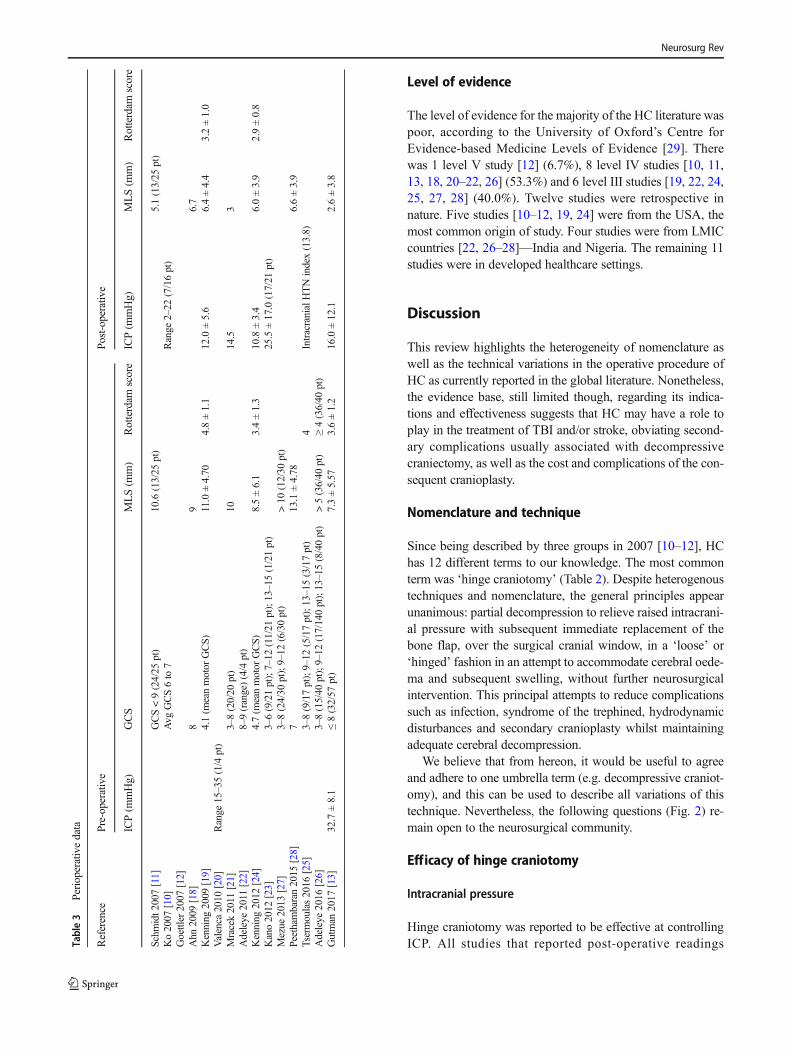
The level of evidence for the majority of the HC literature waspoor, according to the University of Oxford’s Centre forEvidence-based Medicine Levels of Evidence [29]. Therewas 1 level V study [12] (6.7%), 8 level IV studies [10, 11,13, 18, 20–22, 26] (53.3%) and 6 level III studies [19, 22, 24,25, 27, 28] (40.0%). Twelve studies were retrospective innature. Five studies [10–12, 19, 24] were from the USA, themost common origin of study. Four studies were from LMICcountries [22, 26–28]—India and Nigeria. The remaining 11studies were in developed healthcare settings.
Discussion
This review highlights the heterogeneity of nomenclature aswell as the technical variations in the operative procedure ofHC as currently reported in the global literature. Nonetheless,the evidence base, still limited though, regarding its indica-tions and effectiveness suggests that HC may have a role toplay in the treatment of TBI and/or stroke, obviating second-ary complications usually associated with decompressivecraniectomy, as well as the cost and complications of the con-sequent cranioplasty.
Nomenclature and technique
Since being described by three groups in 2007 [10–12], HChas 12 different terms to our knowledge. The most commonterm was ‘hinge craniotomy’ (Table 2). Despite heterogenoustechniques and nomenclature, the general principles appearunanimous: partial decompression to relieve raised intracrani-al pressure with subsequent immediate replacement of thebone flap, over the surgical cranial window, in a ‘loose’ or‘hinged’ fashion in an attempt to accommodate cerebral oede-ma and subsequent swelling, without further neurosurgicalintervention. This principal attempts to reduce complicationssuch as infection, syndrome of the trephined, hydrodynamicdisturbances and secondary cranioplasty whilst maintainingadequate cerebral decompression.
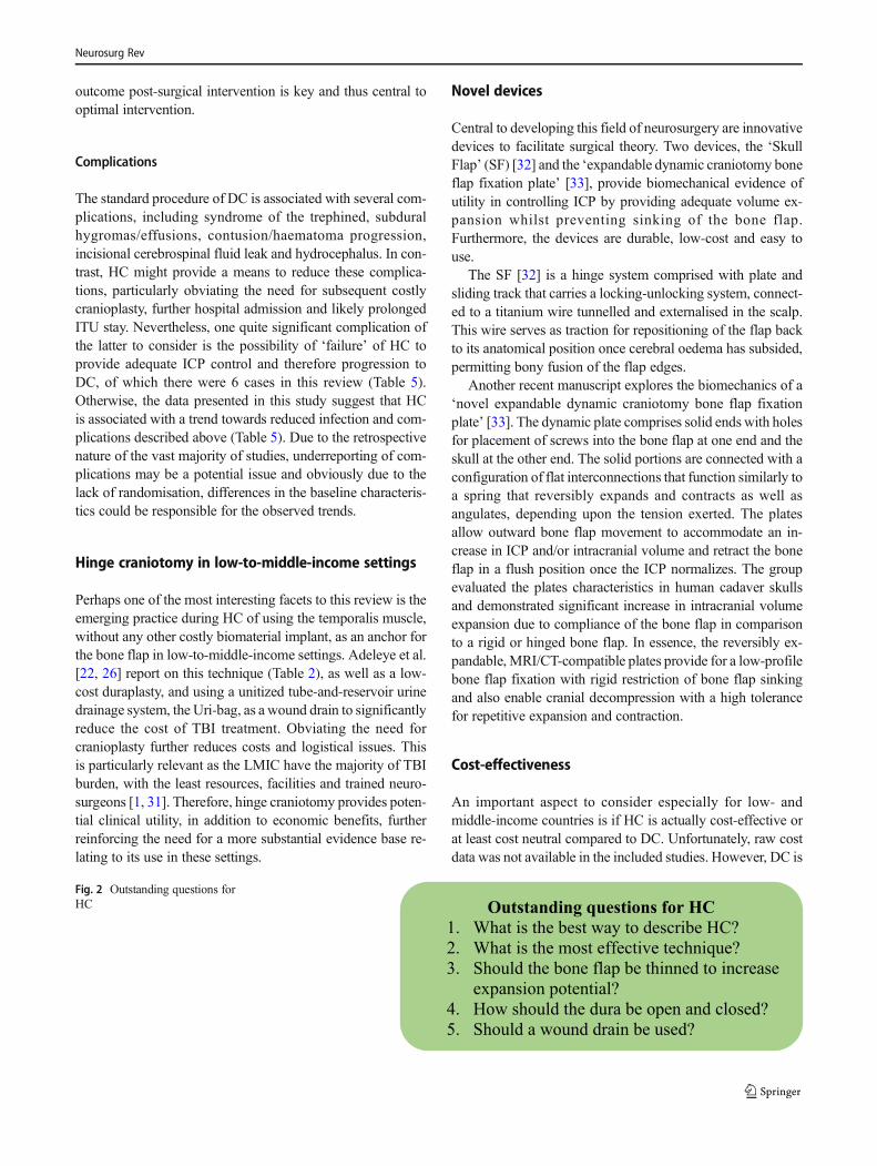
We believe that from hereon, it would be useful to agreeand adhere to one umbrella term (e.g. decompressive craniot-omy), and this can be used to describe all variations of thistechnique. Nevertheless, the following questions (Fig. 2) re-main open to the neurosurgical community.
Efficacy of hinge craniotomy
Intracranial pressure
Hinge craniotomy was reported to be effective at controllingICP. All studies that reported post-operative readingsTa
ble3
Perioperativedata
Reference
Pre-operativ
ePo
st-operativ
e
ICP(m
mHg)
GCS
MLS(m
m)
Rotterdam
score
ICP(m
mHg)
MLS(m
m)
Rotterdam
score
Schm
idt2
007[11]
GCS<9(24/25
pt)
10.6(13/25
pt)
5.1(13/25
pt)
Ko2007
[10]
Avg
GCS6to
7Range
2–22
(7/16pt)
Goettler2007
[12]
Ahn
2009
[18]
89
6.7
Kenning
2009
[19]
4.1(m
eanmotor
GCS)
11.0±4.70
4.8±1.1
12.0±5.6
6.4±4.4
3.2±1.0
Valenca
2010
[20]
Range
15–35(1/4
pt)
Mracek2011
[21]
3–8(20/20
pt)
1014.5
3Adeleye
2011
[22]
8–9(range)(4/4
pt)
Kenning
2012
[24]
4.7(m
eanmotor
GCS)
8.5±6.1
3.4±1.3
10.8±3.4
6.0±3.9
2.9±0.8
Kano2012
[23]
3–6(9/21pt);7–12
(11/21
pt);13–15(1/21pt)
25.5±17.0(17/21
pt)
Mezue
2013
[27]
3–8(24/30
pt);9–12
(6/30pt)
>10
(12/30
pt)
Peethambaran2015
[28]
713.1±4.78
6.6±3.9
Tsermoulas2016
[25]
3–8(9/17pt);9–12
(5/17pt);13–15(3/17pt)
4IntracranialHTNindex(13.8)
Adeleye
2016
[26]
3–8(15/40
pt);9–12
(17/140pt);13–15(8/40pt)
>5(36/40
pt)
≥4(36/40
pt)
Gutman
2017
[13]
32.7±8.1
≤8(32/57
pt)
7.3±5.57
3.6±1.2
16.0±12.1
2.6±3.8
Neurosurg Rev
demonstrated a decrease in ICP and associated reduction inmidline shift (Table 3). The studies comparing HC to DCsuggest that HC is at least as effective as DC in this respect(Table 6). Kano et al. [23] compared HC with DC in TBI andstroke, and summarising their data, averred ‘the hinge tech-nique with ICP monitoring was effective and safe for the man-agement for head trauma and stroke’. Furthermore, Kenninget al. [19, 24] suggested that ‘hinge craniotomy was at least asgood as decompressive craniectomy in providing post-operative ICP control’. Furthermore, out of 283 patients un-dergoing HC, only 9 patients (3.2%) required subsequent DC,suggesting that adequate cerebral decompression wasachieved by HC alone. In the RESCUEicp trial, which en-rolled patients with severe TBI and ICP raised and refractoryto medical management, of the patients who were randomisedand received a DC (n = 187), 12 required a barbiturate infu-sion post-DC due to ongoing issues with ICP control (6.4%).Obviously, the patients included in the studies of the presentscoping review are not directly comparable to theRESCUEicp population but this figure can give an indicationas to the proportion of patients with refractory ICP despiteDC.
This concept is further reinforced by a biomechanical studyin human cadaver skulls [30], which compared DC to HC and‘dynamic decompressive craniotomy’ and the effect on ICP
after abrupt increase in intracranial volume. They found thatboth the dynamic craniotomy and the HC techniques providedsignificant control of ICP during 120 ml increase in intracra-nial volume as compared with craniotomy rigidly fixed withplates (4.86 mmHg, 8.36 mmHg, 44.84 mmHg, respectively).
Clinical and functional outcomes
Central to the clinical management of raised ICP in TBI orstroke is the patient’s quality of life after receiving treatmentfor the injury. This was highlighted in the recent RESCUEicptrial [4] which demonstrated that more patients with refractoryICP (> 25 mmHg) who underwent decompressivecraniectomy had decreased mortality but increased disabilitywhen compared with medical therapy alone. In RESCUEicp[4], the survival of the DC patients at 6 months was 73.1%.The overall survival of patients undergoing HC was 74.6% (n= 211) (Table 4) and thus very similar to the RESCUEicpmortality, although it is important to appreciate thatRESCUEicp was looking at secondary DC. The studies in-cluded in this manuscript had limited long-term functionaloutcome reported (Tables 4 and 6). Furthermore, the presenceof extracranial injuries or injuries of different severity makesdirect summary of outcomes difficult. Lastly, as the evidenceinforming treatment strategies in TBI is developing, morbidity
Table 4 Functional outcome data. GOS: ‘Good’ = 4–5; ‘Poor’ = 1–3. mRS: ‘Good’ = 0–2; ‘Moderate’ = 3–4; ‘Poor’ = 5–6
Reference Survival Functional outcome at discharge unless otherwise stated Length offollow-up(months)n, % of total pt GOS mRS
Schmidt 2007 [11] 13, 52% NR NR NR
Ko 2007 [10] 14, 87.5% NR NR 10
Goettler 2007 [12] 2, 66.6% NR NR NR
Ahn 2009 [18] 6, 85.7% Good: 2 (28.6%); Poor: 5 (71.4%) NR NR
Kenning 2009 [19] 15, 75% NR NR NR
Valenca 2010 [20] 4, 100% NR NR 2–14
Mracek 2011 [21] 16, 80% Good: 8 (40%); Poor: 12 (60%) NR Up to 6
Adeleye 2011 [22] 4, 100% GOSE ‘near normal’ NR 3–18
Kenning 2012 [24] 5, 56% 3.6 ± 0.6 (at 1–3 months) 2.8 ± 1.1 (at 1–3 months) 12
Kano 2012 [23] 19, 90.4% Good: 3 (43%); Poor: 4 (57%) Good: 1 (7%); Moderate: 9 (64%); Poor: 4 (29%) 13.7 ± 11.2(18 cases)
Mezue 2013 [27] 24, 80% Good: 16 (53%); Poor: 14 (47%) NR NR
Peethambaran 2015 [28] 3, 30% NR NR 6
Tsermoulas 2016 [25] 14, 82.0% Good: 11 (64.7%); Poor: 6 (35.3%) NR 6
Adeleye 2016 [26] 28, 70% Good: 27 (67.5%); Poor: 13 (32.5%) NR 11
Gutman 2017 [13] 44, 77.2% NR Pre-discharge:Good: 31 (54.3%);Moderate: 10 (17.5%);Poor: 14 (24.6%);NA: 2 (3.5%)
Post-discharge:Good: 22 (38.6%);Moderate: 3 (5.3%);Poor: 13 (22.8%);NA: 10 (17.5%)
NR
GOS: ‘Good’ = 4–5; ‘Poor’ = 1–3. mRS: ‘Good’ = 0–2; ‘Moderate’ = 3–4; ‘Poor’ = 5–6. NR = not reported by authors.
Neurosurg Rev
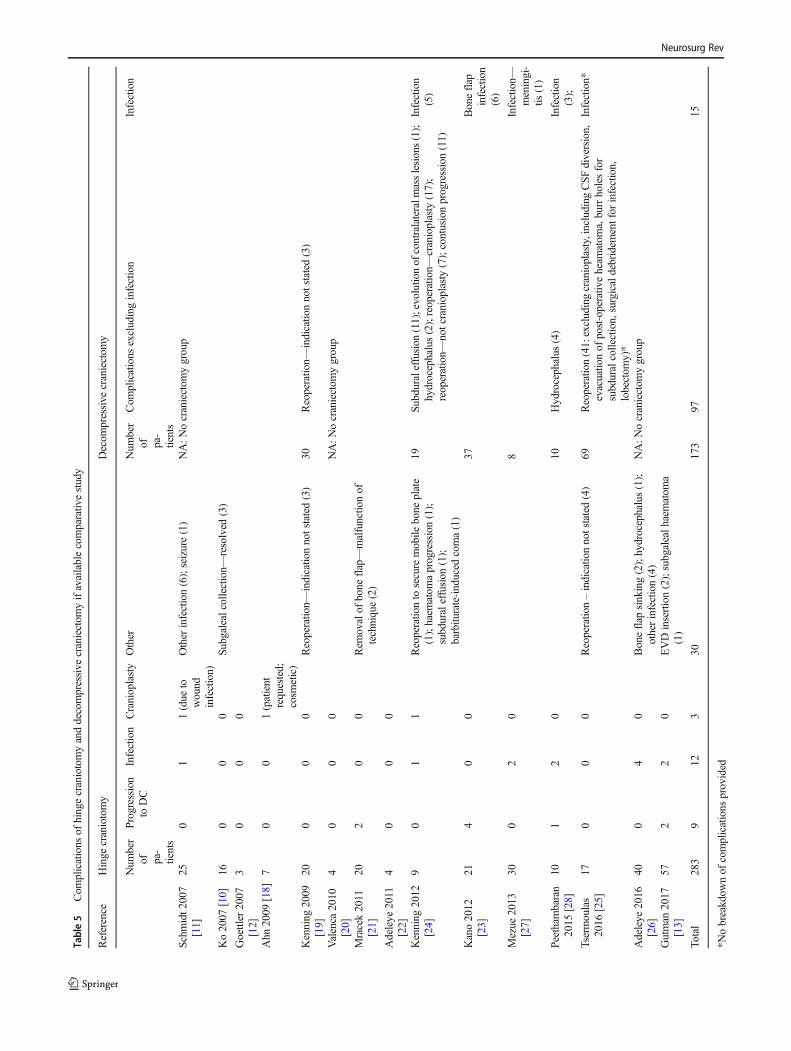
Table5
Com
plications
ofhingecraniotomyanddecompressive
craniectom
yifavailablecomparativ
estudy
Reference
Hinge
craniotomy
Decom
pressive
craniectom
y
Num
ber
of pa-
tients
Progression
toDC
Infection
Cranioplasty
Other
Num
ber
of pa-
tients
Com
plications
excludinginfection
Infection
Schm
idt2
007
[11]
250
11(due
towound
infection)
Other
infection(6);seizure(1)
NA:N
ocraniectom
ygroup
Ko2007
[10]
160
00
Subgalealcollection—
resolved
(3)
Goettler
2007
[12]
30
00
Ahn
2009
[18]
70
01(patient
requested;
cosm
etic)
Kenning
2009
[19]
200
00
Reoperatio
n—indicatio
nnotstated(3)
30Reoperatio
n—indicatio
nnotstated(3)
Valenca
2010
[20]
40
00
NA:N
ocraniectom
ygroup
Mracek2011
[21]
202
00
Rem
ovalof
bone
flap—malfunctio
nof
technique(2)
Adeleye
2011
[22]
40
00
Kenning
2012
[24]
90
11
Reoperatio
nto
secure
mobile
bone
plate
(1);haem
atom
aprogression(1);
subduraleffusion
(1);
barbitu
rate-induced
coma(1)
19Su
bduraleffusion
(11);evolutio
nof
contralateralm
asslesions(1);
hydrocephalus(2);reoperation—
cranioplasty
(17);
reoperation—
notcranioplasty(7);contusionprogression(11)
Infection
(5)
Kano2012
[23]
214
00
37Boneflap
infection
(6)
Mezue
2013
[27]
300
20
8Infection—
meningi-
tis(1)
Peethambaran
2015
[28]
101
20
10Hydrocephalus
(4)
Infection
(3);
Tsermoulas
2016
[25]
170
00
Reoperatio
n–indicatio
nnotstated(4)
69Reoperatio
n(41:excludingcranioplasty,including
CSF
diversion,
evacuatio
nof
post-operativ
eheam
atom
a,burrholesfor
subduralcollection,surgicaldebridem
entfor
infection,
lobectom
y)*
Infection*
Adeleye
2016
[26]
400
40
Boneflap
sinking(2);hydrocephalus(1);
otherinfection(4)
NA:N
ocraniectom
ygroup
Gutman
2017
[13]
572
20
EVDinsertion(2);subgalealh
aematom
a(1)
Total
283
912
330
173
9715
*Nobreakdow
nof
complications
provided
Neurosurg Rev
outcome post-surgical intervention is key and thus central tooptimal intervention.
Complications
The standard procedure of DC is associated with several com-plications, including syndrome of the trephined, subduralhygromas/effusions, contusion/haematoma progression,incisional cerebrospinal fluid leak and hydrocephalus. In con-trast, HC might provide a means to reduce these complica-tions, particularly obviating the need for subsequent costlycranioplasty, further hospital admission and likely prolongedITU stay. Nevertheless, one quite significant complication ofthe latter to consider is the possibility of ‘failure’ of HC toprovide adequate ICP control and therefore progression toDC, of which there were 6 cases in this review (Table 5).Otherwise, the data presented in this study suggest that HCis associated with a trend towards reduced infection and com-plications described above (Table 5). Due to the retrospectivenature of the vast majority of studies, underreporting of com-plications may be a potential issue and obviously due to thelack of randomisation, differences in the baseline characteris-tics could be responsible for the observed trends.
Hinge craniotomy in low-to-middle-income settings
Perhaps one of the most interesting facets to this review is theemerging practice during HC of using the temporalis muscle,without any other costly biomaterial implant, as an anchor forthe bone flap in low-to-middle-income settings. Adeleye et al.[22, 26] report on this technique (Table 2), as well as a low-cost duraplasty, and using a unitized tube-and-reservoir urinedrainage system, the Uri-bag, as a wound drain to significantlyreduce the cost of TBI treatment. Obviating the need forcranioplasty further reduces costs and logistical issues. Thisis particularly relevant as the LMIC have the majority of TBIburden, with the least resources, facilities and trained neuro-surgeons [1, 31]. Therefore, hinge craniotomy provides poten-tial clinical utility, in addition to economic benefits, furtherreinforcing the need for a more substantial evidence base re-lating to its use in these settings.
Novel devices
Central to developing this field of neurosurgery are innovativedevices to facilitate surgical theory. Two devices, the ‘SkullFlap’ (SF) [32] and the ‘expandable dynamic craniotomy boneflap fixation plate’ [33], provide biomechanical evidence ofutility in controlling ICP by providing adequate volume ex-pansion whilst preventing sinking of the bone flap.Furthermore, the devices are durable, low-cost and easy touse.
The SF [32] is a hinge system comprised with plate andsliding track that carries a locking-unlocking system, connect-ed to a titanium wire tunnelled and externalised in the scalp.This wire serves as traction for repositioning of the flap backto its anatomical position once cerebral oedema has subsided,permitting bony fusion of the flap edges.
Another recent manuscript explores the biomechanics of a‘novel expandable dynamic craniotomy bone flap fixationplate’ [33]. The dynamic plate comprises solid ends with holesfor placement of screws into the bone flap at one end and theskull at the other end. The solid portions are connected with aconfiguration of flat interconnections that function similarly toa spring that reversibly expands and contracts as well asangulates, depending upon the tension exerted. The platesallow outward bone flap movement to accommodate an in-crease in ICP and/or intracranial volume and retract the boneflap in a flush position once the ICP normalizes. The groupevaluated the plates characteristics in human cadaver skullsand demonstrated significant increase in intracranial volumeexpansion due to compliance of the bone flap in comparisonto a rigid or hinged bone flap. In essence, the reversibly ex-pandable, MRI/CT-compatible plates provide for a low-profilebone flap fixation with rigid restriction of bone flap sinkingand also enable cranial decompression with a high tolerancefor repetitive expansion and contraction.
Cost-effectiveness
An important aspect to consider especially for low- andmiddle-income countries is if HC is actually cost-effective orat least cost neutral compared to DC. Unfortunately, raw costdata was not available in the included studies. However, DC is
Outstanding questions for HC1. What is the best way to describe HC?
2. What is the most effective technique?
3. Should the bone flap be thinned to increase
expansion potential?
4. How should the dura be open and closed?
5. Should a wound drain be used?
Fig. 2 Outstanding questions forHC
Neurosurg Rev
thought to be an expensive procedure with a mean patient costof over US$ 20,000 when performed in a developing country[34]. HC is potentially a cheaper alternative due to autologoustissues being utilised and reducing the need for a second op-eration for a cranioplasty [26]. A formal cost-effectivenessanalysis evaluating the many different factors that influencecost needs to be undertaken, which is one of the future aims ofour neurotrauma study group at the University of Cambridge.
Limitations of the present study
The lack of robust comparative HC vs DC data, the combinedindications (TBI and stroke) for HC in some of the reports,heterogenous pre-operative and radiological metrics, paucityof pre-operative ICP monitoring, short-term follow-up andpoor reporting of long-term functional outcomes makes abso-lute conclusion difficult. Furthermore, GOS recorded at dis-charge is not necessarily a true representative of long-termoutcomes, and the paucity of robust follow-up investigatingassociated morbidity, a key metric, in most reports is also ofnote. Additionally, mRS is the most commonly used outcomescale in the field of stroke but many of the included studiesused the GOS to assess outcomes in the TBI but also strokepatients. The two scales examine slightly different aspects offunctional outcome but the GlasgowOutcome Scale correlateswell with the mRS in patients with stroke [35].
Developing the evidence base
To continue developing the evidence base for HC, we wouldadvocate following the IDEALMethodology [36]. This is a 5-stage description of the surgical development process, a cru-cial tool for systematic evaluation of surgical innovation andthat is instrumental for achieving improved design, conductand reporting of surgical research. Currently, HC is betweenstage IIb and III ‘Exploration’ and ‘Assessment’, i.e. the tech-nique is stable, has been replicated by numerous study groupsand there is some literature demonstrating comparison toexisting practice (DC).
It must be appreciated that HC cannot, as yet, be consideredan alternative to all the DCs, but rather an alternative to pri-mary DC, not to secondary DC. Whereas HC has been per-formed most often for persistent brain swelling after evacua-tion of intracranial hematoma, DC has been performed mostoften as a part of second- or third-tier therapy as in DECRA[3] and RESCUEicp [4] trials for patients with medically re-fractory intracranial hypertension. In such cases, it is unlikelythat a HC will be performed instead of DC.
To further develop HC, additional evaluation of the tech-nique prospectively and co-operatively may help mature con-sensus over definition, quality and indications. Ultimately, aninternational effort, with a multi-centre randomised controlledtrial, with participation from low- and middle-incomeTa
ble6
Sum
marytableof
studiescomparing
HCto
DC
Reference
Pt(n)
Indicatio
nOutcomes
TBI
Stroke
ICP(m
mHg)
MeanMLS(m
m)
GOS
mRS
Survivaln(%
)
HC
DC
HC
DC
HC
DC
HC
DC
HC
DC
HC
DC
HC
DC
Kenning
2009
[19]
5012
188
1212.1±2.6
15.0±6.3
6.4±4.4
5.5±4.6
Not
stated
15(75)
21(70)
Kenning
2012
[24]
28–
919
10.8±3.4
11.9±3.5
6.0±3.9
5.3±5.4
NA
2.8±1.1at30–90days
post-op
4.4±0.9at30–90
days
post-op
5(56)
17(89)
Kano2012
[23]
587
1914
1825.5±17.0
Not
reported
Good:
3(50%
);Po
or:3
(50%
)Good:
4(21%
);Po
or:1
5(79%
)Good:
1(7%);
Moderate:9(64%
);Po
or:4
(29%
)
Good:
1(6%);
Moderate:6(33%
);Po
or:11(61%
)
19(94)
*
Mezue
2013
[27]
3830
8–
Not
reported
Good:
16(53%
);Po
or:1
4(47%
)Good:
2(25%
);Po
or:6
(75%
)24
(80)
6(75)
Peethambaran2015
[28]
2010
91
Not
reported
6.6±3.9
6.0±2.1
Not
stated
3(30)
5(50)
Tsermoulas2016
[25]
8617
69–
13.8^
16.6^
Not
reported
Good:
11(64.7%
);Po
or:6
(35.3%
)Good:
28(40.1%
);Po
or:4
1(59.9%
)NA
14(82)
43(62)
^Expressed
asintracranialhypertension
index
*Unableto
extractm
ortalityas
notexplicitlystated,onlygroupedas
mRS5–6
Neurosurg Rev
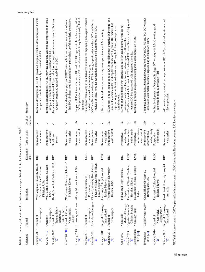
Table7
Summaryof
evidence.L
evelof
evidence
asperOxfordCentreforEvidenceMedicine2009
29
Reference
Journal
Institu
tion
Economic
status
Type
ofstudy
Levelof
evidence
Sum
mary
Schm
idt2
007
[11]
Journalo
fNeurosurgery
WestV
irginiaUniversity
Health
SciencesCenterCharleston
Division,USA
HIC
Retrospectiv
ecase
series
IVTechnicald
escriptio
nof
HC;H
Cprovided
adequatecerebraldecompression
Ism
all
sample;no
complications
usually
associated
with
DC.
Ko2007
[10]
Operativ
eNeurosurgery
WeillCornellMedicalCollege,
USA
HIC
Retrospectiv
ecase
series
IVTechnicaldescriptio
nof
HC;H
Cprovided
adequatecerebraldecompression
insm
all
sample;no
complications
usually
associated
with
DC.
Goettler
2007
[12]
Journalo
fTraum
a-Injury
Infection&
CriticalCare.
Brody
School
ofMedicine,USA
HIC
Smallcase
series
VTechnicald
escriptio
nof
TF;
provides
less
decompression
volumethan
DCbutw
asadequate;suggested
reducedmorbidity
vsDC.
Ahn
2009
[18]
Journalo
fKorean
Neurosurgical
Society
Wonkw
angUniversity
Schoolo
fMedicine,Korea
HIC
Retrospectiv
ecase
series
IVTechnicald
escriptio
n;perhapsISRFC
betterableto
accommodatecerebralodem
athan
HC;o
bviatesneed
forcranioplasty;p
articularly
useful
inelderlypopulatio
n.
Kenning
2009
[19]
NeurosurgicalFo
cus
AlbanyMedicalCentre,USA
HIC
Retrospectiv
ecase
control
IIIb
Com
paredICPoutcom
esbetweenHCandDC:H
Cappearsto
beatleastasgood
asDCinprovidingpost-operativ
eICPcontroland
results
inequivalentearlyclinical
outcom
es.
Valenca
2010
[20]
Journalo
fNeurosurgery
FederalU
niversity
ofPernam
buco,B
razil
UMIC
Retrospectiv
ecase
series
IV‘In-window’craniotomyisan
alternativesolutio
nfordeployingautologous
material
andobviates
need
forsecondarysurgery.
Mracek2011
[21]
ActaNeurochirurgica
Charles
University
Hospitaland
Faculty
ofMedicinein
Pilsen,
Czech
Republic
HIC
Retrospectiv
ecase
series
IVODCiseffectiveatreducing
ICPin
asubgroup
ofpatientswhere
DCwould
betoo
radical;obviates
need
forreoperationandassociated
complications
ofDC.
Adeleye
2011
[22]
SurgicalNeurology
International
University
College
Hospital,
Ibadan,N
igeria
LMIC
Retrospectiv
ecase
series
IVEffectiv
ecerebraldecompression
usingautologous
tissuein
LMIC
setting
Kenning
2012
[24]
Journalo
fNeurosurgery
Thomas
JeffersonUniversity
Hospital,USA
HIC
Retrospectiv
ecase
control
IIIa
HCappearstobe
atleastasgood
asDCinprovidingpost-operativ
eICPcontrolata
similartherapeutic
index;
in-hospitalm
ortalitywas
higher
inHCpatientsbut
superior
long-term
functio
nalo
utcomes;H
Cmay
help
limitpost-operativ
ecomplications.
Kano2012
[23]
Neurologia
Medico-Chirurgic
FukayaRed
Cross
Hospital,
Japan
HIC
Prospectiv
ecohortstudy
IIIa
HCwith
ICPmonito
ring
was
effectiveandsafe
forhead
traumaor
stroke;n
otassociated
with
bone
flap
infection;
follo
wup
of13–40months.
Mezue
2013
[27]
NigerianJournalo
fClin
icalPractice
University
ofNigeriaTeaching
Hospital,Nigeria
LMIC
Retrospectiv
ecohortstudy
IIIa
HCsufficient
andableto
controlICPin
selected
TBIcases.Severehead
injury
still
requires
DCbutassociatedwith
increasedmortality.
Peethambaran
2015
[28]
Neurology
India
Governm
entM
edicalCollege,
India
LMIC
Prospective
observational
case
control
IIIb
Techniqueprovides
adequateandcomparabledecompression
toDC
Tsermoulas
2016
[25]
World
Neurosurgery
Queen
ElizabethHospital,
Birmingham
,UK
HIC
Retrospectiv
eobservational
cohortstudy
IIIb
Com
paredreoperations,functionaloutcomeandICPinDC,R
CandFC
;DCwas
not
associated
with
betteroutcom
es;replace
flap
ifconditionsallow
Adeleye
2016
[26]
Journalo
fNeurological
Surgery
PartA
University
College
Hospital,
Ibadan,N
igeria
LMIC
Prospective
cohortstudy
IVFu
rtherdevelopm
ento
ftemporalis
hingeDCtechniquein
LMIC
setting;g
ood
outcom
esin
mild
tomoderateTBI
Gutman
2017
[13]
SurgicalNeurology
International
GoldCoastUniversity
Hospital,
Australia
HIC
Retrospectiv
ecase
series
IVFA
Cprovides
symmetricald
ecom
pression
vsHC;FACprovided
adequatecerebral
decompression
HIC
high-incom
ecountry,UMIC
upper-middle-incomecountry,LM
IClow-to-middle-incomecountry,LIClow-incom
ecountry
Neurosurg Rev
countries is required. The trial could compare HC to DC withcriteria for progression from HC to DC in selected cases. Inaddition, ICP monitoring, if already used clinically, would aidmeaningful comparison. Importantly, such a study would aimto compare long-term functional outcomes and surgical mor-bidity (Table 7).
Conclusion
Hinge craniotomy has a potential role in the surgical manage-ment of TBI/stroke, yielding adequate cerebral decompressionin the majority of reported cases, a reduction in complicationsand potentially offers substantial economic savings (both op-erative costs and the cost of living with significant morbidity).It is likely that HC offers an intermediate intervention betweentreatment-refractive medical therapy and traditional decom-pressive craniectomy. Future work should aim to facilitate aglobal consensus about HC and its utility as treatment, ulti-mately paving the way for a randomised controlled trial.
Acknowledgements Many thanks to Dr Veronica Phillips, MedicalLibrary, University of Cambridge for her assistance with the searchstrategy.
Funding information This research was commissioned by the NationalInstitute for Health Research (NIHR) Global Health Research Group onNeurotrauma (16/137/105) using UK aid from the UK Government.Aswin Chari is supported by a Great Ormond Street Hospital (GOSH)Children’s Charity Surgeon Scientist Fellowship and the NationalInstitute for Health Research GOSH Biomedical Research Centre. PeterHutchinson is supported by a Research Professorship from the NationalInstitute for Health Research (NIHR), the NIHR Cambridge BiomedicalResearch Centre, a European Union Seventh Framework Program grant(CENTER-TBI; grant no. 602150) and the Royal College of Surgeons ofEngland. Angelos Kolias is supported by a Clinical Lectureship, Schoolof Clinical Medicine, University of Cambridge.
Compliance with ethical standards
Conflict of interest Franco Servadei has received personal fees fromTakeda Pharmaceutical Company, grants and personal fees from IntegraLifeSciences and grants and personal fees from Finceramica SpA. Therest of the authors declare that they have no conflict of interest related tothis manuscript.
Ethical approval Ethical approval is not applicable for a scoping review.
Informed consent Informed consent is not applicable for a scopingreview.
Disclaimer The views expressed in this publication are those of theauthor(s) and not necessarily those of the NIHR or the Department ofHealth and Social Care.
Open Access This article is distributed under the terms of the CreativeCommons At t r ibut ion 4 .0 In te rna t ional License (h t tp : / /creativecommons.org/licenses/by/4.0/), which permits unrestricted use,distribution, and reproduction in any medium, provided you give
appropriate credit to the original author(s) and the source, provide a linkto the Creative Commons license, and indicate if changes were made.
References
1. Dewan, M. C. et al. (2018) Estimating the global incidence oftraumatic brain injury. J Neurosurg 1–18. https://doi.org/10.3171/2017.10.jns17352
2. Kolias AG et al (2018) The current status of decompressivecraniectomy in traumatic brain injury.Curr Trauma Rep 4:326–332
3. Cooper JD et al (2011) Decompressive craniectomy in diffuse trau-matic brain injury. New Engl J Med 364:1493–1502
4. Hutchinson PJ et al (2016) Trial of decompressive craniectomy fortraumatic intracranial hypertension. New Engl J Med 375:1119–1130
5. Vahedi K et al (2007) Sequential-design, multicenter, randomized,controlled trial of early decompressive craniectomy in malignantmiddle cerebral artery infarction (DECIMAL Trial). Stroke 38:2506–2517
6. Jüttler E et al (2007) Decompressive surgery for the treatment ofmalignant infarction of the middle cerebral artery (DESTINY).Stroke 38:2518–2525
7. Hofmeijer J et al (2009) Surgical decompression for space-occupying cerebral infarction (the Hemicraniectomy After MiddleCerebral Artery infarction with Life-threatening Edema Trial[HAMLET]): a multicentre, open, randomised trial. LancetNeurol 8:326–333
8. Cruz-Flores S, Berge E, Whittle IR (2012) Surgical decompressionfor cerebral oedema in acute ischaemic stroke. Cochrane Db SystRev. https://doi.org/10.1002/14651858.cd003435.pub2
9. Kolias AG, Kirkpatrick PJ, Hutchinson PJ (2013) Decompressivecraniectomy: past, present and future. Nat Rev Neurol 9:405
10. Ko K, Segan S (2007) In situ hinge craniectomy. Oper Neurosurg60:ONS-255–ONS-259
11. Schmidt JH, Reyes BJ, Fischer R, Flaherty SK (2007) Use of hingecraniotomy for cerebral decompression. J Neurosurg 107:678–682
12. Goettler CE, Tucci KA (2007) Decreasing the morbidity of decom-pressive craniectomy: the Tucci Flap. J Trauma Acute Care 62:777
13. Gutman M, How E, Withers T (2017) The floating anchored crani-otomy. Surg Neurol Int 8:130
14. Armstrong R, Hall BJ, Doyle J, Waters E (2011) ‘Scoping thescope’ of a cochrane review. J Public Health 33:147–150
15. Munn Z et al (2018) Systematic review or scoping review?Guidance for authors when choosing between a systematic or scop-ing review approach. BMC Med Res Methodol 18(143)
16. Munn Z, Stern C, Aromataris E, Lockwood C, Jordan Z (2018)What kind of systematic review should I conduct? A proposedtypology and guidance for systematic reviewers in the medicaland health sciences. BMC Med Res Methodol 18(5)
17. Tricco AC et al (2018) PRISMA extension for Scoping Reviews(PRISMA-ScR): checklist and explanation. Ann InternMed. https://doi.org/10.7326/m18-0850
18. Ahn D-H, Kim D-W, Kang S-D (2009) In situ floating resincranioplasty for cerebral decompression. J Korean Neurosurg S46:417–420
19. Kenning TJ, Gandhi RH, German JW (2009) A comparison ofhinge craniotomy and decompressive craniectomy for the treatmentof malignant intracranial hypertension: early clinical and radio-graphic analysis. Neurosurg Focus 26:E6
20. Valença MM, Martins C, da Silva J (2010) “In-window” cranioto-my and “bridgelike” duraplasty: an alternative to decompressivehemicraniectomy. J Neurosurg 113:982–989
Neurosurg Rev
21. Mracek J, ChocM, Mork J, Vacek P, Mracek Z (2011) Osteoplasticdecompressive craniotomy—an alternative to decompressivecraniectomy. Acta Neurochir 153:2259
22. Adeleye AO, Azeez A (2011) Decompressive craniectomy boneflap hinged on the temporalis muscle: a new inexpensive use foran old neurosurgical technique. Surg Neurol Int 2:150
23. Kano T, Kurosaki S, Wada H (2012) Retrospective analysis ofhinge technique for head trauma or stroke. Neurol Med-Chir 52:816–821
24. Kenning TJ et al (2012) Cranial decompression for the treatment ofmalignant intracranial hypertension after ischemic cerebral infarc-tion: decompressive craniectomy and hinge craniotomy. JNeurosurg 116:1289–1298
25. Tsermoulas G et al (2016) Surgery for acute subdural hematoma:replace or remove the bone flap? World Neurosurg 88:569–575
26. Adeleye AO (2016) Clinical and radiologic outcome of a less inva-sive, low-cost surgical technique of osteoplastic decompressivecraniectomy. J Neurol Surg Part Central European Neurosurg 77:167–175
27. Mezue W, Ndubuisi C, Ohaegbulam S, Chikani M, Erechukwu U(2013) Cranial bony decompressions in the management of headinjuries: decompressive craniotomy or craniectomy? Niger J ClinPract 16:343–347
28. (2015) Four-quadrant osteoplastic decompressive craniotomy: anovel technique for refractory intracranial hypertension - A pilotstudy. Neurol India 63:895–902
29. (2009) Oxford Centre for Evidence-based Medicine – Levels ofEvidence (March 2009)
30. Khanna R, Ferrara L (2016) Dynamic telescopic craniotomy: acadaveric study of a novel device and technique. J Neurosurg125:674–682
31. Dewan MC, et al. (2018) Global neurosurgery: the current capacityand deficit in the provision of essential neurosurgical care.Executive Summary of the Global Neurosurgery Initiative at theProgram in Global Surgery and Social Change. J Neurosurg 1–10.https://doi.org/10.3171/2017.11.jns171500
32. Chibbaro S et al (2013) The ‘Skull Flap’ a new conceived device fordecompressive craniectomy/cranioplasty: feasibility study on ca-daver specimen. J Neurosci Rural Pract 4:283–287
33. Khanna R, Ferrara L, Khanna S (2019) Biomechanics of a novelreversibly expandable dynamic craniotomy bone flap fixation plate.J Neurosurg:1–8. https://doi.org/10.3171/2018.8.jns172614
34. Badke G et al (2018) Analysis of direct costs of decompressivecraniectomy in victims of traumatic brain injury. ArqNeuropsiquiatr 76(4):257–264
35. Kasner SE (2006) Clinical interpretation and use of stroke scales.Lancet Neurol 5(7):603–612
36. McCulloch P et al (2009) No surgical innovation without evalua-tion: the IDEAL recommendations. Lancet 374:1105–1112
Publisher’s note Springer Nature remains neutral with regard to juris-dictional claims in published maps and institutional affiliations.
Neurosurg Rev