Embed Size (px)
DESCRIPTION
Higher Processing of Visual Information: Lecture I --- April 2, 2007 by Mu-ming Poo Overview of the Mammalian Visual System Structure of Lateral Geniculate Nucleus (LGN) LGN Receptive Fields Retinotopic Maps. Three subcortical areas in the visual pathway - PowerPoint PPT Presentation
Citation preview

Higher Processing of Visual Information: Lecture I
--- April 2, 2007 by Mu-ming Poo
1. Overview of the Mammalian Visual System
2. Structure of Lateral Geniculate Nucleus (LGN)
3. LGN Receptive Fields
4. Retinotopic Maps
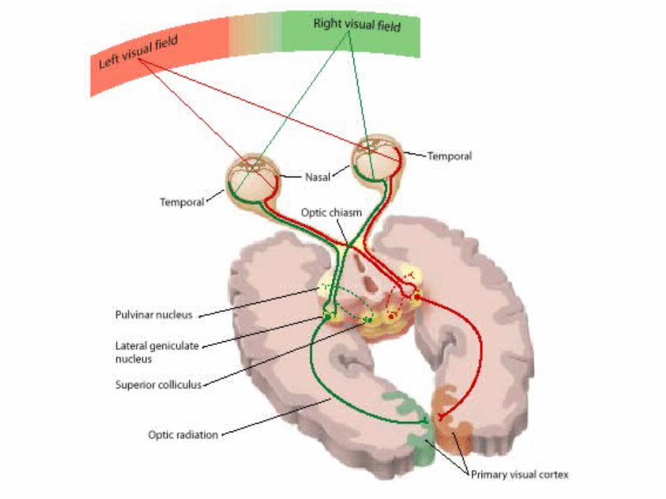
Three subcortical areas in the visual pathway
Pretectal area, superior colliculus, and lateral geniculate nucleus (LGN)
Pretectal area mediates pupillary light reflexRetina – pretectal area – Edinger- Westphal nuclei (on both sides) – IIIrd cranial nerve – pupillary constrictor muscles.
Superior colliculus controls saccadic eye movements coordinates visual, somatic and auditory information and adjusts movement
of the head and eyes towards a stimulus 1. Superior colliculus – brain stem – eye muscles (oculomotor reflex) 2. Superior colliculus – tectospinal and tectopontine tracts – head and neck
muscles
Lateral Geniculate Nucleus (a subregion of the thalamus) relays visual information from retina to cortex
-

Projection from retina to LGNfixation point
fovea
• Nasal RGC: axons crossover at optic chiasm, project to contralateral LGN
• Temporal RGC: axons stay on the same side (ipsilateral)
• Left visual field: right LGN, right V1
• Right visual field: left LGN, left V1
1-6: lesion that produce distinct visual defects

Structure of LGN
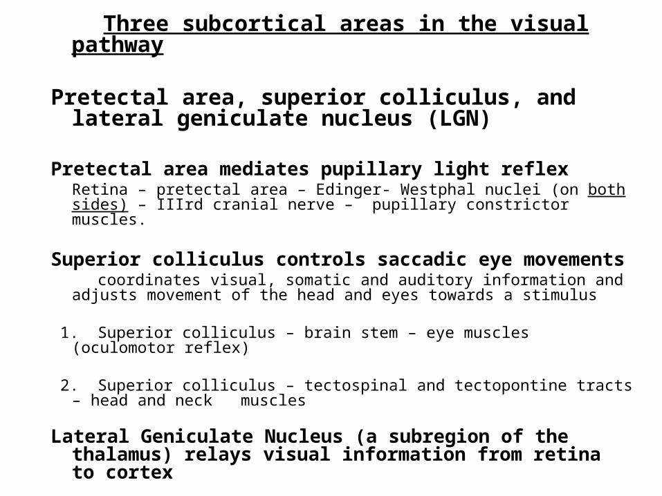
Layers 1,3,6 receive contralateral input and Layers 2,3,5 receive ipsilateral input.
Each LGN serves the contralateral visual field. The retinotopic maps from two eyes are in register across the layers. (Axons from RGCs responding to the same visual field innervate LGN cells that are aligned vertically across the layers)

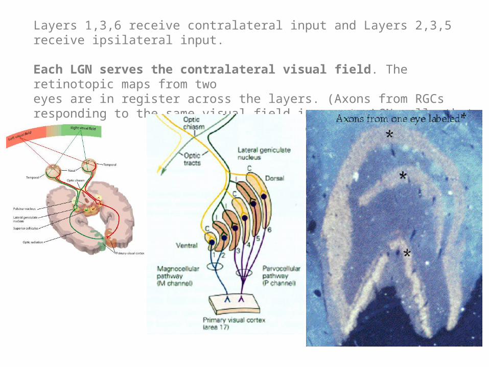
Magnocellular layers (motion) Layer 1 & 2, large cells, color blind, low
spatial resolution (large RF), high temporal resolution (good for processing motion stimuli), receive inputs from M type RGC cells.
Parvocellular layers (form and color):
Layer 3 to 6, small cells, color sensitive, high spatial resolution (small RF), low temporal resolution (does not see fast flickers of light). receive inputs from P type RGC cells.
Interlaminar koniocellular (K) Layers between each of the M and P layers. K cells are functionally and neurochemically distinct from M and P cells and provide a third channel to the visual cortex, receive inputs from blue/yellow RGCs.
Layers of LGN

Lesion studies
(after selective lesion)
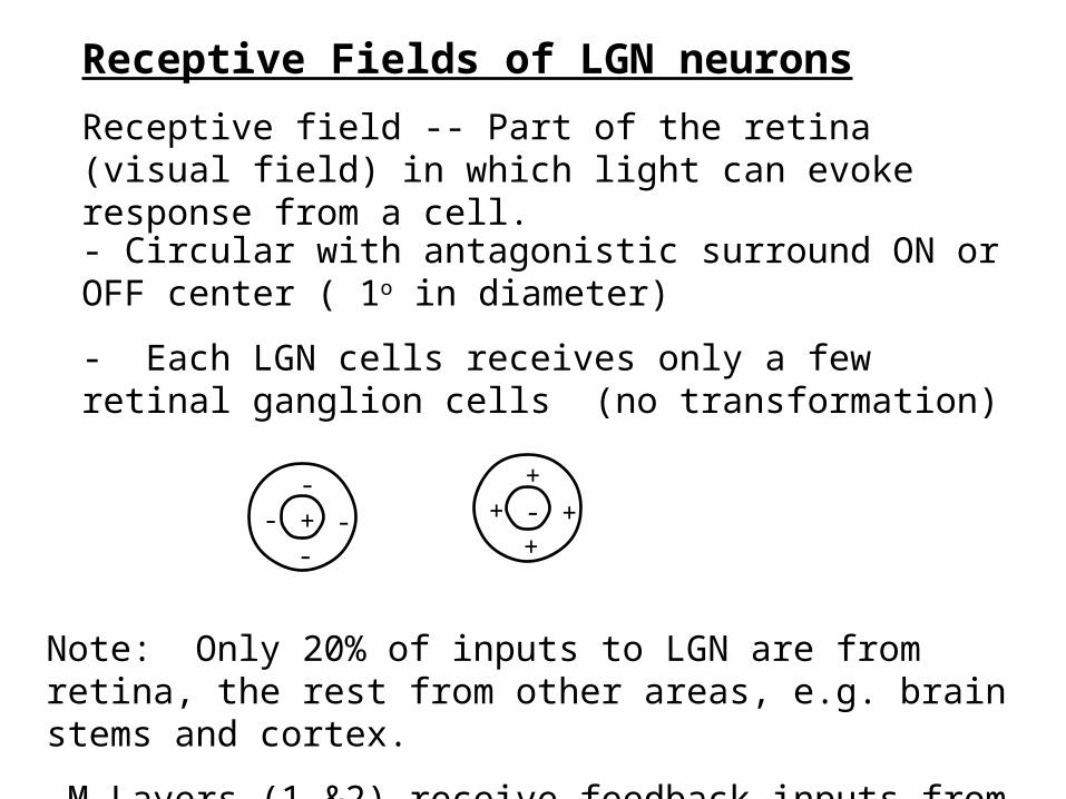
Receptive Fields of LGN neurons
Receptive field -- Part of the retina (visual field) in which light can evoke response from a cell.
+-
-- - -
+
++ +
- Circular with antagonistic surround ON or OFF center ( 1o in diameter)
- Each LGN cells receives only a few retinal ganglion cells (no transformation)
Note: Only 20% of inputs to LGN are from retina, the rest from other areas, e.g. brain stems and cortex.
-M Layers (1 &2) receive feedback inputs from extrastriate cortex
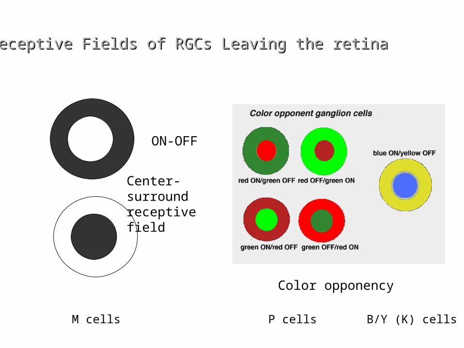
Receptive Fields of RGCs Leaving the retinaReceptive Fields of RGCs Leaving the retina
Color opponency
ON-OFF
Center-surround receptive field
M cells P cells B/Y (K) cells
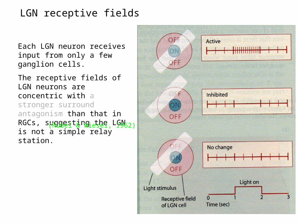
Each LGN neuron receives input from only a few ganglion cells.
The receptive fields of LGN neurons are concentric with a stronger surround antagonism than that in RGCs, suggesting the LGN is not a simple relay station.
(Hubel & Wiesel, 1962)
LGN receptive fields

Retinotopic Maps
Adjacent neurons in the retina project to adjecent neurons in higher-order brain regions.
Mapping of LGN:
1. Recording parallel to the layer showed that adjacentcells are excited by adjacent RGCs of the same retina
2. Recording perpendicular to the layers showed thatcells in different layers are excited by RGCs in either right or left retina but having the same receptive field location.
LGN Cells in different layers are in “topographic register”.

medial visual field lateral V1
lower visual field anterior V1
upper visual field posterior V1
1 2 3
23
1
23
1
23
1
1 2 3
retina
LGN
V1
FPvisual field
From visual field to V1
left right
V2V2


![SummaryMap ward2 [Converted] · 2019-10-01 · MU-2 MU-6 MU-16 MU-14 MU-6 MU-2 MU-20 MU-9 MU-4 MU-13 MU-15 MU-13 MU-16 MU-18 MU-22 MU-19 MU-16 MU-27 MU-4 MU-3A MU-17 MU-13 MU-4](https://img.pdfslide.us/doc/110x75/5f5e4f591750d150e9633369/summarymap-ward2-converted-2019-10-01-mu-2-mu-6-mu-16-mu-14-mu-6-mu-2-mu-20.jpg)