Embed Size (px)
Citation preview
8182019 High-Voltage Electrically Head Injury Presenting underlying Calvarial Osteomyelitis Single Indonesian Tertiary Hosphellip
httpslidepdfcomreaderfullhigh-voltage-electrically-head-injury-presenting-underlying-calvarial-osteomyelitis 14
Journal of Surgery and Surgical Research
Citation Yudoyono F Sutiono AB Arin MZ (2016) High-Voltage Electrically Head Injury Presenting underlying Calvarial Osteomyelitis Single Indonesian
Tertiary Hospital Experience J Surg Surgical Res 2(1) 010-013010
Abst ract
Objective To demonstrate the characteristic of high-voltage electrically head injury patientspresenting underlying calvarial osteomyelitis
Methods Retrospectively report of patients high-voltage electrically head injured with calvarial
osteomyelitis from January 1st 2011 to December 31st 2013 The demographic variable namely age
sex place of accident present of calvarial osteomyelitis Glasgow coma scale surgical treatment type
grading of burn injury and total body surface area of burn (TBSA)
Results Eleven high-voltage electrically injured patients presenting with calvarial osteomyelitis
who admitted to the Emergency Unit Department of Neurosurgery Hasan Sadikin Hospital All patients
were males (100) Their ages ranged between 24 and 51 years (average 237 years old) All patient
(100) suffered from calvarial osteomyelitis Eight patient (777) were high building worker at the
time of incident two patients were electric installation worker (1818) Entry point of electric wave 11
patients (100) from head and outlet 11 patients (100) from leg
Conclusion Bone debridement in calvarial osteomyelitis is a difcult to treat infectious diseasewith a high relapse risk cure is possible with appropriate treatment choices Antibiotic treatment will
provide more benet if it is combined with appropriate and timely surgical treatment for both scalp andcalvarial
socioeconomic conditions and immunodeficiency syndromes
[8] and also due to preexisting inectious ocus Local vascular
insufficiency or hematogenous spread Te treatment involves a
surgical and long term antibiotics or causative agent [9]
Methods
Retrospectively report o high-voltage electrically head injured
with underlying calvarial osteomyelitis rom January 1st 2011 to
December 31st 2013 Demographic inormation and the mechanism
o injury complications hospitalization period surgical interventions
and grading o burn injury and total body surace area o burn(BSA) were recorded Incomplete records and patients who had
lef the Hospital with written consent beore the termination o their
treatment course were excluded rom the study (able 1)
Criteria or surgical procedures were high-voltage electrically
head injured patients with underlying calvarial osteomyelitis who do
not ulfill these criteria or admission are treated in the outpatient
clinic until healing o their burn wounds or becoming ready or
grafing
Results
Eleven adult patients (11 males) were managed or high-voltage
electrically head injured with underlying calvarial osteomyelitis
Introduction
Electrically head injured are extremely rare and pose a difficult
challenge or neurosurgeon Electrical injuries account or less than
5 o admissions to major burn centers Te mortality is reported
to be between 3 and 15 with about 1000 deaths a year in the
United States attributed to electrical injury [1-4] Te disability o
electrotrauma depends not only the nature o the voltage current
(DC or AC) but also the length o exposure location and contact
resistance o different tissues Electrotrauma is divided between
higher (gt1000 volts) and lower (1000 volts) voltage injuries [125] High-voltage (gt1000 volts) electrical current it ofen causes deep
scalp and calvarial burns when enters into the body rom the
head present serious challenges in early and late stages o healing
underlying dura and cerebrum may be severely injured moreover
neurological deficits caused by intracranial hemorrhage leading to
loss o consciousness sensory and motor deficiencies may occur
No difference management but cerebral involvement are more
devastating and requently end with a destructive condition [6]
Osteomyelitis is an inection process accompanied by bone
destruction caused by a microorganism [7] Te incidence is on
the rise in developing countries because o malnutrition poor
Research Article
High-Voltage Electrically HeadInjury Presenting underlying
Calvarial Osteomyelitis Single
Indonesian Tertiary Hospital
Experience
Farid Yudoyono Agung Budi Sutiono
and M Zafrullah Arifin
Department of Neurosurgery Faculty of MedicineUniversitas PadjadjaranndashDr Hasan Sadikin
Hospital Bandung West Java Indonesia
Dates Received 23 December 2015 Accepted
25 January 2016Published 27 January 2016
Corresponding author Farid Yudoyono MD
Department of Neurosurgery Faculty of Medicine
Universitas PadjadjaranndashDr Hasan Sadikin Hospital
Jl Pasteur No 38 Bandung 40161 Indonesia
Tel +62222041694 Fax +62222041694 E-mail
wwwpeertechzcom
ISSN 2455-2968
Keywords Calvarial Osteomyelitis High voltageelectrical injuries Scalp ap
8182019 High-Voltage Electrically Head Injury Presenting underlying Calvarial Osteomyelitis Single Indonesian Tertiary Hosphellip
httpslidepdfcomreaderfullhigh-voltage-electrically-head-injury-presenting-underlying-calvarial-osteomyelitis 24
Citation Yudoyono F Sutiono AB Arin MZ (2016) High-Voltage Electrically Head Injury Presenting underlying Calvarial Osteomyelitis Single Indonesian
Tertiary Hospital Experience J Surg Surgical Res 2(1) 010-013
Yudoyono et al (2016)
011
All patients were males (100) Teir ages ranged between 24
and 51 years (average 237 years old) and the surace extent o burn
ranged rom 4 to 40 otal Body Surace Area (BSA) average2372 Wound care average 15 days included 5 patient loss ollow
up caused by orced discharged due to financial problem and not
controlled in outpatient clinic GCS admission was 15 included one
patient paraphrases caused by spinal cord injury All patient (100)
suffered rom calvarial osteomyelitis ound radiographically or
clinical assessment Head C scan not available or all patient due
to limitation o insurance coverage Eight patient (777) were high
building worker at the time o incident two patients were electric
installation worker (1818) Entry point o electric wave 11 patients
(100) rom head and outlet 11 patients (100) rom leg
Diagnosis o the type o electrical injury either as a low-voltage or
a high-voltage injury was made according to the clinical examination
and the history given by the patient an attendant o the accidentor the medical report supplied by the medical acility that provided
first aid management andor transportation Assessment o the type
and depth scalp is confirmed subsequent debridements and dressing
changes later intra-operatively during debridement
Discussion
Electrical injuries account or less than 5 o admissions to
major burn centers Te mortality is reported to be between 3 and
15 with about 1000 deaths a year in the United States attributed
to electrical injury [126] Electrical burns remain an important
issue in developing countries due to its higher prevalence and
complications mortality rate reported in literature as high as 59
[67] Electrotrauma is requently caused by work-related accidents
Handschin et al show a large number o high voltage electrotrauma
in connection with accidents at work (72) [2]
Prolonged High-voltage electrical head injuries can be damage various types o tissue such as skin subcutaneous tissue muscles
nerves tendons and blood vessels although rare calvarial destroys
requently both sof tissues and bony parts o the head and inection
can occurs such as calvarial osteomyelitis on the inlet part (Figures
2AB) [1011]
Grading o burn injury divided to First degree includes only the
outer layer o skin epidermis usually appear red and very painul
Second degree divided into two partial thickness involve entire
epidermis ull thickness involve entire epidermis and most o the
dermis with diminished sensation third degree involve all layers o
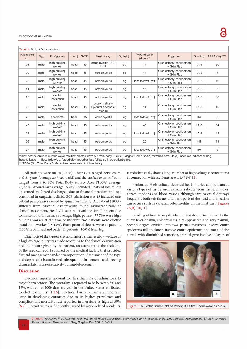
Tabel 1 Patient Demographic
Age (y ears
oldSex Profession Inlet dagger GCS Skull X ray Outlet Dagger
Wound care
(days)Treatment Grading TBSA () 2
24 malehigh building
worker head 15
osteomyelitis+ SCI
L1-2leg 14
Craniectomy debridement
+ Skin FlapIIA-B 30
30 malehigh building
worker head 15 osteomyelitis leg 11
Craniectomy debridement
+ Skin FlapIIA-B 4
32 malehigh building
worker head 15 osteomyelitis leg loss follow Updaggerdagger
Craniectomy debridement
+ Skin FlapIIA-B 40
51 malehigh building
worker head 15 osteomyelitis leg 15
Craniectomy debridement
+ Skin FlapIIA-B 5
32 maleelectric
instalationhead 15 osteomyelitis leg loss follow Updaggerdagger
Craniectomy debridement
+ Skin FlapIIA-B 38
30 maleelectric
instalationhead 15
osteomyelitis +
Epidural Abcess at
Vertex
leg 14Craniectomy debridement
+ Skin FlapIIA-B 40
45 male accidental head 15 osteomyelitis leg loss follow UpdaggerdaggerCraniectomy debridement
+ Skin FlapIIA 39
45 malehigh building
worker head 15 osteomyelitis leg 45
Craniectomy debridement
+ Skin FlapIIA-B 34
33 malehigh building
worker head 15 osteomyelitis leg loss follow Updaggerdagger
Craniectomy debridement
+ Skin FlapIIA-B 13
26 malehigh building
worker head 15 osteomyelitis leg 25
Craniectomy debridement
+ Skin FlapII-III 13
27 malehigh building
worker head 15 osteomyelitis leg loss follow Updaggerdagger
Craniectomy debridement
+ Skin FlapIIA 5
daggerinlet port de entre of electric wave Daggeroutlet electric wave out from body GCS Glasgow Coma Scale Wound care (days) open wound care during
hospitalization daggerdaggerloss follow Up forced discharged or loss follow up in outpatient clinic
TBSA () Total Body Surface Area Area extent of burn injury
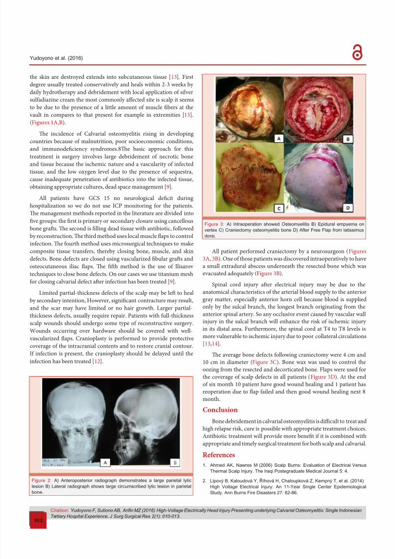
Figure 1 A Electric Source inlet on Vertex B Outlet Electric wave on pedis
8182019 High-Voltage Electrically Head Injury Presenting underlying Calvarial Osteomyelitis Single Indonesian Tertiary Hosphellip
httpslidepdfcomreaderfullhigh-voltage-electrically-head-injury-presenting-underlying-calvarial-osteomyelitis 34
Citation Yudoyono F Sutiono AB Arin MZ (2016) High-Voltage Electrically Head Injury Presenting underlying Calvarial Osteomyelitis Single Indonesian
Tertiary Hospital Experience J Surg Surgical Res 2(1) 010-013
Yudoyono et al (2016)
012
the skin are destroyed extends into subcutaneous tissue [13] First
degree usually treated conservatively and heals within 2-3 weeks by
daily hydrotherapy and debridement with local application o silver
suladiazine cream the most commonly affected site is scalp it seemsto be due to the presence o a little amount o muscle fibers at the
vault in compares to that present or example in extremities [13]
(Figures 1AB)
Te incidence o Calvarial osteomyelitis rising in developing
countries because o malnutrition poor socioeconomic conditions
and immunodeficiency syndromes8Te basic approach or this
treatment is surgery involves large debridement o necrotic bone
and tissue because the ischemic nature and a vascularity o inected
tissue and the low oxygen level due to the presence o sequestra
cause inadequate penetration o antibiotics into the inected tissue
obtaining appropriate cultures dead space management [9]
All patients have GCS 15 no neurological deficit during
hospitalization so we do not use ICP monitoring or the patients
Te management methods reported in the literature are divided into
five groups the first is primary or secondary closure using cancellous
bone grafs Te second is filling dead tissue with antibiotic ollowed
by reconstruction Te third method uses local muscle flaps to control
inection Te ourth method uses microsurgical techniques to make
composite tissue transers thereby closing bone muscle and skin
deects Bone deects are closed using vascularized fibular grafs and
osteocutaneous iliac flaps Te fifh method is the use o Ilisarov
techniques to close bone deects On our cases we use titanium mesh
or closing calvarial deect afer inection has been treated [9]
Limited partial-thickness deects o the scalp may be lef to healby secondary intention However signi1047297cant contracture may result
and the scar may have limited or no hair growth Larger partial-
thickness deects usually require repair Patients with ull-thickness
scalp wounds should undergo some type o reconstructive surgery
Wounds occurring over hardware should be covered with well-
vascularized 1047298aps Cranioplasty is perormed to provide protective
coverage o the intracranial contents and to restore cranial contour
I inection is present the cranioplasty should be delayed until the
inection has been treated [12]
All patient perormed craniectomy by a neurosurgeon (Figures
3A 3B) One o those patients was discovered intraoperatively to have
a small extradural abscess underneath the resected bone which was
evacuated adequately (Figure 3B)
Spinal cord injury afer electrical injury may be due to the
anatomical characteristics o the arterial blood supply to the anteriorgray matter especially anterior horn cell because blood is supplied
only by the sulcal branch the longest branch originating rom the
anterior spinal artery So any occlusive event caused by vascular wall
injury in the sulcal branch will enhance the risk o ischemic injury
in its distal area Furthermore the spinal cord at 4 to 8 levels is
more vulnerable to ischemic injury due to poor collateral circulations
[1314]
Te average bone deects ollowing craniectomy were 4 cm and
10 cm in diameter (Figure 3C) Bone wax was used to control the
oozing rom the resected and decorticated bone Flaps were used or
the coverage o scalp deects in all patients (Figure 3D) At the end
o six month 10 patient have good wound healing and 1 patient has
reoperation due to flap ailed and then good wound healing next 8month
Conclusion
Bone debridement in calvarial osteomyelitis is difficult to treat and
high relapse risk cure is possible with appropriate treatment choices
Antibiotic treatment will provide more benefit i it is combined with
appropriate and timely surgical treatment or both scalp and calvarial
References
1 Ahmed AK Nawres M (2006) Scalp Burns Evaluation of Electrical Versus
Thermal Scalp Injury The Iraqi Postegraduate Medical Journal 5 4
2 Lipovyacute B Kaloudovaacute Y Řiacutehovaacute H Chaloupkovaacute Z Kempnyacute T et al (2014) High Voltage Electrical Injury An 11-Year Single Center Epidemiological
Study Ann Burns Fire Disasters 27 82-86
Figure 2 A) Anteroposterior radiograph demonstrates a large parietal lytic
lesion B) Lateral radiograph shows large circumscribed lytic lesion in parietal
bone
Figure 3 A) Intraoperation showed Osteomyelitis B) Epidural empyema on
vertex C) Craniectomy osteomyelitis bone D) After Free Flap from latissimus
dorsi
8182019 High-Voltage Electrically Head Injury Presenting underlying Calvarial Osteomyelitis Single Indonesian Tertiary Hosphellip
httpslidepdfcomreaderfullhigh-voltage-electrically-head-injury-presenting-underlying-calvarial-osteomyelitis 44
Citation Yudoyono F Sutiono AB Arin MZ (2016) High-Voltage Electrically Head Injury Presenting underlying Calvarial Osteomyelitis Single Indonesian
Tertiary Hospital Experience J Surg Surgical Res 2(1) 010-013
Yudoyono et al (2016)
013
Copyright copy 2016 Yudoyono F et al This is an open-access article distributed under the terms of the Creative Commons Attribution License which permits
unrestricted use distribution and reproduction in any medium provided the original author and source are credited
3 Makboul M Abdel-Rahim M (2013) Simple aps for reconstruction of pediatric scalp defects after electrical burn Chin J Traumatol 16 204-206
4 Kaif M Singh Panwar D Chandra A Chandra N (2009) High-voltage electrical
burn of the headReport of an unusual case Indian Journal of Neurotrauma (IJNT) 6 163-164
5 Lee J Sinno H Perkins A Tahiri Y Luc M (2010) 14000 Volt Electrical Injury
to Bilateral Upper Extremities A Case Report Mcgill J Med 13 18-21
6 Ghavami Y Reza Mobayen M Vaghardoost R (2014) Electrical Burn Injury
A Five-Year Survey of 682 Patients Trauma Mon 19 e18748
7 Soo Kim M (2012) Skull Osteomyelitis Osteomyelitis Prof Mauricio S
Baptista (Ed) 45-88
8 Raut AA Nagar AM Muzumdar D Chawla AJ Narlawar RS et al (2004)
Imaging Features of Calvarial Tuberculosis A Study of 42 Cases AJNR Am
J Neuroradiol 25 409ndash414
9 Suumlmer S Karamese M Koumlktekir E Ural O (2013) Chronic osteomyelitis of
skull associated with necrotic injury after trauma A case report Journal of
Microbiology and Infectious Diseases 3 218-222
10 N Guumlmuumlş (2012) Negative pressure dressing combined with a traditional approach for the treatment of skull burn Niger J Clin Pract 15 494-497
11 Abd Al-Aziz H Ahmad A Al-Leithy I Abu Alfotoh S (2004) Evaluation of the
Treatment protocol of electrical injuries in Ain Shams University Burn Unit
Egypt J Palst Reconstr Surg 28 149-158
12 Samuel JL Matthew MH Roman JS (2008) Scalp and Calvarial
Reconstruction Seminars in Plastic Surgery 22 281-293
13 Ko SH Chun W Kim HC (2004) Delayed spinal cord injury following electrical
burns a 7-year experience Burns 30 691ndash695
14 Johl HK Olshansky A Beydoun SR Rison RA (2012) Cervicothoracic spinal
cord and pontomedullary injury secondary to high-voltage electrocution a
case report J Med Case Rep 6 296
8182019 High-Voltage Electrically Head Injury Presenting underlying Calvarial Osteomyelitis Single Indonesian Tertiary Hosphellip
httpslidepdfcomreaderfullhigh-voltage-electrically-head-injury-presenting-underlying-calvarial-osteomyelitis 24
Citation Yudoyono F Sutiono AB Arin MZ (2016) High-Voltage Electrically Head Injury Presenting underlying Calvarial Osteomyelitis Single Indonesian
Tertiary Hospital Experience J Surg Surgical Res 2(1) 010-013
Yudoyono et al (2016)
011
All patients were males (100) Teir ages ranged between 24
and 51 years (average 237 years old) and the surace extent o burn
ranged rom 4 to 40 otal Body Surace Area (BSA) average2372 Wound care average 15 days included 5 patient loss ollow
up caused by orced discharged due to financial problem and not
controlled in outpatient clinic GCS admission was 15 included one
patient paraphrases caused by spinal cord injury All patient (100)
suffered rom calvarial osteomyelitis ound radiographically or
clinical assessment Head C scan not available or all patient due
to limitation o insurance coverage Eight patient (777) were high
building worker at the time o incident two patients were electric
installation worker (1818) Entry point o electric wave 11 patients
(100) rom head and outlet 11 patients (100) rom leg
Diagnosis o the type o electrical injury either as a low-voltage or
a high-voltage injury was made according to the clinical examination
and the history given by the patient an attendant o the accidentor the medical report supplied by the medical acility that provided
first aid management andor transportation Assessment o the type
and depth scalp is confirmed subsequent debridements and dressing
changes later intra-operatively during debridement
Discussion
Electrical injuries account or less than 5 o admissions to
major burn centers Te mortality is reported to be between 3 and
15 with about 1000 deaths a year in the United States attributed
to electrical injury [126] Electrical burns remain an important
issue in developing countries due to its higher prevalence and
complications mortality rate reported in literature as high as 59
[67] Electrotrauma is requently caused by work-related accidents
Handschin et al show a large number o high voltage electrotrauma
in connection with accidents at work (72) [2]
Prolonged High-voltage electrical head injuries can be damage various types o tissue such as skin subcutaneous tissue muscles
nerves tendons and blood vessels although rare calvarial destroys
requently both sof tissues and bony parts o the head and inection
can occurs such as calvarial osteomyelitis on the inlet part (Figures
2AB) [1011]
Grading o burn injury divided to First degree includes only the
outer layer o skin epidermis usually appear red and very painul
Second degree divided into two partial thickness involve entire
epidermis ull thickness involve entire epidermis and most o the
dermis with diminished sensation third degree involve all layers o
Tabel 1 Patient Demographic
Age (y ears
oldSex Profession Inlet dagger GCS Skull X ray Outlet Dagger
Wound care
(days)Treatment Grading TBSA () 2
24 malehigh building
worker head 15
osteomyelitis+ SCI
L1-2leg 14
Craniectomy debridement
+ Skin FlapIIA-B 30
30 malehigh building
worker head 15 osteomyelitis leg 11
Craniectomy debridement
+ Skin FlapIIA-B 4
32 malehigh building
worker head 15 osteomyelitis leg loss follow Updaggerdagger
Craniectomy debridement
+ Skin FlapIIA-B 40
51 malehigh building
worker head 15 osteomyelitis leg 15
Craniectomy debridement
+ Skin FlapIIA-B 5
32 maleelectric
instalationhead 15 osteomyelitis leg loss follow Updaggerdagger
Craniectomy debridement
+ Skin FlapIIA-B 38
30 maleelectric
instalationhead 15
osteomyelitis +
Epidural Abcess at
Vertex
leg 14Craniectomy debridement
+ Skin FlapIIA-B 40
45 male accidental head 15 osteomyelitis leg loss follow UpdaggerdaggerCraniectomy debridement
+ Skin FlapIIA 39
45 malehigh building
worker head 15 osteomyelitis leg 45
Craniectomy debridement
+ Skin FlapIIA-B 34
33 malehigh building
worker head 15 osteomyelitis leg loss follow Updaggerdagger
Craniectomy debridement
+ Skin FlapIIA-B 13
26 malehigh building
worker head 15 osteomyelitis leg 25
Craniectomy debridement
+ Skin FlapII-III 13
27 malehigh building
worker head 15 osteomyelitis leg loss follow Updaggerdagger
Craniectomy debridement
+ Skin FlapIIA 5
daggerinlet port de entre of electric wave Daggeroutlet electric wave out from body GCS Glasgow Coma Scale Wound care (days) open wound care during
hospitalization daggerdaggerloss follow Up forced discharged or loss follow up in outpatient clinic
TBSA () Total Body Surface Area Area extent of burn injury
Figure 1 A Electric Source inlet on Vertex B Outlet Electric wave on pedis
8182019 High-Voltage Electrically Head Injury Presenting underlying Calvarial Osteomyelitis Single Indonesian Tertiary Hosphellip
httpslidepdfcomreaderfullhigh-voltage-electrically-head-injury-presenting-underlying-calvarial-osteomyelitis 34
Citation Yudoyono F Sutiono AB Arin MZ (2016) High-Voltage Electrically Head Injury Presenting underlying Calvarial Osteomyelitis Single Indonesian
Tertiary Hospital Experience J Surg Surgical Res 2(1) 010-013
Yudoyono et al (2016)
012
the skin are destroyed extends into subcutaneous tissue [13] First
degree usually treated conservatively and heals within 2-3 weeks by
daily hydrotherapy and debridement with local application o silver
suladiazine cream the most commonly affected site is scalp it seemsto be due to the presence o a little amount o muscle fibers at the
vault in compares to that present or example in extremities [13]
(Figures 1AB)
Te incidence o Calvarial osteomyelitis rising in developing
countries because o malnutrition poor socioeconomic conditions
and immunodeficiency syndromes8Te basic approach or this
treatment is surgery involves large debridement o necrotic bone
and tissue because the ischemic nature and a vascularity o inected
tissue and the low oxygen level due to the presence o sequestra
cause inadequate penetration o antibiotics into the inected tissue
obtaining appropriate cultures dead space management [9]
All patients have GCS 15 no neurological deficit during
hospitalization so we do not use ICP monitoring or the patients
Te management methods reported in the literature are divided into
five groups the first is primary or secondary closure using cancellous
bone grafs Te second is filling dead tissue with antibiotic ollowed
by reconstruction Te third method uses local muscle flaps to control
inection Te ourth method uses microsurgical techniques to make
composite tissue transers thereby closing bone muscle and skin
deects Bone deects are closed using vascularized fibular grafs and
osteocutaneous iliac flaps Te fifh method is the use o Ilisarov
techniques to close bone deects On our cases we use titanium mesh
or closing calvarial deect afer inection has been treated [9]
Limited partial-thickness deects o the scalp may be lef to healby secondary intention However signi1047297cant contracture may result
and the scar may have limited or no hair growth Larger partial-
thickness deects usually require repair Patients with ull-thickness
scalp wounds should undergo some type o reconstructive surgery
Wounds occurring over hardware should be covered with well-
vascularized 1047298aps Cranioplasty is perormed to provide protective
coverage o the intracranial contents and to restore cranial contour
I inection is present the cranioplasty should be delayed until the
inection has been treated [12]
All patient perormed craniectomy by a neurosurgeon (Figures
3A 3B) One o those patients was discovered intraoperatively to have
a small extradural abscess underneath the resected bone which was
evacuated adequately (Figure 3B)
Spinal cord injury afer electrical injury may be due to the
anatomical characteristics o the arterial blood supply to the anteriorgray matter especially anterior horn cell because blood is supplied
only by the sulcal branch the longest branch originating rom the
anterior spinal artery So any occlusive event caused by vascular wall
injury in the sulcal branch will enhance the risk o ischemic injury
in its distal area Furthermore the spinal cord at 4 to 8 levels is
more vulnerable to ischemic injury due to poor collateral circulations
[1314]
Te average bone deects ollowing craniectomy were 4 cm and
10 cm in diameter (Figure 3C) Bone wax was used to control the
oozing rom the resected and decorticated bone Flaps were used or
the coverage o scalp deects in all patients (Figure 3D) At the end
o six month 10 patient have good wound healing and 1 patient has
reoperation due to flap ailed and then good wound healing next 8month
Conclusion
Bone debridement in calvarial osteomyelitis is difficult to treat and
high relapse risk cure is possible with appropriate treatment choices
Antibiotic treatment will provide more benefit i it is combined with
appropriate and timely surgical treatment or both scalp and calvarial
References
1 Ahmed AK Nawres M (2006) Scalp Burns Evaluation of Electrical Versus
Thermal Scalp Injury The Iraqi Postegraduate Medical Journal 5 4
2 Lipovyacute B Kaloudovaacute Y Řiacutehovaacute H Chaloupkovaacute Z Kempnyacute T et al (2014) High Voltage Electrical Injury An 11-Year Single Center Epidemiological
Study Ann Burns Fire Disasters 27 82-86
Figure 2 A) Anteroposterior radiograph demonstrates a large parietal lytic
lesion B) Lateral radiograph shows large circumscribed lytic lesion in parietal
bone
Figure 3 A) Intraoperation showed Osteomyelitis B) Epidural empyema on
vertex C) Craniectomy osteomyelitis bone D) After Free Flap from latissimus
dorsi
8182019 High-Voltage Electrically Head Injury Presenting underlying Calvarial Osteomyelitis Single Indonesian Tertiary Hosphellip
httpslidepdfcomreaderfullhigh-voltage-electrically-head-injury-presenting-underlying-calvarial-osteomyelitis 44
Citation Yudoyono F Sutiono AB Arin MZ (2016) High-Voltage Electrically Head Injury Presenting underlying Calvarial Osteomyelitis Single Indonesian
Tertiary Hospital Experience J Surg Surgical Res 2(1) 010-013
Yudoyono et al (2016)
013
Copyright copy 2016 Yudoyono F et al This is an open-access article distributed under the terms of the Creative Commons Attribution License which permits
unrestricted use distribution and reproduction in any medium provided the original author and source are credited
3 Makboul M Abdel-Rahim M (2013) Simple aps for reconstruction of pediatric scalp defects after electrical burn Chin J Traumatol 16 204-206
4 Kaif M Singh Panwar D Chandra A Chandra N (2009) High-voltage electrical
burn of the headReport of an unusual case Indian Journal of Neurotrauma (IJNT) 6 163-164
5 Lee J Sinno H Perkins A Tahiri Y Luc M (2010) 14000 Volt Electrical Injury
to Bilateral Upper Extremities A Case Report Mcgill J Med 13 18-21
6 Ghavami Y Reza Mobayen M Vaghardoost R (2014) Electrical Burn Injury
A Five-Year Survey of 682 Patients Trauma Mon 19 e18748
7 Soo Kim M (2012) Skull Osteomyelitis Osteomyelitis Prof Mauricio S
Baptista (Ed) 45-88
8 Raut AA Nagar AM Muzumdar D Chawla AJ Narlawar RS et al (2004)
Imaging Features of Calvarial Tuberculosis A Study of 42 Cases AJNR Am
J Neuroradiol 25 409ndash414
9 Suumlmer S Karamese M Koumlktekir E Ural O (2013) Chronic osteomyelitis of
skull associated with necrotic injury after trauma A case report Journal of
Microbiology and Infectious Diseases 3 218-222
10 N Guumlmuumlş (2012) Negative pressure dressing combined with a traditional approach for the treatment of skull burn Niger J Clin Pract 15 494-497
11 Abd Al-Aziz H Ahmad A Al-Leithy I Abu Alfotoh S (2004) Evaluation of the
Treatment protocol of electrical injuries in Ain Shams University Burn Unit
Egypt J Palst Reconstr Surg 28 149-158
12 Samuel JL Matthew MH Roman JS (2008) Scalp and Calvarial
Reconstruction Seminars in Plastic Surgery 22 281-293
13 Ko SH Chun W Kim HC (2004) Delayed spinal cord injury following electrical
burns a 7-year experience Burns 30 691ndash695
14 Johl HK Olshansky A Beydoun SR Rison RA (2012) Cervicothoracic spinal
cord and pontomedullary injury secondary to high-voltage electrocution a
case report J Med Case Rep 6 296
8182019 High-Voltage Electrically Head Injury Presenting underlying Calvarial Osteomyelitis Single Indonesian Tertiary Hosphellip
httpslidepdfcomreaderfullhigh-voltage-electrically-head-injury-presenting-underlying-calvarial-osteomyelitis 34
Citation Yudoyono F Sutiono AB Arin MZ (2016) High-Voltage Electrically Head Injury Presenting underlying Calvarial Osteomyelitis Single Indonesian
Tertiary Hospital Experience J Surg Surgical Res 2(1) 010-013
Yudoyono et al (2016)
012
the skin are destroyed extends into subcutaneous tissue [13] First
degree usually treated conservatively and heals within 2-3 weeks by
daily hydrotherapy and debridement with local application o silver
suladiazine cream the most commonly affected site is scalp it seemsto be due to the presence o a little amount o muscle fibers at the
vault in compares to that present or example in extremities [13]
(Figures 1AB)
Te incidence o Calvarial osteomyelitis rising in developing
countries because o malnutrition poor socioeconomic conditions
and immunodeficiency syndromes8Te basic approach or this
treatment is surgery involves large debridement o necrotic bone
and tissue because the ischemic nature and a vascularity o inected
tissue and the low oxygen level due to the presence o sequestra
cause inadequate penetration o antibiotics into the inected tissue
obtaining appropriate cultures dead space management [9]
All patients have GCS 15 no neurological deficit during
hospitalization so we do not use ICP monitoring or the patients
Te management methods reported in the literature are divided into
five groups the first is primary or secondary closure using cancellous
bone grafs Te second is filling dead tissue with antibiotic ollowed
by reconstruction Te third method uses local muscle flaps to control
inection Te ourth method uses microsurgical techniques to make
composite tissue transers thereby closing bone muscle and skin
deects Bone deects are closed using vascularized fibular grafs and
osteocutaneous iliac flaps Te fifh method is the use o Ilisarov
techniques to close bone deects On our cases we use titanium mesh
or closing calvarial deect afer inection has been treated [9]
Limited partial-thickness deects o the scalp may be lef to healby secondary intention However signi1047297cant contracture may result
and the scar may have limited or no hair growth Larger partial-
thickness deects usually require repair Patients with ull-thickness
scalp wounds should undergo some type o reconstructive surgery
Wounds occurring over hardware should be covered with well-
vascularized 1047298aps Cranioplasty is perormed to provide protective
coverage o the intracranial contents and to restore cranial contour
I inection is present the cranioplasty should be delayed until the
inection has been treated [12]
All patient perormed craniectomy by a neurosurgeon (Figures
3A 3B) One o those patients was discovered intraoperatively to have
a small extradural abscess underneath the resected bone which was
evacuated adequately (Figure 3B)
Spinal cord injury afer electrical injury may be due to the
anatomical characteristics o the arterial blood supply to the anteriorgray matter especially anterior horn cell because blood is supplied
only by the sulcal branch the longest branch originating rom the
anterior spinal artery So any occlusive event caused by vascular wall
injury in the sulcal branch will enhance the risk o ischemic injury
in its distal area Furthermore the spinal cord at 4 to 8 levels is
more vulnerable to ischemic injury due to poor collateral circulations
[1314]
Te average bone deects ollowing craniectomy were 4 cm and
10 cm in diameter (Figure 3C) Bone wax was used to control the
oozing rom the resected and decorticated bone Flaps were used or
the coverage o scalp deects in all patients (Figure 3D) At the end
o six month 10 patient have good wound healing and 1 patient has
reoperation due to flap ailed and then good wound healing next 8month
Conclusion
Bone debridement in calvarial osteomyelitis is difficult to treat and
high relapse risk cure is possible with appropriate treatment choices
Antibiotic treatment will provide more benefit i it is combined with
appropriate and timely surgical treatment or both scalp and calvarial
References
1 Ahmed AK Nawres M (2006) Scalp Burns Evaluation of Electrical Versus
Thermal Scalp Injury The Iraqi Postegraduate Medical Journal 5 4
2 Lipovyacute B Kaloudovaacute Y Řiacutehovaacute H Chaloupkovaacute Z Kempnyacute T et al (2014) High Voltage Electrical Injury An 11-Year Single Center Epidemiological
Study Ann Burns Fire Disasters 27 82-86
Figure 2 A) Anteroposterior radiograph demonstrates a large parietal lytic
lesion B) Lateral radiograph shows large circumscribed lytic lesion in parietal
bone
Figure 3 A) Intraoperation showed Osteomyelitis B) Epidural empyema on
vertex C) Craniectomy osteomyelitis bone D) After Free Flap from latissimus
dorsi
8182019 High-Voltage Electrically Head Injury Presenting underlying Calvarial Osteomyelitis Single Indonesian Tertiary Hosphellip
httpslidepdfcomreaderfullhigh-voltage-electrically-head-injury-presenting-underlying-calvarial-osteomyelitis 44
Citation Yudoyono F Sutiono AB Arin MZ (2016) High-Voltage Electrically Head Injury Presenting underlying Calvarial Osteomyelitis Single Indonesian
Tertiary Hospital Experience J Surg Surgical Res 2(1) 010-013
Yudoyono et al (2016)
013
Copyright copy 2016 Yudoyono F et al This is an open-access article distributed under the terms of the Creative Commons Attribution License which permits
unrestricted use distribution and reproduction in any medium provided the original author and source are credited
3 Makboul M Abdel-Rahim M (2013) Simple aps for reconstruction of pediatric scalp defects after electrical burn Chin J Traumatol 16 204-206
4 Kaif M Singh Panwar D Chandra A Chandra N (2009) High-voltage electrical
burn of the headReport of an unusual case Indian Journal of Neurotrauma (IJNT) 6 163-164
5 Lee J Sinno H Perkins A Tahiri Y Luc M (2010) 14000 Volt Electrical Injury
to Bilateral Upper Extremities A Case Report Mcgill J Med 13 18-21
6 Ghavami Y Reza Mobayen M Vaghardoost R (2014) Electrical Burn Injury
A Five-Year Survey of 682 Patients Trauma Mon 19 e18748
7 Soo Kim M (2012) Skull Osteomyelitis Osteomyelitis Prof Mauricio S
Baptista (Ed) 45-88
8 Raut AA Nagar AM Muzumdar D Chawla AJ Narlawar RS et al (2004)
Imaging Features of Calvarial Tuberculosis A Study of 42 Cases AJNR Am
J Neuroradiol 25 409ndash414
9 Suumlmer S Karamese M Koumlktekir E Ural O (2013) Chronic osteomyelitis of
skull associated with necrotic injury after trauma A case report Journal of
Microbiology and Infectious Diseases 3 218-222
10 N Guumlmuumlş (2012) Negative pressure dressing combined with a traditional approach for the treatment of skull burn Niger J Clin Pract 15 494-497
11 Abd Al-Aziz H Ahmad A Al-Leithy I Abu Alfotoh S (2004) Evaluation of the
Treatment protocol of electrical injuries in Ain Shams University Burn Unit
Egypt J Palst Reconstr Surg 28 149-158
12 Samuel JL Matthew MH Roman JS (2008) Scalp and Calvarial
Reconstruction Seminars in Plastic Surgery 22 281-293
13 Ko SH Chun W Kim HC (2004) Delayed spinal cord injury following electrical
burns a 7-year experience Burns 30 691ndash695
14 Johl HK Olshansky A Beydoun SR Rison RA (2012) Cervicothoracic spinal
cord and pontomedullary injury secondary to high-voltage electrocution a
case report J Med Case Rep 6 296


![Periacetabular Brucella Osteomyelitis - file.scirp.org · spondylitis, bursitis, tenosynovitis and osteomyelitis [3-6]. Brucella osteomyelitis may appear as a radiolucent area and](https://img.pdfslide.us/doc/110x75/5d52ce1188c993277b8b9aaa/periacetabular-brucella-osteomyelitis-filescirporg-spondylitis-bursitis.jpg)