Embed Size (px)
Citation preview

High-throughput screening of small molecules in miniaturizedmammalian cell-based assays involving post-translationalmodificationsBrent R Stockwell, Stephen J Haggarty and Stuart L Schreiber
Background: Fully adapting a forward genetic approach to mammalian systemsrequires efficient methods to alter systematically gene products without priorknowledge of gene sequences, while allowing for the subsequentcharacterization of these alterations. Ideally, these methods would also allowfunction to be altered in a temporally controlled manner.
Results: We report the development of a miniaturized cell-based assay format thatenables a genetic-like approach to understanding cellular pathways in mammaliansystems using small molecules, rather than mutations, as the source of gene-product alterations. This whole-cell immunodetection assay can sensitively detectchanges in specific cellular macromolecules in high-density arrays of mammaliancells. Furthermore, it is compatible with screening large numbers of smallmolecules in nanoliter to microliter culture volumes. We refer to this assay formatas a ‘cytoblot’, and demonstrate the use of cytoblotting to monitor biosyntheticprocesses such as DNA synthesis, and post-translational processes such asacetylation and phosphorylation. Finally, we demonstrate the applicability of theseassays to natural-product screening through the identification of marine spongeextracts exhibiting genotype-specific inhibition of 5-bromodeoxyuridineincorporation and suppression of the anti-proliferative effect of rapamycin.
Conclusions: We show that cytoblots can be used for high-throughputscreening of small molecules in cell-based assays. Together with small-molecule libraries, the cytoblot assay can be used to perform chemical geneticscreens analogous to those used in classical genetics and thus should beapplicable to understanding a wide variety of cellular processes, especiallythose involving post-transitional modifications.
IntroductionA genetic approach to analyzing biological systems isextremely powerful in that novel gene products involved ina biological process of interest can be identified. Thegenetic approach entails determining the phenotypic conse-quences of mutations in genes and ordering genes intofunctional pathways. Such an approach has been widelyused in a large number of genetically tractable organismsincluding fruit flies, nematodes, yeast, plants, and evencomplex vertebrates such as zebrafish and mice [1]. Fur-thermore, the genetic approach can be subdivided into‘forward’ genetics, which involves phenotype-based screen-ing of random mutations, and ‘reverse’ genetics, whichinvolves studying the phenotypic consequences of muta-tions in a known gene.
Methods for comprehensive genetic analysis of mammaliansystems are currently limited [2]. With whole organismssuch as mice, the space requirement and expense of largenumbers of animals, their long generation time, small littersize and the difficulty inherent in identifying and mapping
recessive mutations (mutations that only result in a pheno-typic effect when all copies of a gene are altered) are prob-lematic. In addition, many gene products are essential,redundant or expressed in a temporal- or tissue-specificmanner. Tissue-culture systems can alleviate some of theseproblems and yet still provide a suitable model system forunderstanding mammalian physiological and developmen-tal pathways [3,4]. Reverse genetic techniques that arecompatible with these systems, such as the use of antisenseconstructs, ribozymes or gene targeting, require prior knowl-edge of gene sequences and are not widely applicable tonovel gene discovery or large-scale ‘forward genetic’ pheno-type-based screens [4–7].
Fully adapting a forward genetic approach to mammaliansystems requires efficient methods to alter gene productssystematically without prior knowledge of gene sequences,while still allowing the subsequent recovery and character-ization of these alterations. Ideally, these methods shouldalso allow the conditional alteration of a gene product,through both loss and gain of function, in a temporally
Addresses: Howard Hughes Medical Institute,Harvard Institute of Chemistry and Cell Biology,Department of Chemistry and Chemical Biology,and Department of Molecular and Cellular Biology,Harvard University, 12 Oxford Street, Cambridge,MA 02138, USA.
Correspondence: Stuart L SchreiberE-mail: [email protected]
Key words: cell-based assay, chemical genetics,high-throughput screening, post-translational,profiling
Received: 5 October 1998Revisions requested: 27 October 1998Revisions received: 9 November 1998Accepted: 10 November 1998
Published: 19 January 1999
Chemistry & Biology February 1999, 6:71–83http://biomednet.com/elecref/1074552100600071
© Elsevier Science Ltd ISSN 1074-5521
Research Paper 71
defined manner. In addition, it would be useful to be ableto perform suppressor and enhancer screens, which seekto identify genes that, when mutated, suppress orenhance a previously identified phenotype of interest.The advantage of such screens, as compared with usingwild-type conditions, is that the pathway is sensitized tofurther perturbation, rendering the mutations identifiedmore relevant to the pathway of interest.
A forward chemical genetic approach (use of phenotype-based screens) using small molecules that alter proteinfunction directly has the potential to overcome many ofthe current limitations in genetic analyses of mammaliansystems. This process is akin to the generation of muta-tions in genes, but relies on small molecules, often in theform of a chemical library, as the source of perturbation.As a small molecule in a cell-based assay can alter specif-ically the function of a gene product from all copies of agene, a small molecule can be used analogously to aninducible dominant or homozygous recessive mutation.These characteristics circumvent the difficulty of gener-ating these types of mutations in mammalian systems.Also, just as mutation sites can identify functionally rele-vant coding sequences of genes, small molecules can iden-tify functionally relevant residues of proteins, based ontheir mechanism of interaction [8]. We reason that, byusing small-molecule libraries in an appropriate cell-basedassay, it should be possible to identify novel gene prod-ucts on pathways of interest, as well as novel biologicallyactive small molecules from either natural sources or lab-oratory syntheses ([9] and the Schreiber group website,http://www-schreiber.chem.harvard.edu). This idea is sup-ported by the existence of a wide variety of small moleculesthat cause a loss of function of their cognate targets, includ-ing kinases [10], phosphatases [11], membrane receptors[12], proteases [8], isoprenyl transferases [13] and poly-merases [14]. To a lesser extent, small molecules that resultin a gain of function in a protein of interest have also beendiscovered or invented [15–17].
Once a new small-molecule modulator of a gene productis discovered, a reverse chemical genetic approach (use ofsmall molecules whose target is known) is possible. Thisentails using the small molecule as a tool to alter the func-tion of the gene product and subsequently observing thephenotypic effects. For example, an inhibitor of an essen-tial gene product can be used to eliminate conditionallythat gene product’s function. The demonstrated effec-tiveness of the reverse chemical genetic approach makesthis an attractive option, but this approach is often unavail-able because the vast majority of gene products currentlylack a small-molecule partner. A forward chemical geneticapproach is therefore required. With the aim of perform-ing forward chemical genetic screens to discover newsmall-molecule partners, we sought to develop a minia-turized cell-based assay format that would be suitable for
high-throughput screening. We report here the develop-ment of such a format and we highlight its applicability toa broad range of genetic-like screens in mammalian cells.This assay format is capable of detecting post-translationaland biosynthetic events, in a high-throughput manner,without the use of engineered cell lines or radioactivity.
ResultsA whole-cell immunodetection assay related to ELISAs andwestern blotting We have developed a high-throughput whole-cell immuno-detection assay that is similar to both enzyme-linkedimmunosorbent assays (ELISAs) [18] and western blotting[19,20] (Figure 1). We refer to this technique as a cytoblotbecause, as in a western blot, an antibody is used to detectthe presence of an antigen immobilized in the solid phase.In a cytoblot, a phosphorylated protein or other cellularmolecule of interest is detected in whole, fixed cells onthe bottom of a well using a specific primary antibody anda secondary antibody covalently linked to horseradish per-oxidase (HRP) [21] (Figure 1). As in a western blot, theretention of this complex in the solid phase is detectedby adding luminol, hydrogen peroxide and p-iodophenol(Figure 2a). The amount of antigen present is visualizeddirectly on film using this chemiluminescent reaction(Figure 2a). Although antibodies are routinely used toanalyze the cellular state of mammalian cells in conven-tional procedures such as western blotting, dot blotting,ELISAs, flow cytometry and immunocytochemistry, theseprocedures are not practical for high-throughput screen-ing of small-molecule libraries in mammalian cells.
Detection of biosynthetic events in high-density arraysWe were interested in assaying for the extent of cellularbiosynthetic events, such as DNA synthesis. We testedthe ability of cytoblotting to detect changes in DNA syn-thesis by measuring the incorporation of 5-bromodeoxy-uridine (BrdU, Figure 2a) in the presence or absence oftransforming growth factor β (TGF-β), which arrests manycell types in the G1 phase of the cell cycle [22,23]. BrdUis a thymidine analog in which the methyl group at the5-position is replaced with bromine (Figure 2a). Thisanalog is efficiently incorporated into DNA during DNAreplication, and can be detected with an antibody raisedspecifically against this modified form [24–26]. We seeded2000 mink lung cells, which are responsive to TGF-β [22],in each well of an opaque, white 384-well plate, treatedthe cells with varying concentrations of TGF-β for 16 hand then added 10 µM BrdU for 16 h. We found thatTGF-β treatment effectively prevented BrdU incorpor-ation and that background staining in the presence ofTGF-β was negligible (Figure 2b). We also tested theability of small-molecule cell-cycle-arresting agents toinhibit BrdU incorporation in this assay. We found thathydroxyurea, nocodazole, trapoxin and rapamycin effi-ciently prevented BrdU incorporation, but that FK506, as
72 Chemistry & Biology 1999, Vol 6 No 2

expected, failed to prevent BrdU incorporation (Figure 2c)— cell-cycle progression and DNA synthesis are indepen-dent of FKBP12 and calcineurin, the protein targets ofFK506, in mink lung epithelial cells (B.R.S. and S.L.S.,unpublished observations). A cytoblot assay in this formatcan therefore be used to measure the extent of cell prolif-eration and to perform a chemical genetic screen for cell-cycle-arresting agents. Furthermore, the cell-cycle-arrestingagents identified by this type of screen are likely to be thesmall-molecule partners of many of the gene products iden-tified in classical yeast cell-division cycle (cdc) screens [27].
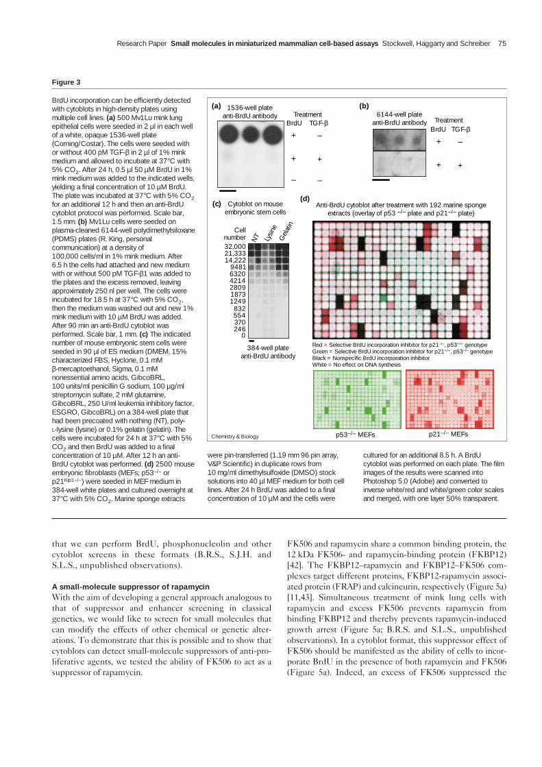
We tested whether these results could be extended to1536- and 6144-well plates. We seeded mink lung cellswith or without TGF-β in opaque, white 1536-well plates.TGF-β effectively prevented BrdU incorporation andbackground staining was negligible (Figure 3a). We thentested the anti-BrdU cytoblot assay in a plate containing6144 arrayed ‘nanowells’ ([28]; R. King, personal commun-ication). Again, TGF-β effectively prevented BrdU incor-poration, background staining was negligible and individualwells were easily resolved, indicating that interwell crosstalkdid not occur (Figure 3b).
The cytoblot assay is compatible with a variety of celltypes, both primary and transformed. We found thatMv1Lu mink lung epithelial cells (Figure 2c), A549human lung carcinoma cells (data not shown), HeLaS3human cervical carcinoma cells (data not shown) andmouse embryonic stem (ES) cells (Figure 3c) were com-patible with the BrdU cytoblot. We found that pretreatingthe wells was important for allowing attachment of some
cell lines. For example, pretreating a 384-well plate with0.1% gelatin was the most efficient method of allowing EScell attachment and growth (Figure 3c).
Although general inhibitors of DNA synthesis could beuseful and interesting compounds [29], genotype-specificinhibitors of DNA synthesis would be even more useful.We obtained 192 marine sponge extracts from ProfessorP. Crews and M. Sanders, University of California, SantaCruz (UCSC) and we tested these crude organic extracts(in duplicate rows) for their ability to inhibit BrdU incor-poration in either p53–/– [30] or p21Kip1–/– [31] mouseembryonic fibroblasts (MEFs) using a BrdU cytoblot(Figure 3d). By overlaying the results of these experiments,we were able to identify extracts that were genotype-independent BrdU-incorporation inhibitors (black wells),p21Kip1–/– p53+/+-specific BrdU incorporation inhibitors(red wells), and possibly some weak p21+/+ p53–/–-specificBrdU incorporation inhibitors (green wells).
Detection of post-translational modifications ofendogenous proteinsAs many biologically interesting and therapeutically impor-tant signaling pathways (including cell-cycle progression[32], gene expression [33], and determination of cell fate[34]) involve the reversible covalent modification of pro-teins as a means of post-translational regulation, it wouldbe useful to screen small-molecule libraries for regulatorsof such modifications. Towards this end, we used an anti-acetylated histone H4 antibody in the cytoblot format todetect an increase in the acetylation of histone H4 inresponse to the histone-deacetylase inhibitors trapoxin
Research Paper Small molecules in miniaturized mammalian cell-based assays Stockwell, Haggarty and Schreiber 73
Figure 1
Schematic of whole-cell immunodetectionassay using chemiluminescent detection (acytoblot). A cytoblot involves growing cells onthe bottom of a well, fixing the cells andprobing the cells for the presence of aparticular antigen using a specific primaryantibody in solution. A secondary antibodycovalently linked to horseradish peroxidase(HRP) is added and the presence of the entirecomplex is detected through thechemiluminescent reaction caused by additionof luminol, hydrogen peroxide and anenhancer such as p-iodophenol. Ab, antibody.
Cells Cells
Cells Cells
HRP
Secondary Ab
Primary Ab
HRP
Secondary Ab
Primary Ab
Signaldetected
No signaldetected
Antigen
Chemistry & Biology

and trichostatin [35]. Treatment of A549 cells withtrapoxin or trichostatin, but not other cell-cycle-arrestingagents, caused an increase in acetylation of histone H4(Figure 4a). An identical cytoblot in HaCaT cells [36],which do not upregulate p21 in response to trichostatin A(C. Grozinger, C. Hassig and S.L.S., unpublished observa-tions), did not result in an increase in histone H4 acetyla-tion (Figure 4a). As few as 500 cells could be detected withthe anti-acetylated histone H4 antibody after just a 4 htreatment (Figure 4b).
Histone H3 and nucleolin are both phosphorylated inmitosis and antibodies against these phospho-epitopeshave been used to detect mitotic cells [37–39]. TG-3, amonoclonal antibody that recognizes the phosphorylatedform of nucleolin, has previously been used to identifynovel small molecules from natural-product extracts thatare capable of arresting cells in mitosis (M. Roberge andR. Anderson, personal communication) or inhibiting theDNA damage-induced G2 checkpoint [40]. Here weextend the applicability of these antibodies as markers ofmitosis and as tools for screening for anti-mitotic com-pounds in the cytoblot format. We treated cells with eitherthe microtubule inhibitor nocodazole (Figure 4c,d) or
other anti-mitotic agents (data not shown), and then usedthese antibodies to assay for mitotic cells. Both antibodieswere capable of detecting mitotic cells (Figure 4c,d). Withan anti-phosphonucleolin cytoblot we were able to detectas few as 500 cells in 250 nl cell culture in a 6144-wellplate, after an 8 h nocodazole treatment (Figure 4e).
The use of these antibodies in the cytoblot format enablesscreening of small-molecule libraries for direct inhibitorsand activators of histone deacetylases and acetyl trans-ferases, as well as nucleolin and histone H3 kinases andphosphatases. Alternatively, one can use the alterations inacetylated histone H4, phosphohistone H3 and phospho-nucleolin levels as more general markers of the cellularstate, allowing one to screen for small molecules that indi-rectly induce these molecular changes (S.J.H., A. You andS.L.S., unpublished observations).
In order to screen rapidly the many hundreds of thou-sands or millions of compounds that can be synthe-sized using modern split-pool synthesis [41], we need athroughput even greater than that obtained using 384-well plates. We are currently optimizing methods of enmasse compound delivery to 1536- and 6144-well plates so
74 Chemistry & Biology 1999, Vol 6 No 2
Figure 2
A cytoblot assay for DNA synthesis in high-density arrays of mammalian cells. (a) Aschematic of an anti-BrdU cytoblot. Thethymidine analog 5-bromodeoxyuridine (BrdU)is incorporated into the DNA of adherent cellsthat are actively replicating their DNA. BrdU isdetected using a two-step antibody-bindingprocedure. The secondary antibody isconjugated to HRP. In the presence ofluminol, hydrogen peroxide and p-iodophenol,light of wavelength 428 nm is generated. Thelight emission can be detected by exposingthe plate to film. (b) A cytoblot can detectTGF-β’s ability to prevent BrdU incorporationin mink lung epithelial cells. 2000 Mv1Lu minklung epithelial cells were seeded in each wellof a white, opaque 384-well plate. The cellswere seeded in the indicated concentrationsof TGF-β in 45 µl of 1% mink medium andincubated at 37°C with 5% CO2. After 16 h,5 µl of 100 µM BrdU in 1% mink medium wasadded to each well, for a final concentration of10 µM BrdU. The cells were incubated at37°C with 5% CO2 for an additional 16 h andthen an anti-BrdU cytoblot protocol wasperformed. (c) A cytoblot can detect theability of numerous anti-proliferative agents toinhibit BrdU incorporation. Anti-BrdUcytoblots were performed as in (b) on cellstreated for 43 h with the indicatedconcentrations of the anti-proliferative agentsshown. BrdU treatment was for 7 h. Scalebars, 4 mm.
Br
Br
Br
BrBr
Br
DNA with BrdU
HN
N
BrO
OO
HO
HO
BrdU
Anti-BrdU Ab (mouse)
Anti-mouse Ab
(a)
DNAsynthesis
1) Mouse anti- BrdU antibody
2) Anti-mouse antibody
HRP
Luminol
NH2
NHNH
O
O
NH2
O–
O–
O
O
H2O2
TGF-β (pM)
0
0.5
1.5
5.0
15
50
150
500
(b) (c)
Hydroxyurea
Nocodazole
Trapoxin
TGF-β
Rapamycin
FK506
Fold dilution
–
512
256
128
64
32 16 8 4 2 1
10 mM
13 µM
100 nM
250 pM
100 nM
1 µM
MaximumconcentrationAnti-BrdU antibody
Br
Br
Br
BrBr
Br
+ hυ
Chemistry & Biology
that we can perform BrdU, phosphonucleolin and othercytoblot screens in these formats (B.R.S., S.J.H. andS.L.S., unpublished observations).
A small-molecule suppressor of rapamycinWith the aim of developing a general approach analogous tothat of suppressor and enhancer screening in classicalgenetics, we would like to screen for small molecules thatcan modify the effects of other chemical or genetic alter-ations. To demonstrate that this is possible and to show thatcytoblots can detect small-molecule suppressors of anti-pro-liferative agents, we tested the ability of FK506 to act as asuppressor of rapamycin.
FK506 and rapamycin share a common binding protein, the12 kDa FK506- and rapamycin-binding protein (FKBP12)[42]. The FKBP12–rapamycin and FKBP12–FK506 com-plexes target different proteins, FKBP12-rapamycin associ-ated protein (FRAP) and calcineurin, respectively (Figure 5a)[11,43]. Simultaneous treatment of mink lung cells withrapamycin and excess FK506 prevents rapamycin frombinding FKBP12 and thereby prevents rapamycin-inducedgrowth arrest (Figure 5a; B.R.S. and S.L.S., unpublishedobservations). In a cytoblot format, this suppressor effect ofFK506 should be manifested as the ability of cells to incor-porate BrdU in the presence of both rapamycin and FK506(Figure 5a). Indeed, an excess of FK506 suppressed the
Research Paper Small molecules in miniaturized mammalian cell-based assays Stockwell, Haggarty and Schreiber 75
Figure 3
BrdU incorporation can be efficiently detectedwith cytoblots in high-density plates usingmultiple cell lines. (a) 500 Mv1Lu mink lungepithelial cells were seeded in 2 µl in each wellof a white, opaque 1536-well plate(Corning/Costar). The cells were seeded withor without 400 pM TGF-β in 2 µl of 1% minkmedium and allowed to incubate at 37°C with5% CO2. After 24 h, 0.5 µl 50 µM BrdU in 1%mink medium was added to the indicated wells,yielding a final concentration of 10 µM BrdU.The plate was incubated at 37°C with 5% CO2for an additional 12 h and then an anti-BrdUcytoblot protocol was performed. Scale bar,1.5 mm. (b) Mv1Lu cells were seeded onplasma-cleaned 6144-well polydimethylsiloxane(PDMS) plates (R. King, personalcommunication) at a density of100,000 cells/ml in 1% mink medium. After6.5 h the cells had attached and new mediumwith or without 500 pM TGF-β1 was added tothe plates and the excess removed, leavingapproximately 250 nl per well. The cells wereincubated for 18.5 h at 37°C with 5% CO2,then the medium was washed out and new 1%mink medium with 10 µM BrdU was added.After 90 min an anti-BrdU cytoblot wasperformed. Scale bar, 1 mm. (c) The indicatednumber of mouse embryonic stem cells wereseeded in 90 µl of ES medium (DMEM, 15%characterized FBS, Hyclone, 0.1 mMβ-mercaptoethanol, Sigma, 0.1 mMnonessential amino acids, GibcoBRL,100 units/ml penicillin G sodium, 100 µg/mlstreptomycin sulfate, 2 mM glutamine,GibcoBRL, 250 U/ml leukemia inhibitory factor,ESGRO, GibcoBRL) on a 384-well plate thathad been precoated with nothing (NT), poly-L-lysine (lysine) or 0.1% gelatin (gelatin). Thecells were incubated for 24 h at 37°C with 5%CO2 and then BrdU was added to a finalconcentration of 10 µM. After 12 h an anti-BrdU cytoblot was performed. (d) 2500 mouseembryonic fibroblasts (MEFs; p53–/– orp21Kip1–/–) were seeded in MEF medium in384-well white plates and cultured overnight at37°C with 5% CO2. Marine sponge extracts
were pin-transferred (1.19 mm 96 pin array,V&P Scientific) in duplicate rows from10 mg/ml dimethylsulfoxide (DMSO) stocksolutions into 40 µl MEF medium for both celllines. After 24 h BrdU was added to a finalconcentration of 10 µM and the cells were
cultured for an additional 8.5 h. A BrdUcytoblot was performed on each plate. The filmimages of the results were scanned intoPhotoshop 5.0 (Adobe) and converted toinverse white/red and white/green color scalesand merged, with one layer 50% transparent.
p53–/– MEFs p21–/– MEFs
TreatmentBrdU TGF-β
+
+ +
–
– –
(a) (b)
TreatmentBrdU TGF-β
+
+ +
–
(c)
Cellnumber32,00021,33314,222
948163204214280918731249
832554370246
0
NT Lysin
e
1536-well plateanti-BrdU antibody 6144-well plate
anti-BrdU antibody
384-well plateanti-BrdU antibody
Cytoblot on mouseembryonic stem cells
(d)Anti-BrdU cytoblot after treatment with 192 marine sponge
extracts (overlay of p53 –/– plate and p21–/– plate)
Red = Selective BrdU incorporation inhibitor for p21–/–, p53+/+ genotypeGreen = Selective BrdU incorporation inhibitor for p21+/+, p53–/– genotypeBlack = Nonspecific BrdU incorporation inhibitorWhite = No effect on DNA synthesis
Gel
atin
Chemistry & Biology

anti-proliferative effect of rapamycin in an anti-BrdU cyto-blot (Figure 5b). FK506 should not, however, and did not,suppress the anti-proliferative activity of the unrelated mol-ecule trapoxin, which does not require FKBP12 for its anti-proliferative effects (Figure 5b) [35].
To demonstrate that it is possible to identify natural prod-ucts that act as suppressors of anti-proliferative agents, wescreened 192 marine sponge extracts for suppressors of theanti-proliferative effect of rapamycin. We identified twocrude organic extracts that allowed mink lung cells toincorporate BrdU in the presence of 20 nM rapamycin(Figure 5c), a concentration that otherwise prevents BrdUincorporation in these cells (Figure 5b). The extracts weretested in duplicate rows and the two hits are shown in redboxes (Figure 5c). A third extract with weak suppressoractivity was visible upon longer exposure to film (data notshown). All three hits were confirmed by retesting theextracts in duplicate. These active extracts were generatedfrom Indo-Pacific marine sponges, collected by the Crews
group. Two of these samples came from sponges in thefamily Petrosiidae, and the third originates from a speci-men most closely resembling Callyspongia ramosa. All threesponges belong to the order Haplosclerida. Taxonomicidentification of the source organisms and further chemi-cal analysis of the active extracts are now underway. Incollaboration with Crews and Sanders, we hope to testfurther these extracts in this suppressor assay. This sup-pressor-screening strategy can also be applied to otheranti-proliferative or cytostatic proteins and small mol-ecules such as TGF-β, hydroxyurea, mimosine, lovastatin,nocodazole, benomyl and depudicin, as well as DNA-damaging agents such as mitomycin, bleomycin, cisplatin,ultraviolet light and gamma irradiation [29].
Screens for small-molecule suppressors of other cell-cyclearresting agentsWe attempted to extend the above demonstration of small-molecule suppressors, as well as the previous use of TG-3in monitoring the G2 checkpoint [40], to other cytoblot
76 Chemistry & Biology 1999, Vol 6 No 2
Figure 4
Cytoblot assays for the accumulation ofhyperacetylated histone H4, phosphonucleolinand phosphorylated histone H3. (a) 4000A549 human lung carcinoma cells wereseeded in 40 µl in a white 384-well plate,allowed to attach overnight and then eithernot treated (NT), or washed once and treatedwith 0.5% serum, 80 pM TGF-β, 300 nMtrichostatin A (TSA), 100 nM trapoxin (trap) or250 nM nocodazole (ncdz) and incubated for24 h at 37°C with 5% CO2 in a final volume of50 µl. A cytoblot was performed and thepresence of the hyperacetylated form ofhistone H4 was detected using anti-acetylated H4 antibody and a secondaryantibody conjugated to HRP. (b) HumanHeLaS3 cells were seeded in 40 µl in a white384-well plate, allowed to attach overnightand either not treated (NT) or treated withtrapoxin at a final concentration of 100 nM forthe times indicated and incubated at 37°Cwith 5% CO2 in a final volume of 50 µl. Acytoblot was performed as in (a). (c) A549cells were seeded in 40 µl in a white 384-wellplate, allowed to attach overnight and eithernot treated (NT) or treated with nocodazole(ncdz) at a final concentration of 250 nM forthe times indicated and incubated at 37°Cwith 5% CO2 in a final volume of 50 µl. Acytoblot was performed and the presence ofthe phosphorylated form of histone H3detected using anti-phospho histone H3mitosis marker and a secondary antibodyconjugated to HRP. (d) HeLaS3 cells wereseeded in 40 µl in a white 384-well plate,allowed to attach overnight and eitheruntreated (NT) or treated with nocodazole(ncdz) at a final concentration of 500 nM forthe times indicated and incubated at 37°Cwith 5% CO2 in a final volume of 50 µl. A
cytoblot was performed and the presence ofthe phosphorylated form of nucleolin detectedusing the TG-3 antibody and a secondaryantibody conjugated to HRP. (e) A sample ofwells from a 6144-well plate were collectivelyincubated in 1 ml of A549 cells (500 cells per
well) and were either not treated (NT) ortreated with nocodazole (ncdz) at a finalconcentration of 500 nM and incubated for24 h at 37°C with 5% CO2 in a final volume of50 µl. A cytoblot was performed. Scale bars(a–d), 4 mm; (e), 1 mm.
NT
NT no 1º Ab
0.5% serum
80 pM TGF-β
300 nM TSA
100 nM trap
250 nM ncdz
(a)
A549 HaCaT
500 1000 2000 4000
(d)
NT
250 nM ncdz
(b)
500 1000 2000 4000
(c)
Number of cells
Number of cells
Number of cells
500 1000 2000 4000
4 h
8 h
12 h
24 h
NT
(e)
Anti-acetylated H4 antibody
Anti-acetylated H4 antibody
Anti-phosphonucleolinantibody (mitotic marker)
Anti-phosphonucleolinantibody (mitotic marker)
Anti-phospho histone H3antibody (mitotic marker)
6144-well plate
ncdz
trea
tmen
t
4 h
8 h
12 h
24 h
NT
ncdz
trea
tmen
t
4 h
8 h
12 h
24 h
NT
Trap
tre
atm
ent
Chemistry & Biology

Research Paper Small molecules in miniaturized mammalian cell-based assays Stockwell, Haggarty and Schreiber 77
Figure 5
FK506 (nM)
0 1 3
10 30
100
300
1000
Rapamycin(nM)
3010
31
0.30.1
0.030
Trapoxin(nM)
300100
3010
31
0.30
(a)
(b) (c)Anti-BrdU antibody
Anti-BrdU antibody
20 nM rapamycin + marine sponge extracts
CNFK506
O
OMe
N
O
O
OMeMe
HOMe
Me
OH
MeMe
H
HO
MeO
H
O
OHO
FKBP12
FK506
FRAPRapamycinFKBP12
Rapamycin
FKBP12
1 eq. rapamycin100 eq. FK506
CNFK506
O
OMe
N
O
O
OMeMe
HOMe
Me
OH
MeMe
H
HO
MeO
H
O
OHO
Unbound FRAPUnbound rapamycin
+ +
Effect on mink lung cells
None
None
Growth inhibition
Chemistry & Biology
O
O
O
N
OO
O
O
Me
OH HMeO
Me
Me
Me
OH
OMeMeMe
Me
H
HO
MeO
H
O
O
O
N
OO
O
O
Me
OH HMeO
Me
Me
Me
OH
OMeMeMe
Me
H
HO
MeO
H
Genetic-like screens using small molecules. (a) A schematic of theability of excess FK506 to suppress the anti-proliferative effect ofrapamycin. Excess FK506 binds all available FKBP and therebyprevents rapamycin from binding FKBP. Rapamycin cannot bind FRAP,and therefore does not inhibit proliferation, in the absence of FKBP.(b) FK506 suppresses the anti-proliferative activity of rapamycin, butnot trapoxin. 2000 6F mink lung epithelial cells (6F cells, a stable cellline in which the small molecule FK1012 activates TGF-β signaling [61],are more responsive to the growth inhibitory effects of rapamycin thanthe parental Mv1Lu cell line [B.R.S. and S.L.S., unpublishedobservations]) were seeded in each well of a white, opaque 384-wellplate. The cells were seeded in the indicated concentrations ofrapamycin or trapoxin in 40 µl of 1% mink medium and immediately40 µl of 2× stocks of the indicated concentrations of FK506 was added
to each well and then the cells were allowed to incubate at 37°C with5% CO2. After 24 h, 9 µl of 100 µM BrdU in 1% mink medium wasadded to each well, for a final concentration of 10 µM BrdU. The cellswere incubated at 37°C with 5% CO2 for an additional 16 h and thenan anti-BrdU cytoblot protocol was performed. (c) Identification ofcrude organic marine sponge extracts that are capable of suppressingrapamycin’s anti-proliferative effect. 2000 6F mink lung epithelial cellswere seeded in 50 µl of 1% mink media containing 20 nM rapamycin ineach well of a white 384-well plate. 192 marine natural product extracts(10 mg/ml stock solution in DMSO) were assayed in duplicate rows bytransferring approximately 50 nl to each assay well using a 96 pin array(V&P Scientific, catalog number VP409). After 49 h, 10 µl of 6X BrdUwas added to each well, yielding a final concentration of 10 µM BrdU.After 13 h an anti-BrdU cytoblot was performed. Scale bars, 4 mm.

assays. We took advantage of the fact that treatment ofcells with any small molecule that arrests cells outside ofmitosis will suppress the ability of nocodazole to arrestcells in mitosis. Recently, the natural-product juglone [44]was demonstrated to be a selective, covalent inhibitor ofPin1 in vitro. Pin1 is a member of the parvulin family ofpeptidyl-prolyl cis/trans isomerases (PPIases), whose activ-ity has been implicated in progression through mitosis inXenopus and yeast (Figure 6a) [45–47]. We demonstratedthat pretreatment of cells with > 25 µM juglone preventsaccumulation of phosphonucleolin upon nocodazole treat-ment (Figure 6b). Similar results were obtained usingtrapoxin and camptothecin (data not shown), both ofwhich arrest cells outside of M phase.
To demonstrate that it is possible to find small-moleculesuppressors of G2-arresting agents (i.e. small molecules thatallow entry into mitosis in the presence of a G2-arrestingagent), we tested the ability of purine analogs to suppressthe G2 arrest caused by the topoisomerase II (Top2)inhibitor ICRF-193 [48] (Figure 7a). These purine analogsare known to be nonspecific competitive inhibitors of5′-adenosine-triphosphate binding and therefore are likelyto inhibit a wide range of kinases in the cell [29,48]. Asexpected, cells treated with nocodazole arrested in mitosisand therefore contained substantial phosphonucleolin(Figure 7b, row 1, column 2). The simultaneous addition ofeither ICRF-193 (row 2, column 3), or roscovitine (row 3,column 2), prevented this mitotic arrest, presumably byarresting the cells in interphase. The simultaneous addition
of 2-aminopurine or caffeine, however, prevented the arrestin response to ICRF-193 and allowed cells to accumulate inmitosis in the presence of nocodazole (rows 4–5, column 4).As expected, nocodazole was required for the accumulationof cells in mitosis (rows 4–5, column 3). It is, therefore, pos-sible to screen for inhibitors of cell-cycle-arresting agentsusing both anti-BrdU and anti-phosphonucleolin antibodiesin the cytoblot format.
The juglone experiment (Figure 6b) demonstrates that it ispossible to find molecules that suppress the effects of noco-dazole treatment by preventing entry into mitosis. It shouldalso be possible to find small molecules that induce exitfrom mitosis, resulting in a reduction in phosphonucleolinlevels. As the nocodazole-induced arrest of cells in mitosisrequires cyclin-dependent kinase (CDK) activity for themaintenance of the spindle-assembly checkpoint, inhibitionof CDK activity should suppress nocodazole-induced mitoticarrest (Figure 7a). We pretreated cells with nocodazole for14 h to arrest cells in mitosis (resulting in the accumulationof phosphonucleolin) and then added increasing amounts ofroscovitine, a small-molecule inhibitor of CDKs [49], for8 h. As a result of roscovitine treatment, cells exited fromthe nocodazole-induced arrest, as measured by the disap-pearance of phosphonucleolin (Figure 7c) and flow cytome-try (data not shown). On the basis of a similar anti-BrdUcytoblot (Figure 7c), however, these cells did not begin toincorporate BrdU, indicating that the ability to proliferatewas not restored by roscovitine treatment. This is not sur-prising, as CDK activity is required for S-phase progression.
78 Chemistry & Biology 1999, Vol 6 No 2
Figure 6
The ability of anti-proliferative agents such asjuglone to suppress the effects of nocodazole-induced mitotic arrest can be detected in acytoblot. (a) Schematic of the ability ofjuglone to inhibit the effects of the cis/transpeptidyl-prolyl isomerase Pin1. The activity ofPin1 is required for proper mitoticprogression. If inhibition of Pin1 activity resultsin an interphase arrest, juglone treatmentshould prevent the entry of cells into mitosis[46,47]. (b) A549 human lung carcinomacells were seeded at a density of 4000 cellsin 40 µl of DMEM+ in each well of a white,opaque 384-well plate and incubatedovernight at 37°C with 5% CO2. Cells wereeither untreated or pretreated with juglone atthe indicated concentrations for 8 h.Subsequently, nocodazole at a finalconcentration of 250 nM was added to allwells in a final volume of 50 µl and the cellsincubated for a further 12 h. Aphosphonucleolin cytoblot was performed.Equivalent concentrations of methanol had noeffect on phosphonucleolin levels (data notshown). Scale bar, 4 mm.
Juglone
Juglonepre-treatment
(interphase arrest)8 h
Nocodazole added(M phase arrest)
12 h
Phosphonucleolincytoblot
(marker of mitosis)
Nocod
azole
on
ly
Juglone concentration (µM)
269207
159123
94 72 5643 2533 111519 579 234 1.8 1.4 1.1 0.8
(b)
(a)
Interphase
Cyclin/CDKphosphorylation
Inactivesubstrate
Activesubstrate
Inactivesubstrate
PIN1 peptidylprolyl isomerase
Mitotic state
OPO32–
OPO32–
O
OOH
Chemistry & Biology

DiscussionThere are several existing approaches for the discovery ofbiologically active small molecules. In one approach, smallmolecules are designed to interact with the active sites ofproteins whose structures have been elucidated [50]. In asecond approach, screening methods are developed toidentify novel protein–small-molecule interactions [51,52].
These and related methods can be used to detect specificprotein–ligand interactions using the effects of recombi-nant proteins or small molecules on the proliferation ofcells, without concern for the specific cellular pathwaysinvolved [53,54]. Neither of these approaches can iden-tify novel pathway components, however. In contrast,appropriately designed cell-based assays targeting specific
Research Paper Small molecules in miniaturized mammalian cell-based assays Stockwell, Haggarty and Schreiber 79
Figure 7
NT
ncdz
Roscovitine
2-AminopurineCaffeine
Anti-phosphonucleolin antibody
NT ncdzICRF ncdz +
ICRF
(b)
(a)
(c)
Anti-phosphonucleolinantibody
Anti-BrdUantibody
Phosphonucleolin
Interphase chromatin
Nucleolin
Mitotic chromatin
Cell cycle
N
N N
HN
H2N
2-Aminopurine
N
N N
NMeO
MeO
Me
Caffeine
N NHMe
MeNHN
O
O
O
OICRF-193
S
O
NH
N
NH
O
OMe
N
N N
N
NH
HN
HO
Roscovitine (µM)
NT 0 2 10 20 30 60 90 120
180
Nocodazole (ncdz)
Roscovitine
Top2
Column 1 2 3 4
Row
1
2
3
4
5
S G2
G1
M
P
Chemistry & Biology
Cytoblots can be used to screen for small-molecule suppressors of anti-proliferative agents using the presence of phosphonucleolin, and forsmall molecules that induce exit from mitosis using the absence ofphosphonucleolin. (a) Schematic of the topoisomerase II (Top2)-dependent change in chromatin conformation required for entry intomitosis and the ability of caffeine and 2-aminopurine to suppress theeffects of the Top2 inhibitor ICRF-193. (b) The ability of caffeine and2-aminopurine to suppress the DNA-damage-independent,topoisomerase inhibitor-induced G2-checkpoint arrest [48] can bedetected in a cytoblot. A549 human lung carcinoma cells were seededat a density of 4000 cells in 40 µl of DMEM+ in each well of an opaque384-well plate and incubated for 24 h at 37°C with 5% CO2. Cells werethen either left untreated (NT) or treated with 250 nM nocodazole (ncdz),20 µM roscovitine, 1 mM 2-aminopurine or 2 mM caffeine, and
simultaneously treated with either DMEM+ (NT), 250 nM nocodazole(ncdz), 14 µM ICRF-193 or both 250 nM nocodazole and 14 µM ICRF-193 (ICRF-193 + ncdz) in a final volume of 50 µl. Cells were thenincubated for 18 h at 37°C with 5% CO2 and a phosphonucleolincytoblot was performed. (c) HeLaS3 cells were seeded at a density of4000 cells in 40 µl of DMEM+ in each well of an opaque 384-well plateand incubated for 24 h at 37°C with 5% CO2. Cells were either leftuntreated or treated with 554 nM nocodazole for 14 h to arrest cells inmitosis. Roscovitine was then added to the final concentrations indicatedin a final volume of 50 µl and cells were incubated for 4 h at 37°C with5% CO2. Finally, BrdU was added to a final concentration of 10 µM tothose wells that were assayed for BrdU incorporation, and the cellsincubated for an additional 6 h at 37°C with 5% CO2. BrdU andphosphonucleolin cytoblots were performed.

pathways can probe a cell’s complete complement of geneproducts simultaneously.
To date, most cell-based assays have used reporter genesas an indicator of cellular activity [55]. Reporter genes,however, restrict the detection of cellular processes tothose that cause changes in transcriptional events. Itwould be useful to screen directly for small molecules thatcan affect post-translational events such as protein glyco-sylation, methylation, lipidation, isoprenylation, ubiqui-tination, phosphorylation and acetylation. The cytoblotformat is capable of detecting such post-translationalevents, given the existence of an appropriate antibody,and therefore should be a useful tool for cell-based screensinvolving such modifications. In addition, in contrast toreporter-gene-based assays, cytoblots can detect changesin protein concentration directly without the need to engi-neer a stable reporter-gene-expressing cell line. This facil-itates simultaneous assaying of transformed or primary celllines that are from different tissue types or from differentgenetic backgrounds (e.g., comparing p21Kip1–/– with p53–/–
mouse embryonic fibroblasts [Figure 3d]). It is also possi-ble to grow, and perform cytoblot assays on, mouse embry-onic stem cells (Figure 3c) using BrdU. It should thereforebe possible to use other antibodies against markers of dif-ferentiation in ES cells, thereby allowing screens for dif-ferentiation-inducing agents.
The changes in mRNA levels in a cell that result fromtreatment with a small molecule can be used as a finger-print [56]. Although mRNA detection in this manner is apowerful tool, many cellular events, including all post-translational events, cannot be detected with this method.Cytoblots can provide a post-translational profile of theeffect of a small molecule on cells, however, and thereforecan be used to classify compounds functionally.
The demonstrated ability of cytoblots to report oninduced post-translational events makes them a valuabletool for chemical genetic screening. There are, however,several important differences between small moleculesand mutations as the source of phenotypic variation in achemical-genetic or genetic screen. First, unlike mostmutagenic methods, the use of small-molecule librarieswill not generally produce heritable alterations in genes.Because a small molecule can generally be added andremoved from an experiment at will, this means that theperturbations induced by small molecules are generallyconditional and reversible. Second, it is large numbers ofsmall molecules and not mutations that must be gener-ated in order to perturb potentially the complete com-plement of cellular gene products. Third, determiningwhich gene product is phenotypically altered in a cyto-blot assay will require identifying the target of the smallmolecule, as opposed to the mapping of a mutation orsequencing of a gene.
The use of small-molecules to alter gene-product func-tion already permeates many routine aspects of biologyincluding the synchronization of cells, growth selection ofrecombinant bacteria or engineered cells, and inhibitionof proteases and phosphatases during biochemical purifi-cation of proteins to maintain protein integrity. Tradition-ally, the source of such useful small molecules has beenextracts containing natural products [57]. The advent ofmodern split-pool synthesis now adds to this repertoirecomplex ‘natural-product-like’ libraries, the products oflaboratory syntheses, as a source of small molecules to bescreened for novel compounds with biological activity[41]. These libraries will allow one to access a vast arrayof chemical structures that have not been selected bynature over time and therefore could allow the identifica-tion of structurally distinct and novel regulators of geneproducts for which there exist no natural-product part-ners. In order to screen the hundreds of thousands or mil-lions of molecules in such libraries, in analogy to ageneticist’s need to screen such numbers of mutations toachieve saturation of the genome, an appropriate assayformat is required. Although the conventional format forhigh-throughput screening is a 96-well plate, this through-put will probably be insufficient to screen very largelibraries. Moreover, in order to achieve a reasonable con-centration of limited reagents and to minimize expense,space requirements, reagent consumption and time, smallassay volumes must be used. To satisfy this need, com-mercial vendors have developed 384-well and 1536-welltissue culture plates. In addition, a miniaturized tissue-culture plate containing 6500 ‘nanowells’ with 1 mmdiameter wells was recently reported [28].
These small-assay formats themselves impose a number ofconstraints on assay design. First, culturing mammaliancells in small volumes can be problematic because of nutri-ent depletion, toxin accumulation and evaporation (B.R.S.and S.L.S., unpublished observations). Second, detectionof the limited number of cells that can be grown in nano-liter volumes requires advances in optical imaging tech-nology, including the ability to resolve individual wellsand to detect weak signals. Finally, in dense arrays, withthe current technology, sequential detection of a largenumber of wells is unacceptably slow, meaning that paralleldetection is necessary. We have demonstrated here thatcytoblots, which can be detected rapidly in parallel, arecompatible with culturing cells in nanoliter volumes indense arrays. Cells cultured in 384-, 1536- and 6144-wellplates still retained the ability to replicate their DNA, asevidenced by BrdU incorporation (Figures 2b,c, 3a–c). Thisindicates that normal cellular proliferation is occurring onthe time scale of the cytoblot assay and that cells cultured innanowells remain sufficiently viable. As well, the use ofchemiluminescent detection allows parallel imaging ofwells regardless of the density, and provides an inexpen-sive, visual and quantifiable record of the assay results.
80 Chemistry & Biology 1999, Vol 6 No 2

Once a small-molecule suppressor or enhancer of a pheno-type is discovered, the identification of its protein targetwill be a high priority, and will rely upon target identifica-tion methods that employ the small molecule as bait. Onemethod involves preparing radiolabeled derivatives of thesmall molecule and determining the molecular targets thatare labeled, perhaps covalently, by these radioactive probes[58]. A second method identifies the target protein bio-chemically, by fractionating cellular extracts with an affin-ity matrix covalently modified with the biologically activesmall molecule [35]. A third method uses a ‘three-hybrid’transcriptional activation system, which anchors a deriva-tive of the active ligand for display against a library ofcDNAs fused to a transcriptional activation domain [51,59].Finally, expression cloning can be used to test for the pres-ence of the target within a small pool of proteins [60].
It is worth comparing cytoblots, western blots and dotblots to see their relative advantages. Unlike a westernblot, a cytoblot does not reveal the molecular weight ofthe target antigen and therefore cannot easily distinguishbetween specific and nonspecific reactivity. Therefore,when performing a cytoblot with an antibody that has notpreviously been characterized, it is important to confirmthat the reactivity is due to binding to the desired targetantigen. It might be possible to perform cytoblots againstsmall-molecule second messengers in the cell that wouldnot be visible in a western blot. It might also be possibleto distinguish between subcellular localizations with acytoblot but not a western blot or dot blot, on the basis ofaccessibility of antigens to the primary antibody or selec-tive dissolution of the contents of the cell. Cytoblots arealso preferable to western and dot blots in the analysis oflarge numbers of compounds.
Cytoblots could also be useful alternatives to western anddot blots in the routine characterization of recombinantprotein expression levels, and the extent of activation ofsignaling pathways. These applications, as well as antibodyscreening and optimization are easily realized in the cyto-blot format because of its high-throughput nature, minimaluse of reagents, and short time requirement. Finally, incomparing cytoblots and dot blots, cytoblots have theadvantage of preserving cellular architecture, as well as notrequiring cell lysis and subsequent lysate transfer, whichare potentially rate-limiting and variable steps when dealingwith large numbers of samples. Cytoblots could thereforebecome a useful new tool in the arsenal of chemical geneti-cists and molecular biologists.
SignificanceTo date, most cell-based assays have used reporter genes,which detect transcriptional events, as an indicator of cel-lular activity. New cell-based assays that are both compat-ible with high-throughput screening and capable ofdetecting post-translational events are desirable. We have
developed a miniaturized cell-based assay format, referredto as a cytoblot, that visualizes changes in biosyntheticprocesses, such as DNA synthesis, and post-translationalprotein modifications, such as acetylation and phosphory-lation. Such post-translational alterations cannot bedetected with reporter gene assays. The cytoblot assaydoes not require the generation of stably transfected celllines, the use of radioactivity or the use of sensitive opticalimaging systems, but simply uses a specific primary anti-body directed against the endogenous cellular molecule ofinterest. High-throughput screens can therefore be per-formed simply using plates, cells, antibodies and film. Thecytoblot assay, which can be performed in 384-, 1536- or6144-well plates, can be used in a genetic-like approach tounderstanding cellular pathways in mammalian cells.Novel biologically active chemicals can be classified func-tionally by using cytoblots to simultaneously detect avariety of biosynthetic and post-translational changes in acell. Finally, cytoblots could be convenient and efficientalternatives to western blotting, dot blotting and reporter-gene assays in the routine analysis of the function andpresence of recombinant proteins in mammalian cells.
Materials and methodsCell cultureMv1Lu mink lung epithelial cells were obtained from the AmericanType Culture Collection (ATCC, catalog number CCL64) and culturedat 37°C with 5% CO2 in Dulbecco’s Modified Eagle Medium (DMEM)with 10% fetal bovine serum (FBS), 100 units/ml penicillin G sodium,100 µg/ml streptomycin sulfate and 100 µM each of the amino acidsalanine, aspartate, glutamine, glycine, asparagine and proline (Gibco-BRL) (referred to as 10% mink medium). Clone 6f mink lung epithelialcells, reported previously [61], were cultured in 10% mink mediumsupplemented with 700 µg/ml G418 sulfate (GibcoBRL). HeLaS3 cer-vical carcinoma and A549 lung carcinoma cells were cultured at 37°Cwith 5% CO2 in DMEM with 10% FBS, 100 units/ml penicillin Gsodium, 100 µg/ml streptomycin sulfate and 2 mM glutamine (Gibco-BRL). p53–/– and p21Kip1–/– MEFs were grown in DMEM with 10%fetal calf serum (GibcoBRL), 2 mM glutamine (GibcoBRL), 100 µM ofthe nonessential amino acids (GibcoBRL), 100 units/ml penicillin Gsodium, and 100 µg/ml streptomycin sulfate (MEF medium).
384-well anti-BrdU cytoblot2000 cells were seeded in each well of a white, opaque 384-well plate(Nalge Nunc) using a Multidrop 384 liquid dispenser (Labsystems). Thecells were seeded in 40–50 µl of 1% medium containing the reagent ofinterest. After 16–36 h at 37°C with 5% CO2, BrdU was added, usingthe Multidrop 384, from a 10× stock in medium (prepared from a 1000×stock in phosphate-buffered saline (PBS), pH 7.4) to a final concentra-tion of 10 µM BrdU. The cells were incubated for an additional 4–16 hat 37°C with 5% CO2. The plate was cooled on ice for 15 min andkept under aluminum foil to minimize light exposure for all remainingoperations. The supernatant was removed from each well with a 24-channel wand (V&P Scientific), used throughout the protocol for aspira-tion, attached to a house vacuum source. The cells were fixed with 50 µlof cold (4°C) 70% ethanol/30% PBS, incubated for 1 h on ice, washedwith 90 µl of cold (4°C) PBS and then 25 µl of 2 M HCl / 0.5% Tween20 / ddH2O was added. The cells were incubated at room temperaturefor 20 min, then washed with 90 µl of 10% 2 M NaOH / 90% Hank’sBalanced Salt Solution (HBSS, GibcoBRL), twice with 90 µl of HBSS,and once with 75 µl of PBSTB (PBS, 0.1% Tween 20 (Sigma), 0.5%bovine serum albumin (Sigma)). Then 20 µl of antibody solution wasadded containing 0.5 µg/ml mouse anti-BrdU antibody (1:1000 dilution
Research Paper Small molecules in miniaturized mammalian cell-based assays Stockwell, Haggarty and Schreiber 81

of stock, Pharmingen) and 1:2000 dilution of anti-mouse Ig antibodyconjugated to HRP (Amersham) in PBSTB. The cells were incubated for1 h at room temperature, washed twice with 90 µl PBS and then 20 µlHRP substrate solution was added to each well. The HRP substratesolution was obtained by mixing equal volumes of solutions 1 and 2 fromthe Amersham ECL detection kit. The plate was placed on a flat surfacein a dark room, film (X-OMAT AR, Kodak Corporation) was placed ontop of the plate for 1–5 min, and then developed in a Kodak M35AX-OMAT processor.
1536- and 6144-well anti-BrdU cytoblotFor 1536-well plates, 500 cells were seeded in 2 µl in each well of awhite, opaque 1536-well plate (Corning/Costar) (cell density was250,000 cells/ml). The cells were seeded in 1% medium containing thereagent of interest (e.g. 400 pM TGF-β1, Sigma). After 40 h at 37°C with5% CO2, 0.5 µl of BrdU was added, using a multichannel pipette, from a5× stock in medium (prepared from a 1000× stock in PBS pH 7.4) to afinal concentration of 10 µM BrdU. The cells were incubated for an addi-tional 7 h at 37°C with 5% CO2. See below for fixing and staining steps.For the 6144-well plate, 400 cells per well were seeded in excess 1%mink medium (cell density was 100,000 cells/ml). After 6.5 h, the mediawas changed to 1% mink medium with or without 400 pM TGF-β1(Sigma) and the excess media was aspirated off, leaving 250 nl per well.After 18.5 h at 37°C with 5% CO2, the medium was washed out andfresh 1% mink medium containing 10 µM BrdU was added. The cellswere incubated for an additional 1.5 h at 37°C with 5% CO2. For either1536-well or 6144-well plates, the plate was cooled on ice for 15 minand kept under aluminum foil to minimize light exposure for all remainingoperations. The plate was immersed in PBS and excess PBS waspoured off. The cells were fixed by immersing the plate in a cold (4°C)solution of 70% ethanol/30% PBS and incubating for 1 h on ice. Theplate was washed with cold (4°C) PBS and then 2 M HCl / 0.5% Tween-20 / ddH2O was added. The plate was incubated at room temperaturefor 20 min. The cells were washed once with a solution of 10% 2 MNaOH / 90% HBSS (GibcoBRL), twice with HBSS and once withPBSTB. Then antibody solution was added by layering onto the platesurface. The plate was incubated for 1 h at room temperature, washedtwice with PBS and then HRP substrate solution was added. The platewas incubated for 1 min at room temperature and then wrapped in saranwrap and laid face down on a piece of film (X-OMAT AR, Kodak Corpo-ration). Exposures of 1 min and 5 min were sufficient for detecting BrdUactivity in mink lung cells. The film was developed in a Kodak M35AX-OMAT processor.
384-well acetylated histone H4, phospho histone H3 andphosphonucleolin cytoblotsCells were seeded in 40–45 µl at the indicated cell density (typically4000 cells/well), allowed to attach overnight (12–14 h) and then aknown biological agent (e.g. trapoxin, 100 nM in DMSO for anti-acety-lated histone H4 or 250 nM to 500 nM nocodazole for anti-phosphonu-cleolin) was added. After 4–24 h, the supernatant was removed fromeach well with a 24-channel wand attached to a vacuum source. 50 µl ofcold (4°C) Tris-buffered saline (TBS, 10 mM Tris, pH 7.4, 0.15 M NaCl)was added to each well. The TBS was aspirated off and 40 µl of a cold(4°C) fixing solution of 3.7% formaldehyde in TBS was added to eachwell. The plates were incubated for 1 h at 4°C. The fixing solution wasaspirated off and 30 µl of cold (–20°C) 100% methanol was added toeach well. The plates were incubated at 4% C for 5 min. The methanolwas aspirated off and each well was washed with 90 µl of 3% milk inTBS, then 25 µl of an antibody solution was added. Antibody solutioncontained appropriately either: 1:100 dilution of anti-acetylated H4 anti-body (Upstate Biotechnology, Lake Placid, NY catalog number 06599)and 1:1000 dilution of anti-rabbit Ig antibody conjugated to HRP (Amer-sham) in 3% milk/TBS, a 1:100 dilution of anti-phospho Histone H3Mitosis Marker antibody (Upstate Biotechnology, Lake Placid, NY,catalog number 06-570) and 1:500 dilution of anti-rabbit Ig antibodyconjugated to HRP (Amersham) in 3% milk/TBS, or a 1:250 dilution ofTG-3 monoclonal supernatant and 1:7500 dilution of anti-mouse IgMantibody conjugated to HRP (Calbiochem catalog number 401225). The
plates were incubated for 2–24 h at 4°C. The antibody solution was aspi-rated off and the plates were washed twice with 90 µl of TBS. 30 µl HRPsubstrate solution was added to each well. The plates were allowed toincubate for 5 min at room temperature. The plate was placed on a flatsurface in a dark room and a piece of film (Kodak X-OMAT AR) wasplaced on top of the plate. Exposures of 5–10 min were sufficient fordetecting acetylation of histone H4 in A549 cells and 1–3 min were suffi-cient for detecting phosphorylation of histone H3 or nucleolin.
6144-well TG-3 cytoblotFor the 6144-well plate (Randy King, personal communication), cellswere seeded on a 1 cm disk from an actual plate in a 24-well dish plateso as to have a final density of 300–500 cells per well by flooding thesurface of the plate with cells in media. Cells were cultured overnight at37°C with 5% CO2. Media to a final volume of 1 ml was added and cellswere either untreated or treated with 250 nM nocodazole for a 4–24 hperiod. Cells were washed and fixed as for above, except that solutionsflooded onto the plate were aspirated off directly. Antibody solutionswere added by layering onto the plate surface and incubated overnightat 4°C. The plates were then washed at room temperature overnight inTBS, the HRP substrate solution added by flooding the plate, and theplate was wrapped in saran wrap and laid face down on a piece of film.
AcknowledgementsWe thank Randy King and Angie You for helpful discussions and valuableadvice, Christian Hassig and Christina Grozinger for suggestions on the useof the anti-acetylated H4 antibody, James C. Wang for ICRF-193, PeterDavies for the TG-3 antibody, Douglas Melton for mouse embryonic stemcells, Phillip Crews and Miranda Sanders for the marine sponge extracts,Tyler Jacks for p53–/– and p21Kip1–/– MEFs, Norbert E. Fusenig for HaCaTcells, and Michel Roberge for discussions concerning the use of the TG-3antibody in screening for small molecules that affect mitotic progression.B.R.S. is a Howard Hughes Medical Institute predoctoral fellow. Thisresearch was supported by grants from the National Cancer Institute (1 P01CA78048-01), the National Institutes of Health (CA 47135, CA52955) andthe Human Frontier Science Program (RG0293). S.L.S. is an Investigator atthe Howard Hughes Medical Institute in the department of Chemistry andChemical Biology at Harvard University.
References1. Nadeau, J.H. & Dunn, P.J. (1998). Genomic strategies for defining and
dissecting developmental and physiological pathways. Curr. Opin.Genet. Dev. 8, 311-315.
2. Schimenti, J. & Bucan, M. (1998). Functional genomics in the mouse:phenotype-based mutagenesis screens. Genome Res. 8, 698-710.
3. Harrison, R.G. (1910). The outgrowth of the nerve fiber as a mode ofprotoplasmic movement. J. Exp. Zool. 9, 787-848.
4. Xiong, J.-W., Battaglino, R., Leahy, A. & Stuhlmann, H. (1998). Large-scale screening for developmental genes in embryonic stem cells andembryoid bodies using retroviral entrapment vectors. Dev. Dyn. 212,181-197.
5. Dymecki, S.M. (1996). A modular set of Flp, FRT and lacZ fusionvectors for manipulating genes by site-specific recombination. Gene171, 197-201.
6. Capecchi, M.R. (1989). Altering the genome by homologousrecombination. Science 244, 1288-1292.
7. Couture, L.A. & Stinchcomb, D.T. (1996). Anti-gene therapy: the useof ribozymes to inhibit gene function. Trends Genet. 12, 510-515.
8. Fenteany, G., Standaert, R.F., Lane, W.S., Choi, S., Corey, E.J. &Schreiber, S.L. (1995). Inhibition of proteasome activities and subunit-specific amino-terminal threonine modification by lactacystin. Science268, 726-731.
9. Mitchison, T.J. (1994). Towards a pharmacological genetics. Chem.Biol. 1, 3-6.
10. Pang, L., Sawada, T., Decker, S.J. & Saltiel, A.R. (1995). Inhibition ofMAP kinase kinase blocks the differentiation of PC-12 cells inducedby nerve growth factor. J. Biol. Chem. 270, 13585-13588.
11. Liu, J., Farmer, J.D., Lane, W.S., Friedman, J., Weissman, I. &Schreiber, S.L. (1991). Calcineurin is a common target of cyclophilin-cyclosporin A and FKBP-FK506 complexes. Cell 66, 807-815.
12. Panek, R.L., et al., & Brown, K.J. (1998). In vitro biologicalcharacterization and antiangiogenic effects of PD 166866, a selectiveinhibitor of the FGF-1receptor tyrosine kinase. J. Pharmacol. Exp.Ther. 286, 569-577.
82 Chemistry & Biology 1999, Vol 6 No 2

13. Kohl, N.E., et al., & Lee, T.J. (1994). Protein farnesyltransferaseinhibitors block the growth of ras-dependent tumors in nude mice.Proc. Natl Acad. Sci. USA 91, 9141-9145.
14. Tantillo, C., et al., & Arnold, E. (1994). Locations of anti-AIDS drugbinding sites and resistance mutations in the three-dimensionalstructure of HIV-1 reverse transcriptase. Implications for mechanismsof drug inhibition and resistance. J. Mol. Biol. 243, 369-387.
15. Crabtree, G.R. & Schreiber, S.L. (1996). Three-part inventions:intracellular signaling and induced proximity. Trends Biochem. Sci.21, 418-422.
16. Tian, S.S., et al., & Rosen, J. (1998). A small, nonpeptidyl mimic ofgranulocyte-colony-stimulating factor. Science 281, 257-259.
17. Rohrer, S.P., et al., & Schaeffer, J.M. (1998). Rapid identification ofsubtype-selective agonists of the somatostatin receptor throughcombinatorial chemistry. Science 282, 737-740.
18. Engvall, E. & Perlman, P. (1971). Enzyme-linked immunosorbent assay(ELISA). Quantitative assay of immunoglobulin G. Immunochemistry 9,871-874.
19. Schneppenheim, R., Budde, U., Dahlmann, N. & Rautenberg, P.(1991). Luminography — a new, highly sensitive visualization methodfor electrophoresis. Electrophoresis 12, 367-372.
20. Towbin, H., Staehelin, T. & Gordon, J. (1979). Electrophoretic transferof proteins from polyacrylamide gels to nitrocellulose sheets:Procedure and some applications. Proc. Natl Acad. Sci. USA 76,4350-4354.
21. Tijssen, P. & Kurstak, E. (1984). Highly efficient and simple methodsfor the preparation of peroxidase and active peroxidase-antibodyconjugates for enzyme immunoassay. Anal. Biochem. 136, 451-457.
22. Tucker, R.F., Shipley, G.D., Moses, H.L. & Holley, R.W. (1984).Growth inhibitor from BSC-1 cells is closely related to the platelettype β transforming growth factor. Science 226, 705-707.
23. Laiho, M., DeCaprio, J.A., Ludlow, J.W., Livingston, D.M. & Massague,J. (1990). Growth inhibition by TGF-β linked to suppression ofretinoblastoma protein phosphorylation. Cell 62, 175-185.
24. Magaud, J.-P., Sargent, I. & Mason, D.Y. (1988). Detection of humanwhite cell proliferative responses by immunoenzymatic measurementof bromodeoxyuridine uptake. J. Immunol. Methods 106, 95-100.
25. Muir, D., Varon, S. & Manthorpe, M. (1990). An enzyme-linkedimmunosorbent assay for bromodeoxyuridine incorporation using fixedmicrocultures. Anal. Biochem. 185, 377-382.
26. Gratzner, H.G. (1982). Monoclonal antibody to 5-bromo and5-iododeoxyuridine: a new reagent for detection of DNA replication.Science 218, 474-475.
27. Hartwell, L.H. (1991). Twenty-five years of cell-cycle genetics.Genetics 4, 975-980.
28. You, A.J., Jackman, R.J., Whitesides, G.M. & Schreiber, S.L. (1997). Aminiaturized arrayed assay format for detecting small molecule-proteininteractions in cells. Chem. Biol. 4, 969-975.
29. Hung, D.T., Jamison, T.F. & Schreiber, S.L. (1996). Understanding andcontrolling the cell cycle with natural products. Chem. Biol. 3, 623-640.
30. Jacks, T., et al., & Weinberg, R.A. (1994). Tumor spectrum analysis inp53-mutant mice. Curr. Biol. 4, 1-7.
31. Brugarolas, J., Bronson, R.T. & Jacks, T. (1998). p21 is a criticalCDK2 regulator essential for proliferation control in Rb-deficient cells.J. Cell Biol. 141, 503-514.
32. Lew, D.J. & Kornbluth, S. (1996). Regulatory roles of cyclin dependentkinase phosphorylation in cell cycle control. Curr. Opin. Cell. Biol. 6,795-804.
33. Hassig, C.A. & Schreiber, S.L. (1997). Nuclear histone acetylases anddeacetylases and transcriptional regulation: HATs off to HDACs. Curr.Opin. Chem. Biol. 1, 300-308.
34. Gabay, L., Seger, R. & Shilo, B.Z. (1997). In situ activation ofDrosophila EGF receptor pathway during development. Science 277,1103-1106.
35. Taunton, J., Hassig, C.A. & Schreiber, S.L. (1996). A mammalianhistone deacetylase related to the yeast transcriptional regulatorRpd3p. Science 272, 408-411.
36. Boukamp, P., Petrussevska, R.T., Breitkreutz, D., Hornung, J.,Markham, A. & Fusenig, N.E. (1988). Normal keratinization in aspontaneously immortalized aneuploid human keratinocyte cell line.J. Cell Biol. 106, 761-771.
37. Mahadevan, L.C., Willis, A.C. & Barratt, M.J. (1991). Rapid histone H3phosphorylation in response to growth factors, phorbol esters,okadaic acid and protein synthesis inhibitors. Cell 65, 775-783.
38. Anderson, H.J., de Jong, G., Vincent, I. & Roberge, M. (1998). Flowcytometry of mitotic cells. Exp. Cell Res. 238, 498-502.
39. Vincent, I., Rosado, M. & Davies, P. (1996). Mitotic mechanisms inAlzheimer’s disease. J. Cell Biol. 132, 413-425.
40. Roberge, M., et al., & Anderson, R.J. (1998). High-throughput assay forG2 checkpoint inhibitors and identification of the structurally novelcompound isogranulatimide. Cancer Res 58, 5701-5706.
41. Tan, D.S., Foley, M.A., Shair, M.D. & Schreiber, S.L. (1998).Stereoselective synthesis of over two million compounds havingstructural features both reminiscent of natural products and compatiblewith miniaturized cell-based assays. J. Am. Chem. Soc. 120, 8565-8566.
42. Fretz, H., et al., & Schreiber, S.L. (1991). Rapamycin and FK506 bindingproteins (immunophilins). J. Am. Chem. Soc. 113, 1409-1411.
43. Brown, E.J., et al., & Schreiber, S.L. (1994). A mammalian proteintargeted by G1-arresting rapamycin-receptor complex. Nature369, 756-758.
44. Okada, T.A., Roberts, E. & Brodie, A.F. (1967). Mitotic abnormalitiesproduced by juglone in Ehrlich ascites tumor cells. Proc. Soc. Exp.Biol. Med. 126, 583-588.
45. Hennig, L., et al., & Fischer, G. (1998). Selective inactivation ofparvulin-like peptidyl-prolyl cis/trans isomerases by juglone.Biochemistry 37, 5953-5960.
46. Lu, K.P., Hanes, S.D. & Hunter, T. (1996). A human peptidyl-prolylisomerase essential for regulation of mitosis. Nature 380, 544-547.
47. Shen, M., Stukenberg, P.T., Kirschner, M.W. & Lu, K.P. (1998). Theessential mitotic peptidyl-prolyl isomerase Pin1 binds and regulatesmitosis-specific phosphoproteins. Genes Dev. 12, 706-720.
48. Downes, C.S., Clarke, D.J., Mullinger, A.M., Giménez-Abián, J.F.,Creighton, A.M. & Johnson, R.T. (1994). A topoisomerase II-dependentG2 cycle checkpoint in mammalian cells. Nature 372, 467-470.
49. Meijer, L., et al., & Moulinoux, J.P. (1997). Biochemical and cellular effectsof roscovitine, a potent and selective inhibitor of the cyclin-dependentkinases cdc2, cdk2 and cdk5. Eur. J. Biochem. 243, 527-536.
50. Blundell, T.L. (1996). Structure-based drug design. Nature 384, 23-26.51. Borchardt, A., Liberles, S.D., Biggar, S.R., Crabtree, G.R. & Schreiber,
S.L. (1997). Small molecule-dependent genetic selection in stochasticnanodroplets as a means of detecting protein-ligand interactions on alarge scale. Chem. Biol. 4, 961-968.
52. Huang, J. & Schreiber, S.L. (1997). A yeast genetic system forselecting small molecule inhibitors of protein-protein interactions innanodroplets. Proc. Natl Acad. Sci. USA 94, 13396-13401.
53. Combs, A.P., Kapoor, T.M., Feng, S., Chen, J.K., Snow, L.F. &Schreiber, S.L. (1996). Protein structure-based combinatorialchemistry: discovery of non-peptide binding elements to src SH3domain. J. Am. Chem. Soc. 118, 287-288.
54. Cook, J.A. & Mitchell, J.B. (1989). Viability measurements inmammalian cell systems. Anal. Biochem. 179, 1-7.
55. Silverman, L., Campbell, R. & Broach, J.R. (1998). New assaytechnologies for high-throughput screening. Curr. Opin. Chem. Biol.2, 397-403.
56. DeRisi, J.L., Iyer, V.R. & Brown, P.O. (1997). Exploring the metabolicand genetic control of gene expression on a genomic scale. Science278, 680-686.
57. Clark, A.M. (1996). Natural products as a resource for new drugs.Pharm. Res. 13, 1133-1144.
58. Kwon, H.J., Owa, T., Hassig, C.A., Shimada, J. & Schreiber, S.L.(1998). Depudecin induces morphological reversion of transformedfibroblasts via the inhibition of histone deacetylase. Proc. Natl Acad.Sci. USA 95, 3356-3361.
59. Licitra, E.J. & Liu, J.O. (1996). A three-hybrid system for detectingsmall ligand-protein receptor interactions. Proc. Natl Acad. Sci. USA93, 12817-12821.
60. King, R.W., Lustig, K.D., Stukenberg, P.T., McGarry, T.J. &Kirschner, M.W. (1997). Expression cloning in the test tube. Science277, 973-974.
61. Stockwell, B.R. & Schreiber, S.L. (1998). Probing the role ofhomomeric and heteromeric receptor interactions in TGF-β signalingusing small molecule dimerizers. Curr. Biol. 8, 761-770.
Research Paper Small molecules in miniaturized mammalian cell-based assays Stockwell, Haggarty and Schreiber 83
Because Chemistry & Biology operates a ‘Continuous
Publication System’ for Research Papers, this paper has been
published via the internet before being printed. The paper can
be accessed from http://biomednet.com/cbiology/cmb — for
further information, see the explanation on the contents pages.