Embed Size (px)
Citation preview

Arab Journal of Urology (2017) 15, 78–81
Arab Journal of Urology(Official Journal of the Arab Association of Urology)
www.sciencedirect.com
PEDIARTRIC UROLOGY
ORIGINAL ARTICLE
High single scrotal-incision orchidopexy as the
standard technique in infants aged 6–24 months
* Corresponding author.
E-mail address: [email protected] (R.G. Omar).
Peer review under responsibility of Arab Association of Urology.
Production and hosting by Elsevier
http://dx.doi.org/10.1016/j.aju.2016.11.0072090-598X � 2017 Arab Association of Urology. Production and hosting by Elsevier B.V.This is an open access article under the CC BY-NC-ND license (http://creativecommons.org/licenses/by-nc-nd/4.0/).
Ahmed Mohey, Tarek M. Gharib, Rabea G. Omar *, Ahmed Sebaey,
Basheer N. Elmohamady, Wael Kandeel
Department of Urology, Benha University Hospital, Faculty of Medicine, Benha University, Benha, Egypt
Received 16 July 2016, Received in revised form 31 October 2016, Accepted 20 November 2016Available online 8 February 2017
KEYWORDS
Undescended testis;Orchidopexy
ABBREVIATIONS
HSSIO, high singlescrotal-incision orchi-dopexy;(P)UDT, (palpable)undescended testis
Abstract Objective: To prospectively investigate the effectiveness of high singlescrotal-incision orchidopexy (HSSIO) for palpable undescended testis (PUDT) ininfants aged 6–24 months.
Patients and methods: From March 2012 to July 2014, 46 age range-restricted (6–24 months) infants with 57 PUDT underwent HSSIO after obtaining written consentfrom their parents. The exclusion criteria were ectopic, retractile testes and recurrentcases. All infants were examined before surgery in the outpatient department and afteranaesthesia induction immediately before surgery. All infants had general anaesthesiawith a caudal block. The operative time, intraoperative and postoperative complica-tions, and follow-up of the infants at 0.5, 3 and 6 months were recorded and analysed.
Results: Themean (SD; range) operative timewas 23.45 (3.28; 18–29) min.A herniasac was found in 39 (68.4%) UDTs. For postoperative complications, only one infantdeveloped a scrotal haematoma that was managed conservatively. The procedure wassuccessful in 56/57 PUDT (98%). An auxiliary procedure was needed in one case, toobtain more length of the cord by extension of the incision to the external ring.
Conclusion: HSSIO is a safe and feasible technique, withmany benefits, and as suchshould be considered as the standard technique for orchidopexy in infants aged6–24 months.� 2017 Arab Association of Urology. Production and hosting by Elsevier B.V. Thisis an open access article under the CC BY-NC-ND license (http://creativecommons.
org/licenses/by-nc-nd/4.0/).

Fig. 1 High transverse scrotal incision.
Fig. 2 Two stay sutures are placed in the dartos muscle.
Fig. 3 Delivery of the testis with surrounding fascia.
High single scrotal-incision orchidopexy 79
Introduction
Undescended testis (UDT) is a common condition inchildren [1]. The classic technique for repair of anUDT is the inguinal procedure in which two incisionsare made. The first incision, the inguinal incision, ismade to view and dissect the spermatic cord; and thesecond, the scrotal incision, is made to prepare the sitewhere the testicle is to be relocated [2].
The inguinal canal differs anatomically betweenadults and children, as it is shorter and the skin and sub-cutaneous tissue are highly mobile in children. A highpercentage of UDTs are palpable and low lying, andthus considered suitable for scrotal orchidopexy [3].
As regards the inguinal canal length, Parnis et al. [4]reported that the inguinal canal length does not increasemarkedly in the first 10 years of life but remains shortuntil the age of 2 years.
In the present study, we prospectively investigatedthe technique of high single scrotal-incision orchi-dopexy (HSSIO) in infants aged 6–24 months withpalpable UDT (PUDT), with the hypothesis that itwould be a highly effective approach with minimalmorbidity.
Patients and methods
From March 2012 to July 2014, 46 age range-restricted(6–24 months) infants with 57 PUDT from the Outpa-tient Department, Benha University Hospital, under-went HSSIO after obtaining written consent fromtheir parents. The exclusion criteria were ectopic,retractile testes and recurrent cases. All infants wereexamined before surgery in the Outpatient Departmentand after anaesthesia induction immediately before sur-gery. All infants had general anaesthesia with a caudalblock.
Surgical technique
After sterilisation and towelling, a high transverse scro-tal incision was made at one of the scrotal rugal skinfolds (Fig. 1). Then an extra-dartos pouch sufficient toaccommodate the testis was created, two stay sutureswere placed in the dartos muscle (Fig. 2) and then themuscle was incised. The testis was exposed by compres-sion of the inguinal canal, then dissection of its sur-rounding fascia, cutting the gubernaculum (Fig. 3)with identification of the hernia sac if present (39 testes).Cutting the sac into proximal and distal parts, the prox-imal part was subjected to traction for dissection untilreaching the level of the internal ring (Fig. 4), transfixingligature of its proximal part at the level of the internalring and removing the sac. The distal part of the sacwas everted as for hydrocoelectomy. Finally, the testiswas fixed at the previously created extra-dartos pouch
by narrowing the opening of the dartos layer over thecord to prevent re-ascent and fixation of the testis tothe under surface of the scrotal skin, followed by skinclosure with interrupted absorbable sutures (Fig. 5).
The technique was considered successful, if there wasno need for any auxiliary procedure or conversion to thestandard combined inguinal and scrotal approach.
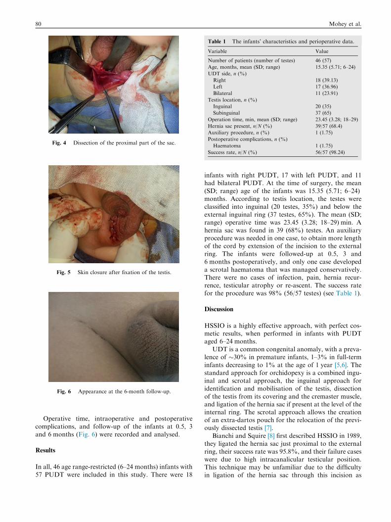
Fig. 4 Dissection of the proximal part of the sac.
Fig. 5 Skin closure after fixation of the testis.
Fig. 6 Appearance at the 6-month follow-up.
Table 1 The infants’ characteristics and perioperative data.
Variable Value
Number of patients (number of testes) 46 (57)
Age, months, mean (SD; range) 15.35 (5.71; 6–24)
UDT side, n (%)
Right 18 (39.13)
Left 17 (36.96)
Bilateral 11 (23.91)
Testis location, n (%)
Inguinal 20 (35)
Subinguinal 37 (65)
Operation time, min, mean (SD; range) 23.45 (3.28; 18–29)
Hernia sac present, n/N (%) 39/57 (68.4)
Auxiliary procedure, n (%) 1 (1.75)
Postoperative complications, n (%)
Haematoma 1 (1.75)
Success rate, n/N (%) 56/57 (98.24)
80 Mohey et al.
Operative time, intraoperative and postoperativecomplications, and follow-up of the infants at 0.5, 3and 6 months (Fig. 6) were recorded and analysed.
Results
In all, 46 age range-restricted (6–24 months) infants with57 PUDT were included in this study. There were 18
infants with right PUDT, 17 with left PUDT, and 11had bilateral PUDT. At the time of surgery, the mean(SD; range) age of the infants was 15.35 (5.71; 6–24)months. According to testis location, the testes wereclassified into inguinal (20 testes, 35%) and below theexternal inguinal ring (37 testes, 65%). The mean (SD;range) operative time was 23.45 (3.28; 18–29) min. Ahernia sac was found in 39 (68%) testes. An auxiliaryprocedure was needed in one case, to obtain more lengthof the cord by extension of the incision to the externalring. The infants were followed-up at 0.5, 3 and6 months postoperatively, and only one case developeda scrotal haematoma that was managed conservatively.There were no cases of infection, pain, hernia recur-rence, testicular atrophy or re-ascent. The success ratefor the procedure was 98% (56/57 testes) (see Table 1).
Discussion
HSSIO is a highly effective approach, with perfect cos-metic results, when performed in infants with PUDTaged 6–24 months.
UDT is a common congenital anomaly, with a preva-lence of �30% in premature infants, 1–3% in full-terminfants decreasing to 1% at the age of 1 year [5,6]. Thestandard approach for orchidopexy is a combined ingu-inal and scrotal approach, the inguinal approach foridentification and mobilisation of the testis, dissectionof the testis from its covering and the cremaster muscle,and ligation of the hernia sac if present at the level of theinternal ring. The scrotal approach allows the creationof an extra-dartos pouch for the relocation of the previ-ously dissected testis [7].
Bianchi and Squire [8] first described HSSIO in 1989,they ligated the hernia sac just proximal to the externalring, their success rate was 95.8%, and their failure caseswere due to high intracanalicular testicular position.This technique may be unfamiliar due to the difficultyin ligation of the hernia sac through this incision as

High single scrotal-incision orchidopexy 81
described by Redman [9], Iyer et al. [10] reported on 367cases of high scrotal orchidopexies, their success ratewas 96.2%.
Misra et al. [7] adapted the Bianchi and Squire [8]approach and used a low transverse scrotal incision,but their technique was criticised as it cannot create asufficient extra-dartos pouch and cannot reach intra-canalicular testis [11]. Also Parson et al. [12] conducted71 orchidopexies of palpable testes through a low scrotalincision; however, when there was a hernia sac they con-verted to the classic approach. So they proposed that thesingle scrotal incision is indicated only in cases withouthernia sacs.
Parnis et al. [4] documented that between the ages of6 and 24 months, the inguinal canal is short (0.7–1.1 cm), so proper dissection and ligation of a herniasac does not need opening of the canal. Dayanc et al.[13] categorised their study according to the UDTs posi-tion into: the inguinal canal and below the external ingu-inal ring, with success rates of 89.7% and 97.6%,respectively.
The presence of a hernia sac in conjugation with anUDT ranges from 20% to 70% [12,14]. In the presentseries, a hernia sac was found in 68.4% of the UDTsand all were ligated properly at the level of the internalring.
The conversion rate from HSSIO to the standardcombined inguinal and scrotal approach ranges from0% to 13% [11], in the present study there were no con-versions to the standard approach. For postoperativecomplications, we had only one case of scrotal wall hae-matoma that was managed conservatively. All patientscomplied with follow-up and there were no cases of tes-ticular atrophy, re-ascent or hernia recurrence duringfollow up of the infants at 0.5, 3 and 6 months.
In the present study, our success rate was very high at98%, irrespective of the testicular position, either ingu-inal or below the external inguinal ring, and this is prob-ably due to the age being restricted to between 6 and24 months, as the inguinal canal is short and thus theundescended testis can be reached even if intracanalicu-lar through the high scrotal transverse incision. We suc-ceeded in completing the procedure safely without theneed for any auxiliary procedure, except in one casewhere the external ring was opened as more cord lengthwas needed.
None of the infants presented with bothersome pain,so there was no need for analgesics postoperatively. Theprocedure was considered as day case surgery. The lim-itations of the present study include the relatively fewinfants included and the lack of a control group (i.e.
standard combined inguinal and scrotal approach). Wehope that a larger number of patients in this age rangewill be available for future studies.
In conclusion, HSSIO is a safe and feasible technique,with good cosmesis and apparently less discomfort forinfants aged between 6 and 24 months. The very highsuccess rate and few complications encountered, suggestthat HSSIO should be considered as the standard tech-nique for orchidopexy in this age group.
Conflicts of interest
None.
References
[1] Berkowitz GS, Lapinski RH, Dolgin SE, Gazella JG, Bodian CA,
Holzman IR. Prevalence and natural history of cryptorchidism.
Pediatrics 1993;92:44–9.
[2] Ritchey ML, Bloom DA. Modified dartos pouch orchiopexy.
Urology 1995;45:136–8.
[3] Callewaert PR, Rahnama’i MS, Biallosterski BT, van Kerre-
broeck PE. Scrotal approach to both palpable and impalpable
undescended testes: should it become our first choice? Urology
2010;76:73–6.
[4] Parnis SJ, Roberts JP, Hutson JM. Anatomical landmarks of the
inguinal canal in prepubescent children. Aust NZ J Surg
1997;67:335–7.
[5] Caruso AP, Walsh RA, Wolach JW, Koyle MA. Single scrotal
incision orchiopexy for the palpable undescended testicle. J Urol
2000;164:156–8.
[6] Kanemoto K, Hayashi Y, Kojima Y, Tozawa K, Mogami T,
Kohri K. The management of nonpalpable testis with combined
groin exploration and subsequent transinguinal laparoscopy. J
Urol 2002;167:674–6.
[7] Misra D, Dias R, Kapila L. Scrotal fixation. A different surgical
approach in the management of the low undescended testes.
Urology 1997;49:762–5.
[8] Bianchi A, Squire BR. Transscrotal orchidopexy: orchidopexy
revised. Pediatr Surg Int 1989;4:189–92.
[9] Redman JF. Simplified technique for scrotal pouch orchiopexy.
Urol Clin North Am 1990;17:9–12.
[10] Iyer KR, Kumar V, Huddart SN, Bianchi A. The scrotal
approach. Pediatr Surg Int 1995;10:58–60.
[11] Gordon M, Cervellione RM, Morabito A, Bianchi A. 20 years of
transcrotal orchidopexy for undescended testis: results and
outcomes. J Pediatr Urol 2010;6:506–12.
[12] Parsons JK, Ferrer F, Docimo SG. The low scrotal approach to
the ectopic or ascended testicle: prevalence of a patent processus
vaginalis. J Urol 2003;169:1832–3.
[13] Dayanc M, Kibar Y, Irkilata HC, Demir E, Tahmaz L, Peker AF.
Long-term outcome of scrotal incision orchiopexy for unde-
scended testis. Urology 2007;70:786–8.
[14] Russinko PJ, Siddiq FM, Tackett LD, Caldamone AA. Prescro-
talorchiopexy: an alternative surgical approach for the palpable
undescended testis. J Urol 2003;170:2436–8.


![Circumcision-incision orchidopexy: A novel technique for ... · childhood for the treatment of phimosis, paraphimosis, or recurrent balanoposthitis [3]. Furthermore, recent literature](https://img.pdfslide.us/doc/110x75/5c923dc109d3f244438d130b/circumcision-incision-orchidopexy-a-novel-technique-for-childhood-for-the.jpg)