Embed Size (px)
Citation preview
High salt-induced conversion of Escherichia coli GroEL into a fully functional
thermophilic chaperonin
Andrew R. Kusmierczyk and Jörg Martin
Department of Molecular Biology, Cell Biology, and Biochemistry, Brown University,
Box G-J2, Providence, RI 02912
Running Title: GroEL chaperonin activity at high temperatures
1
Copyright 2000 by The American Society for Biochemistry and Molecular Biology, Inc.
This is the author's manuscript of the article published in final edited form as:
Kusmierczyk, A. R., & Martin, J. (2000). High Salt-induced Conversion of Escherichia coliGroEL into a Fully Functional Thermophilic Chaperonin. Journal of Biological Chemistry, 275(43), 33504–33511. Available from: http://dx.doi.org/10.1074/jbc.M006256200
brought to you by COREView metadata, citation and similar papers at core.ac.uk
provided by IUPUIScholarWorks

Corresponding Author: Jörg Martin
Tel: (401) 863 3540
Fax: (401) 863 1201
E-mail: [email protected]
2
The GroE chaperonin system can adapt to and function at various environmental folding
conditions. To examine chaperonin-assisted protein folding at high salt concentrations,
we characterized Escherichia coli GroE chaperonin activity in 1.2 M ammonium sulfate.
Our data are consistent with GroEL undergoing a conformational change at this salt
concentration, characterized by elevated ATPase activity and increased exposure of
hydrophobic surface, as indicated by increased binding of the fluorophore bis-(5,5’)-8-
anilino-1-naphthalene sulfonic acid (bisANS) to the chaperonin. The presence of the salt
results in increased substrate stringency and dependence on the full GroE system for
release and productive folding of substrate proteins. Surprisingly, GroEL is fully
functional as a thermophilic chaperonin in high concentrations of ammonium sulfate and
is stable at temperatures up to 75°C. At these extreme conditions, GroEL can suppress
aggregation and mediate refolding of non-native proteins.
3
INTRODUCTION
The chaperonin GroEL from Escherichia coli belongs to a class of proteins, termed
molecular chaperones, whose collective function is to assist in the folding of newly
synthesized proteins and in the refolding of non-native polypeptides generated under
conditions of stress (reviewed in 1, 2). Like its homologs, CCT1 in eukaryotes and the
thermosome in archaea, GroEL forms a multi-subunit assembly arranged into twin rings
stacked end-to-end (3-5). The resultant homotetradecamer of 57 kDa subunits provides
a deep cavity where non-native protein species may bind and undergo productive folding
(6). GroEL is assisted in its chaperoning function by GroES, a heptamer composed of
identical 10 kDa subunits arranged into a single ring (7, 8). One of the functions of
GroES is to act as a lid on the GroEL cylinder, thereby providing an enclosed
environment for the folding polypeptide. However, its binding to GroEL plays other
important roles too, such as modulating the low intrinsic ATPase activity of GroEL by
coordinating the actions of nucleotide binding and hydrolysis (7-9). Crystallographic
data has enabled the visualization of the GroEL complex and individual subunit
architecture (10-12). It has revealed the regions implicated in both nucleotide and
substrate binding. It is now known that each subunit of GroEL is arranged into three
domains. An equatorial domain forms the majority of inter-subunit interactions and is the
site of nucleotide binding. The apical domain contains the substrate binding and GroES
binding residues (13). The two domains are connected by a hinge region, which transmits
4
information on the status of GroES, polypeptide, and nucleotide binding (14). Since
GroEL/GroES has been the most widely studied chaperonin system, its mechanism of
protein folding is known in some detail. Briefly, GroEL binds substrate protein in one of
its two ring cavities. The bound substrate protein is in a molten-globule state
characterized by the presence of secondary structure but lacking well-defined tertiary
structure (8, 15). Cooperative binding of seven molecules of ATP to the same (cis) ring as
the bound polypeptide is immediately followed by the binding of GroES, also to the cis
ring, which causes the polypeptide to be displaced into the cavity (16, 17). The released
polypeptide now folds in the protective environment of the enclosed chaperonin complex.
Hydrolysis of the seven ATP molecules primes the cis complex for disassembly, and the
binding of seven ATP molecules to the opposite (trans) ring of GroEL causes the release
of both GroES and the folded substrate (18). The system is now reset and ready to either
accept a new substrate or rebind the just-released but not yet native polypeptide for
another round of folding.
Folding by GroEL involves a complex interplay of three ligands (i.e. substrate,
GroES, and nucleotide), and all three are capable of inducing allosteric conformational
changes within GroEL, either individually or in concert (19-21). Attempts to perturb the
GroEL system by mutagenesis (13, 22-24), chemical modification (14, 25), substrate
modification (26, 27) or solvent manipulation (28-30), have often resulted in chaperonins
with altered functional properties. These can provide a wealth of information on the inner
workings of the system as a whole. Here we report on the functional properties of one
such altered state, induced by the presence of high concentrations of ammonium sulfate.
5
Our work was prompted by structural data on both GroEL and the thermosome
from the archaeon Thermoplasma acidophilum, which have been obtained from the
analysis of crystals grown in high concentrations of ammonium sulfate (10, 31).
Furthermore, the in vitro assembly of functional chaperonins from certain species of
archaea has required the presence of ammonium sulfate (32), and the ATPase activity of
at least one archaeal chaperonin has been shown to be dependant on a relatively high
concentration of ammonium ions (33). We find that high ammonium sulfate
concentrations alter the functional properties of GroEL, resulting in, among other things,
an increased hydrophobic surface area and increased stringency for protein folding. Most
surprisingly, these conditions allow the extension of E. coli chaperonin action to
thermophilic conditions.
1 The abbreviations used are: CCT, chaperonin containing TCP-1; bisANS, bis-(5,5’)-
6

8-anilino-1-naphthalene sulfonic acid; DTT, dithiothreitol; EDTA, ethylenediamine-
tetraacetic acid; GFP, green fluorescent protein; MOPS, 3-(N-morpholino)
propanesulfonic acid; NEM, N-ethylmaleimide; Pi , inorganic phosphate.
7
EXPERIMENTAL PROCEDURES
Proteins —GroEL, and GroES, were expressed in and purified from E. coli as previously
described (8, 17). In a final additional step in purification of GroEL, contaminating
peptides were removed on a Reactive Red 120 column (34). Removal of tryptophan-
containing impurities was confirmed by measuring fluorescence emission spectra of
eluted fractions (excitation 295 nm, emission from 325 to 355 nm). Protein
concentrations were determined spectrophotometrically based on the procedure outlined
by Gill and von Hippel (35), using the following extinction coefficients: ε276 = 8700
M-1cm-1 for GroEL; ε276 = 1450 M-1cm-1 for GroES. Recombinant green fluorescent
protein GFP was purified as described (36).
GroEL ATPase activity GroEL (125 nM) was equilibrated for 10 min at a given
temperature in 60 µL of buffer A (25 mM MOPS-NaOH pH 7.5, 5 mM MgCl2, 2 mM
DTT) supplemented with various salts as described in the figure legends. Ionic strength
of the salts was calculated according to I = 1/2Σcz2 where c is the concentration and z is
the charge of each ionic species generated by a given salt in solution. Where indicated,
GroES was present at a four-fold molar excess over GroEL, and αs1-casein (Sigma)
was present at a five-fold molar excess over GroEL. ATPase activity was initiated by the
addition of ATP to 2 mM and allowed to proceed for 10 min. The reaction was stopped
by the addition of CDTA to 10 mM and 50 µL were withdrawn for quantification of
8
liberated inorganic phosphate by the malachite green assay as previously described (37).
The absorbance was measured at 640 nm.
Analysis of GroEL Stability – GroEL (530 nM) was diluted at 75°C into buffer
containing 25 mM MOPS-NaOH pH 7.6, 5 mM MgCl2, and increasing concentrations of
(NH4)2SO4 as indicated in the figure legends. Protein denaturation was followed as
aggregation by measuring light scattering at 320 nm. In a parallel experiment, GroEL
(300 nM) was diluted at 75°C into buffer containing 25 mM MOPS-NaOH pH 7.6, 5
mM MgCl2, and supplemented with 50 mM KCl or increasing concentrations of
(NH4)2SO4. GroEL samples were incubated for 20 min at 75°C and centrifuged for 5 min at
10,000 g to remove aggregates. The supernatants were desalted over a NICK column
(Amersham Pharmacia) into 25 mM MOPS-NaOH pH 7.2, 100 mM NaCl, and were
analyzed by non-denaturing polyacrylamide gel electrophoresis as described (4).
Analysis of GFP Folding — All GFP experiments were carried out either at room
temperature or at 70°C. Acid-denatured GFP was prepared according to the method of
Makino et al. (38). Briefly, GFP (22 µM) was incubated for 1 hour in 8 mM Tris-Cl pH
7.5, 12.5 mM HCl, 1 mM DTT, 0.3 mM EDTA. For spontaneous refolding, denatured
GFP was diluted 200-fold into 1 mL of buffer B (25 mM MOPS-NaOH pH 7.5, 5 mM
MgCl2, 5 mM DTT) supplemented with either 50 mM KCl or 1.2 M (NH4)2SO4. For
chaperonin-mediated refolding, denatured GFP was diluted as above into buffer B
containing a two-fold molar excess of GroEL. As indicated in the figure legends, folding
was initiated by the addition of nucleotide (2 mM) or nucleotide plus GroES (at a four-
9
fold molar excess over GroEL). Where indicated, αs1-casein was added at a five-fold
molar excess over GroEL. Intrinsic GFP fluorescence was measured at 508 nm in 10 sec
intervals using a FluoroMax-2 spectrofluorometer (Instruments S.A.) equipped with a
thermostatted cuvette cell-holder. Samples were excited at 398 nm. The excitation and
emission slit-widths were set at 5 and 3 nm of bandpass respectively. The refolding
solution was mixed continuously with a built-in magnetic stirrer.
Thermal Aggregation of Citrate Synthase —Porcine citrate synthase (173 nM; Roche
Molecular Biochemicals) was added to buffer C (25 mM MOPS-NaOH pH 7.6, 5 mM
MgCl2, 1.2 M (NH4)2SO4), pre-equilibrated to 70°C, in the absence or presence of
various amounts of GroEL as indicated in the figure legends. Aggregation was followed
by monitoring light scattering at 320 nm in an Ultrospec300 spectrophotometer
(Amersham-Pharmacia Biotech) equipped with a thermostatted cuvette cell-holder.
Where indicated, release of GroEL-bound citrate synthase was accomplished by the
addition of ATP to 2 mM.
α-Glucosidase Folding α-Glucosidase (270 nM) from Bacillus stearothermophilus
(Sigma) was heat-inactivated in buffer C, pre-equilibrated to 70°C, in the absence or
presence of chaperonins and ATP as indicated in the figure legends. Aliquots (50 µL)
were removed at the indicated times and assayed for α-glucosidase activity. For
reactivation experiments, α-glucosidase was first heat-inactivated for 60 min at 70°C in
the presence of a three-fold molar excess of GroEL. Refolding was initiated by the
addition of ATP (2 mM) and GroES (at a four-fold molar excess over GroEL).
10
Immediately upon addition, the sample was split in half. One half was shifted to 50°C,
while the other remained at 70°C. At the indicated time points, 50 µL aliquots were
mixed with 550 µL of assay buffer (20 mM MOPS-NaOH pH 7.2, 100 mM NaCl, 2 mM
EDTA, 3 mM p-nitrophenyl-α-glucoside) and incubated for 20 min at room
temperature. α-Glucosidase enzymatic activity was measured as increase in absorbance
at 400 nm.
GroEL Binding of bisANS A dilution series of bis-(5,5)-8-anilino-1-naphthalene
sulfonic acid dipotassium salt (bisANS; ICN) at various concentrations was prepared in
water. 3 µL of each dilution was added to 300 µL of buffer (250 nM GroEL, 25 mM
MOPS-NaOH pH 7.5, 5 mM MgCl2, and 50 mM KCl or 1.2 M (NH4)2SO4) and the
fluorescence emission spectrum recorded. Intensity maxima were plotted as a function of
bisANS concentration. Scatchard analysis was performed to determine the amount of
bisANS bound and the approximate affinity. To determine the maximum fluorescence
intensity of a given amount of bisANS that is totally bound by protein, 250 nM of
bisANS in KCl- or (NH4)2SO4-containing buffer was titrated with increasing amounts
of GroEL and graphical analysis of the resultant fluorescence emission spectra was
carried out according to Bohnert et al (39). Fluorescence emission spectra were recorded
at room temperature from 450 to 580 nm. Samples were excited at 397 nm. The
excitation and emission slit-widths were set at 5 and 3 nm of bandpass respectively.
11
RESULTS
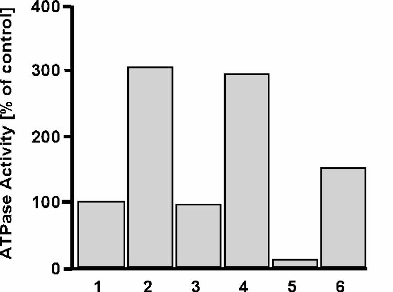
Increased GroEL ATPase Activity at High Concentrations of Ammonium Ions — A key
feature of the GroEL mechanism of action is its ATPase activity, which is modulated by
interaction of GroES and substrate protein with the chaperonin (4, 7, 8, 40, 41). The rate
of ATP hydrolysis responds to conformational changes in the chaperonin, and can
therefore serve as a sensitive indicator of possible structural alterations in GroEL. We
investigated the effect of high salt concentrations on GroEL ATPase activity. It has been
demonstrated previously that ammonium ions can support the ATPase activity of GroEL,
although less efficiently than potassium ions (40). In contrast, we find that at high
concentrations, ammonium ions have a stimulatory effect exceeding that of potassium
ions considerably (Fig. 1). The ATPase activity of GroEL in 1.2 M (NH4)2SO4 (lane 2)
and 1.2 M NH4Cl (lane 4) was found to be approximately three-fold higher than that in
50 mM KCl (lane 1). The effect was not solely dependent upon ionic strength, as 1.2 M
KCl (lane 3; same ionic strength as lane 4) failed to stimulate ATP hydrolysis above
control. However, the stimulatory effect was ammonium-specific, as 1.2 M Na2SO4
alone (data not shown) did not support ATPase activity. It actually had an inhibitory
effect on the ATPase activity of GroEL in the presence of 50 mM KCl (lane 5). It is
noteworthy that the level of stimulation of ATPase activity is roughly the same for both
1.2 M (NH4)2SO4 and 1.2 M NH4Cl even though the latter salt supplies only half as
12
many ammonium ions on a per mole basis. The increased concentration of NH4+
provided by the ammonium sulfate may be necessary to counteract the inhibitory effect of
the SO42- anions, with a strongly elevated ATPase activity as a net result. In agreement
with this explanation, we find that the ATPase activity of GroEL in 0.6 M (NH4)2SO4
plus 0.6 M Na2SO4 is only 80% of that in 1.2 M (NH4)2SO4 (data not shown) even
though the two buffers have the same ionic strength and same SO42- concentration.
Despite sulfate having an inhibitory effect at lower temperatures, the stabilizing effect of
this anion becomes important for the chaperone activity of GroEL at high temperatures,
as we will demonstrate below. For this reason, in the remainder of this article we will
focus on the effects of ammonium sulfate.
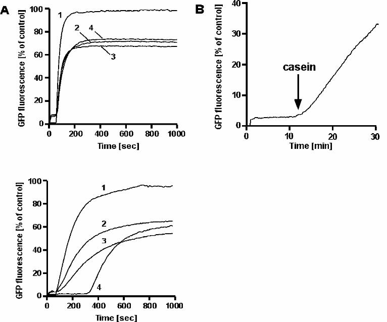
GroES-dependence of GroEL-mediated Refolding of Proteins in Ammonium Sulfate —
How does the changed ATPase rate in ammonium sulfate affect the ability of GroEL to
bind substrate protein and mediate folding? We determined the ability of GroEL to fold
proteins in the presence of 1.2 M (NH4)2SO4 using GFP, a monomeric 29 kDa protein,
which is an established GroEL substrate in vitro. Because GFP fluoresces only in the
native state, folding can be monitored by following its intrinsic fluorescence (38, 42).
Acid-denatured GFP folds spontaneously upon dilution into a renaturation buffer (38)
containing 50 mM KCl (Fig. 2 A, upper panel). When GroEL was present in the
renaturation buffer at a two-fold molar excess, folding was suppressed as the non-native
13
GFP was bound by GroEL. The addition of ATP alone was sufficient to release GFP
from GroEL for productive folding (ref. 38, Fig. 2 A). The full GroEL/GroES system in
the presence of ATP or ADP also supported folding in 50 mM KCl. In 1.2 M ammonium
sulfate, GFP folded spontaneously upon dilution into renaturation buffer and the presence
of GroEL in this buffer also suppressed folding (Fig. 2 A, lower panel). However, unlike
in 50 mM KCl, ATP alone was not sufficient to mediate the folding of GFP by GroEL in
1.2 M (NH4)2SO4. Instead, the full GroE system was required to reactivate GFP under
these conditions (trace 4). Under high salt conditions, where ATPase rates are strongly
increased, the time-span in which the chaperonin is in a low affinity state for unfolded
polypeptide is expected to be shortened. After the rate-limiting ATP hydrolysis step,
GroEL would then regain the acceptor state for substrate protein before released GFP
could internalize its hydrophobic structure elements. It would be re-bound immediately,
resulting in a steady-state association with the chaperonin. In that case it should be
possible to prevent rebinding of GFP by adding a competitor molecule. αs1-Casein is a
relatively hydrophobic, yet soluble protein that binds readily to chaperonins and has been
used as an effective substrate competitor under refolding conditions (8). We found that
addition of αs1-casein to a preformed GroEL-GFP complex resulted in release of the
substrate in the presence of ATP and its subsequent productive refolding (Fig. 2 B). The
inability of GroEL to release GFP in the presence of ATP alone can thus be explained by
a cycle of release and rapid recapture of substrate. This situation is reminiscent of that
observed with N-ethylmaleimide-modified GroEL (NEM-GroEL; ref. 14). It was shown
14
that covalent modification of a cysteine residue (C138) in the intermediate domain of
GroEL with N-ethylmaleimide results in a chaperonin with increased basal ATPase
activity and a more stringent requirement for the folding of substrate proteins (i.e.
previously GroES-independent substrates like dihydrofolate reductase now were GroES-
dependent). The altered properties of GroEL were attributed to a disruption in the
communication between the apical and equatorial domains (14). Typically, binding of
GroES to GroEL attenuates its ATPase activity by about 50%, whereas substrate binding
enhances it (4, 7, 8, 40, 41). The latter effect is expected because if ATP binding and
hydrolysis affect the affinity of GroEL for substrate protein, the reverse should apply as
well. In contrast, in an uncoupled system the two ligands, ATP and substrate protein,
should not affect each other. We were curious to see if the ammonium-induced changes
in the functional properties of GroEL were, as in NEM-GroEL, the result of an
uncoupling of the chaperonin system, and investigated the ATPase activity of GroEL in
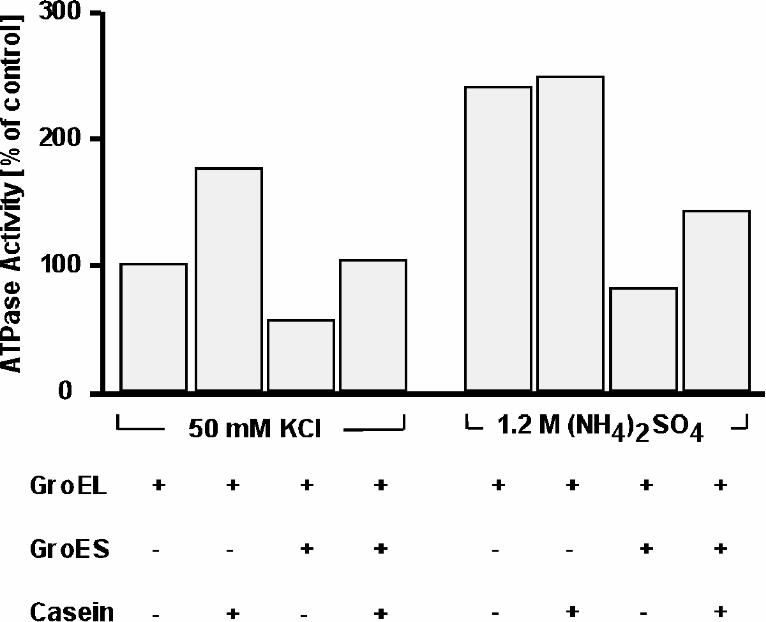
1.2 M (NH4)2SO4 in the presence of GroES or substrate proteins. As expected, in 50
mM KCl, a four-fold molar excess of GroES over GroEL reduced the ATPase activity to
56%, whereas a five-fold molar excess of αs1-casein over GroEL stimulated the ATPase
activity by 74% (Fig. 3). When αs1-casein was added to GroEL/GroES, stimulation of
84% over GroEL/GroES alone was observed. In ammonium sulfate, a different picture
emerged. While the addition of GroES inhibited the ATPase activity three-fold, the
addition of αs1-casein to GroEL alone was without effect (Fig. 3). However, when αs1-
casein was added to GroEL/GroES, we again observed stimulation of ATPase activity by
15
78% over that of GroEL/GroES alone. We conclude that, in contrast to NEM-GroEL,
communication between the apical and equatorial domains is not compromised in 1.2 M
(NH4)2SO4 because substrate can still elicit the expected increase in ATPase activity
when GroES is present.
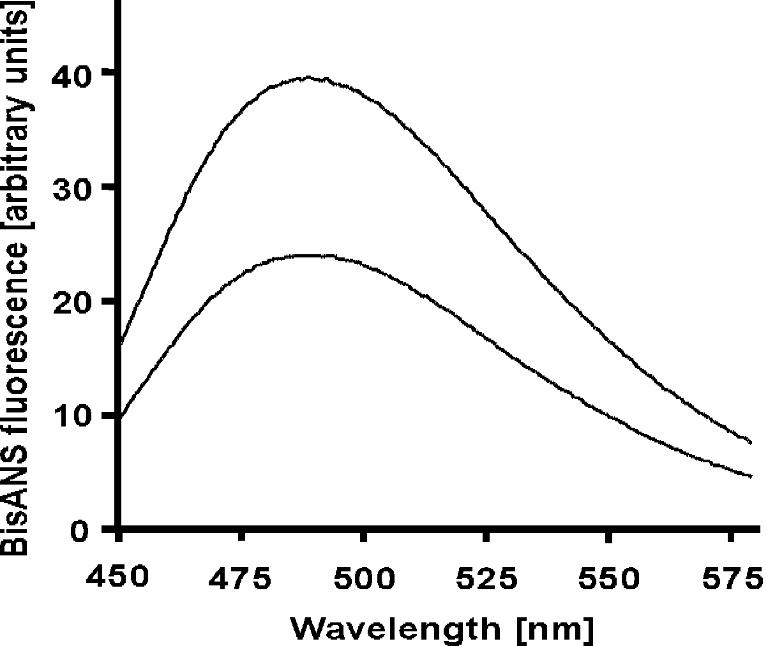
Increased Binding of bisANS to GroEL in 1.2 M (NH4)2SO4 — To determine in more
detail the nature of the altered state of GroEL in the presence of 1.2 M (NH4)2SO4, we
revisited the observation that ATP alone was not sufficient to release GFP from GroEL
(Fig. 2B) and tested whether or not other factors, in addition to a shortened time-window
for folding, could be contributing to this phenomenon. Because substrate binding to
GroEL has been demonstrated to involve numerous hydrophobic interactions (13, 43), we
considered the possibility that the substrate has a higher affinity for GroEL. This could be
because binding sites in GroEL are now more hydrophobic or because there are more
binding sites for substrate available in GroEL. A method of choice for studying GroEL-
mediated folding has been the use of chemical probes such as bisANS. BisANS is a
fluorescent probe whose quantum yield increases with increasing hydrophobicity of its
environment. It binds readily and noncovalently to exposed hydrophobic regions on
proteins and has thus been used extensively to probe both conformational changes and
folding in a number of protein systems (28, 30, 39, 44, 45). It has been demonstrated that
GroEL exposes a region of hydrophobic residues in its apical domain implicated in
substrate binding (11, 13) and that bisANS binds readily to GroEL, presumably to this
very region (45). We titrated a fixed amount of GroEL in both 50 mM KCl and 1.2 M
16
(NH4)2SO4 with increasing concentrations of bisANS. At each concentration tested, the
bisANS fluorescence was markedly higher in 1.2 M (NH4)2SO4 than in 50 mM KCl
(Fig. 4; representative spectrum). Also, the emission λmax was at the same wavelength
in both 50 mM KCl and 1.2 M (NH4)2SO4. Since λmax of fluorescence emission is an
indicator of the degree of hydrophobicity of the fluorophore environment (46), these
findings taken together suggest that the bisANS binding sites were not more hydrophobic
in character in 1.2 M (NH4)2SO4 than in low salt. Rather, more bisANS molecules were
bound per GroEL complex in 1.2 M (NH4)2SO4. To control for the possibility that
ammonium sulfate was enhancing bisANS fluorescence or that potassium chloride was
acting as a quencher, we titrated a fixed amount of bisANS with increasing amounts of
GroEL in both 50 mM KCl and in 1.2 M (NH4)2SO4. When bisANS is completely
bound by GroEL, its fluorescence should be the same in both salts provided that the local
binding environment is equally hydrophobic. Since λmax is the same in both 50 mM
KCl and 1.2 M (NH4)2SO4 (Fig. 4), we are confident that the binding environment
offered by GroEL is equally hydrophobic in both salts, and therefore any difference
observed should be due to buffer composition alone. Having titrated 250 nM bisANS
with increasing amounts of GroEL in both 50 mM KCl and 1.2 M (NH4)2SO4, we
graphically extrapolated the resultant fluorescence intensity maxima to infinite protein
17
concentration, as described by Bohnert et al (39). The values for all-bound bisANS
obtained in this way in KCl and (NH4)2SO4-containing buffers were found to differ by
no more than 5% (data not shown). We conclude that buffer composition has no effect on
the observed bisANS fluorescence. Using the fluorescence values obtained for the all-
bound bisANS, we applied Scatchard analysis to the titration data and found the number
of bisANS binding sites per 14-mer GroEL to be 21.6 in 50 mM KCl and 29.6 in 1.2 M
(NH4)2SO4, with apparent dissociation constants of 13.7 and 10.9 µM respectively.
These values are comparable to, although somewhat higher, than those obtained in
previous studies (28, 30). Thus, the increased bisANS fluorescence observed in 1.2 M
(NH4)2SO4 genuinely reflects an increase in the hydrophobic area available for bisANS
binding. The increased exposure of solvent-accessible hydrophobic sites may at least in
part be responsible for the increased substrate stringency observed with GFP.
Heat-stability and ATPase Activity of GroEL at 70°C in Ammonium Sulfate — It had
previously been reported that GroEL ATPase activity reached a maximum near 60°C and
that at higher temperatures, the protein rapidly denatured and became non-functional (47,
48). We describe here conditions under which the operational range of GroEL can be
extended well beyond the previously reported maximum. While investigating the
functional properties of GroEL in 1.2 M (NH4)2SO4, we noticed the chaperonin to be
stable and active at elevated temperatures. Figure 5 A shows that GroEL possesses
significant ATPase activity at 70°C in 1.2 M (NH4)2SO4 that was unequaled in other
18
buffers. This activity could be inhibited by GroES (data not shown). We were able to
demonstrate similar ATPase activity even at a temperature of 75°C (data not shown).
Notably, 1.2 M NH4Cl (lane 4) was virtually unable to support ATPase activity at 70°C
even though it was as effective as 1.2 M (NH4)2SO4 at 30°C (refer to Fig. 1). Likewise,
the ability of 3.6 M NH4Cl (Fig. 5 A, lane 5), which has the same ionic strength as 1.2 M
(NH4)2SO4, to support ATPase activity was significantly lower by comparison. In
accordance with earlier observations (48), GroEL in 50 mM KCl is virtually inactive at
70°C (lane 1). We ascribe this novel feature of E. coli GroEL, functioning as a
thermophilic protein, to stabilization provided by the sulfate ions. This is inferred from
the observation that unlike in 50 mM KCl alone (Fig. 5 A, lane 1), GroEL at 70°C in 50
mM KCl plus 1.2 M Na2SO4 exhibited ATPase activity significantly higher than that of
GroEL in 50 mM KCl at 30°C (lane 5). The stabilization effect was also observed
directly by following the time course of unfolding of GroEL at 75°C. At 50 mM KCl
(data not shown) and at medium concentrations of (NH4)2SO4 (0.6 - 0.8 M), GroEL
aggregated rapidly at this high temperature (Fig. 5 B). Increasing the salt concentration
had a protective effect such that at 1.2 M (NH4)2SO4, GroEL remained fully soluble and
active over the course of the experiment. Non-denaturing polyacrylamide gel
electrophoresis confirmed this result (Fig. 5 C). When GroEL was incubated for 20 min
at 75°C in buffer supplemented with 1.0 or 1.2 M (NH4)2SO4, all of it was soluble and
19
fully assembled as judged by its migration on a native gel (Fig. 5 C, lanes 4 and 5). In
contrast, only a small fraction of GroEL remained soluble and assembled in 0.6 M
(NH4)2SO4. No soluble GroEL was detected in 50 mM KCl in agreement with previous reports
that GroEL is unstable at temperatures above 60°C in low salt buffers (47, 48).
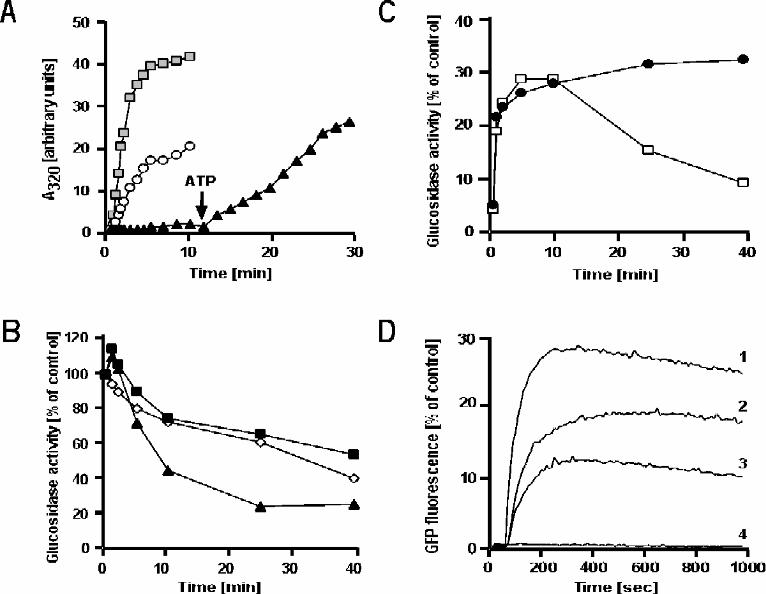
GroEL Chaperone Activity at 70°C in Ammonium Sulfate — The finding that E.
coli GroEL was stable and possessed ATPase activity under thermophilic conditions
opened the exciting possibility that it could function as a chaperone under these
conditions as well. First we asked if GroEL can bind unfolded polypeptides and prevent
them from thermal aggregation. As a model substrate, we used porcine citrate synthase, a
homodimer of 48 kDa subunits. Citrate synthase is a thermolabile protein with a mid-
point transition temperature for thermal denaturation of only 43°C and as such aggregates
rapidly and irreversibly (49). When citrate synthase was diluted into buffer containing 1.2
M (NH4)2SO4 at 70°C, it aggregated immediately (Fig. 6 A). Aggregation was inhibited
by GroEL in a concentration-dependent manner, and was completely suppressed at a
three-fold molar excess of GroEL. When ATP was added, citrate synthase was released
from GroEL and aggregated rapidly. Interestingly, under these conditions ATP alone
appears sufficient to affect release of substrate from GroEL, in contrast to the results
obtained with GFP at room temperature (Fig. 2). At 70°C the thermal energy is expected
to be sufficiently high to allow aggregation to occur on a faster scale than rebinding to
GroEL. Unlike citrate synthase, GFP does not aggregate when released from GroEL and
there is no side reaction to compete with rebinding to the chaperonin. We reasoned that
20
we should be able to reestablish increased substrate stringency in 1.2 M (NH4)2SO4 at
70°C by using a more thermotolerant substrate for our experiments. The results described
below show that this is indeed the case. α-Glucosidase from Bacillus stearothermophilus,
a moderate thermophile with a growth optimum of 55°C (50), exhibited a gradual loss of
activity upon incubation at 70°C in 1.2 M (NH4)2SO4 (Fig. 6B). In the presence of
GroEL, GroES, and ATP, only a marginal stabilization was observed. In the presence of a
three-fold molar excess of GroEL alone, the loss of activity was accelerated. The faster
loss of α-glucosidase activity in the presence of GroEL can be attributed to the binding
of heat-inactivated intermediates of α-glucosidase to the chaperonin. As α-glucosidase
slowly inactivates, an equilibrium condition likely exists between the native form and the
non-native intermediate(s). GroEL binding to these intermediates would shift the
equilibrium away from the native form, thereby causing a faster decline in α-glucosidase
activity. After 60 min of heat-inactivation, residual α-glucosidase activity was no more
than 5% of control (Fig. 6 C). At this point (t = 0), we attempted refolding of the protein
by addition of ATP and GroES. ATP alone was unable to affect any recovery of α-
glucosidase activity (data not shown). Only the full chaperonin system was capable of
restoring α-glucosidase activity (Fig. 6 C). Enzymatic activity began to decline again
after 10 min, presumably because the protein is thermodynamically unstable under these
conditions. When refolding in a parallel sample was done at 50°C, a temperature at which
the protein is stable, activity did not decline. These results confirm that the GroEL/GroES
chaperonin system is functional at thermophilic conditions in 1.2 M (NH4)2SO4.
21
Finally, we studied the folding of GFP at 70°C in 1.2 M (NH4)2SO4. This protein
appeared to be the most robust of the substrates tested in this study, as the fluorescence of
native GFP under these conditions was nearly identical to that at room temperature and
declined by no more than 5 to 10% over the course of the experiment (data not shown).
Acid-denatured GFP refolded spontaneously upon dilution into 1.2 M (NH4)2SO4-
containing buffer at 70°C with a recovery of about 30% (Fig. 6 D, trace 1). Dilution of
GFP into buffer containing a two-fold molar excess of GroEL prevented spontaneous
refolding caused by binding of the non-native protein to the chaperonin. Like at room
temperature, addition of ATP alone was unable to release GFP from GroEL (trace 4). In
the presence of GroES, together with ATP or ADP, folding resumed. We conclude that
the presence of 1.2 M (NH4)2SO4 extends considerably the range of GroEL action,
preserving its chaperone activity at high temperatures.
22
DISCUSSION
In this study, we have characterized the functional properties of the GroEL chaperonin
system in the presence of high salt concentrations. The most surprising result is the ability
of GroEL to function as a chaperone under thermophilic conditions in 1.2 M
(NH4)2SO4. GroEL is able to suppress thermally induced aggregation of citrate synthase. It can bind
to intermediates of heat-inactivated α-glucosidase and mediate their refolding in a
GroES-dependant manner. And, as at room temperature, it can bind and refold acid-
denatured GFP with the same increased substrate stringency. We find that high
concentrations of ammonium ions have a stimulatory effect on the ATPase activity of
GroEL. At 30°C, NH4Cl and (NH4)2SO4 are equally effective, even though the former
contributes only half the number of ammonium ions on a per mole basis. A higher
concentration of ammonium ions may counteract negative effects of sulfate. In fact,
Na2SO4 has an inhibitory effect on the ATPase activity of GroEL. At high concentrations,
some salts are thought to make a protein more rigid (51), and are widely used as protein
stabilizers. This is particularly true of SO42-, which has a high charge density and
resides high on the Hofmeister series of anions. Sulfate ions, by virtue of making the
protein more rigid, may hinder the ability of the chaperonin to hydrolyze ATP.
Nevertheless, the stabilizing effect of the sulfate ions enables GroEL to function at
thermophilic conditions by preventing its denaturation and keeping it soluble. This is
23
evident in the fact that at temperatures of up to 75°C, only ammonium sulfate is able to
support a markedly enhanced chaperonin activity.
Several features of the GroEL/GroES chaperonin system can be explained in
terms of a Monod-Wyman-Changeux representation (52). Each of the two rings can
either be in a tense acceptor state (T), in which GroEL has high affinity for substrate
protein, low affinity for ATP, and high ATP hydrolysis rates, or in a relaxed state (R)
with high affinity for ATP and low affinity for protein substrate. With increasing ATP
concentrations, the equilibrium of conformations shifts first to the TR state, and when
most GroEL subunits are occupied by ATP, the RR state dominates in which ATP
hydrolysis rates are slightly decreased and substrate protein is released (19, 20, 24, 53,
54). Our data are consistent with the possibility that (NH4)2SO4, rather than acting as an
uncoupler, induces a conformational change in GroEL to a TT-like state in which the
ATPase activity is at or near capacity. Unlike in low concentrations of KCl, where the
substrate protein αs1-casein stimulates the ATPase activity of GroEL, no such increase is
observed in (NH4)2SO4. Indeed, with GroEL already in a TT-like state, substrate
protein should have no further effect. It has been established that GroES binding to
GroEL regulates the ATPase activity of the chaperonin (4, 7, 8, 40, 41). When GroES
binds to GroEL, it is able to shift the chaperonin conformation to TR and RR states of
sub-maximal ATP hydrolysis. Consequently, in the presence of GroES, substrate should
show more pronounced effects in ammonium sulfate by trying to shift the T/R
equilibrium back toward the TT state. This is exactly what we have observed with αs1-
24
casein which stimulates ATP hydrolysis in 1.2 M (NH4)2SO4 in the presence of GroES.
GroEL is fully able to fold proteins under these conditions at both ambient and
thermophilic temperatures. Interestingly, the presence of the high concentration of
ammonium sulfate increases substrate stringency such that GFP, capable of folding in a
GroES-independent manner under low salt conditions, now becomes strictly GroES-
dependent. A strongly favored TT-like state in 1.2 M (NH4)2SO4 would explain this
inability of ATP hydrolysis alone to mediate GFP release from GroEL. In the absence of
GroES, the nucleotide is not able to induce on its own the conformational shifts toward
the TR and RR state that are necessary to dissociate the substrate. The predominantly
present TT-like GroEL form can thus be seen as locked in a conformation with high
substrate affinity. Moreover, in this conformation GroEL exposes more hydrophobic
binding surface than in low salt, which may affect the interaction with substrate protein.
GroEL-bound substrates are typically in a molten globule-like state (8, 15). This quasi-
ordered condition, in which secondary structure is present but tertiary structure is
undefined, is characterized by the exposure of hydrophobic residues which would
normally be buried in a native protein. GroEL contains a number of hydrophobic residues
in its apical domain that have been demonstrated to be necessary for binding of non-
native substrate protein (13). It has been demonstrated that perturbation of the ionic
strength of the solvent can increase exposure of hydrophobic residues on GroEL (28, 30).
Ammonium sulfate seems to elicit a similar change in the chaperonin. Titration data
presented here suggest that GroEL can bind more bisANS per tetradecamer in 1.2 M
25
(NH4)2SO4 than in low salt. Moreover, the hydrophobic nature of the binding sites is
comparable, as the emission λmax is the same in both salts. There may well be
additional reasons for the inability of ATP alone to mediate GFP release in (NH4)2SO4.
For example, increased GFP stringency could be the result of some change within GFP
itself induced by the high salt. The fluorescence of native GFP is virtually the same in
both 50 mM KCl and 1.2 M (NH4)2SO4, and the spontaneous recovery of fluorescence
of acid-denatured GFP is essentially complete in both buffers. This suggests that GFP
behaves similarly in both buffers. Nevertheless, it is conceivable that non-native GFP
intermediate(s) bound by GroEL upon dilution from denaturant are different in nature
such that those in 50 mM KCl are more amenable to release from GroEL by ATP alone
than those in 1.2 M (NH4)2SO4.
The presence of high concentrations of ammonium ions and ammonium sulfate
pertaining to chaperonin structure and function has surfaced a few times in recent
literature (10, 31-33). Notably, high ammonium sulfate concentrations were used to
obtain crystals for the determination of the structures of both E. coli GroEL and the
thermosome from T. acidophilum. Although the structures represent well the overall
architecture of the chaperonins, questions have arisen as to the nature of the actual state,
in terms of functional properties, that these structures represent. For instance, the
unliganded thermosome from T. acidophilum was crystallized in 2 M (NH4)2SO4 in a
“closed” conformation said to represent the Mg-ATP bound form (31). However,
26
Gutsche et al have recently demonstrated by small angle neutron scattering that in
solution, the Mg-ATP bound thermosome favors the “open” conformation in low salt
buffer (55). The closed conformation occurs only after ATP hydrolysis, but before release
of Pi. Surprisingly, Gutsche et al also showed that the crystallization buffer can induce
the closed conformation in solution (55). The crystals for the unliganded structure of
GroEL were grown in similarly high (NH4)2SO4 concentrations as those employed in
this study (10). Based on our results it is conceivable that the GroEL represented in that
crystal structure has solution properties similar to the functional state observed here.
Although these salt conditions are not physiologically relevant for E. coli in vivo, the
changes in chaperonin function that they induce are nevertheless informative. For
example, it was noted that high concentrations of sodium sulfate resulted in a stimulation
of the ATPase activity of the archaeal chaperonin because of the aforementioned
induction of the “closed” conformation which occurs after ATP hydrolysis (55). The
situation is different with bacterial chaperonins, as our findings show that sulfate inhibits
the ATPase of GroEL; an effect that is in turn counterbalanced by high concentrations of
ammonium ions. This difference serves to underscore the likelihood that despite having
structurally conserved ATP binding domains, the molecular basis of ATP hydrolysis in
the two chaperonin systems may differ in some respects.
It appears that examination of the solution properties of chaperonins, under the
solvent conditions used for crystallization, is a worthwhile endeavor in order to better
assign the functional state that the respective structures represent. Moreover, the ability to
27
convert a mesophilic chaperonin into a thermophilic chaperonin opens interesting
possibilities for direct comparison to and study of homologs from naturally occurring
thermophiles. Some methanogenic archaea use increased intracellular ion concentrations
to stabilize their proteins in vivo (56). Whether or not a similar method of
thermoadaptation is used by some extremophilic bacteria remains to be seen, but the
results presented here suggest that this is a distinct possibility.
28
REFERENCES
1. Martin, J., and Hartl, F.U. (1997) Curr. Opin. Struct. Biol. 7, 41-52
2. Fenton, W. A., and Horwich, A. L. (1997) Prot. Sci. 6, 743-760
3. Hemmingsen, S. M., Woolford, C., van der Vies, S. M., Tilly, K., Dennis, D. T.,
Georgopoulos, C. P., Hendrix, R., W., and Ellis, R. J. (1988) Nature 333, 330-334
4. Langer, T., Pfeifer, G., Martin, J., Baumeister, W., and Hartl, F.U. (1992) EMBO J. 11,
4757-4765
5. Gutsche, I., Essen L.-O., and Baumeister, W. (1999) J. Mol. Biol. 293, 295-312
6. Braig, K., Furuya, F., Hainfeld, J., and Horwich, A. L. (1993) Proc. Natl. Acad. Sci.
U.S.A. 90, 3978-3982
7. Chandrasekhar, G. N., Tilly, K., Woolford, C., Hendrix, R., and Georgopoulos, C.
(1986) J. Biol. Chem. 261, 12414-12419
8. Martin, J., Langer, T., Boteva, R., Schramel, A., Horwich, A. L., and Hartl, F.U.
(1991) Nature 352, 36-42
9. Gray, T.E., and Fersht, A.R. (1991) FEBS Lett. 292, 254-258
10. Braig, K., Otwinowski, Z., Hegde, R., Boisvert, D. C., Joachimiak, A., Horwich, A.
L., and Sigler, P. B. (1994) Nature 371, 578-586
11. Boisvert, D. C., Wang, J. M., Otwinowski, Z., Horwich, A. L., and Sigler, P. B.
(1996) Nature Struct. Biol. 3, 170-177
12. Xu, Z., Horwich, A. L., and Sigler, P. B. (1997) Nature 388, 741-749
13. Fenton, W. A., Kashi, Y., Furtak, K., and Horwich A. L. (1994) Nature 371, 614-619
29
14. Martin, J. (1998) J. Biol. Chem. 273, 7351-7357
15. Robinson, C.V., Gross, M., Eyles, S.J., Ewbank, J.J., Mayhew, M., Hartl, F.U.,
Dobson, C.M., and Radford, S.E. (1994) Nature 372, 646-651
16. Langer, T., Pfeifer, G., Martin, J., Baumeister, W., and Hartl, F. U. (1992) EMBO J.
11, 4757-4765
17. Martin, J., Mayhew, M., Langer, T., and Hartl, F. U. (1993) Nature 366, 228-233
18. Rye, H.S., Burston, S.G., Fenton, W.A., Beechem, J.M., Xu, Z., Sigler, P.B., and
Horwich, A.L. (1997) Nature 388, 792-798
19. Yifrach, O., and Horovitz, A. (1995) Biochemistry 34, 5303-5308
20. Yifrach, O., and Horovitz, A. (1996) J. Mol. Biol. 255, 356-361
21. Rye, H.S., Roseman, A.M., Chen, S., Furtak, K., Fenton, W.A., Saibil, H.R., and
Horwich, A.L. (1999) Cell 97, 325-338
22. Horovitz, A., Bochkareva, E.S., Kovalenko, O., and Girshovich, A.S. (1993) J. Mol.
Biol. 231, 58-64
23. Weissman, J.S., Kashi, Y., Fenton, W.A., and Horwich, A.L. (1994) Cell 78, 693-
702
24. Yifrach, O., and Horovitz, A. (2000) Proc. Natl. Acad. Sci. U.S.A. 97, 1521-1524
25. Gibbons, D.L., and Horowitz, P.M. (1995) J. Biol. Chem. 270, 7335-7340
26. Luo, G.-X., and Horowitz, P. M. (1994) J. Biol. Chem. 269, 32151-32154
27. Mayhew, M., da Silva, A.C.R., Erdjument-Bromage, H., Tempst, P., and Hartl, F.-U.
(1996) Nature 379, 420-426
28. Horowitz, P.M., Hua, S., and Gibbons, D.L. (1995) J. Biol. Chem. 270, 1535-1542
30
29. Perrett, S., Zahn, R., Stenberg, G., and Fersht, A.R. (1997) J. Mol. Biol. 269, 892-
901
30. Brazil, B.T., Ybarra, J., and Horowitz, P.M. (1998) J. Biol. Chem. 273, 3257-3263
31. Ditzel, L., Löwe, J., Stock, D., Stetter, K.-O., Huber, H., Huber, R., and Steinbacher,
S. (1998) Cell 93, 125-138
32. Furutani, M., Iida, T., Yoshida, T., and Maruyama, T. (1998) J. Biol. Chem. 273,
28399-28407
33. Andrä, S., Frey, G., Jaenicke, R., and Stetter, K.O. (1998) Eur. J. Biochem. 255, 93-
99
34. Clark, A.C., Ramanathan, R., and Frieden, C. (1998) Meth. Enzym. 290, 100-118
35. Gill, S.C., and von Hippel, P.H. (1989) Anal. Biochem. 182, 319-326
36. Deschamps J.R., Miller C.E., and Ward K.B. (1995) Protein Expr. Purif. 6, 555-558
37. Lanzetta, P.A., Alvarez, L.J., Reinach, P.S., and Candia, O.A. (1979) Anal. Biochem.
100, 95-97
38. Makino, Y., Amada, K., Taguchi, H., and Yoshida, M. (1997) J. Biol. Chem. 272,
12468-12474
39. Bohnert, J.L., Malencik, D.A., Anderson, S.R., Teller, D., and Fischer, E.H. (1982)
Biochemistry 21, 5570-5576
40. Viitanen, P. V., Lubben, T. H., Reed, J., Goloubinoff, P., O’Keefe, D. P., and
Lorimer, G. H. (1990) Biochemistry 29, 5665-5671
41. Jackson, G.S., Staniforth, R.A., Halsall, D.J., Atkinson, T., Holbrook, J.J., Clarke,
A.R., and Burston, S.G. (1993) Biochemistry 32, 2554-2563
31
42. Weissman, J.S., Rye, H.S., Fenton, W.A., Beechem, J.M., and Horwich, A.L. (1996)
Cell 84, 481-490
43. Hayer-Hartl, M. K., Ewbank J. J., Creigthon, T. E., and Hartl, F. U. (1994) EMBO J.
13, 3192-3202
44. Rosen, C.G., and Weber, G. (1969) Biochemistry 8, 3915-3920
45. Seale, J.W., Martinez, J.L., and Horowitz, P.M. (1995) Biochemistry 34, 7443-7449
46. Freifelder, D. (1982) Chapter 15: Fluorescence Spectroscopy in “Biophysical
Chemistry: Applications to Biotechnology and Molecular Biology”. W.H. Freeman and
Company, New York
47. Lissin N.M., Venyaminov S.Y., and Girshovich A.S. (1990) Nature 348, 339-342
48. Mendoza, J.A., Warren, T., and Dulin, P. (1996) Biochem. Biophys. Res. Commun.
229, 271-274
49. Minuth, T., Frey, G., Lindner, P., Rachel, R., Stetter, K.O., and Jaenicke, R. (1998)
Eur. J. Biochem. 258, 837-845
50. Takii, Y., Daimon, K., and Suzuki, Y. (1992) Appl. Microbiol. Biotechnol. 38, 243-
247
51. Timasheff, S.N., and Arakawa, T. (1997) Chapter 14: Stabilization of Protein
Structure by Solvents in “Protein Structure: A Practical Approach”. Oxford University
Press Inc., New York
52. Monod, J., Wyman, J., and Changeux, J.P. (1965) J. Mol. Biol. 12, 88-118
53. Ma, J., and Karplus, M. (1998) PNAS 95, 8502-8507
54. Cliff, M.J., Kad, N.M., Hay, N., Lund, P.A., Webb, M.R., Burston, S.G., and Clarke,
32
A.R. (1999) J. Mol. Biol. 293, 667-684
55. Gutsche, I., Holzinger, J., Rössle, M., Heumann, H., Baumeister, W., and May, R.P.
(2000) Curr. Biol. 10, 405-408
56. Hensel, R., König, H. (1988) FEMS Microbiol. Lett. 49, 75-79
Acknowledgements: We thank Jennifer Carr for critically reading the manuscript. This
research was supported by National Institutes of Health Research Grant GM54534.
33
FIGURE LEGENDS
FIG. 1. ATPase activity of GroEL under various salt conditions. GroEL (125 nM) in
buffer A was supplemented with various salts as indicated below. ATPase activity was
initiated at 30°C by addition of ATP (2 mM). The ATPase activity of GroEL in 50 mM
KCl (lane 1) is set as 100%. Lane 1: 50 mM KCl; Lane 2: 1.2 M (NH4)2SO4; Lane 3:
1.2 M KCl; Lane 4: 1.2 M NH4Cl; Lane 5: 1.2 M Na2SO4, 50 mM KCl; Lane 6: 3.6 M
NH4Cl.
FIG. 2. Chaperone activity of GroEL at high concentrations of ammonium sulfate. A)
Denatured GFP was diluted 200-fold into buffer B supplemented with 50 mM KCl
(upper panel) or 1.2 M (NH4)2SO4 (lower panel) containing 0.22 µM GroEL. To initiate
folding, the following additions were made at 60 sec: 2 mM ATP and 0.88 µM GroES
(trace 2); 2 mM ADP and 0.88 µM GroES (trace 3); 2 mM ATP (trace 4). Spontaneous
folding of GFP was observed upon dilution of denatured GFP into buffer without
chaperonins (trace 1). Fluorescence of native GFP in buffer is set as 100%. B) A
competing substrate protein can mediate release of GroEL-bound GFP in the presence of
ATP at a high ammonium sulfate concentration. Denatured GFP was diluted 200-fold
into buffer B supplemented with 1.2 M (NH4)2SO4 and 0.22 µM GroEL. ATP (2 mM)
and αs1-casein (1.1 µM) were added at 1 and 11 min respectively. The refolding of GFP
34
was followed by monitoring fluorescence at 508 nm.
FIG. 3. Effects of GroES and substrate protein on GroEL ATPase activity. The ATPase
of GroEL (125 nM) was assayed at 25°C in the presence or absence of αs1-casein (0,6
µM). The reaction was carried out in buffer A plus 50 mM KCl or 1.2 M (NH4)2SO4.
Where indicated, GroES was added at 0.5 µM. ATPase activity was initiated by the
addition of ATP (2 mM). The ATPase activity of GroEL in 50 mM KCl is set as 100%.
FIG. 4. Increased binding of bisANS to GroEL in elevated concentrations of ammonium
sulfate. BisANS (10 µM) was added to GroEL (250 nm) in 25 mM MOPS-NaOH pH
7.5, 5 mM MgCl2, supplemented with either 50 mM KCl (bottom trace) or 1.2 M
(NH4)2SO4 (top trace). Fluorescence emission spectra were recorded from 450 to 580 nm.
Samples were excited at 397 nm.
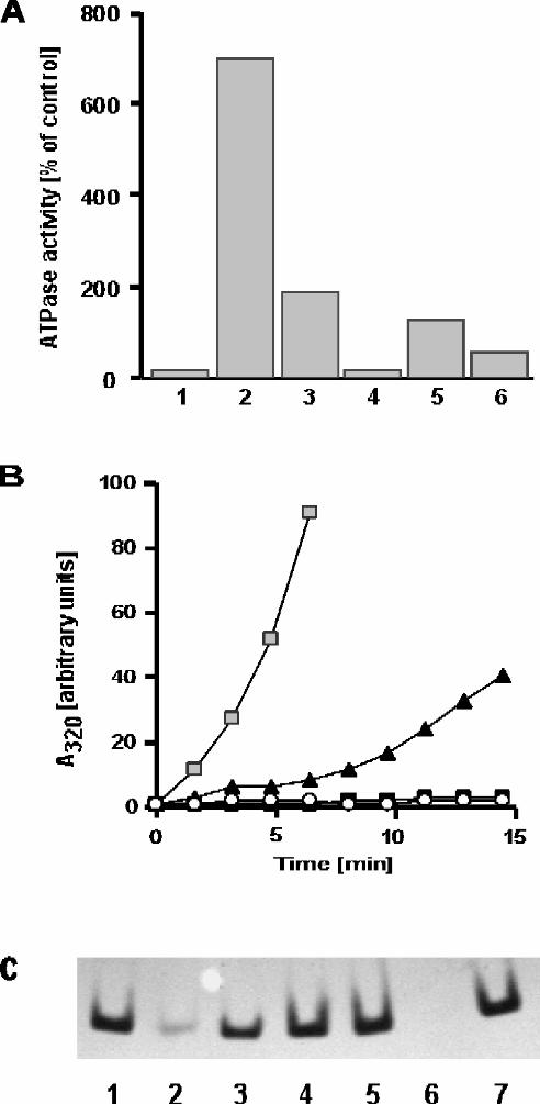
FIG. 5. GroEL stability and ATPase activity at high temperatures in elevated
concentrations of ammonium sulfate. A) GroEL (125 nM) was added at 70°C to buffer A
supplemented with various salts as indicated below. ATPase activity was initiated by
addition of ATP (2 mM). The ATPase activity of GroEL in 50 mM KCl at 30°C (as
shown in Fig. 1, lane 1) is set as 100%. Lane 1: 50 mM KCl; Lane 2: 1.2 M (NH4)2SO4;
Lane 3: 1.2 M KCl; Lane 4: 1.2 M NH4Cl; Lane 5: 1.2 M Na2SO4, 50 mM KCl; Lane
35
6: 3.6 M NH4Cl. B) GroEL (530 nM) was diluted at 75°C into buffer containing 25 mM
MOPS-NaOH pH 7.6, 5 mM MgCl2, and increasing concentrations of (NH4)2SO4: 0.6
M (grey squares); 0.8 M (black triangles); 1.0 M (black squares); 1.2 M (open circles).
Denaturation of GroEL was followed as protein aggregation by measuring light scattering
at 320 nm. C) GroEL (300 nM) was diluted at 75°C into buffer containing 25 mM
MOPS-NaOH pH 7.6, 5 mM MgCl2, and supplemented with the indicated salts. The
samples were incubated for 20 min at 75°C. Control samples were incubated at room
temperature. After centrifugation to remove aggregates, samples were desalted,
concentrated, and electrophoresed on a non-denaturing polyacrylamide gel. Lane 1: 1.2
M (NH4)2SO4 (control); Lane 2: 0.6 M (NH4)2SO4; Lane 3: 0.8 M (NH4)2SO4; Lane 4:
1.0 M (NH4)2SO4; Lane 5: 1.2 M (NH4)2SO4; Lane 6: 50 mM KCl; Lane 7: 50 mM
KCl (control).
FIG. 6. Chaperone activity of GroEL at high temperatures. A) Suppression of citrate
synthase aggregation. Citrate synthase (176 nM) was diluted at 70°C into buffer C, in the
absence or presence of various amounts of GroEL. Where indicated (arrow), ATP was
added to 2 mM. Protein aggregation was followed by measuring light scattering at 320
nm. Citrate synthase (grey squares); GroEL : Citrate synthase = 1 : 1 (open circles);
GroEL : Citrate synthase = 3 : 1 (black triangles). B) GroEL binding of heat-inactivated
α-glucosidase. α-Glucosidase (270 nM) was diluted into buffer C at 70°C in the absence
or presence of chaperonins (810 nM GroEL; 3.2 µM GroES) and ATP (2 mM). At the
36
indicated time points, aliquots were withdrawn and assayed for α-glucosidase activity.
α-Glucosidase (open diamonds); α-glucosidase plus GroEL (black triangles); α-
glucosidase plus GroEL/GroES and ATP (black squares). C) GroEL-mediated refolding
of heat-inactivated α-glucosidase. α-Glucosidase (270 nM) was heat-inactivated for 60
min at 70°C in the presence of a three-fold molar excess of GroEL in buffer C. At 60
min (t=0), refolding was initiated by the addition of 3.2 µM GroES and 2 mM ATP and
the sample was split in half. One of the two halves was shifted to 50°C (black circles)
while the other remained at 70°C (open squares). At the indicated time points, aliquots
were withdrawn and analyzed for α-glucosidase activity. D) Refolding of acid-denatured
GFP by GroEL at 70°C. Denatured GFP was diluted 200-fold into buffer B at 70°C
supplemented with 1.2 M
(NH4)2SO4 and 0.22 µM GroEL. To initiate folding,the following additions were made
at 60 sec: 2 mM ATP and 0.88 µM GroES (trace 2); 2 mM ADP and 0.88 µM GroES
(trace 3); 2 mM ATP (trace 4). Spontaneous folding of GFP was observed upon dilution of
denatured GFP into buffer B (trace 1). The amount recovered was set as 100%,
representing approximately 30% of the fluorescence of native GFP at 70°C. GFP
fluorescence was monitored at 508 nm.
37
at Indiana University School of M
edicine on October 3, 2014
http://ww
w.jbc.org/
Dow
nloaded from


![2012_very Good Review_drought Salt Temp Stress Induced Metabolic Rearragements[1]](https://img.pdfslide.us/doc/110x75/5450da2db1af9f19098b50c8/2012very-good-reviewdrought-salt-temp-stress-induced-metabolic-rearragements1.jpg)
