Embed Size (px)
Citation preview
ARTICLE
High-resolution protein structure determination startingwith a global fold calculated from exact solutionsto the RDC equations
Jianyang Zeng Æ Jeffrey Boyles Æ Chittaranjan Tripathy ÆLincong Wang Æ Anthony Yan Æ Pei Zhou ÆBruce Randall Donald
Received: 6 January 2009 / Accepted: 15 July 2009 / Published online: 27 August 2009
� Springer Science+Business Media B.V. 2009
Abstract We present a novel structure determination
approach that exploits the global orientational restraints
from RDCs to resolve ambiguous NOE assignments. Unlike
traditional approaches that bootstrap the initial fold from
ambiguous NOE assignments, we start by using RDCs to
compute accurate secondary structure element (SSE) back-
bones at the beginning of structure calculation. Our structure
determination package, called RDC-PANDA (RDC-based SSE
PAcking with NOEs for Structure Determination and NOE
Assignment), consists of three modules: (1) RDC-EXACT; (2)
PACKER; and (3) HANA (HAusdorff-based NOE Assignment).
RDC-EXACT computes the global optimal solution of backbone
dihedral angles for each secondary structure element by
exactly solving a system of quartic RDC equations derived
by Wang and Donald (Proceedings of the IEEE computa-
tional systems bioinformatics conference (CSB), Stanford,
CA, 2004a; J Biomol NMR 29(3):223–242, 2004b), and
systematically searching over the roots, each of which is a
backbone dihedral /- or w-angle consistent with the RDC
data. Using a small number of unambiguous inter-SSE
NOEs extracted using only chemical shift information,
PACKER performs a systematic search for the core structure,
including all SSE backbone conformations. HANA uses a
Hausdorff-based scoring function to measure the similarity
between the experimental spectra and the back-computed
NOE pattern for each side-chain from a statistically-diverse
rotamer library, and drives the selection of optimal position-
specific rotamers for filtering ambiguous NOE assignments.
Finally, a local minimization approach is used to compute
the loops and refine side-chain conformations by fixing the
core structure as a rigid body while allowing movement of
loops and side-chains. RDC-PANDA was applied to NMR data
for the FF Domain 2 of human transcription elongation
factor CA150 (RNA polymerase II C-terminal domain
interacting protein), human ubiquitin, the ubiquitin-binding
zinc finger domain of the human Y-family DNA polymerase
Eta (pol g UBZ), and the human Set2-Rpb1 interacting
domain (hSRI). These results demonstrated the efficiency
and accuracy of our algorithm, and show that RDC-PANDA can
be successfully applied for high-resolution protein structure
determination using only a limited set of NMR data by first
computing RDC-defined backbones.
Keywords Nuclear magnetic resonance � Nuclear
overhauser effect assignment � Residual dipolar coupling �Structure determination � Packing
Abbreviations
NMR Nuclear magnetic resonance
ppm Parts per million
The methodology developed in this paper has been applied to
compute the ensemble of structures for FF2. The atomic coordinates
have been deposited into the Protein Data Bank (PDB ID: 2KIQ).
Electronic supplementary material The online version of thisarticle (doi:10.1007/s10858-009-9366-3) contains supplementarymaterial, which is available to authorized users.
J. Zeng � C. Tripathy � L. Wang � A. Yan � B. R. Donald (&)
Department of Computer Science, Duke University, Durham,
NC 27708, USA
e-mail: [email protected]
J. Boyles � P. Zhou (&) � B. R. Donald
Department of Biochemistry, Duke University Medical Center,
Durham, NC 27708, USA
e-mail: [email protected]
Present Address:L. Wang
Medicinal Chemistry, Boehringer Ingelheim Pharmaceuticals,
Inc., Ridgefield, CT 06877, USA
123
J Biomol NMR (2009) 45:265–281
DOI 10.1007/s10858-009-9366-3
RMSD Root mean square deviation
HSQC Heteronuclear single quantum coherence
spectroscopy
NOE Nuclear Overhauser effect
NOESY Nuclear Overhauser and exchange
spectroscopy
RDC Residual dipolar coupling
PDB Protein Data Bank
pol g UBZ Ubiquitin-binding zinc finger domain of the
human Y-family DNA polymerase Eta
CH Ca-Ha
hSRI Human Set2-Rpb1 interacting domain
FF2 FF Domain 2 of human transcription
elongation factor CA150 (RNA polymerase
II C-terminal domain interacting protein)
POF Principal order frame
SA Simulated annealing
MD Molecular dynamics
SSE Secondary structure element
C0 Carbonyl carbon
WPS Well-packed satisfying
vdW van der Waals
SM Supplementary Material
Introduction
One of the main bottlenecks in protein NMR structure
determination lies in the acquisition and interpretation of a
sufficient number of accurate distance restraints from
nuclear Overhauser effect (NOE) data, which is often
obstructed by assignment ambiguities due to chemical shift
degeneracy. Traditional NMR structure determination
approaches (Guntert 2003; Mumenthaler et al. 1997;
Gronwald et al. 2002; Huang et al. 2006; Kuszewski et al.
2004) rely on heuristic techniques such as molecular
dynamics (MD) and simulated annealing (SA), and may
use a variety of data, including chemical shifts, scalar
couplings, NOEs and residual dipolar couplings (RDCs). In
these approaches, however, RDCs are typically not
employed in the annealing protocol until the end of struc-
ture calculation (i.e. refinement). Moreover, SA/MD based
structure determination algorithms are used in these
approaches as a subroutine in an iterative NOE assignment
protocol. The SA/MD structure determination subroutine
must typically be carefully initialized the first time using
only reliable NOE assignments. The computed structures
are then used to prune ambiguous NOE assignments. The
SA/MD subroutine is used in an iterative fashion with new
NOE assignments until convergence is reached.
Since NOEs provide merely local distance restraints, the
correctness of an NOE assignment usually depends on the
accuracy of other NOE assignments in its neighborhood.
Thus, an error in an incorrect NOE assignment can be
propagated, and hence influence the assignments of other
NOEs. In contrast to NOE restraints, RDCs provide global
orientational restraints on internuclear vectors, for exam-
ple, backbone NH and Ca–Ha (henceforth abbreviated to
CH) bond vectors with respect to a global alignment frame
(Tolman et al. 1995; Tjandra and Bax 1997). In addition, in
solution NMR RDC data can be collected with high pre-
cision, and assigned much faster than NOEs. Although
several attempts have been made to find self-consistent
NOE assignments (Guntert 2003; Mumenthaler et al. 1997;
Herrmann et al. 2002; Linge et al. 2003; Gronwald et al.
2002; Huang et al. 2006; Kuszewski et al. 2004), little
work has been done on exploiting other constraints such as
residual dipolar couplings (RDCs) for resolving ambiguous
NOE assignments. NOAH (Mumenthaler et al. 1997; Guntert
2003), for example, uses the structure determination
package DYANA (Herrmann et al. 2002), and starts with an
initial set of NOE assignments with putatively one or two
possible assignments. ARIA (Linge et al. 2003) and CANDID
(Herrmann et al. 2002) improved on NOAH by incorporat-
ing better modeling of ambiguous distance constraints.
For instance, in both programs, the form of a (P
r-6)-1/6
sum is used for handling ambiguous distances when
multiple possible assignments exist for an NOE crosspeak.
In AUTO-STRUCTURE (Huang et al. 2006), several heuristic
rules that simulate the expertise of manual assignment are
included to generate an initial fold. In PASD (Kuszewski
et al. 2004) several strategies were proposed to reduce the
chance of invoking the structure calculation into a biased
path due to the incorrect initial global fold. None of the
above NOE assignment approaches applied the global
constraints from RDC data to filter ambiguous NOE
assignments.
In traditional NMR structure determination approaches,
stochastic techniques such as SA/MD are employed in a
tight inner-loop and are invoked several times to filter
ambiguous NOE assignments (Guntert 2003). The objec-
tive function used in these stochastic techniques models
both the empirical molecular mechanics energy and the
satisfaction of experimental data for a protein structure.
Unfortunately, these stochastic techniques can be trapped
into local minima in the energy landscape of the objective
function. Missing the global minimum structure solution
during the iterative process can subsequently lead to
incorrect NOE assignments. Furthermore, most previous
approaches for automated NOE assignment (Herrmann
et al. 2002; Linge et al. 2003) heavily depend on an
accurate initial fold. However, the acquisition of a suffi-
cient number of initial NOE assignments for computing a
reliable starting fold is non-trivial, mainly due to the
chemical shift degeneracy. Manual intervention and human
266 J Biomol NMR (2009) 45:265–281
123
expertise are often required in assigning these important
initial NOEs in order to obtain a reliable initial fold.
To address the above issues, a polynomial time de novo
algorithm, called RDC-EXACT, has been proposed to compute
high-resolution backbone structures for secondary structure
elements (SSEs) using a minimum amount of residual
dipolar coupling (RDC) data (Wang and Donald 2004a, b;
Wang et al. 2006). The accurate backbone conformations
computed by this algorithm enable us to propose a new
strategy for NOE assignment. For example, a novel NOE
assignment algorithm (Wang and Donald 2005) was pro-
posed to filter ambiguous NOE assignments based on an
ensemble of distance intervals computed using intra-resi-
due vectors mined from a rotamer database, against inter-
residue vectors from the backbone structure determined
from RDCs. This algorithm uses a triangle-like inequality
between the intra-residue and inter-residue vectors to prune
incorrect assignments for side-chain NOEs. However, the
algorithm (Wang and Donald 2005) has the following
deficiencies: (a) it does not exploit the diversity of the
rotamers in the library, (b) it does not exploit the rotamer
patterns observable in NOE spectra, and (c) uncertainty in
NOE peak position suggests a probabilistic model with
provable properties which the previous algorithm (Wang
and Donald 2005) did not capture.
In this paper, we propose a new structure determination
framework, called RDC-PANDA (RDC-based SSE PAcking
with NOEs for Structure Determination and NOE Assign-
ment), by starting from an accurate global fold computed
from RDCs. RDC-PANDA consists of three modules: (1)
RDC-EXACT, which computes orientations and conformations
of SSE backbones; (2) PACKER, which packs SSE backbones
using sparse NOE restraints; and (3) HANA (HAusdorff-
based NOE Assignment), which uses the SSE backbones to
place side-chains and assign NOEs. Unlike previous
approaches that randomly sample the conformation space
to find solutions that satisfy experimental restraints (Tian
et al. 2001; Hus et al. 2001; Andrec et al. 2004), RDC-EXACT
solves a system of low-degree polynomial equations in
closed-form formulated from RDC restraints and protein
backbone kinematics, to compute the backbone dihedral
angle solutions. RDC-EXACT employs a systematic search
over the roots of the polynomial system to find the global
optimal solution for each secondary structure element.
PACKER first extracts a small number of unambiguous inter-
SSE NOEs from the NOESY spectra using only chemi-
cal shift information, and then performs a systematic
3-dimensional (3D) grid search over relative translations
for the core structures, including all SSE backbone con-
formations. It considers all possible rotamers and discrete
translation points that satisfy these sparse inter-SSE NOEs.
The HANA module uses a Hausdorff-based scoring function
to measure the similarity between the experimental spectra
and the back-computed NOE pattern for each rotamer from
a statistically-diverse rotamer library (Lovell et al. 2000),
and selects optimal position-specific rotamers for filtering
ambiguous NOE assignments. Finally, a local minimiza-
tion approach is used to compute loops, refine side-chain
conformations, and eliminate steric clashes among side-
chains. HANA views the NOE assignment process as a
pattern-recognition problem, where the objective is to
establish a match by explicitly modeling the uncertainty in
NOE peak positions, and thereby to choose an ensemble of
rotamers with the best match scores between the experi-
mental NOE data and the back-computed NOE pattern.
Unlike previous, stochastic algorithms (Guntert 2003;
Herrmann et al. 2002; Huang et al. 2006; Linge et al.
2003; Mumenthaler et al. 1997; Kuszewski et al. 2004) for
NOE assignment, HANA uses the reliable initial fold com-
puted primarily from RDCs, and hence can effectively
prune ambiguous NOE assignments. Our strategy for
computing the global fold is similar to the hierarchical
approaches in (Hayes-Roth et al. 1986; Chen et al. 1998;
Delaglio et al. 2000), which apply the ‘‘local-to-global’’
idea and start with the SSEs to construct the global fold. In
these approaches, however, SSEs are either determined by
sampling the conformation space to find solutions satisfy-
ing the distance restraints (mainly from assigned NOEs)
(Hayes-Roth et al. 1986; Chen et al. 1998), or selected
from a structure database (Delaglio et al. 2000), while in
RDC-PANDA the global fold is defined from exact solutions
to the RDC equations.
We applied RDC-PANDA to four proteins: the FF Domain
2 of human transcription elongation factor CA150 (RNA
polymerase II C-terminal domain interacting protein)
(FF2), human ubiquitin, the ubiquitin-binding zinc finger
domain of the human Y-family DNA polymerase Eta (pol gUBZ), and the human Set2-Rpb1 interacting domain
(hSRI). Our results show that RDC-PANDA can achieve an
accuracy of more than 90% for NOE assignment, and can
calculate an ensemble of structures with backbone RMSD
of 0.97 ± 0.30 A and all-heavy-atom RMSD of 1.74 ±
0.36 A from the reference structures. These results show
that RDC-PANDA can be successfully applied for high-
resolution protein structure determination using only a
limited set of NMR data by first computing RDC-defined
backbones.
Methods
Overview
Figure 1 shows a schematic illustration of the RDC-PANDA
approach (The flow chart of the RDC-PANDA algorithm is
given in SM (Supplementary Material), Section S1 and Fig.
J Biomol NMR (2009) 45:265–281 267
123
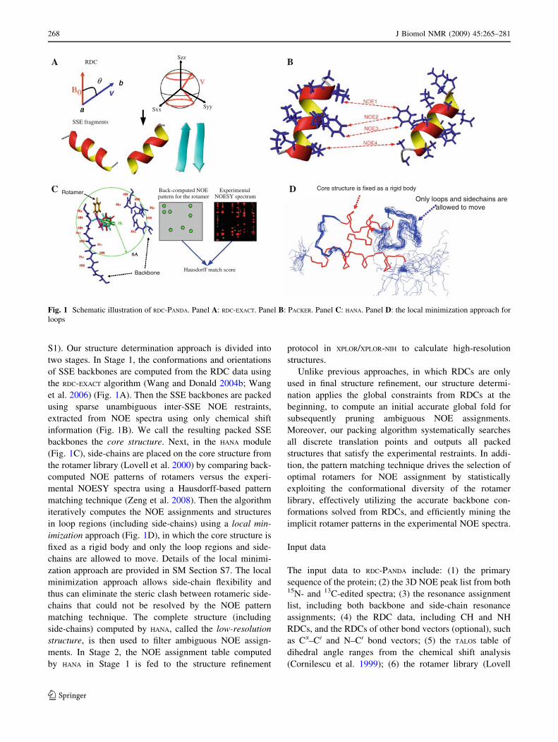
S1). Our structure determination approach is divided into
two stages. In Stage 1, the conformations and orientations
of SSE backbones are computed from the RDC data using
the RDC-EXACT algorithm (Wang and Donald 2004b; Wang
et al. 2006) (Fig. 1A). Then the SSE backbones are packed
using sparse unambiguous inter-SSE NOE restraints,
extracted from NOE spectra using only chemical shift
information (Fig. 1B). We call the resulting packed SSE
backbones the core structure. Next, in the HANA module
(Fig. 1C), side-chains are placed on the core structure from
the rotamer library (Lovell et al. 2000) by comparing back-
computed NOE patterns of rotamers versus the experi-
mental NOESY spectra using a Hausdorff-based pattern
matching technique (Zeng et al. 2008). Then the algorithm
iteratively computes the NOE assignments and structures
in loop regions (including side-chains) using a local min-
imization approach (Fig. 1D), in which the core structure is
fixed as a rigid body and only the loop regions and side-
chains are allowed to move. Details of the local minimi-
zation approach are provided in SM Section S7. The local
minimization approach allows side-chain flexibility and
thus can eliminate the steric clash between rotameric side-
chains that could not be resolved by the NOE pattern
matching technique. The complete structure (including
side-chains) computed by HANA, called the low-resolution
structure, is then used to filter ambiguous NOE assign-
ments. In Stage 2, the NOE assignment table computed
by HANA in Stage 1 is fed to the structure refinement
protocol in XPLOR/XPLOR-NIH to calculate high-resolution
structures.
Unlike previous approaches, in which RDCs are only
used in final structure refinement, our structure determi-
nation applies the global constraints from RDCs at the
beginning, to compute an initial accurate global fold for
subsequently pruning ambiguous NOE assignments.
Moreover, our packing algorithm systematically searches
all discrete translation points and outputs all packed
structures that satisfy the experimental restraints. In addi-
tion, the pattern matching technique drives the selection of
optimal rotamers for NOE assignment by statistically
exploiting the conformational diversity of the rotamer
library, effectively utilizing the accurate backbone con-
formations solved from RDCs, and efficiently mining the
implicit rotamer patterns in the experimental NOE spectra.
Input data
The input data to RDC-PANDA include: (1) the primary
sequence of the protein; (2) the 3D NOE peak list from both15N- and 13C-edited spectra; (3) the resonance assignment
list, including both backbone and side-chain resonance
assignments; (4) the RDC data, including CH and NH
RDCs, and the RDCs of other bond vectors (optional), such
as Ca–C0 and N–C0 bond vectors; (5) the TALOS table of
dihedral angle ranges from the chemical shift analysis
(Cornilescu et al. 1999); (6) the rotamer library (Lovell
RDC
Back-computed NOE pattern for the rotamer
ExperimentalNOESY spectrum
Hausdorff match score
C
SyySxx
Szz
SSE fragments
A B
Core structure is fixed as a rigid body
Only loops and sidechains areallowed to move
D
6A6A
Backbone
Rotamer
a
bθ
a
b
Fig. 1 Schematic illustration of RDC-PANDA. Panel A: RDC-EXACT. Panel B: PACKER. Panel C: HANA. Panel D: the local minimization approach for
loops
268 J Biomol NMR (2009) 45:265–281
123
et al. 2000). Only CH and NH RDCs in one medium are
required by RDC-PANDA to compute the backbone confor-
mation and orientation, but other additional RDCs such as
Ca–C0 and N–C0 RDCs can be also included. This additional
data is used to prune the (/, w) angles in the RDC-EXACT step.
SSE backbone determination from residual dipolar
couplings
Residual dipolar couplings (Tjandra and Bax 1997; Tolman
et al. 1995) provide global orientational restraints on the
internuclear bond vectors, such as NH and CH bond vec-
tors, with respect to a global coordinate frame. Given NH
RDCs in two aligning media, the associated NH vector
must lie on the intersection of two conic curves (Skrynni-
kov and Kay 2000; Wedemeyer et al. 2002; Wang and
Donald 2004b). In Wang et al. (2006), Wang and Donald
(2004a, b), the authors proposed the first polynomial-time
de novo algorithm, which we henceforth refer to as
RDC-EXACT, to compute high-resolution protein backbone
structures from RDC data. RDC-EXACT takes as input two
RDCs per residue (e.g., assigned NH RDCs in two media
or NH and CH RDCs in a single medium). In Wang and
Donald (2004a, b), the authors also showed that given a
peptide plane, the orientation of the next peptide plane can
have at most 16 possible orientations; a related theorem
was shown by (Hus et al. 2008). Given NH and CH RDCs
in one medium, the associated NH and CH vectors can be
solved from the RDC curve equations (see SM Section S2)
and the protein backbone geometry (Wang and Donald
2004a; Wang et al. 2006). For the one-medium case,
detailed proofs can be found in Wang and Donald (2004a,
b), Wang et al. (2006), and for completeness we provide a
somewhat simpler derivation in SM Section S2. The deri-
vation closely mirrors our new (open-source) software
implementation, and the clearer equations therein are easier
to interpret and build upon. For a detailed review of this
method see (Donald and Martin 2008).
RDC-EXACT does not randomly search the conformation
space to find solutions consistent with the RDC data.
Rather, it formulates the problem such that the structures
computed are exact solutions of a system of quartic
monomial equations derived from the RDC equation.
Hence, these roots, and therefore the conformations, are
discrete, finite and algebraic. All dihedral angles for each
residue are solved exactly from the quartic RDC equations.
A depth-first systematic search algorithm is applied to
search over all possible roots (conformations) to find an
optimal solution for a SSE. The scoring function used in
the depth-first systematic search contains RDC RMSD
(namely the sum of the squared differences between
experimental and back-computed RDCs over all RDCs in
the SSE), Ramachandran suitability, van der Waals
packing, and other common empirical molecular mechanics
energy terms (Wang and Donald 2004b, Wang et al. 2006).
As previously described in those references, the computed
dihedral angles for a residue are not solely dependent on the
local RDC information at that residue. Rather, the algorithm
searches over all SSE residues and finds the global mini-
mum of the scoring function for each SSE.
Before applying the RDC-EXACT algorithm to solve
backbones for the core structure, we first identify the SSE
boundaries based on the dihedral angle ranges computed
from TALOS (Cornilescu et al. 1999) based on chemical shift
information. When the TALOS dihedral intervals for a resi-
due are within the favored or allowed Ramachandran area
of a-helix or b-strand, we consider this residue as part of
that SSE. We note that other experimental data such as the
J-coupling data from the NMR experiment HNHA (Vuister
and Bax 1993) or the NOE patterns from automated
assignment (Bailey-Kellogg et al. 2000) of SSEs can also
be used to determine the SSE boundaries.
We have extended the RDC-EXACT algorithm (Wang and
Donald 2004b, Wang et al. 2006) to incorporate CaC0 and
NC0 RDCs with CH and NH RDCs for the backbone cal-
culation. The alignment tensor is estimated from NH and
CH RDCs using the same approach as in (Wang and Donald
2004b,Wang et al. 2006). The CaC0 and NC0 RDCs are
excluded in the alignment tensor computation, because they
are in general noisier than NH and CH RDCs. In the new
version of RDC-EXACT, NH and CH RDCs are first applied to
compute and enumerate conformations as solutions to the
polynomial systems derived from the RDC equations. Then
the remaining CaC0 and NC0 RDCs are used to prune
solutions for which the RMSD between back-computed and
experimental RDCs is larger than thresholds 5.0 Hz (after
being scaled to NH RDC). In addition, the outlier (/, w)
angle solutions are pruned by intersecting the TALOS ranges
with the Ramachandran regions. At this stage, every residue
(except glycine and proline) in the backbone structure is
replaced with alanine. Such a scheme is called alanine
replacement. A serious steric clash is defined between
atoms i and j when the distance between them satisfies
dij \ (ri ? rj) - e, where ri and rj are van der Waals radii,
and e is the overlap threshold between two atoms. Currently
we choose e = 0.5 A for the steric clash checking. We
prune those (/, w) solutions that result in serious steric clash
after alanine replacement, and ensure that no serious clashes
occur in our computed backbones. Later (below), we will
replace the alanines with proper side-chain rotamers.
SSE packing from sparse NOE restraints
Once the conformations and orientations of individual SSE
backbones have been solved by RDC-EXACT, their translations
can be determined using a small number of inter-SSE NOE
J Biomol NMR (2009) 45:265–281 269
123
distance restraints (Fowler et al. 2000, Wang and Donald
2004b). Although the NOE restraint provides only an
interval bound on the distance between atoms, in general a
small number of NOE distance restraints can confine the
translation into a bounded conformation space in which all
discretized solutions within the parameterized resolution
can be enumerated using a systematic grid search.
When packing a pair of SSEs solved from RDCs, we
must consider the fourfold orientational ambiguity.
Although the orientational ambiguity certainly can be
resolved given a sufficient number of independent media, it
cannot be resolved based only on RDCs in a single medium.
Suppose that we pack all SSEs sequentially, that is, we first
assemble the first two SSEs, and then pack their combina-
tion with the third SSE, and so on, as previously described
(Wang and Donald 2004b). Before packing each pair of
SSEs, sparse inter-SSE NOE restraints were extracted using
chemical shift information alone (details are provided in
SM Section S3). Using these sparse NOEs plus a van der
Waals packing score, we can prune the orientational
ambiguity (Georgiev et al. 2008, Potluri et al. 2006).
Since most inter-SSE NOEs involve side-chain interac-
tions, we must consider all of the possible side-chain rot-
amer conformations when using these unambiguous NOE
restraints in packing each pair of the SSE backbones. Let
H1 and H2 denote two SSE backbones to be packed toge-
ther. Let D be the set of inter-SSE NOE restraints between
H1 and H2, where for di ¼ ðhi1; hi2; ‘i; uiÞ 2 D; hi1; hi2 are
the NOE-interaction protons in H1 and H2 respectively, and
‘i; ui are the lower and upper bounds of the NOE. Our goal
is to find all possible translations t 2 R3 between H1 and
H2, such that there exists a pair of rotamers in H1 and H2 in
which the distance between the corresponding protons hi1
and hi2, denoted by di, satisfies the NOE restraint, namely
‘i� di� ui:
Without loss of generality, we choose the centers of H1
and H2, denoted by a0 and b0, as the representative points
for H1 and H2 respectively. Then the translation t between
H1 and H2 can be represented by the translation between
points a0 and b0, namely t = a0 - b0. Let a be a proton of
a residue in a particular rotamer state in H1 and b be a
proton of a residue in a particular rotamer state in H2.
Suppose that an NOE ða; b; ‘i; uiÞ gives the distance
restraint between proton a and b. Then we have
‘i�ka� bk� ui: ð1Þ
Let ta be the vector from a0 to a, namely a = ta ? a0.
Let tb be the vector from b0 to b, namely b = tb ? b0.
Substituting into Eq. 1, we have
‘i�kt� ðtb � taÞk� ui: ð2Þ
Equation (2) restricts the translation t to a spherical shell
with center at tb - ta and radii of ‘i and ui. Let q be the
number of inter-SSE NOE restraints. Given the ith NOE
restraint, let ki be the total number of rotamer pairs
between two corresponding residues. Let Aij denote the
spherical shell that represents the ith NOE restraint given
the jth pair of rotamers at corresponding residues, where
1 B i B q and 1 B j B ki. Then the complete space
of translation between H1 and H2 that satisfies all inter-SSE
NOE restraints is represented by
\q
i¼1
[ki
j¼1
Aij; ð3Þ
where q is the total number of NOE restraints, and ki is the
total number of rotamer pairs between two corresponding
residues.
We are interested in finding an ensemble of packed
structures (within a parameterized resolution) that satisfy
all NOE restraints, rather than just one single maximum-
likelihood solution. Below we describe our algorithm for
computing an ensemble of packed structures:
(1) When packing each pair of SSE backbones, consider
all four-fold symmetries of SSE orientations due to
the symmetry of the dipolar operator reflected in the
RDCs.
(2) Apply a 3D grid search over relative translations t 2R
3 with a resolution of 0.2 A to find all discrete
translation points in which the set of solutions in
Eq. 3 is not empty.
(3) Prune those packed structures containing steric
clashes between atoms from difference SSE
fragments.
(4) Cluster all packed structures from Step (2) using an
agglomerative hierarchical clustering algorithm (Han
and Kamber 2006), in which two packed structures
are allowed to be in a cluster only if their backbone
RMSD is within 0.4 A. The centroids of all clusters
form the final set of representative packed structures.
Our packing algorithm finds all of the discrete transla-
tion solutions within a parameterized resolution (i.e. 0.4 A)
that satisfy SSE orientations determined from RDCs and all
inter-SSE NOE restraints. The time complexity analysis for
PACKER is given in SM Section S4. In practice, PACKER runs
in 30–60 minutes on a 3 GHz single-processor Linux
workstation.
After we obtained the set of packed SSEs, which are
called the initial packed structures, we computed a subset
of well-packed satisfying (WPS) structures that had both
high-quality van der Waals (vdW) score and good NOE
satisfaction score (Potluri et al. 2006, 2007). We used a 6-
12 Lennard-Jones potential to compute the packing score
between SSEs, and a square-well potential (Brunger 1992)
to calculate the NOE satisfaction score.
270 J Biomol NMR (2009) 45:265–281
123
NOE assignment using a pattern matching technique
The NOE assignment process can be divided into three
phases, viz. initial NOE assignment (phase 1), rotamer
selection (phase 2), and filtration of ambiguous NOE
assignments (phase 3). The initial NOE assignment (phase
1) is done by considering all pairs of protons that are
possibly assigned to an NOE cross peak if the resonances
of corresponding atoms fall within a tolerance window
around the NOE peak. We use error windows of 0.4 ppm
for heavy atoms (15N and 13C), and 0.04 ppm for protons in
the NOE assignment. In the rotamer selection phase (phase
2), we place all the side-chain rotamers of each residue into
the backbone and compute all expected NOEs for protons
within 6 A apart. Based on the set of the expected NOEs
and the resonance assignment list, we back-compute the
expected NOE peak pattern for each rotamer. By matching
the back-computed NOE pattern with the experimental
NOE spectrum using an extended model of the Hausdorff
distance (Huttenlocher and Jaquith 1995), we measure how
well a rotamer fits the actual side-chain conformation when
interpreted in terms of the NOE data (Fig. 1C). We then
select the top k rotamers with highest fitness scores at each
residue, and obtain a ‘‘low-resolution’’ structure, which
typically has approximately 2.0–3.0 A (all heavy atom)
RMSD from the reference structures solved by X-ray or
traditional NMR approaches. The low-resolution structure
is then used (in phase 3) to filter ambiguous NOE assign-
ments. The details of the NOE assignment algorithm are
provided in SM Section S5.
NOE pattern matching based on the Hausdorff distance
measure
The Hausdorff distance measures the closeness between
two sets of points by computing the distance from the point
in one set that is farthest from any point in the other set, and
vice versa. Two sets of points have a small Hausdorff dis-
tance if every point in one set is close to some point in the
other set. Thus, the Hausdorff distance is suitable for
determining the degree of resemblance between two sets of
points when they are superimposed on each other. The
Hausdorff distance has been widely used in image pro-
cessing and computer vision, e.g., visual correspondence,
pattern recognition, and shape matching (Huttenlocher and
Kedem 1992). Unlike many other pattern-recognition
algorithms, Hausdorff-based algorithms are combinatori-
ally precise, and provide a robust method for measuring the
similarity between two point sets or image patterns (Hut-
tenlocher and Kedem 1992) in the presence of noise and
positional uncertainties. In the NOE assignment problem,
let B denote a back-computed NOE pattern, i.e., the set of
back-computed NOE peaks, and let Y denote the set of
experimental NOE peaks. The Hausdorff distance between
B and Y can be measured by H(B,Y) = max(h(B,Y), h(Y,B)),
where hðB; YÞ ¼ maxb2Bminy2Ykb� yk: The operations of
nested Min and Max in the Hausdorff definition compute
the point b 2 B that is farthest from any point in Y, and
measures the distance from point b to its closest neighbor y
in Y. Thus, the Hausdorff distance H(B,Y) describes the
discrepancy between the configurations of the two point sets
B and Y, since it actually measures the the distance from a
point in B that is farthest from any point in Y, and vice versa.
A review article on the Hausdorff distance can be found in
(Huttenlocher and Kedem 1992).
We apply an extended model of Hausdorff distance
(Huttenlocher and Kedem 1992, Huttenlocher and Jaquith
1995) to measure the match between the back-computed
NOE pattern and experimental NOE spectrum. Below, we
assume 3D NOE spectra without loss of generality. Given
the back-computed NOE pattern B with m peaks, and the
set of NOE peaks Y with w peaks, the s-th Hausdorff dis-
tance from B to Y is defined as
hsðB; YÞ ¼ sthb2B
miny2Ykb� yk;
where sth is the s-th largest of m values. We call s = s/m
the similarity score between the back-computed NOE
pattern B and the experimental peak set Y, after fixing the
Hausdorff distance hs(B,Y) = d, which is the error in
chemical shift. The similarity score for a rotamer given dcan be computed using a scheme similar to that of
(Huttenlocher and Jaquith 1995):
s ¼ jB \ YdjjBj ; ð4Þ
where Yd denotes the union of all balls obtained by
replacing each point in Y with a ball of radius d, B \ Yd
denotes the intersection of sets B and Yd, and j � j denotes
the size of a set.
Let (a1, a2, a3, d) represent a distance restraint back-
computed from a structure, where a1 and a3 are the
involved protons in the structure, a2 is the heavy atom
covalently bound to the proton a1, and d is the distance
between protons a1 and a3. Let (p1, p2, p3, Ip) denote an
experimental NOE peak from a 3D NOESY spectrum,
where p1 and p3 are frequencies of a pair of (unassigned)
interacting protons, p2 is the frequency of the heavy atom
covalently bound to the first proton, and Ip is the intensity
of the cross peak. Let bi = (x(a1), x(a2), x(a3), I(d))
denote the back-computed NOE peak for a distance
restraint (a1, a2, a3, d) back-computed from a structure,
where x(aj) is the assigned chemical shift of atom aj, 1 B j
B 3, and I(d) is the back-computed peak intensity of dis-
tance d. The equation I(d) = kd-6 is used to back-compute
the peak intensity I(d) from the distance d, where the
J Biomol NMR (2009) 45:265–281 271
123
constant k is calculated using the same strategy as in
(Mumenthaler et al. 1997). The likelihood for a back-
computed peak bi = (x(a1), x(a2), x(a3), I(d)) in the NOE
pattern B to match an experimental NOE peak within the
distance d in Yd can be defined as
N iðbiÞ ¼ N ðjIðdÞ � Ipj; rIÞ �Y3
j¼1
N jxðajÞ � pjj; rj
� �;
where (p1, p2, p3, Ip) is the experimental NOE peak mat-
ched to the back-computed NOE peak (x(a1), x(a2), x(a3),
I(d)) under the Hausdorff distance measure, and Nðjx�lj; rÞ is the probability of observing the difference |x-l| in
a normal distribution with mean l and standard deviation
r. We note that the normal distribution and other similar
distribution families have been widely used to model the
noise in the NMR data, e.g., see (Rieping et al. 2005) and
(Langmead and Donald 2004).
The expected number of peaks in B \ Yd can be bounded
byPm
i¼1N iðbiÞ. Thus, we obtain the following equation
for the similarity score:
s ¼ 1
m
Xm
i¼1
N iðbiÞ: ð5Þ
When back-computing the NOE pattern for each
rotamer, HANA also considers the stereospecific assign-
ment ambiguity for Ha in glycine, all b-methylene protons,
and methyl groups in leucine and valine. HANA back-
computes all possible NOE patterns resulting from the
different possible stereospecific assignments for all protons
in a residue, and chooses the stereospecific assignment with
the best match score for each rotamer when compared
versus the experimental data.
We provide the detailed pseudocode for computing the
similarity score and for HANA in SM Section S5. The
time complexity analysis (see SM Section S6) shows
that HANA runs in polynomial time. In practice, HANA runs
in 1–2 minutes on a 3 GHz single-processor Linux
workstation.
Incorporation of rotamer probabilities into the scoring
function
Different side-chain rotamers for each residue occur at
different probabilities (Lovell et al. 2000). To consider the
occurrence rates of different rotamers, the following
inference is applied to extend our scoring function in Eq. 5.
Let Xj be the boolean proposition that the j-th rotamer is
selected. Let Pr(D|Xj) be the likelihood function that
quantifies the likelihood of observing data D given the j-th
rotamer. Then Pr(D|Xj) is equivalent to the similarity score
in Eq. 5, that is,
PrðDjXjÞ ¼1
m
Xm
i¼1
N iðbiÞ: ð6Þ
By Bayes’ Theorem, the posterior probability, Pr(Xj|D) is
given by
PrðXjjDÞ/PrðDjXjÞ �PrðXjÞ/1
m
Xm
i¼1
N iðbiÞ !
PrðXjÞ: ð7Þ
Equation (7) is used as the scoring function for computing
the ensemble of rotamers that best fit the experimental data.
Results
We tested RDC-PANDA on four proteins: the FF Domain 2 of
human transcription elongation factor CA150 (RNA poly-
merase II C-terminal domain interacting protein) (FF2),
the ubiquitin-binding zinc finger domain of the human
Y-family DNA polymerase Eta (pol g UBZ), human
ubiquitin, and the human Set2-Rpb1 interacting domain
(hSRI). The lengths (number of amino acid residues) of
these proteins are 62 for FF2, 39 for pol g UBZ, 76 for
ubiquitin, and 112 for hSRI.
All NMR data except the RDC data of ubiquitin were
recorded and collected using Varian 600 and 800 MHz
spectrometers at Duke University. The NMR spectra were
processed using the program NMRPIPE (Delaglio et al.
1995). All NMR peaks were picked by the programs
NMRVIEW (Johnson and Blevins 1994) or XEASY/CARA
(Bartels et al. 1995), followed by manual editing. Back-
bone assignments were obtained from the set of triple
NMR experiments HNCA, HN(CO)CA, HN(CA)CB,
HN(COCA)CB, and HNCO, combined with the HSQC
spectra using the program PACES (Coggins and Zhou 2003),
followed by manual checking. The side-chain resonances
were assigned from the HA(CA)NH, HA(CACO), HCCH-
TOCSY, and HC(CCO)NH-TOCSY spectra. The NOE
cross peaks were picked from three-dimensional 15N- and13C-edited NOESY-HSQC spectra. In addition, we
removed the diagonal cross peaks and water artifacts from
the picked NOE peak list. The NH and CH RDC data of
FF2, pol g UBZ and hSRI were measured from a 2D1H–15N IPAP experiment (Ottiger et al. 1998) and a
modified (HACACO)NH experiment (Ball et al. 2006)
respectively. The CaC0 and NC0 RDC data of FF2 were
measured from a set of HNCO-based experiments (Permi
et al. 2000). The CH and NH RDC data of ubiquitin were
obtained from the Protein Data Bank (PDB ID: 1D3Z).
The solution structures of FF2, pol g UBZ, hSRI and
ubiquitin have been solved using conventional NMR
structure determination approaches (PDB ID of ubiquitin
NMR reference structure: 1D3Z; PDB ID of FF2 NMR
272 J Biomol NMR (2009) 45:265–281
123
reference structure: 2E71; PDB ID of pol g UBZ NMR
reference structure: 2I5O; PDB ID of hSRI NMR reference
structure: 2A7O). In addition, the X-ray structure of human
ubiquitin (PDB ID: 1UBQ) was available. We used these
previously-solved NMR or X-ray structures as the refer-
ence structures for evaluating the structure calculation
results from RDC-PANDA.
Evaluation of SSE backbones determined from RDCs
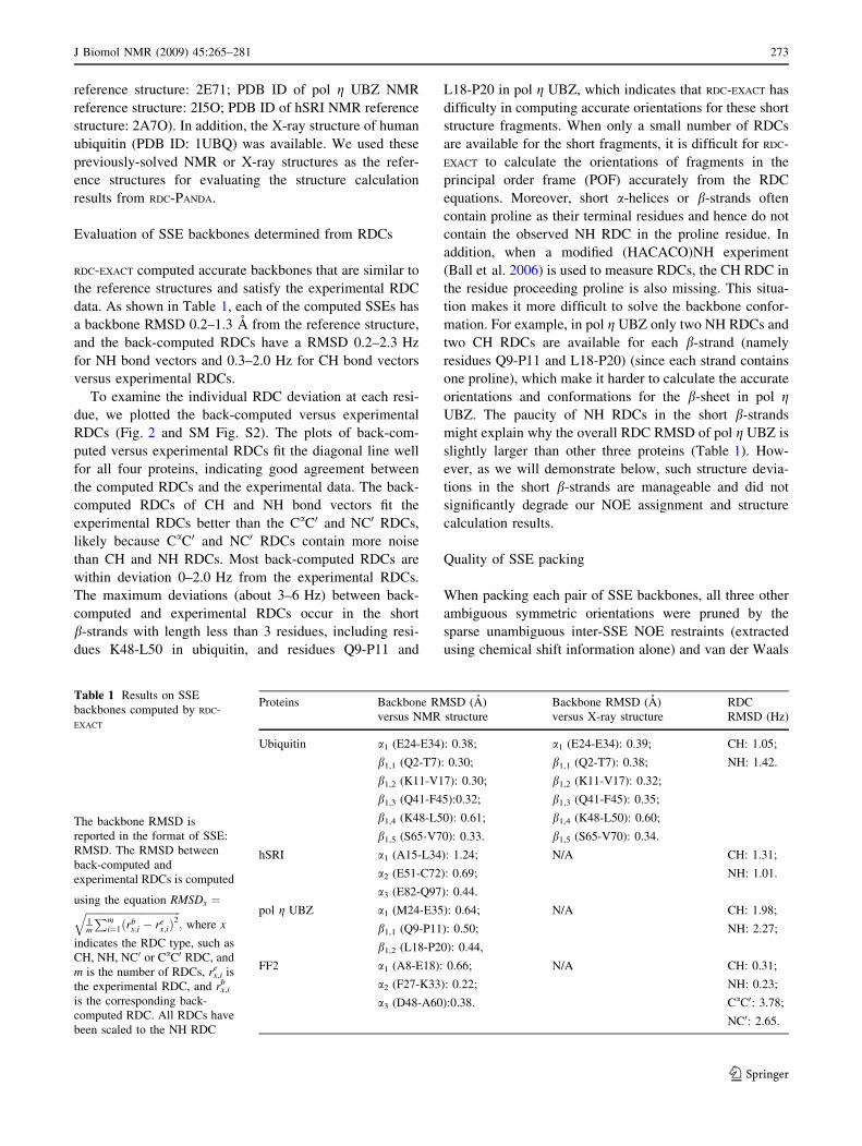
RDC-EXACT computed accurate backbones that are similar to
the reference structures and satisfy the experimental RDC
data. As shown in Table 1, each of the computed SSEs has
a backbone RMSD 0.2–1.3 A from the reference structure,
and the back-computed RDCs have a RMSD 0.2–2.3 Hz
for NH bond vectors and 0.3–2.0 Hz for CH bond vectors
versus experimental RDCs.
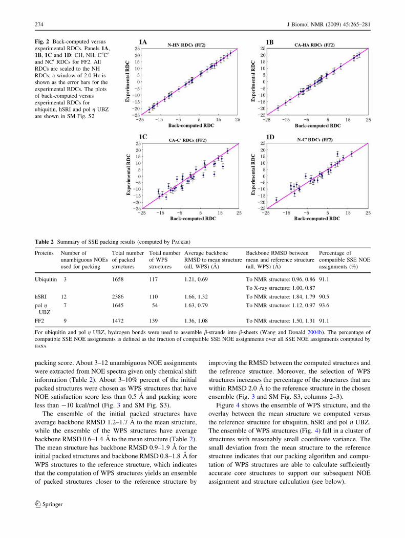
To examine the individual RDC deviation at each resi-
due, we plotted the back-computed versus experimental
RDCs (Fig. 2 and SM Fig. S2). The plots of back-com-
puted versus experimental RDCs fit the diagonal line well
for all four proteins, indicating good agreement between
the computed RDCs and the experimental data. The back-
computed RDCs of CH and NH bond vectors fit the
experimental RDCs better than the CaC0 and NC0 RDCs,
likely because CaC0 and NC0 RDCs contain more noise
than CH and NH RDCs. Most back-computed RDCs are
within deviation 0–2.0 Hz from the experimental RDCs.
The maximum deviations (about 3–6 Hz) between back-
computed and experimental RDCs occur in the short
b-strands with length less than 3 residues, including resi-
dues K48-L50 in ubiquitin, and residues Q9-P11 and
L18-P20 in pol g UBZ, which indicates that RDC-EXACT has
difficulty in computing accurate orientations for these short
structure fragments. When only a small number of RDCs
are available for the short fragments, it is difficult for RDC-
EXACT to calculate the orientations of fragments in the
principal order frame (POF) accurately from the RDC
equations. Moreover, short a-helices or b-strands often
contain proline as their terminal residues and hence do not
contain the observed NH RDC in the proline residue. In
addition, when a modified (HACACO)NH experiment
(Ball et al. 2006) is used to measure RDCs, the CH RDC in
the residue proceeding proline is also missing. This situa-
tion makes it more difficult to solve the backbone confor-
mation. For example, in pol g UBZ only two NH RDCs and
two CH RDCs are available for each b-strand (namely
residues Q9-P11 and L18-P20) (since each strand contains
one proline), which make it harder to calculate the accurate
orientations and conformations for the b-sheet in pol gUBZ. The paucity of NH RDCs in the short b-strands
might explain why the overall RDC RMSD of pol g UBZ is
slightly larger than other three proteins (Table 1). How-
ever, as we will demonstrate below, such structure devia-
tions in the short b-strands are manageable and did not
significantly degrade our NOE assignment and structure
calculation results.
Quality of SSE packing
When packing each pair of SSE backbones, all three other
ambiguous symmetric orientations were pruned by the
sparse unambiguous inter-SSE NOE restraints (extracted
using chemical shift information alone) and van der Waals
Table 1 Results on SSE
backbones computed by RDC-
EXACT
The backbone RMSD is
reported in the format of SSE:
RMSD. The RMSD between
back-computed and
experimental RDCs is computed
using the equation RMSDx ¼ffiffiffiffiffiffiffiffiffiffiffiffiffiffiffiffiffiffiffiffiffiffiffiffiffiffiffiffiffiffiffiffiffiffiffiffiffi1m
Pmi¼1ðrb
x;i � rex;iÞ
2q
; where x
indicates the RDC type, such as
CH, NH, NC0 or CaC0 RDC, and
m is the number of RDCs, rx,ie is
the experimental RDC, and rx,ib
is the corresponding back-
computed RDC. All RDCs have
been scaled to the NH RDC
Proteins Backbone RMSD (A)
versus NMR structure
Backbone RMSD (A)
versus X-ray structure
RDC
RMSD (Hz)
Ubiquitin a1 (E24-E34): 0.38; a1 (E24-E34): 0.39; CH: 1.05;
b1,1 (Q2-T7): 0.30; b1,1 (Q2-T7): 0.38; NH: 1.42.
b1,2 (K11-V17): 0.30; b1,2 (K11-V17): 0.32;
b1,3 (Q41-F45):0.32; b1,3 (Q41-F45): 0.35;
b1,4 (K48-L50): 0.61; b1,4 (K48-L50): 0.60;
b1,5 (S65-V70): 0.33. b1,5 (S65-V70): 0.34.
hSRI a1 (A15-L34): 1.24; N/A CH: 1.31;
a2 (E51-C72): 0.69; NH: 1.01.
a3 (E82-Q97): 0.44.
pol g UBZ a1 (M24-E35): 0.64; N/A CH: 1.98;
b1,1 (Q9-P11): 0.50; NH: 2.27;
b1,2 (L18-P20): 0.44,
FF2 a1 (A8-E18): 0.66; N/A CH: 0.31;
a2 (F27-K33): 0.22; NH: 0.23;
a3 (D48-A60):0.38. CaC0: 3.78;
NC0: 2.65.
J Biomol NMR (2009) 45:265–281 273
123
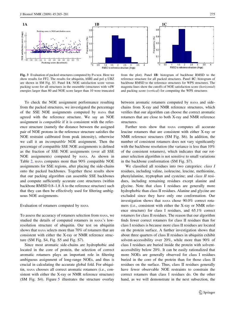
packing score. About 3–12 unambiguous NOE assignments
were extracted from NOE spectra given only chemical shift
information (Table 2). About 3–10% percent of the initial
packed structures were chosen as WPS structures that have
NOE satisfaction score less than 0.5 A and packing score
less than -10 kcal/mol (Fig. 3 and SM Fig. S3).
The ensemble of the initial packed structures have
average backbone RMSD 1.2–1.7 A to the mean structure,
while the ensemble of the WPS structures have average
backbone RMSD 0.6–1.4 A to the mean structure (Table 2).
The mean structure has backbone RMSD 0.9–1.9 A for the
initial packed structures and backbone RMSD 0.8–1.8 A for
WPS structures to the reference structure, which indicates
that the computation of WPS structures yields an ensemble
of packed structures closer to the reference structure by
improving the RMSD between the computed structures and
the reference structure. Moreover, the selection of WPS
structures increases the percentage of the structures that are
within RMSD 2.0 A to the reference structure in the chosen
ensemble (Fig. 3 and SM Fig. S3, columns 2–3).
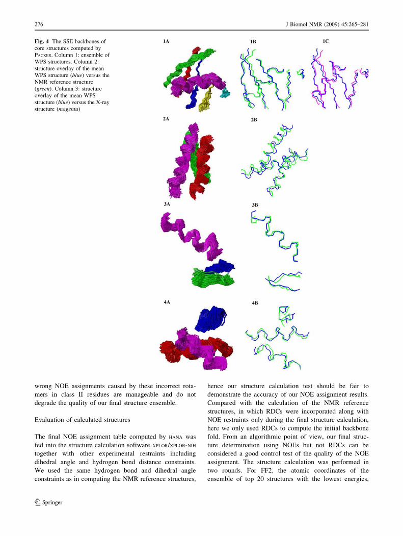
Figure 4 shows the ensemble of WPS structure, and the
overlay between the mean structure we computed versus
the reference structure for ubiquitin, hSRI and pol g UBZ.
The ensemble of WPS structures (Fig. 4) fall in a cluster of
structures with reasonably small coordinate variance. The
small deviation from the mean structure to the reference
structure indicates that our packing algorithm and compu-
tation of WPS structures are able to calculate sufficiently
accurate core structures to support our subsequent NOE
assignment and structure calculation (see below).
Fig. 2 Back-computed versus
experimental RDCs. Panels 1A,
1B, 1C and 1D: CH, NH, CaC0
and NC0 RDCs for FF2. All
RDCs are scaled to the NH
RDCs; a window of 2.0 Hz is
shown as the error bars for the
experimental RDCs. The plots
of back-computed versus
experimental RDCs for
ubiquitin, hSRI and pol g UBZ
are shown in SM Fig. S2
Table 2 Summary of SSE packing results (computed by PACKER)
Proteins Number of
unambiguous NOEs
used for packing
Total number
of packed
structures
Total number
of WPS
structures
Average backbone
RMSD to mean structure
(all, WPS) (A)
Backbone RMSD between
mean and reference structure
(all, WPS) (A)
Percentage of
compatible SSE NOE
assignments (%)
Ubiquitin 3 1658 117 1.21, 0.69 To NMR structure: 0.96, 0.86 91.1
To X-ray structure: 1.00, 0.87
hSRI 12 2386 110 1.66, 1.32 To NMR structure: 1.84, 1.79 90.5
pol gUBZ
7 1645 54 1.63, 0.79 To NMR structure: 1.12, 0.97 93.6
FF2 9 1472 139 1.36, 1.08 To NMR structure: 1.50, 1.31 91.1
For ubiquitin and pol g UBZ, hydrogen bonds were used to assemble b-strands into b-sheets (Wang and Donald 2004b). The percentage of
compatible SSE NOE assignments is defined as the fraction of compatible SSE NOE assignments over all SSE NOE assignments computed by
HANA
274 J Biomol NMR (2009) 45:265–281
123
To check the NOE assignment performance resulting
from the packed structures, we investigated the percentage
of the SSE NOE assignments computed by HANA that
agreed with the reference structure. We say an NOE
assignment is compatible if it is consistent with the refer-
ence structure (namely the distance between the assigned
pair of NOE protons in the reference structure satisfies the
NOE restraint calibrated from peak intensity), otherwise
we call it an incompatible NOE assignment. Then the
percentage of compatible SSE NOE assignments is defined
as the fraction of SSE NOE assignments (over all SSE
NOE assignments) computed by HANA. As shown in
Table 2, HANA computes more than 90% compatible NOE
assignments for SSE regions, after placing the side-chains
onto the packed backbones. Together these results show
that our packing algorithm can assemble SSE backbones
and compute sufficiently accurate core structures (within
backbone RMSD 0.8–1.8 A to the reference structure) such
that they can then be effectively used for filtering ambig-
uous NOE assignments.
Evaluation of rotamers computed by HANA
To assess the accuracy of rotamers selection from HANA, we
studied the details of computed rotamers in HANA’s low-
resolution structure of ubiquitin. Our test on ubiquitin
shows that HANA selects more than 70% of rotamers that are
consistent with either the X-ray or NMR reference struc-
ture (SM Fig. S4, Fig. S5 and Fig. S7).
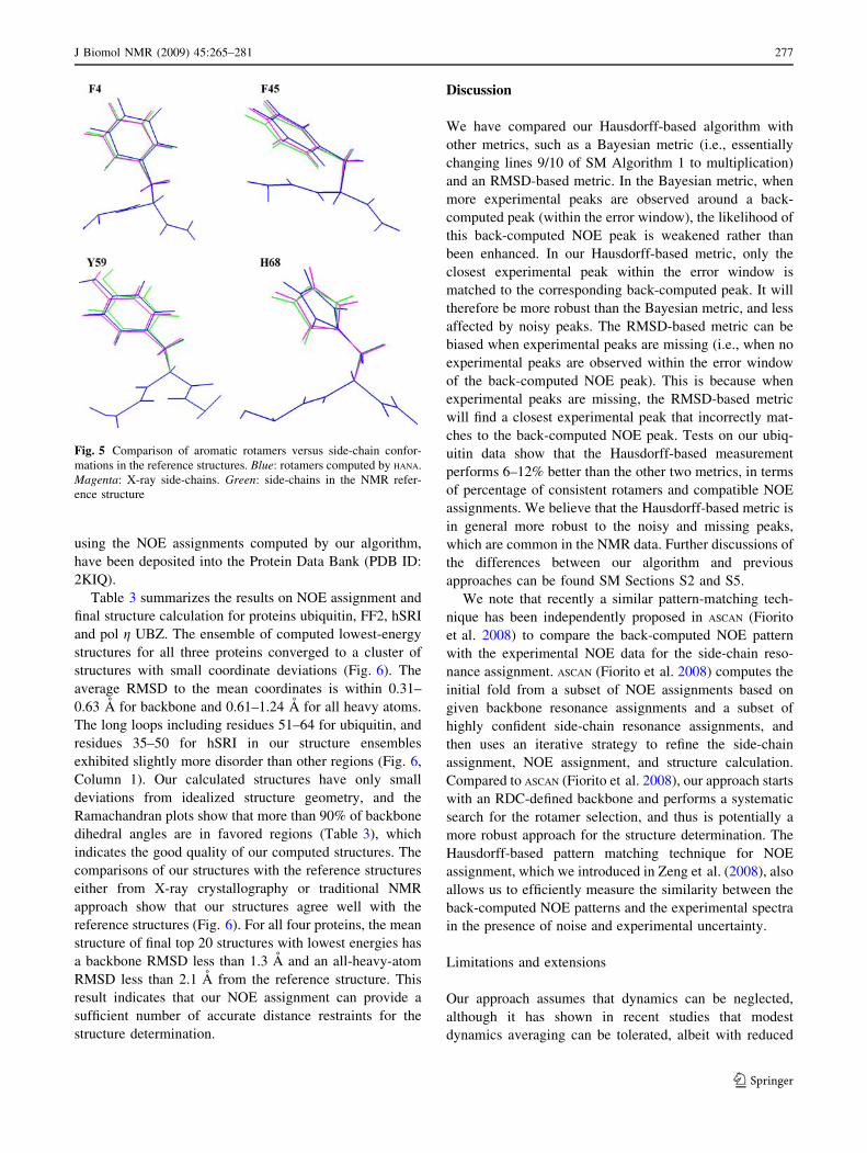
Since most aromatic side-chains are hydrophobic and
located in the core of protein, the selection of correct
aromatic rotamers plays an important role in filtering
ambiguous assignment of long-range NOEs, and thus is
crucial in calculating the accurate global fold. For ubiqui-
tin, HANA chooses all correct aromatic rotamers (i.e., con-
sistent with either the X-ray or NMR reference structure)
(SM Fig. S4). Figure 5 illustrates the structure overlay
between aromatic rotamers computed by HANA and side-
chains from X-ray and NMR reference structures, which
verifies that our algorithm can choose the correct aromatic
rotamers that are close to both X-ray and NMR reference
structures.
Further tests show that HANA computes all accurate
leucine rotamers that are consistent with either X-ray or
NMR reference structures (SM Fig. S6). In addition, the
number of consistent rotamers does not vary significantly
with the backbone resolution (the variance is less than 10%
of the consistent rotamers), which indicates that our rot-
amer selection algorithm is not sensitive to small variations
in the backbone conformation (SM Fig. S7).
We classified all residues into two categories: class I
residues, including valine, isoleucine, leucine, methionine,
phenylalanine, tryptophan and cysteine; and class II resi-
dues, including remaining residues except alanine and
glycine. Note that class I residues are generally more
hydrophobic than class II residues. Alanine and glycine are
excluded since they have only one conformation. Our
investigation shows that HANA chose 90.0% correct rota-
mers (i.e., consistent with either the X-ray or NMR refer-
ence structure) for class I residues, and 65.1% correct
rotamers for class II residues. The reason that our algorithm
finds fewer correct rotamers for class II residues than for
class I residues is because more class II residues are located
on the protein surface. A further investigation shows that
about three quarters of class II residues in ubiquitin exhibit
solvent-accessibility over 20%, while more than 90% of
class I residues are buried inside the protein with solvent-
accessibility below 20%. It can be easily rationalized that
more NOEs are generally observed for class I residues
buried in the core of the protein than for those class II
residues on the surface. Thus, class II residues generally
have fewer observable NOE restraints to constrain the
correct rotamers than class I residues do. On the other
hand, as we will demonstrate in the next subsection, the
Fig. 3 Evaluation of packed structures computed by PACKER. Here we
show results for FF2. The results for ubiquitin, hSRI and pol g UBZ
are shown in SM Fig. S3. Panel 1A: NOE satisfaction score versus
packing score for all structures in the ensemble (structures with vdW
energies larger than 80 and NOE score larger than 10 were truncated
from the plot). Panel 1B: histogram of backbone RMSD to the
reference structure for all packed structures. Panel 1C: histogram of
backbone RMSD to the reference structures for WPS structures. The
magenta lines show the cutoffs of NOE satisfaction score (horizontal)and packing score (vertical) for computing the WPS structures
J Biomol NMR (2009) 45:265–281 275
123
wrong NOE assignments caused by these incorrect rota-
mers in class II residues are manageable and do not
degrade the quality of our final structure ensemble.
Evaluation of calculated structures
The final NOE assignment table computed by HANA was
fed into the structure calculation software XPLOR/XPLOR-NIH
together with other experimental restraints including
dihedral angle and hydrogen bond distance constraints.
We used the same hydrogen bond and dihedral angle
constraints as in computing the NMR reference structures,
hence our structure calculation test should be fair to
demonstrate the accuracy of our NOE assignment results.
Compared with the calculation of the NMR reference
structures, in which RDCs were incorporated along with
NOE restraints only during the final structure calculation,
here we only used RDCs to compute the initial backbone
fold. From an algorithmic point of view, our final struc-
ture determination using NOEs but not RDCs can be
considered a good control test of the quality of the NOE
assignment. The structure calculation was performed in
two rounds. For FF2, the atomic coordinates of the
ensemble of top 20 structures with the lowest energies,
Fig. 4 The SSE backbones of
core structures computed by
PACKER. Column 1: ensemble of
WPS structures. Column 2:
structure overlay of the mean
WPS structure (blue) versus the
NMR reference structure
(green). Column 3: structure
overlay of the mean WPS
structure (blue) versus the X-ray
structure (magenta)
276 J Biomol NMR (2009) 45:265–281
123
using the NOE assignments computed by our algorithm,
have been deposited into the Protein Data Bank (PDB ID:
2KIQ).
Table 3 summarizes the results on NOE assignment and
final structure calculation for proteins ubiquitin, FF2, hSRI
and pol g UBZ. The ensemble of computed lowest-energy
structures for all three proteins converged to a cluster of
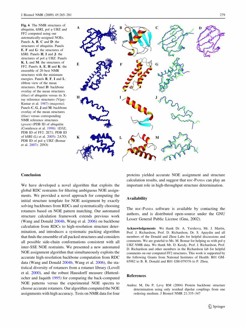
structures with small coordinate deviations (Fig. 6). The
average RMSD to the mean coordinates is within 0.31–
0.63 A for backbone and 0.61–1.24 A for all heavy atoms.
The long loops including residues 51–64 for ubiquitin, and
residues 35–50 for hSRI in our structure ensembles
exhibited slightly more disorder than other regions (Fig. 6,
Column 1). Our calculated structures have only small
deviations from idealized structure geometry, and the
Ramachandran plots show that more than 90% of backbone
dihedral angles are in favored regions (Table 3), which
indicates the good quality of our computed structures. The
comparisons of our structures with the reference structures
either from X-ray crystallography or traditional NMR
approach show that our structures agree well with the
reference structures (Fig. 6). For all four proteins, the mean
structure of final top 20 structures with lowest energies has
a backbone RMSD less than 1.3 A and an all-heavy-atom
RMSD less than 2.1 A from the reference structure. This
result indicates that our NOE assignment can provide a
sufficient number of accurate distance restraints for the
structure determination.
Discussion
We have compared our Hausdorff-based algorithm with
other metrics, such as a Bayesian metric (i.e., essentially
changing lines 9/10 of SM Algorithm 1 to multiplication)
and an RMSD-based metric. In the Bayesian metric, when
more experimental peaks are observed around a back-
computed peak (within the error window), the likelihood of
this back-computed NOE peak is weakened rather than
been enhanced. In our Hausdorff-based metric, only the
closest experimental peak within the error window is
matched to the corresponding back-computed peak. It will
therefore be more robust than the Bayesian metric, and less
affected by noisy peaks. The RMSD-based metric can be
biased when experimental peaks are missing (i.e., when no
experimental peaks are observed within the error window
of the back-computed NOE peak). This is because when
experimental peaks are missing, the RMSD-based metric
will find a closest experimental peak that incorrectly mat-
ches to the back-computed NOE peak. Tests on our ubiq-
uitin data show that the Hausdorff-based measurement
performs 6–12% better than the other two metrics, in terms
of percentage of consistent rotamers and compatible NOE
assignments. We believe that the Hausdorff-based metric is
in general more robust to the noisy and missing peaks,
which are common in the NMR data. Further discussions of
the differences between our algorithm and previous
approaches can be found SM Sections S2 and S5.
We note that recently a similar pattern-matching tech-
nique has been independently proposed in ASCAN (Fiorito
et al. 2008) to compare the back-computed NOE pattern
with the experimental NOE data for the side-chain reso-
nance assignment. ASCAN (Fiorito et al. 2008) computes the
initial fold from a subset of NOE assignments based on
given backbone resonance assignments and a subset of
highly confident side-chain resonance assignments, and
then uses an iterative strategy to refine the side-chain
assignment, NOE assignment, and structure calculation.
Compared to ASCAN (Fiorito et al. 2008), our approach starts
with an RDC-defined backbone and performs a systematic
search for the rotamer selection, and thus is potentially a
more robust approach for the structure determination. The
Hausdorff-based pattern matching technique for NOE
assignment, which we introduced in Zeng et al. (2008), also
allows us to efficiently measure the similarity between the
back-computed NOE patterns and the experimental spectra
in the presence of noise and experimental uncertainty.
Limitations and extensions
Our approach assumes that dynamics can be neglected,
although it has shown in recent studies that modest
dynamics averaging can be tolerated, albeit with reduced
Fig. 5 Comparison of aromatic rotamers versus side-chain confor-
mations in the reference structures. Blue: rotamers computed by HANA.
Magenta: X-ray side-chains. Green: side-chains in the NMR refer-
ence structure
J Biomol NMR (2009) 45:265–281 277
123
accuracy in the calculation of the bond vector orientations
(Ruan et al. 2008). When order parameters S2 are measured
for the same bond vectors as the RDCs (e.g. using relax-
ation experiments), we can neglect the dynamics within the
time scale of the dynamics measurements. Thus, we can
heuristically assume that when S2 is sufficiently uniform
(i.e. the core of the protein is largely rigid), then the
dynamic averaging due to S in the RDC measurement is
safe to tolerate for the structure determination.
Although our current implementation of HANA uses 3D
NOE spectra, HANA is general and can be easily extended to
higher-dimensional (e.g., 4D) NOE data (Coggins and
Zhou 2008). In addition, it would be interesting to extend
the current version of HANA for NOE assignment with
missing resonance assignments. In principle, HANA can be
extended to assign the NOEs with a partially assigned
resonance list, as long as the back-computed NOE patterns
with missing peaks can still support the identification of the
accurate rotamers.
Our structure determination starts with the high-resolu-
tion core structure computed from RDCs. The loop regions
are computed by a local minimization approach, which does
not incorporate the RDC data into the structure calculation.
Thus, the loop regions are less accurate than the SSE
regions in our final structures (see Table 3 and Fig. 6).
Currently we are developing efficient algorithms for com-
puting the loop conformations that satisfy both NOE and
RDC data.
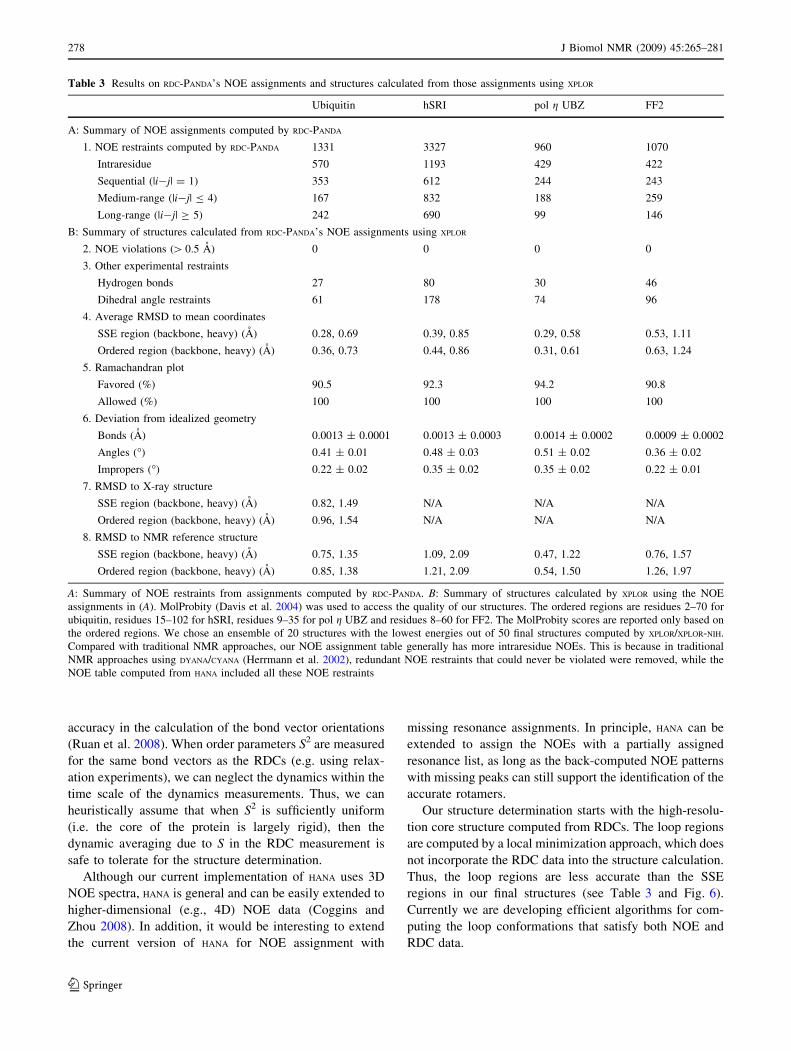
Table 3 Results on RDC-PANDA’s NOE assignments and structures calculated from those assignments using XPLOR
Ubiquitin hSRI pol g UBZ FF2
A: Summary of NOE assignments computed by RDC-PANDA
1. NOE restraints computed by RDC-PANDA 1331 3327 960 1070
Intraresidue 570 1193 429 422
Sequential (|i-j| = 1) 353 612 244 243
Medium-range (|i-j| B 4) 167 832 188 259
Long-range (|i-j| C 5) 242 690 99 146
B: Summary of structures calculated from RDC-PANDA’s NOE assignments using XPLOR
2. NOE violations ([ 0.5 A) 0 0 0 0
3. Other experimental restraints
Hydrogen bonds 27 80 30 46
Dihedral angle restraints 61 178 74 96
4. Average RMSD to mean coordinates
SSE region (backbone, heavy) (A) 0.28, 0.69 0.39, 0.85 0.29, 0.58 0.53, 1.11
Ordered region (backbone, heavy) (A) 0.36, 0.73 0.44, 0.86 0.31, 0.61 0.63, 1.24
5. Ramachandran plot
Favored (%) 90.5 92.3 94.2 90.8
Allowed (%) 100 100 100 100
6. Deviation from idealized geometry
Bonds (A) 0.0013 ± 0.0001 0.0013 ± 0.0003 0.0014 ± 0.0002 0.0009 ± 0.0002
Angles (�) 0.41 ± 0.01 0.48 ± 0.03 0.51 ± 0.02 0.36 ± 0.02
Impropers (�) 0.22 ± 0.02 0.35 ± 0.02 0.35 ± 0.02 0.22 ± 0.01
7. RMSD to X-ray structure
SSE region (backbone, heavy) (A) 0.82, 1.49 N/A N/A N/A
Ordered region (backbone, heavy) (A) 0.96, 1.54 N/A N/A N/A
8. RMSD to NMR reference structure
SSE region (backbone, heavy) (A) 0.75, 1.35 1.09, 2.09 0.47, 1.22 0.76, 1.57
Ordered region (backbone, heavy) (A) 0.85, 1.38 1.21, 2.09 0.54, 1.50 1.26, 1.97
A: Summary of NOE restraints from assignments computed by RDC-PANDA. B: Summary of structures calculated by XPLOR using the NOE
assignments in (A). MolProbity (Davis et al. 2004) was used to access the quality of our structures. The ordered regions are residues 2–70 for
ubiquitin, residues 15–102 for hSRI, residues 9–35 for pol g UBZ and residues 8–60 for FF2. The MolProbity scores are reported only based on
the ordered regions. We chose an ensemble of 20 structures with the lowest energies out of 50 final structures computed by XPLOR/XPLOR-NIH.
Compared with traditional NMR approaches, our NOE assignment table generally has more intraresidue NOEs. This is because in traditional
NMR approaches using DYANA/CYANA (Herrmann et al. 2002), redundant NOE restraints that could never be violated were removed, while the
NOE table computed from HANA included all these NOE restraints
278 J Biomol NMR (2009) 45:265–281
123
Conclusion
We have developed a novel algorithm that exploits the
global RDC restraints for filtering ambiguous NOE assign-
ments. We provided a novel approach for computing the
initial structure template for NOE assignment by exactly
solving backbones from RDCs and systematically choosing
rotamers based on NOE pattern matching. Our automated
structure calculation framework extends previous work
(Wang and Donald 2004b, Wang et al. 2006) on backbone
calculation from RDCs to high-resolution structure deter-
mination, and introduces a systematic packing algorithm
that finds the ensemble of all packed structures and considers
all possible side-chain conformations consistent with all
inter-SSE NOE restraints. We presented a new automated
NOE assignment algorithm that simultaneously exploits the
accurate high-resolution backbone computation from RDC
data (Wang and Donald 2004b, Wang et al. 2006), the sta-
tistical diversity of rotamers from a rotamer library (Lovell
et al. 2000), and the robust Hausdorff measure (Huttenl-
ocher and Jaquith 1995) for comparing the back-computed
NOE patterns versus the experimental NOE spectra to
choose accurate rotamers. Our algorithm computed the NOE
assignments with high accuracy. Tests on NMR data for four
proteins yielded accurate NOE assignment and structure
calculation results, and suggest that RDC-PANDA can play an
important role in high-throughput structure determination.
Availability
The RDC-PANDA software is available by contacting the
authors, and is distributed open-source under the GNU
Lesser General Public License (Gnu, 2002).
Acknowledgements We thank Dr. A. Yershova, Mr. J. Martin,
Prof. J. Richardson, Prof. D. Richardson, Dr. S. Apaydin and all
members of the Donald and Zhou Labs for helpful discussions and
comments. We are grateful to Ms. M. Bomar for helping us with pol gUBZ NMR data. We thank Mr. D. Keedy, Prof. J. Richardson, Prof.
D. Richardson and other members in the Richardson lab for helpful
comments on our computed FF2 structures. This work is supported by
the following Grants from National Institutes of Health: R01 GM-
65982 to B. R. Donald and R01 GM-079376 to P. Zhou.
References
Andrec M, Du P, Levy RM (2004) Protein backbone structure
determination using only residual dipolar couplings from one
ordering medium. J Biomol NMR 21:335–347
Fig. 6 The NMR structures of
ubiquitin, hSRI, pol g UBZ and
FF2 computed using our
automatically-assigned NOEs.
Panels A, B, C and D: the
structures of ubiquitin. Panels
E, F and G: the structures of
hSRI. Panels H, I and J: the
structures of pol g UBZ. Panels
K, L and M: the structures of
FF2. Panels A, E, H and K: the
ensemble of 20 best NMR
structures with the minimum
energies. Panels B, F, I and L:
ribbon view of the mean
structures. Panel D: backbone
overlay of the mean structures
(blue) of ubiquitin versus its X-
ray reference structures (Vijay-
Kumar et al. 1987) (magenta).
Panels C, G, J and M: backbone
overlay of the mean structures
(blue) versus corresponding
NMR reference structures
(green) (PDB ID of ubiquitin
(Cornilescu et al. 1998): 1D3Z;
PDB ID of FF2: 2E71; PDB ID
of hSRI (Li et al. 2005): 2A7O;
PDB ID of pol g UBZ (Bomar
et al. 2007): 2I5O)
J Biomol NMR (2009) 45:265–281 279
123
Bailey-Kellogg C, Widge A, Kelley JJ, Berardi MJ, Bushweller JH,
Donald BR (2000) The NOESY Jigsaw: automated protein
secondary structure and main-chain assignment from sparse,
unassigned NMR data. J Comput Biol 7(3–4):537–558
Ball G, Meenan N, Bromek K, Smith BO, Bella J, Uhrın D (2006)
Measurement of one-bond 13Ca-1Ha residual dipolar coupling
constants in proteins by selective manipulation of Ca Ha spins.
J Magn Reson 180:127–136
Bartels C, Xia T, Billeter M, Guntert P, Wuthrich K (1995) The
program XEASY for computer-supported NMR spectral analysis
of biological macromolecules. J Biomol NMR 6:1–10
Bomar MG, Pai M, Tzeng S, Li S, Zhou P (2007) Structure of the
ubiquitin-binding zinc finger domain of human DNA Y-
polymerase g. EMBO Rep 8:247–251
Brunger AT (1992) X-PLOR, Version 3.1: a system for X-ray
crystallography and NMR. J Am Chem Soc
Chen CC, Singh JP, Altman RB (1998) The hierarchical organiza-
tion of molecular structure computations. J Comput Biol 5:
409–422
Coggins BE, Zhou P (2003) PACES: protein sequential assignment by
computer-assisted exhaustive search. J Biomol NMR 26:93–111
Coggins BE, Zhou P (2008) High resolution 4-D spectroscopy with
sparse concentric shell sampling and FFT-CLEAN. J Biomol
NMR 42: 225–239
Cornilescu G, Marquardt JL, Ottiger M, Bax A (1998) Validation of
protein structure from anisotropic carbonyl chemical shifts in a
dilute liquid crystalline phase. J Am Chem Soc 120:6836–6837
Cornilescu G, Delaglio F, Bax A (1999) Protein backbone angle
restraints from searching a database for chemical shift and
sequence homology. J Biomol NMR 13:289–302
Davis IW, Murray LW, Richardson JS, Richardson DC (2004)
MolProbity: structure validation and all-atom contact analysis
for nucleic acids and their complexes. Nucleic Acids Res
32:W615–W619
Delaglio F, Grzesiek S, Vuister GW, Zhu G, Pfeifer J, Bax A (1995)
NMRPipe: a multidimensional spectral processing system based
on UNIX pipes. J Biomol NMR 6:277–293
Delaglio F, Kontaxis G, Bax A (2000) Protein structure determination
using molecular fragment replacement and NMR dipolar
couplings. J Am Chem Soc 122:2142–2143
Donald BR, Martin J (2009) Automated NMR assignment and protein
structure determination using sparse dipolar coupling con-
straints. Prog NMR Spectrosc 55(2):101–127
Fiorito F, Herrmann T, Damberger F, Wuthrich K (2008) Automated
amino acid side-chain NMR assignment of proteins using (13)C-
and (15)N-resolved 3D [(1)H, (1)H]-NOESY. J Biomol NMR
42:23–33
Fowler CA, Tian F, Al-Hashimi HM, Prestegard JH (2000) Rapid
determination of protein folds using residual dipolar couplings. J
Mol Biol 304:447–460
Georgiev I, Lilien RH, Donald BR (2008) The minimized dead-end
elimination criterion and its application to protein redesign in a
hybrid scoring and search algorithm for computing partition
functions over molecular ensembles. J Comput Chem 29:1527–
1542
Gronwald W, Moussa S, Elsner R, Jung A, Ganslmeier B, Trenner J,
Kremer W, Neidig K-P, Kalbitzer HR (2002) Automated
assignment of NOESY NMR spectra using a knowledge based
method (KNOWNOE). J Biomol NMR 23:271–287
Guntert P (2003) Automated NMR protein structure determination.
Prog Nucl Magn Reson Spectrosc 43:105–125
Han J, Kamber M (2006) Data mining: concepts and techniques.
Morgan Kaufmann Publishers, San Francisco
Hayes-Roth B, Buchanan B, Lichtarge O, Hewtt M, Altman R,
Brinkley J, Cornelius C, Duncan B, Jardetzky O (1986)
PROTEAN: deriving protein structure from constraints. In:
Proceedings of the fifth national conference on artificial intel-
ligence, pp 904–908
Herrmann T, Guntert P, Wuthrich K (2002) Protein NMR structure
determination with automated NOE assignment using the new
software CANDID and the torsion angle dynamics algorithm
DYANA. J Mol Biol 319(1):209–227
Huang YJ, Tejero R, Powers R, Montelione GT (2006) A topology-
constrained distance network algorithm for protein structure
determination from NOESY data. Proteins Struct Funct Bioin-
form 62(3):587–603
Hus JC, Marion D, Blackledge M (2001) Determination of protein
backbone structure using only residual dipolar couplings. J Am
Chem Soc 123:1541–1542
Hus JC, Salmon L, Bouvignies G, Lotze J, Blackledge M,
Bruschweiler R (2008) 16-Fold Degeneracy of peptide plane
orientations from residual dipolar couplings: analytical treatment
and implications for protein structure determination. J Am Chem
Soc 130:15927–15937
Huttenlocher DP, Jaquith EW (1995) Computing visual correspon-
dence: incorporating the probability of a false match. In:
Proceedings of the fifth international conference on computer
vision (ICCV 95), pp 515–522
Huttenlocher DP, Kedem K (1992) Distance metrics for comparing
shapes in the plane. In: Donald BR, Kapur D, Mundy J (eds)
Symbolic and numerical computation for artificial intelligence.
Academic Press, New York, pp 201–219
Johnson BA, Blevins RA (1994) NMRView: a computer program for
the visualization and analysis of NMR data. J Biomol NMR
4:603–614
Kuszewski J, Schwieters CD, Garrett DS, Byrd RA, Tjandra N, Clore
GM (2004) Completely automated, highly error-tolerant macro-
molecular structure determination from multidimensional
nuclear overhauser enhancement spectra and chemical shift
assignments. J Am Chem Soc 126(20):6258–6273
Langmead C, Donald B (2004) An expectation/maximization nuclear
vector replacement algorithm for automated NMR resonance
assignments. J Biomol NMR 29(2):111–138
Li M, Phatnani HP, Guan Z, Sage H, Greenleaf AL, Zhou P (2005)
Solution structure of the Set2-Rpb1 interacting domain of human
Set2 and its interaction with the hyperphosphorylated C-terminal
domain of Rpb1. Proc Natl Acad Sci 102:17636–17641
Linge JP, Habeck M, Rieping W, Nilges M (2003) ARIA: automated
NOE assignment and NMR structure calculation. Bioinformatics
19(2):315–316
Lovell SC, Word JM, Richardson JS, Richardson DC (2000) The
penultimate rotamer library. Proteins Struct Funct Genet 40:
389–408
Mumenthaler C, Guntert P, Braun W, Wuthrich K (1997) Automated
combined assignment of NOESY spectra and three-dimensional
protein structure determination. J Biomol NMR 10(4):351–362
Ottiger M, Delaglio F, Bax A (1998) Measurement of J and dipolar
couplings from simplified two-dimensional NMR spectra.
J Magn Reson 138:373–378
Permi P, Rosevear PR, Annila A (2000) A set of HNCO-based
experiments for measurement of residual dipolar couplings in15N, 13C, (2H)-labeled proteins. J Biomol NMR 17:43–54
Potluri S, Yan AK, Chou JJ, Donald BR, Bailey-Kellogg C (2006)
Structure determination of symmetric homo-oligomers by a
complete search of symmetry configuration space using NMR
restraints and van der Waals packing. Proteins 65:203–219
Potluri S, Yan AK, Donald BR, Bailey-Kellogg C (2007) A complete
algorithm to resolve ambiguity for intersubunit NOE assignment
in structure determination of symmetric homo-oligomers. Pro-
tein Sci 16:69–81
Rieping W, Habeck M, Nilges M (2005) Inferential structure
determination. Science 309:303–306
280 J Biomol NMR (2009) 45:265–281
123
Ruan K, Briggman KB, Tolman JR (2008) De novo determination of
internuclear vector orientations from residual dipolar couplings
measured in three independent alignment media. J Biomol NMR
41:61–76
Skrynnikov NR, Kay LE (2000) Assessment of molecular structure
using frame-independent orientational restraints derived from
residual dipolar couplings. J Biomol NMR 18(3):239–252
Tian F, Valafar H, Prestegard JH. (2001) A dipolar coupling based
strategy for simultaneous resonance assignment and structure
determination of protein backbones. J Am Chem Soc
123:11791–11796
Tjandra N, Bax A (1997) Direct measurement of distances and angles
in biomolecules by NMR in a dilute liquid crystalline medium.
Science 278:1111–1114
Tolman JR, Flanagan JM, Kennedy MA, Prestegard JH (1995)
Nuclear magnetic dipole interactions in field-oriented proteins:
Information for structure determination in solution. Proc Natl
Acad Sci USA 92:9279–9283
Vijay-Kumar S, Bugg CE, Cook WJ (1987) Structure of ubiquitin
refined at 1.8 A resolution. J Mol Biol 194:531–544
Vuister GW, Bax A (1993) Quantitative J correlation: a new approach
for measuring homonuclear three-bond J(HNHa) coupling con-
stants in 15N-enriched proteins. J Am Chem Soc 115:7772–7777
Wang L, Donald BR (2004a) Analysis of a systematic search-based
algorithm for determining protein backbone structure from a
minimal number of residual dipolar couplings. In: Proceedings
of The IEEE computational systems bioinformatics conference
(CSB), Stanford, CA (August 2004), pp 319–330, PMID:
16448025
Wang L, Donald BR (2004b) Exact solutions for internuclear vectors
and backbone dihedral angles from NH residual dipolar
couplings in two media, and their application in a systematic
search algorithm for determining protein backbone structure.
J Biomol NMR 29(3):223–242
Wang L, Donald BR (2005) An efficient and accurate algorithm for
assigning nuclear overhauser effect restraints using a rotamer
library ensemble and residual dipolar couplings. In: The IEEE
computational systems bioinformatics conference (CSB), Stan-
ford, CA (August 2005), pp 189–202, PMID: 16447976
Wang L, Mettu R, Donald BR (2006) A polynomial-time algorithm
for de novo protein backbone structure determination from NMR
data. J Comput Biol 13(7):1276–1288
Wedemeyer WJ, Rohl CA, Scheraga HA (2002) Exact solutions for
chemical bond orientations from residual dipolar couplings. J
Biomol NMR 22:137–151
Zeng J, Tripathy C, Zhou P, Donald BR (2008) A Hausdorff-based
NOE assignment algorithm using protein backbone determined
from residual dipolar couplings and rotamer patterns. In:
Proceedings of the 7th annual international conference on
computational systems bioinformatics, Stanford CA, pp 169–
181. ISBN 1752–7791. PMID: 19122773
J Biomol NMR (2009) 45:265–281 281
123

![Protein structure determination & prediction. Tertiary protein structure: protein folding Three main approaches: [1] experimental determination (X-ray](https://img.pdfslide.us/doc/110x75/56649d605503460f94a41068/protein-structure-determination-prediction-tertiary-protein-structure-protein.jpg)