Embed Size (px)
Citation preview
High-Performance Liquid Chromatographic/Mass Spectrometric Studies on theSusceptibility of Heparin Species to Cleavageby HeparanaseAntonella Bisio, Ph.D.,1 Alessandra Mantegazza, B.Sc.,1 Elena Urso, Ph.D.,1
Annamaria Naggi, Ph.D.,1 Giangiacomo Torri, Ph.D.,1 Christian Viskov, Ph.D.,2
and Benito Casu, Ph.D.1
ABSTRACT
Heparanase is an endo-b-D-glucuronidase that cleaves the heparan sulfate chains ofheparan sulfate proteoglycans and is implicated in angiogenesis andmetastasis.With the aimof establishing a simple and reliable method for studying the susceptibility of heparin/heparan sulfate oligosaccharides to be cleaved by heparanase, an on-line ion pair reversed-phase high-performance liquid chromatographic/electrospray ionization mass spectrometricmethod was set up. The method works in the micromolar range of concentration and doesnot require derivatization of the substrate or of the products. It is based onmass identificationof oligosaccharide fragments generated by heparanase and their quantification with referenceto an internal heparin disaccharide standard. Substrates were (1) the synthetic pentasacchar-ides GlcNNS,6S�GlcA�GlcNNS,3S,6S� IdoA2S�GlcNNS,6S�OMe (AGA*IAM)Q1 andGlcNNS,6S�GlcA�GlcNNS,6S� IdoA2S�GlcNNS,6S�OMe (AGAIAM), correspond-ing to the heparin/heparan sulfate active site for antithrombin, and to the same sequencedevoid of the 3-O-sulfate group in the central glucosamine, respectively; and (2) two naturalheparin octasaccharides containing the AGA*IA sequence in different locations along thechain. The two pentasaccharides exhibited a higher susceptibility to heparanase cleavage withrespect to the octasaccharides. The commercial availability of AGA*IAM makes it an idealsubstrate to determine the specific activity of heparanase preparations. The present methodcould also be used for rapid screening of potential heparanase inhibitors.
KEYWORDS: Heparanase, heparin oligosaccharides, cleavability by heparanase,
mass spectrometry.
Heparan sulfate proteoglycans (HSPGs) aremacromolecules consisting of a core protein to whichlinear heparan sulfate (HS) chains are covalently O-linked. They are found ubiquitously as components ofplasma membrane of a variety of cells; in addition,
HSPGs are major constituents of the extracellularmatrix (ECM), where they play not only a structuralrole contributing to the maintenance of ECM integrityand insolubility, but mediate several important physio-logical functions. Most of the biological properties of
1Institute for Chemical and Biochemical Research ‘‘G. Ronzoni,’’Milan, Italy; 2Sanofi-Aventis, Vitry sur Seine, France.
Address for correspondence and reprint requests: Dr. AntonellaBisio Institute for Chemical and Biochemical Research ‘‘G. Ronzoni,’’Via G. Colombo 81, 20133 Milan, Italy. E-mail: [email protected].
New Anticoagulants; Guest Editor, Job Harenberg, M.D.Semin Thromb Hemost 2007;33:488–495. Copyright # 2007 by
ThiemeMedical Publishers, Inc., 333 Seventh Avenue, New York, NY10001, USA. Tel: +1(212) 584–4662.DOI 10.1055/s-2007-982079. ISSN 0094-6176.
Q1
488
HSPGs are associated with their HS chains, whichtypically consist of repeating hexuronic (either D-glu-curonic or L-iduronic acidQ2) and D-glucosamine dis-accharide units modified at different positions byvarious degrees of O-sulfation and N-sulfation or N-acetylation, yielding characteristic patterns of alternat-ing unmodified and highly modified regions separatedby transition segments.1 By binding to a multitude ofproteins, such as growth factors, cytokines, and chemo-kines on the cell surface and in ECM, the HS sidechains of HSPGs modulate important physiologicaland pathological processes, including morphogenesis,tissue repair, inflammation, vascularization, and cancermetastasis.1–4
Heparanase is an endo-b-D-glucuronidase thatspecifically cleaves HS and hence participates in degra-dation and remodeling of the ECM, also favoring therelease of HS-bound biological mediators.5 The enzymecatalyzes the hydrolytic cleavage of the glycosidic bondbetween glucuronic acid and glucosamine residues,yielding fragments of variable size, typically rangingfrom 10 to 20 sugars.6–8 Heparanase activity has longbeen detected in several cell types and tissues and,importantly, its overexpression has been reported tocorrelate with metastatic potential of tumor cells inseveral studies.9–14 In fact, heparanase activity also isinvolved in neovascularization, inflammation, and auto-immunity, associated with migration of vascular endo-thelial cells and activated cells of the immunesystem.10,15 Recently, the upregulation of heparanasein an increasing number of primary human tumors hasbeen reported, correlating with reduced postoperativesurvival, increased lymph node and distal metastasis, andincreased microvessel density.10,15–17 All of these studiesaccount for the clinical relevance of heparanase andjustify its attractiveness as a therapeutic agent. Attemptsto inhibit the heparanase enzymatic activity were ini-tiated more than 20 years ago.18 However, only with theavailability of recombinant human heparanase11,12,19,20
and the establishment of high-throughput screeningmethods have a variety of inhibitory molecules beendeveloped, including antibodies, peptides, modifiednon-anticoagulant species of heparin, and several otherpolyanionic molecules, such as laminaran sulfate, sur-amin, and PI-88.5
Several assays for evaluating heparanase activityhave been developed, and are based on measurement ofthe cleavage of its substrates, heparin, or HS species,coincubated with the enzyme. Most of these methodsrequire labeling of substrates, either with a radioisotopeusing a chemical or biosynthetic procedure,21–26 bio-tin,27–29 or fluorescent molecules.30,31 In most cases,separation of degraded products from noncleaved sub-strates by gel chromatography,21–25 ultrafiltration,26 oras an alternative, immobilization of the substrate on asolid surface,27–30 is also required. In any case, all of
these methods are time consuming and often yield onlysemiquantitative results.
Two of the reported methods do not requiresubstrate labeling. A recent method, based on an en-zyme-linked immunosorbent assay procedure, has animportant diagnostic value.32 It is a highly sensitiveand reliable method addressed to the detection of hep-aranase protein in tissue extract and body fluids, but it isnot informative on its enzymatic activity. The secondmethod, described in a patent,33 uses fondaparinux, asynthetic pentasaccharide corresponding to the activesite for antithrombin (AT)2 as heparanase substrate: themeasure of its residual anti-Xa activity, in the presence orin the absence of inhibitors, provides an indirect evalua-tion of the heparanase activity.
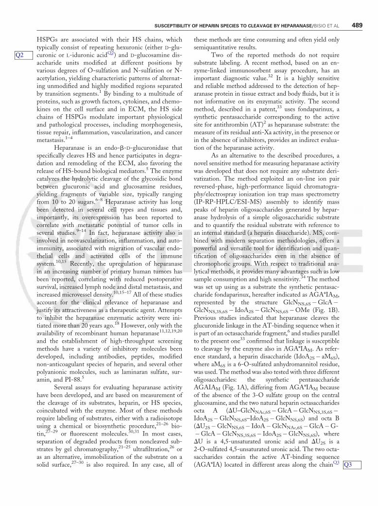
As an alternative to the described procedures, anovel sensitive method for measuring heparanase activitywas developed that does not require any substrate deri-vatization. The method exploited an on-line ion pairreversed-phase, high-performance liquid chromatogra-phy/electrospray ionization ion trap mass spectrometry(IP-RP-HPLC/ESI-MS) assembly to identify masspeaks of heparin oligosaccharides generated by hepar-anase hydrolysis of a simple oligosaccharidic substrateand to quantify the residual substrate with reference toan internal standard (a heparin disaccharide). MS, com-bined with modern separation methodologies, offers apowerful and versatile tool for identification and quan-tification of oligosaccharides even in the absence ofchromophoric groups. With respect to traditional ana-lytical methods, it provides many advantages such as lowsample consumption and high sensitivity.34 The methodwas set up using as a substrate the synthetic pentasac-charide fondaparinux, hereafter indicated as AGA*IAM,represented by the structure GlcNNS,6S�GlcA�GlcNNS,3S,6S� IdoA2S�GlcNNS,6S�OMe (Fig. 1B).Previous studies indicated that heparanase cleaves theglucuronide linkage in the AT-binding sequence when itis part of an octasaccharide fragment,6 and studies parallelto the present one33 confirmed that linkage is susceptibleto cleavage by the enzyme also in AGA*IAM. As refer-ence standard, a heparin disaccharide (IdoA2S� aM6S),where aM6S is a 6-O-sulfated anhydromannitol residue,was used. Themethod was also tested with three differentoligosaccharides: the synthetic pentasaccharideAGAIAM (Fig. 1A), differing from AGA*IAM becauseof the absence of the 3-O sulfate group on the centralglucosamine, and the two natural heparin octasaccharidesocta A (DU–GlcNNAc,6S�GlcA�GlcNNS,3S,6S�IdoA2S�GlcNNS,6S–IdoA2S�GlcNNS,6S) and octa B(DU2S�GlcNNS,6S� IdoA�GlcNNAc,6S�GlcA�G-�GlcA�GlcNNS,3S,6S� IdoA2S�GlcNNS,6S), whereDU is a 4,5-unsaturated uronic acid and DU2S is a2-O-sulfated 4,5-unsaturated uronic acid. The two octa-saccharides contain the active AT-binding sequence(AGA*IA) located in different areas along the chainQ3
Q2
Q3
SUSCEPTIBILITY OF HEPARIN SPECIES TO CLEAVAGE BY HEPARANASE/BISIO ET AL 489
(Figs. 1C,D). Given that these octasaccharides have anatural origin, the first glucosamine residue of the AT-binding sequence bears a N-acetyl group instead of aN-sulfate group.35
METHODAnalyses of kinetics of enzymatic hydrolysis were per-formed by incubating 100 mg of AGA*IAM (66 nmol)with variable amounts (0.5 to 5.0 mg) of recombinanthuman heparanase (kindly provided by Professor IsraelVlodavsky) at 378C in 20 mM ammonium acetateþ 2 mM Ca(OAc)2þ 1 mM b-mercaptoethanol (pH5.8) at a final volume of 125 mL, corresponding to anoligosaccharide concentration of 0.52 nmol/mL. Fivemicroliters of the incubation mixture was taken, atdifferent times (0, 2, 10, 30, and 60 minutes and 2, 4,8, 12, 16, and 24 hours), diluted 40 times with 10 mMammonium acetate containing 31 pmol/mL of referencestandard, IdoA2S� aM6S disaccharide, and treated withformic acid 0.025% to disrupt possible oligosaccharide-protein complexes. Twenty microliters of each step ofdigestion (originally containing 0.4 mg of pentasacchar-ide) were analyzed by IP-RP-HPLC/ESI-MS. Thechromatographic separation was performed on a 3-mmProntosil HypersorbQ5 reversed-phase C18 column(4.6� 250 mm), by eluting with a linear gradient from100% eluent A (MeOH/H2O 20/80) to 100% eluent B(MeOH/H2O 70/30), both in 5 mM dibutylammoniumacetate (DBA), at a flow rate of 0.3 mL/min. Massspectrometric analyses were performed on an Esquire
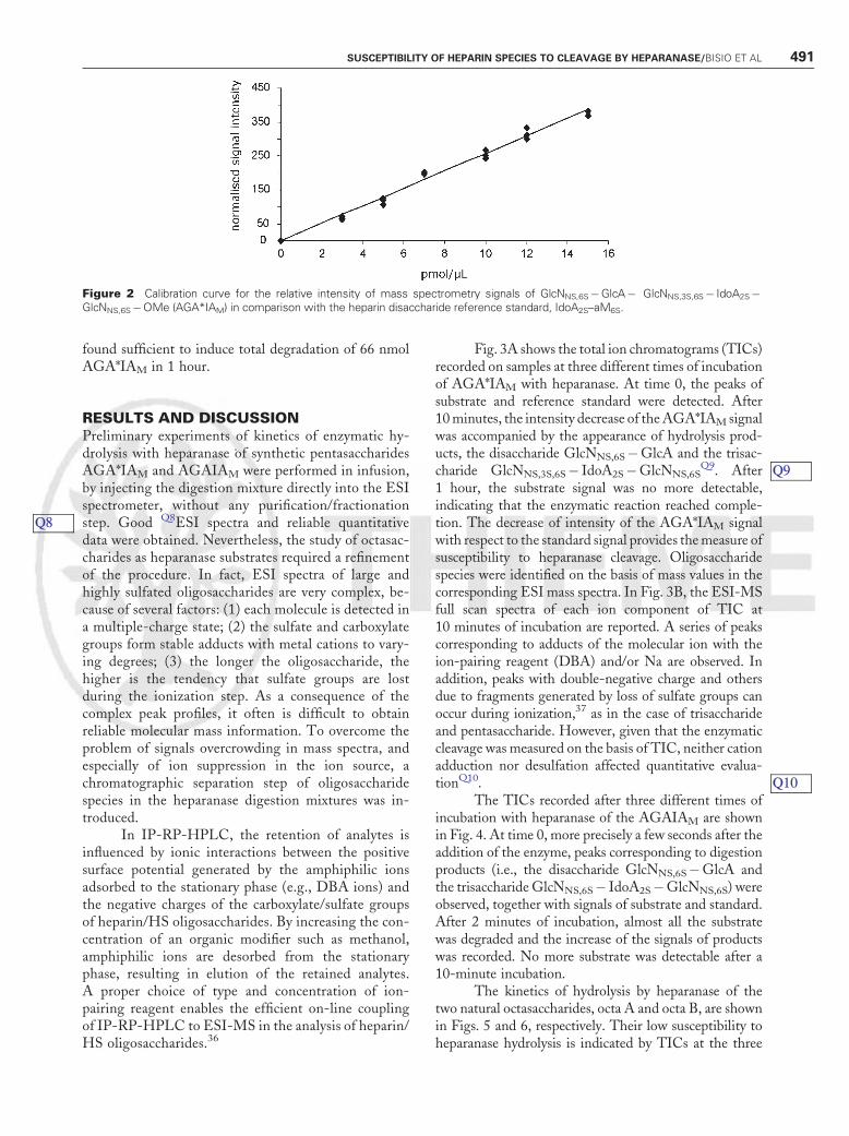
3000 Plus electrospray ion trap (Bruker Daltonics, Bre-men, Germany) equipped with an electrospray sourceworking in negative ion mode in the mass range fromm/z 400 to 1000. Sample ionization was obtained usingthe optimized MS conditions of spray voltage andcapillary temperature (3166 V and 3508C, respectively).Calibration of the mass spectrometer was performedusing an ES tuning mix solution (Agilent Q6acetonitrilesolution) according to a standard procedure. Data wereprocessed by the DataAnalysis software (Version 3.0,Bruker DaltonikQ7). To verify the possible quantitativeapplication of the method, a calibration curve was builtup with different AGA*IAM concentrations (0 to 25pmol/mL) in the presence of a constant amount ofreference standard. Following IP-RP-HPLC/ESI-MSanalysis, the area of AGA*IAM signals calculated fromeach total ion chromatogram was normalized with re-spect to the area of the signal of the internal standard. Asshown in Fig. 2, a good linear correlation of experimen-tal points was found for substrate concentrations in therange 0 to 15 pmol/mL. For each experiment of kineticsof enzymatic hydrolysis, calibration curves with each ofthe oligosaccharides employed as heparanase substrateswere set up. Because of the high sensitivity of thetechnique, such an internal calibration is important toexclude any possible interference due to even minorvariations of instrumental conditions.
For AGAIAM and the two natural heparin octa-saccharides octa A and octa B, the kinetics of enzymatichydrolysis were performed as described above, by incu-bating substrates with the heparanase amount (2.3 mg)
Figure 1 Structure of the heparin oligosaccharides tested as substrate for heparanase. The arrows indicate the site of hydrolyticcleavage. The AGA*IAQ4 sequence is highlighted by a gray background (B, C, D,); N-acetyl groups, typical for natural AGA*IA sequence,are highlighted by a circle comprising a dotted line (C, D).
Q5
Q6
Q7
Q4
490 SEMINARS IN THROMBOSIS AND HEMOSTASIS/VOLUME 33, NUMBER 5 2007
found sufficient to induce total degradation of 66 nmolAGA*IAM in 1 hour.
RESULTS AND DISCUSSIONPreliminary experiments of kinetics of enzymatic hy-drolysis with heparanase of synthetic pentasaccharidesAGA*IAM and AGAIAM were performed in infusion,by injecting the digestion mixture directly into the ESIspectrometer, without any purification/fractionationstep. Good Q8ESI spectra and reliable quantitativedata were obtained. Nevertheless, the study of octasac-charides as heparanase substrates required a refinementof the procedure. In fact, ESI spectra of large andhighly sulfated oligosaccharides are very complex, be-cause of several factors: (1) each molecule is detected ina multiple-charge state; (2) the sulfate and carboxylategroups form stable adducts with metal cations to vary-ing degrees; (3) the longer the oligosaccharide, thehigher is the tendency that sulfate groups are lostduring the ionization step. As a consequence of thecomplex peak profiles, it often is difficult to obtainreliable molecular mass information. To overcome theproblem of signals overcrowding in mass spectra, andespecially of ion suppression in the ion source, achromatographic separation step of oligosaccharidespecies in the heparanase digestion mixtures was in-troduced.
In IP-RP-HPLC, the retention of analytes isinfluenced by ionic interactions between the positivesurface potential generated by the amphiphilic ionsadsorbed to the stationary phase (e.g., DBA ions) andthe negative charges of the carboxylate/sulfate groupsof heparin/HS oligosaccharides. By increasing the con-centration of an organic modifier such as methanol,amphiphilic ions are desorbed from the stationaryphase, resulting in elution of the retained analytes.A proper choice of type and concentration of ion-pairing reagent enables the efficient on-line couplingof IP-RP-HPLC to ESI-MS in the analysis of heparin/HS oligosaccharides.36
Fig. 3A shows the total ion chromatograms (TICs)recorded on samples at three different times of incubationof AGA*IAM with heparanase. At time 0, the peaks ofsubstrate and reference standard were detected. After10minutes, the intensity decrease of theAGA*IAM signalwas accompanied by the appearance of hydrolysis prod-ucts, the disaccharide GlcNNS,6S�GlcA and the trisac-charide GlcNNS,3S,6S� IdoA2S�GlcNNS,6S
Q9. After1 hour, the substrate signal was no more detectable,indicating that the enzymatic reaction reached comple-tion. The decrease of intensity of the AGA*IAM signalwith respect to the standard signal provides themeasure ofsusceptibility to heparanase cleavage. Oligosaccharidespecies were identified on the basis of mass values in thecorresponding ESI mass spectra. In Fig. 3B, the ESI-MSfull scan spectra of each ion component of TIC at10 minutes of incubation are reported. A series of peakscorresponding to adducts of the molecular ion with theion-pairing reagent (DBA) and/or Na are observed. Inaddition, peaks with double-negative charge and othersdue to fragments generated by loss of sulfate groups canoccur during ionization,37 as in the case of trisaccharideand pentasaccharide. However, given that the enzymaticcleavage was measured on the basis of TIC, neither cationadduction nor desulfation affected quantitative evalua-tionQ10.
The TICs recorded after three different times ofincubation with heparanase of the AGAIAM are shownin Fig. 4. At time 0, more precisely a few seconds after theaddition of the enzyme, peaks corresponding to digestionproducts (i.e., the disaccharide GlcNNS,6S�GlcA andthe trisaccharide GlcNNS,6S� IdoA2S�GlcNNS,6S) wereobserved, together with signals of substrate and standard.After 2 minutes of incubation, almost all the substratewas degraded and the increase of the signals of productswas recorded. No more substrate was detectable after a10-minute incubation.
The kinetics of hydrolysis by heparanase of thetwo natural octasaccharides, octa A and octa B, are shownin Figs. 5 and 6, respectively. Their low susceptibility toheparanase hydrolysis is indicated by TICs at the three
Figure 2 Calibration curve for the relative intensity of mass spectrometry signals of GlcNNS,6S�GlcA� GlcNNS,3S,6S� IdoA2S�GlcNNS,6S�OMe (AGA*IAM) in comparison with the heparin disaccharide reference standard, IdoA2S–aM6S.
Q8
Q9
Q10
SUSCEPTIBILITY OF HEPARIN SPECIES TO CLEAVAGE BY HEPARANASE/BISIO ET AL 491
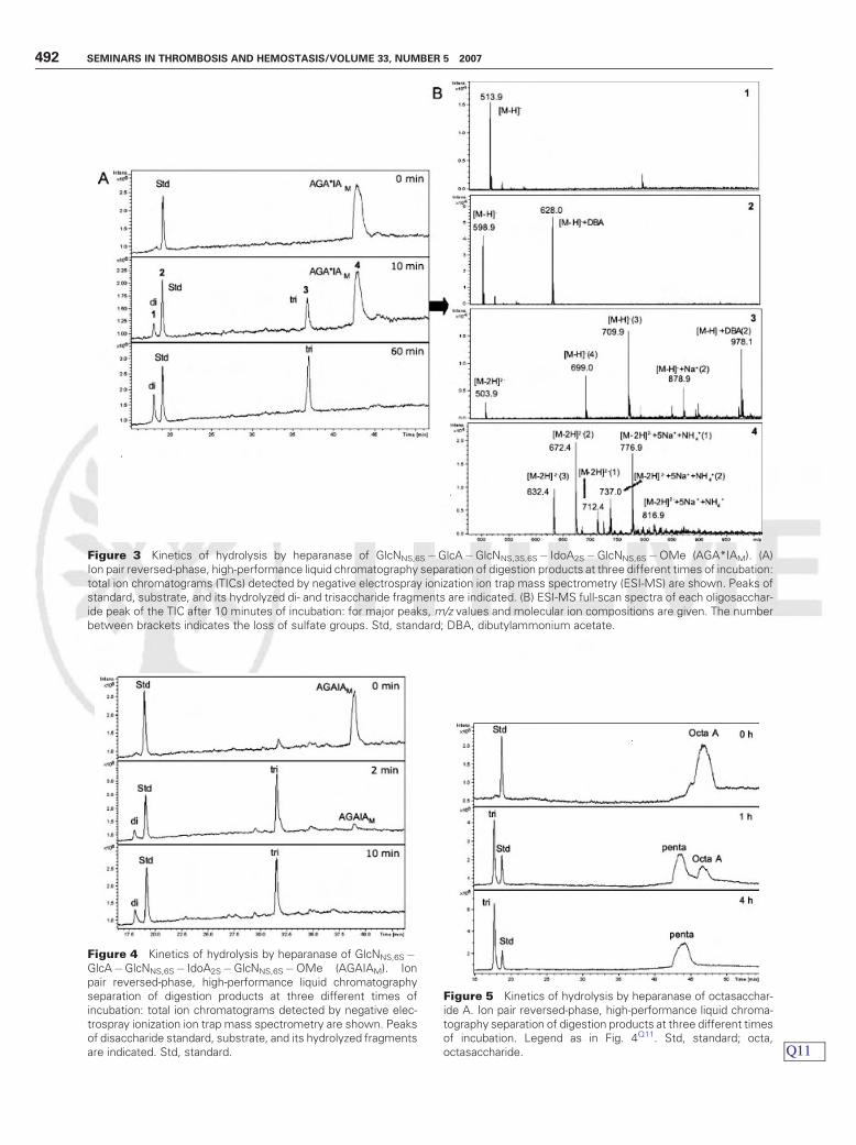
Figure 5 Kinetics of hydrolysis by heparanase of octasacchar-ide A. Ion pair reversed-phase, high-performance liquid chroma-tography separation of digestion products at three different timesof incubation. Legend as in Fig. 4Q11. Std, standard; octa,octasaccharide.
Figure 3 Kinetics of hydrolysis by heparanase of GlcNNS,6S�GlcA�GlcNNS,3S,6S� IdoA2S�GlcNNS,6S�OMe (AGA*IAM). (A)Ion pair reversed-phase, high-performance liquid chromatography separation of digestion products at three different times of incubation:total ion chromatograms (TICs) detected by negative electrospray ionization ion trap mass spectrometry (ESI-MS) are shown. Peaks ofstandard, substrate, and its hydrolyzed di- and trisaccharide fragments are indicated. (B) ESI-MS full-scan spectra of each oligosacchar-ide peak of the TIC after 10 minutes of incubation: for major peaks, m/z values and molecular ion compositions are given. The numberbetween brackets indicates the loss of sulfate groups. Std, standard; DBA, dibutylammonium acetate.
Figure 4 Kinetics of hydrolysis by heparanase of GlcNNS,6S�GlcA�GlcNNS,6S� IdoA2S�GlcNNS,6S�OMe (AGAIAM). Ionpair reversed-phase, high-performance liquid chromatographyseparation of digestion products at three different times ofincubation: total ion chromatograms detected by negative elec-trospray ionization ion trap mass spectrometry are shown. Peaksof disaccharide standard, substrate, and its hydrolyzed fragmentsare indicated. Std, standard. Q11
492 SEMINARS IN THROMBOSIS AND HEMOSTASIS/VOLUME 33, NUMBER 5 2007
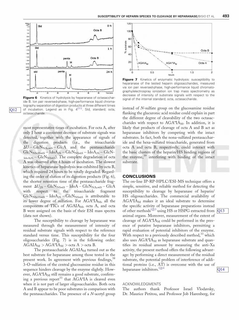
most representative times of incubation. For octa A, afteronly 1 hour a consistent decrease of substrate signals wasdetected, together with the appearance of signals ofthe digestion products (i.e., the trisaccharideDU�GlcNNAc,6S�GlcA and the pentasaccharideGlcNNS,3S,6S� IdoA2S�GlcNNS,6S� IdoA2S�GlcN-
IdoA2S�GlcNNS,6S). The complete degradation of octaA was observed after 4 hours of incubation. The slowestkinetics of heparanase hydrolysis was exhibited by octa B,which required 24 hours to be totally degraded. Regard-ing the order of elution of its digestion products (Fig. 6),the shorter retention time of the pentasaccharide frag-ment DU2S�GlcNNS,6S� IdoA�GlcNNAc,6S�GlcAwith respect to the trisaccharide fragmentGlcNNS,3S,6S� IdoA2S�GlcNNS,6S is attributable toits lower degree of sulfation. For AGA*IAM, all thecomponents of TICs of AGAIAM, octa A, and octaB were assigned on the basis of their ESI mass spectra(data not shown).
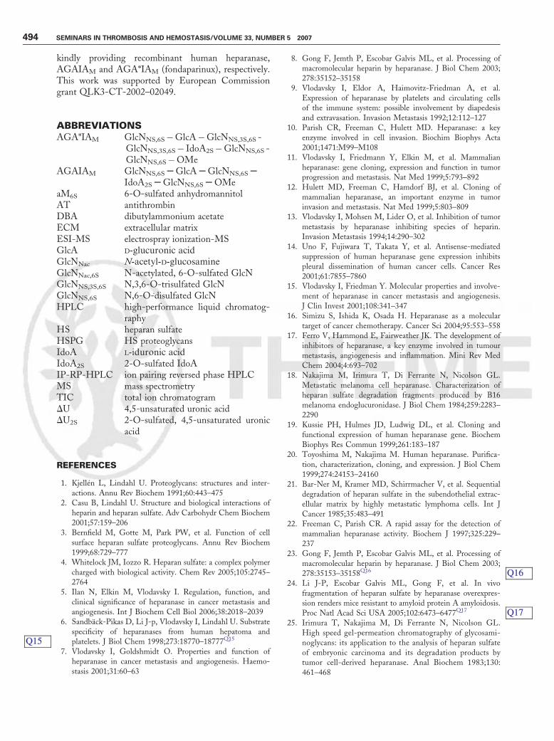
The susceptibility to cleavage by heparanase wasmeasured through the measurement of intensity ofresidual substrate signals with respect to the referencestandard versus time. This susceptibility for the fouroligosaccharides (Fig. 7) is in the following order:AGAIAM >AGA*IAM > octa A > octa B.
The pentasaccharide AGAIAM turned out as thebest substrate for heparanase among those tested in thepresent work. In agreement with previous findings,38
3-O-sulfation of the central glucosamine residue in thissequence hinders cleavage by the enzyme slightly. How-ever, AGA*IAM still remains a good substrate, confirm-ing a previous report33 that AGA*IA is cleaved evenwhen it is not part of larger oligosaccharides. Both octaA and B appear to be poor substrates in comparison withthe pentasaccharides. The presence of a N-acetyl group
instead of N-sulfate group on the glucosamine residueflanking the glucuronic acid residue could explain in partthe different degree of cleavability of the two octasac-charides with respect to AGA*IAM. In addition, it islikely that products of cleavage of octa A and B act asheparanase inhibitors by competing with the intactsubstrates. In fact, both the nona-sulfated pentasacchar-ide and the hexa-sulfated trisaccharide, generated fromocta A and octa B, respectively, could interact withthe basic clusters of the heparin/HS binding regions ofthe enzyme,39 interfering with binding of the intactsubstrate.
CONCLUSIONSThe on-line IP-RP-HPLC/ESI-MS technique offers asimple, sensitive, and reliable method for detecting thesusceptibility to cleavage by heparanase of heparin/HS oligosaccharides. The commercial availability ofAGA*IAM makes it an ideal substrate to determinethe specific activity of heparanase preparations insteadof other methodsQ13 using HS or HSPG extracted fromanimal organs. Moreover, measurement of the extent ofcleavage of AGA*IAM could be performed in the pres-ence of putative heparanase inhibitors, permitting arapid evaluation of potential inhibitors of the enzyme.With respect to a previously described method,33 whichalso uses AGA*IAM as heparanase substrate and quan-tifies its residual amount by measuring the anti-Xaactivity, the present method offers the following advant-age: by performing a direct measurement of the residualsubstrate, the potential problem of interference of addi-tional proteins (i.e., AT) is overcome with the use ofheparanase inhibitors.Q14
ACKNOWLEDGMENTS
The authors thank Professor Israel Vlodavsky,Dr. Maurice Petitou, and Professor Job Harenberg, for
Figure 6 Kinetics of hydrolysis by heparanase of octasacchar-ide B. Ion pair reversed-phase, high-performance liquid chroma-tography separation of digestion products at three different timesof incubation. Legend as in Fig. 4Q12. Std, standard; octa,octasaccharide.
Figure 7 Kinetics of enzymatic hydrolysis: susceptibility toheparanase of the tested heparin oligosaccharides, measuredvia ion pair reversed-phase, high-performance liquid chromato-graphy/electrospray ionization ion trap mass spectrometry asdecrease of intensity of substrate signals with respect to thesignal of the internal standard; octa, octasaccharide.
Q13
Q14
Q12
SUSCEPTIBILITY OF HEPARIN SPECIES TO CLEAVAGE BY HEPARANASE/BISIO ET AL 493
kindly providing recombinant human heparanase,AGAIAM and AGA*IAM (fondaparinux), respectively.This work was supported by European Commissiongrant QLK3-CT-2002–02049.
ABBREVIATIONSAGA*IAM GlcNNS,6S�GlcA�GlcNNS,3S,6S -
GlcNNS,3S,6S� IdoA2S�GlcNNS,6S -GlcNNS,6S�OMe
AGAIAM GlcNNS,6SSGlcASGlcNNS,6SSIdoA2SSGlcNNS,6SSOMe
aM6S 6-O-sulfated anhydromannitolAT antithrombinDBA dibutylammonium acetateECM extracellular matrixESI-MS electrospray ionization-MSGlcA D-glucuronic acidGlcNNac N-acetyl-D-glucosamineGlcNNac,6S N-acetylated, 6-O-sulfated GlcNGlcNNS,3S,6S N,3,6-O-trisulfated GlcNGlcNNS,6S N,6-O-disulfated GlcNHPLC high-performance liquid chromatog-
raphyHS heparan sulfateHSPG HS proteoglycansIdoA L-iduronic acidIdoA2S 2-O-sulfated IdoAIP-RP-HPLC ion pairing reversed phase HPLCMS mass spectrometryTIC total ion chromatogramDU 4,5-unsaturated uronic acidDU2S 2-O-sulfated, 4,5-unsaturated uronic
acid
REFERENCES
1. Kjellen L, Lindahl U. Proteoglycans: structures and inter-actions. Annu Rev Biochem 1991;60:443–475
2. Casu B, Lindahl U. Structure and biological interactions ofheparin and heparan sulfate. Adv Carbohydr Chem Biochem2001;57:159–206
3. Bernfield M, Gotte M, Park PW, et al. Function of cellsurface heparan sulfate proteoglycans. Annu Rev Biochem1999;68:729–777
4. Whitelock JM, Iozzo R. Heparan sulfate: a complex polymercharged with biological activity. Chem Rev 2005;105:2745–2764
5. Ilan N, Elkin M, Vlodavsky I. Regulation, function, andclinical significance of heparanase in cancer metastasis andangiogenesis. Int J Biochem Cell Biol 2006;38:2018–2039
6. Sandback-Pikas D, Li J-p, Vlodavsky I, Lindahl U. Substratespecificity of heparanases from human hepatoma andplatelets. J Biol Chem 1998;273:18770–18777Q15
7. Vlodavsky I, Goldshmidt O. Properties and function ofheparanase in cancer metastasis and angiogenesis. Haemo-stasis 2001;31:60–63
8. Gong F, Jemth P, Escobar Galvis ML, et al. Processing ofmacromolecular heparin by heparanase. J Biol Chem 2003;278:35152–35158
9. Vlodavsky I, Eldor A, Haimovitz-Friedman A, et al.Expression of heparanase by platelets and circulating cellsof the immune system: possible involvement by diapedesisand extravasation. Invasion Metastasis 1992;12:112–127
10. Parish CR, Freeman C, Hulett MD. Heparanase: a keyenzyme involved in cell invasion. Biochim Biophys Acta2001;1471:M99–M108
11. Vlodavsky I, Friedmann Y, Elkin M, et al. Mammalianheparanase: gene cloning, expression and function in tumorprogression and metastasis. Nat Med 1999;5:793–892
12. Hulett MD, Freeman C, Hamdorf BJ, et al. Cloning ofmammalian heparanase, an important enzyme in tumorinvasion and metastasis. Nat Med 1999;5:803–809
13. Vlodavsky I, Mohsen M, Lider O, et al. Inhibition of tumormetastasis by heparanase inhibiting species of heparin.Invasion Metastasis 1994;14:290–302
14. Uno F, Fujiwara T, Takata Y, et al. Antisense-mediatedsuppression of human heparanase gene expression inhibitspleural dissemination of human cancer cells. Cancer Res2001;61:7855–7860
15. Vlodavsky I, Friedman Y. Molecular properties and involve-ment of heparanase in cancer metastasis and angiogenesis.J Clin Invest 2001;108:341–347
16. Simizu S, Ishida K, Osada H. Heparanase as a moleculartarget of cancer chemotherapy. Cancer Sci 2004;95:553–558
17. Ferro V, Hammond E, Fairweather JK. The development ofinhibitors of heparanase, a key enzyme involved in tumourmetastasis, angiogenesis and inflammation. Mini Rev MedChem 2004;4:693–702
18. Nakajima M, Irimura T, Di Ferrante N, Nicolson GL.Metastatic melanoma cell heparanase. Characterization ofheparan sulfate degradation fragments produced by B16melanoma endoglucuronidase. J Biol Chem 1984;259:2283–2290
19. Kussie PH, Hulmes JD, Ludwig DL, et al. Cloning andfunctional expression of human heparanase gene. BiochemBiophys Res Commun 1999;261:183–187
20. Toyoshima M, Nakajima M. Human heparanase. Purifica-tion, characterization, cloning, and expression. J Biol Chem1999;274:24153–24160
21. Bar-Ner M, Kramer MD, Schirrmacher V, et al. Sequentialdegradation of heparan sulfate in the subendothelial extrac-ellular matrix by highly metastatic lymphoma cells. Int JCancer 1985;35:483–491
22. Freeman C, Parish CR. A rapid assay for the detection ofmammalian heparanase activity. Biochem J 1997;325:229–237
23. Gong F, Jemth P, Escobar Galvis ML, et al. Processing ofmacromolecular heparin by heparanase. J Biol Chem 2003;278:35153–35158Q16
24. Li J-P, Escobar Galvis ML, Gong F, et al. In vivofragmentation of heparan sulfate by heparanase overexpres-sion renders mice resistant to amyloid protein A amyloidosis.Proc Natl Acad Sci USA 2005;102:6473–6477Q17
25. Irimura T, Nakajima M, Di Ferrante N, Nicolson GL.High speed gel-permeation chromatography of glycosami-noglycans: its application to the analysis of heparan sulfateof embryonic carcinoma and its degradation products bytumor cell-derived heparanase. Anal Biochem 1983;130:461–468
Q15
Q16
Q17
494 SEMINARS IN THROMBOSIS AND HEMOSTASIS/VOLUME 33, NUMBER 5 2007
26. Tsuchida S, Podyma-Inoue KA, Yanagishita M. Ultra-filtration-based assay for heparanase activity. Anal Biochem2004;331:147–152
27. Nakajima M, Irimura T, Nicolson GL. Tumor metastasis-associated heparanase (heparan sulfate endoglycosidase) activ-ity in human melanoma cells. Cancer Lett 1986;31:277–283
28. Behzad F, Brenkley PEC. A multiwell format assay forheparanase. Anal Biochem 2003;320:207–213
29. Nardella C, Steinkuhler C. Radiolabeled heparan sulfateimmobilized on microplate as substrate for the detection ofheparanase activity. Anal Biochem 2004;332:368–375
30. Huang K-S, Holmgren J, Reik L, et al. High-throughputmethods for measuring heparanase activity and screeningpotential antimetastatic and anti-inflammatory agents. AnalBiochem 2004;333:389–398
31. Enomoto K, Okamoto H, Numata Y, Takemoto H. Asimple and rapid assay for heparanase activity usinghomogeneous time-resolved fluorescence. J Pharm BiomedAnal 2006;41:912–917
32. Shafat I, Zcharia E, Nisman B, et al. An ELISA method fordetection and quantification of human heparanase. BiochemBiophys Res Commun 2006;341:958–963
33. Petitou M, Driguez PA. Derives d’azasucre, inhibiteursd’heparanases, leur procede de preparation, les compositionsen contenant et leur utilisation. FR patent #2873377–A1 2004
34. Capila I, Gunay NS, Schriver Z, Venkataraman G. Methodsfor structural analysis of heparin and heparan sulfate. In:Garg HG, Linhardt RJ, Hales CA eds. Chemistry andBiology of Heparin and Heparan Sulfate. Oxford, UnitedKingdom: Elsevier Ltd; 2005:55–77
35. Mourier PA, Viskov C. Chromatographic analysis andsequencing approach of heparin oligosaccharides usingcethyltrimethylammonium dynamically coated stationaryphase. Anal Biochem 2004;332:299–313
36. Kuberan B, Lech M, Zhang L, et al. Analysis of heparansulfate oligosaccharides with ion pair-reverse phase capillaryhigh performance liquid chromatography-microelectrosprayionization time-of-flight mass spectrometry. J Am Chem Soc2002;124:8707–8718
37. Zaia J, Costello CE. Tandem mass spectrometry of sulfatedheparin-like glycosaminoglycan oligosaccharides. Anal Chem2003;275:2445–2455Q18
38. Okada Y, Yamada S, Toyoshima M, et al. Structuralrecognition by recombinant human heparanase that playscritical roles in tumor metastasis. J Biol Chem 2002;277:42488–44249Q19
39. Levy-Adam F, Abboud-Jarrous G, Guerrini M, et al.Identification and characterization of heparin/heparan sulfatebinding domains of the endoglycosidase heparanase. J BiolChem 2005;280:20457–20466
Q18
Q19
SUSCEPTIBILITY OF HEPARIN SPECIES TO CLEAVAGE BY HEPARANASE/BISIO ET AL 495
Author Query Form (STH/01308)
Special Instructions: Author please write responses to queries directly on proofs and then
return back.
Q1: AU: Please verify all chemical compound names throughout text for accuracy and consistency.All chemical compound acronyms/abbreviations should be defined at first mention in text andwithin each figure and table, if applicable.
Q2: AU: Per standard chemical nomenclature, okay with "D" and "L" for absolute configuration ofcarbohydrates and amino acids as changed to "d" and "l" (small capital letters) throughout?
Q3: AU: Phrase okay as changed from "differently located along the chain"?
Q4: AU: Please define. Is it GlcNNS,6S�GlcA�GlcNNS,6S� IdoA2S�GlcNNS,6S?
Q5: AU: Please provide name and city/state (or city/country if not U.S.) location for manufacturerof column.
Q6: AU: Please provide name and city/state (or city/country if not U.S.) location for manufacturerof Agilent solution.
Q7: AU: Please verify spelling. Earlier in this paragraph, company name is spelled "BrukerDaltonics."
Q8: AU: Please clarify description "good." Do you mean accurate, reproducible, or some othermeasure?
Q9: AU: Are items in this phrase three separate items in a series (we will add a comma before thelast item, per journals style), or are the last two items describing the two hydrolysis products?
Q10: AU: Phrase okay as edited for clarity?
Q11: AU: Please clarify/specify what part of Fig. 4 legend applies to Fig. 5.
Q12: AU: Please clarify/specify what part of Fig. 4 legend applies to Fig. 6.
Q13: AU: Phrase okay as changed for clarity from "... in substitution other methods..."
Q14: AU: Sentence okay as edited for clarity?
Q15: Medline reports the first author "Sandback-Pikas D" is not correct in the reference 6"Sandback-Pikas, Li, Vlodavsky, Lindahl, 1998".
Q16: Medline indexes "J Biol Chem" but cannot find a listing for the reference 23 "Gong, Jemth,Escobar Galvis, et al, 2003". Please check the reference for accuracy.
Q17: Medline reports the first author "Li J" is not correct in the reference 24 "Li, Escobar Galvis,Gong, et al, 2005".
Q18: Medline indexes "Anal Chem" but cannot find a listing for the reference 37 "Zaia, Costello,2003". Please check the reference for accuracy.
Q19: Reference appears to have more than 100 pages. Please verify page numbers (in reference 38"Okada, Yamada, Toyoshima, et al, 2002").
Instructions to Contributors Dear Contributor: Enclosed in this document please find the page proofs, copyright transfer agreement (CTA), and offprint order form for your article in the Seminars in Thrombosis and Hemostasis, Volume 33, Number 5, 2007. Please print this document and complete and return the CTA and offprint form, along with corrected proofs, within 72 hours (3 business days). 1) Please read proofs carefully for typographical and factual errors only; mark corrections in the margins of the proofs in pen. Answer (on the proofs) all editor’s queries indicated in the margins of the proofs. Check references for accuracy. Please check on the bottom of the 1st page of your article that your titles and affiliations are correct. Avoid elective changes, as these are costly and time consuming and will be made at the publisher’s discretion. 2) Please pay particular attention to the proper placement of figures, tables, and legends. Provide copies of any formal letters of permission that you have obtained. 3) Please return the corrected proofs, signed copyright transfer agreement, and your offprint order form, with color prints of figures, if you received any. 4) As a contributor to this journal you will receive one copy of the journal, at no charge. • If you wish to order offprints or e-prints, please circle the quantity required (left column) and the number of pages in your article. If you wish to order additional copies of the journal please enter the number of copies on the indicated line. • If you do not want to order offprints or journals simply put a slash through the form, but please return the form. Please send all materials back via overnight mail, within 72 hours of receipt, to:
Xenia Golovchenko Production editor
Thieme Medical Publishers 77 Gregory Blvd.
Norwalk, CT 06855 Tel: 845-548-8127 Fax: +1 (203) 857 4996
E-mail: [email protected] Please do not return your materials to the editor, or the compositor. Please note: Due to a tight schedule, if the publisher does not receive the return of your article within 7 business days of the mail date (from the compositor), the publisher reserves the right to proceed with publication without author changes. Such proofs will be proofread by the editor and the publisher. Thank you for your contribution to this journal. Xenia Golovchenko, Production Editor, Journal Production Department Thieme Medical Publishers, Inc.
Thieme Medical Publishers, Inc. (the “Publisher”) will be pleased to publish your article (the “Work”) entitled _____________________________ in the Seminars in Thrombosis and Hemostasis, Volume 33, Number 5, 2007. The undersigned Author(s) hereby assigns to the Publisher all rights to the Work of any kind, including those rights protected by the United States Copyright laws. The Author(s) will be given permission by the Publisher, upon written request, to use all or part of the Work for scholarly or academic purposes, provided lawful copyright notice is given. If the Work, subsequent to publication, cannot be reproduced and delivered to the Author(s) by the publisher within 60 days of a written request, the Author(s) is given permission to reprint the Work without further request. The Publisher may grant third parties permission to reproduce all or part of the Work. The Author(s) will be notified as a matter of courtesy, not as a matter of contract. Lawful notice of copyright always will be given. Check appropriate box below and affix signature. [ ] I Sign for and accept responsibility for transferring copyright of this article to Thieme Medical Publishers, Inc. on behalf of any and all authors. Author’s full name, degrees, professional title, affiliation, and complete address: __________________________________ ____________________________ Author’s printed name, degrees Professional title
Complete professional address
______________________________ ___________________ Author’s signature Date [ ] I prepared this article as part of my official duties as an employee of the United States Federal Government. Therefore, I am unable to transfer rights to Thieme Medical Publishers, Inc. ______________________________ _________________ Author’s signature Date

Order Form for Offprints and additional copies of the Seminars in Thrombosis and Hemostasis(Effective October 2005)
Please circle the cost of the quantity/page count you require (orders must be in increments of 100)
Pages in Article/CostQuantity 1 to 4 5 to 8 9 to 12 13 to 16 17 to 20
100 $198 $317 $440 $578 $693200 $277 $444 $615 $809 $970300 $356 $570 $791 $1,041 $1,247400 $396 $634 $879 $1,156 $1,386500 $446 $713 $989 $1,301 $1,559
1000 $792 $1,267 $1,758 $2,313 $2,772
Volume/Issue #: Page Range (of your article):
Article Title:
MC/Visa/AmEx No: Exp. Date:
Signature:
Name:
Address:
City/State/Zip/Country:
Corresponding author will receive one complimentary copy of the issue in which the manuscript is published.Number of additional copies of the journal, at the discounted rate of $20.00 each:
Notes1. The above costs are valid only for orders received before publication of the issue. Please return the completed form, even if your institution intends to send a Purchase Order (the P.O. may sometimes be supplied after the issue has been printed).
2. Orders from outside the U.S. must be accompanied by payment.
3. A shipping charge will be added to the above costs.
4. Reprints are printed on the same coated paper as the journal and side-stapled.
5. For larger quantities or late orders, please contact reprints dept. Phone: +1(212) 584-4662Fax: +1(212) 947-1112 E-mail: [email protected]
As an added benefit to all contributing authors, a discount is offered on all Thieme books.See below for details or go to www.thieme.com

As a Thieme author you are entitled to a 25% discount for new books and a 35% discount for forthcoming books. We selected two books that might be of interest for you: new! 25% forthcoming! 35%
Thurlbeck's Pathology of the Lung 3rd Edition Andrew M. Churg, M.D. Ph. D. Professor of Pathology, University of British Columbia; Pathologist, Vancouver Hospital & Health Sciences Center, Vancouver, BC, Canada
Vascular Diagnosis with Ultrasound Cerebral and Peripheral Vessels Michael Hennerici, M.D. Professor and Chairman, Department of Neurology, University of Heidelberg, Mannheim, Germany
Thurlbeck's cornerstone textbook and reference on pulmonarypathology returns in a brand new edition! Updated with the latestadvances in the field, you will save time with all-inclusive coverage ofneoplastic, non-neoplastic, infectious, occupational/environmental, anddevelopmental pathologies in one book, learn how molecular biologyprovides a greater understanding of lung development, gain newinsights into the diagnosis of neoplastic and non-neoplastic lungdisease, find pertinent information on clinical features, epidemiology,and pathogenetic mechanisms of lung disease and much more!Comprehensive in its scope and authoritative in its scholarship,Thurlbeck's Pathology of the Lung is a virtual one-volume encyclopediawritten by a ''who's who'' list of specialists. It is the one text that nopathologist, pulmonologist, or resident in either specialty can afford tobe without.
Covering the entire venous and body circulation as examinedby vascular ultrasound, this unique text/atlas is invaluable fordiagnosing arterial and venous disease. It includescomprehensive chapters on vascular ultrasonography in thearteries and veins of the cerebral circulation and theperipheral upper and lower limb circulation, systematiccoverage of all available ultrasound technologies, includingcontinuous and pulsed-wave Doppler mode, b-mode, andconventional and color-coded duplex analysis in frequencyand amplitude power modes, anatomy and physiology,normal and abnormal findings, test accuracy and sensitivity,pitfalls, and comparison with other diagnostic tests in eachvascular region and special, difficult-to-interpret casesdiscussed in a separate section
2005, app.1032 pp., app.1064 illus., hardcover $249.95 $187.46 2006, 336 pp., 530 illus., hardcover, $149.95 $97.47 ISBN 1-58890-288-9 ISBN 1-58890-144-0
If you want to view more Thieme books, fell free to visit Thieme Books
Thieme Author order form For faster service, call TOLL-FREE 1-800-782-3488 or fax this order form to 212-947-1112
Quantity ISBN (last 4-digits only) Author/Title Price
Subtotal:
Shipping & Handling (Add $7.50 for the first book and $1.00 for each additional book):
NY and PA residents add applicable sales tax:
TOTAL:
Enclosed is my check for $____________________ Charge my: AMEX MasterCard VISA Discover Card#____________________________________________________________________Exp.____________________________________________________ First Name_____________________________________ MI ________________________Last Name_______________________________________________ Address__________________________________________________________________________________________________________________________ City/State/Zip______________________________________________________________________________________________________________________ Telephone________________________________________________________________FAX_____________________________________________________ e-mail____________________________________________________________________________________________________________________________ Signature_________________________________________________________________________________________________________________________ EM1-05