Embed Size (px)
Citation preview

Proc. Nati. Acad. Sci. USAVol. 89, pp. 12003-12007, December 1992Applied Biological Sciences
High-level expression of a heterologous protein in the milk oftransgenic swine using the cDNA encoding human protein CWILLIAM H. VELANDER*t, JOHN L. JOHNSONf, RAYMOND L. PAGE*, CHRISTOPHER G. RUSSELL*,ANURADHA SUBRAMANIAN*, TRACY D. WILKINSf, FRANCIS C. GWAZDAUSKAS§, CHRISTOPH PITTIUS$ 11,AND WILLIAM N. DROHAN¶Departments of *Chemical Engineering, tAnaerobic Microbiology, and §Dairy Science, Virginia Polytechnic Institute and State University, Blacksburg, VA24061; and 1The Holland Laboratory, The American Red Cross, Rockville, MD 20855
Communicated by Kenneth M. Brinkhous, July 31, 1992
ABSTRACT Transgenic pigs were generated that pro-duced human protein C in their milk at up to 1 g/liter. Thegene construct was a fusion gene consisting of the cDNA forhuman protein C inserted into the first exon of the mouse wheyacidic protein gene. These results demonstrate that the mousewhey acidic protein gene contains regulatory elements that candirect cDNA expression at high levels in the pig mammarygland. Recombinant human protein C that was produced atabout 380 pg/ml per hr in transgenic pig milk possessedanticoagulant activity that was equivalent to that of protein Cderived from human plasma. These studies provide evidencethat y-carboxylation can occur at high levels in the mammarygland of a pig.
Several different regulatory sequences have been identifiedfor milk protein genes that enable the expression of heterol-ogous proteins in the milk of transgenic animals (1-4).However, expression levels from cDNAs (1, 4) and genomicsequences have been variable, with genomic sequences fre-quently producing much higher levels of protein (2, 3). Forexample, the cDNA encoding human factor IX (hFIX) wasexpressed in sheep milk at only 25 ng/ml using a fusion geneconsisting of 4.0 kilobase pairs (kbp) of 5' flanking sequencefrom the sheep /3-lactoglobulin gene (BLG), 1.5 kbp of hFIXcDNA, and 4.9 kbp containing the BLG transcription unitand 3' flanking sequence (1). In contrast, secretion of up to30 mg/ml of human a1-antitrypsin (hAAT) into sheep milkwas achieved using the same 4.0 kbp of BLG 5' flankingsequence fused to 6.5 kbp of hAAT minigene (first intronremoved) coding sequence (3). Additionally, there is nodirect correlation between the level of expression in trans-genic mice compared to livestock for a given genetic con-struct (2, 4). For example, transgenic pigs expressed a7.2-kbp genomic fragment of mouse whey acidic protein(WAP) at a 2- to 100-fold greater level (2, 5) than transgenicmice with the same construct. Therefore, the choice ofemploying a cDNA versus a genomic construct to synthesizea given protein in the mammary gland of livestock canbecome complex.Human protein C (hPC) is a regulator of hemostasis,
suggesting its potential use as a therapy for many diseasestates (6). Protein C is a zymogen of a serine protease that isactivated by thrombin (6). The structure of hPC is complexand its level of expression in recombinant mammalian cells(7) and transgenic mice (8) has been limited to <1 ,ug/ml perhr. In this report, we detail the expression of functionalrecombinant hPC (rhPC) in the milk of transgenic swine at1000 ,ug/ml per hr using a hybrid genetic construct consistingof the cDNA of hPC regulated by the mouse WAP gene.
MATERIALS AND METHODSTransgenic Swine. Crossbred gilts served as embryo donors
and recipients of microinjected eggs. Estrus synchronization,surgical procedures, and pronuclear microinjection of cen-trifuged zygotes were performed as described by Wall et al.(9). About 1-3 pl ofDNA solution (3.3 ,ug ofDNA per ml in10mM Tris HCl/0.25 mM EDTA, pH 7.4) was microinjectedas described by Brinster et al. (10). The hybrid transgene(WAPPC-1) consists of the cDNA for hPC inserted into theunique Kpn I site in the first exon of the mouse WAP gene(11), as illustrated in Fig. 1.
Tail tissue was biopsied from 2-day-old piglets and DNAwas isolated using a modification of the procedure developedby Marmur (12). Transgenic founder animals were identifiedinitially by polymerase chain reaction (PCR) (13) using hPC-specific primers and later confirmed by Southern analysisusing 32P-labeled hPC cDNA.Mammary gland biopsies were performed on two trans-
genic females and a control female on days 55 and 35 oflactation, respectively. Biopsies were washed briefly in ster-ile saline and immediately frozen in liquid nitrogen. TotalRNA was isolated from the pig mammary gland biopsies andhuman liver tissue samples (14). RNA samples (10 ,ug) werefractionated using agarose/formaldehyde gels and then trans-ferred by vacuum onto nylon membranes. The membraneswere probed with 32P random primer-labeled hPC cDNA orWAP cDNA.
Pig Milk Collection and Preparation. Piglets were removedfrom the sows for -30 min prior to milking to allow for milkaccumulation. Milk letdown was induced by intramuscularadministration of 20-30 international units of oxytocin. Milkwas collected directly into Tris-buffered saline/EDTA buffer(2x TBS/EDTA: 100 mM Tris HCl, 300 mM NaCl, 200 mMEDTA, pH 6.5, chilled to 0-2°C) in a 1:1 buffer to milk ratio.Fat and precipitate were removed by centrifugation at 15,000x g for 20 min at 0-2°C. The diluted whey was then filteredthrough sterile gauze to remove residual solids and stored at-90°C. Control pig milk was treated identically.Analysis of rhPC. Expression levels in the whey were
determined by ELISA, using a monoclonal antibody (HPC4-Mab) that has a calcium-dependent binding epitope in theactivation peptide (15). The rhPC captured by the HPC4-Mabwas detected using horseradish peroxidase conjugated togoat anti-rabbit IgG following a 3-hr incubation at roomtemperature with a rabbit polyclonal antibody to hPC. Asecond ELISA procedure was also used in which rabbitpolyclonal anti-hPC antiserum was used to immunocapture
Abbreviations: hPC, human protein C; rhPC, recombinant hPC;WAP, whey acidic protein; nt, nucleotide(s); APTT, activated partialthromboplastin time; NRPP, normal reference plasma pool.tTo whom reprint requests should be addressed.'Present address: Hoechst AG, PGE Stoffwechsel/H 825, Postfach80 03 20, 6230 Frankfurt 80, Federal Republic of Germany.
12003
The publication costs of this article were defrayed in part by page chargepayment. This article must therefore be hereby marked "advertisement"in accordance with 18 U.S.C. §1734 solely to indicate this fact.

12004 Applied Biological Sciences: Velander et al.
K -I- 2 7 bp -- ATC sl p--I1 0 bp p7olyA --K
EcoRl top.--.- -- -f i-- ----]' ... _---H~h-fF----{} al~r
A
20_EcoRI
10 _pf~~~~~~~~~~~~~~~~~~~~~~~~~~~~~~~~~~~~~~~~~~~~
1 2 3 4 5i 6 7 8 9 SO<10 1?H------2.6 kb -a 1.6 kb -- -------30kb
WAP hPC cDNA WAP CODINGPROMOTER
FIG. 1. Diagram of the WAPPC-1 construct. The 1.6-kbp cDNAfor hPC [including 110 bases ofpoly(A)] was inserted at the Kpn I siteat the first exon ofWAP, using Kpn I linker DNA. The genomic WAPgene consisted of 2.6 kbp of 5' flanking promoter sequence, 3.0 kbpof coding sequence (exons and introns), and 1.6 kbp of 3' flankingDNA. Noncoding DNA segments and introns are indicated by darklines. The open box is the linker DNA, the filled box is the cDNA forhPC, and stippled boxes areWAP exons. PlasmidDNA was isolated,digested with EcoRI, and purified using HPLC to remove all tracesof cloning vector DNA (8).
rhPC followed by detection with a sandwich ofgoat anti-hPCantiserum and rabbit anti-goat antiserum conjugated to horse-radish peroxidase.The rhPC from transgenic pig whey was purified and
fractionated by using three successive immunosorptions byHPC4-Mab immobilized onto Affi-Prep 10 (BioRad). Briefly,the whey was diluted to 15 mg oftotal protein per ml and thenloaded onto the column in the presence of50mM Ca2+ in TBS(pH 7.2) at 3-fold excess of column capacity assuming 0.2 mgof hPC antigen per ml of immunosorbent. The rhPC that wasbound to the immunosorbent for each of three successiveimmunodepletions of the starting whey was specificallyeluted with 50 mM EDTA in TBS (pH 7.2). The fall-throughfrom the first immunopurification was applied to the sameimmunosorbent but at 10% of column capacity based uponantigen levels detected by polyclonal ELISA. The fall-through of the second immunopurification was adjusted topH 8.5 and applied to the HPC4-Mab immunosorbent at 10%of column capacity based upon antigen levels detected bypolyclonal ELISA.The biological activity of the rhPC was measured using an
activated partial thromboplastin time (APTT) assay (16). TheAPTT reagent included Protac (Agkistrodon contortrixvenom, American Diagnostica, Greenwich, CT) to specifi-cally activate hPC or rhPC prior to adding CaCl2 to initiatecoagulation.
RESULTSDNA Analysis. A total of 26 piglets were born from 8
recipients that had received embryos microinjected with theWAPPC-1 construct. Screening of genomic DNA from tailbiopsies using the PCR indicated that 7 piglets contained thetransgene, for an integration frequency of 27%6. Southernanalysis ofDNA from these pigs (Fig. 2A) identified only 5
positive for the transgene, including a male that died shortlyafter birth (not shown in Fig. 2A). Assuming hemizygosity,female founder animals 29-2, 83-1, 83-2, and 83-3 wereestimated (by PCR of serial DNA dilutions) to have 10, 20, 5,and 1 copy of the transgene per genome, respectively (Table1). Two other founder gilts (29-1 and 83-6) appeared to bemosaic.
Southern blot analysis of tail DNA (digested with EcoRI,which excises the entire WAPPC-1 transgene) from severalfounder pigs is shown in Fig. 2A. Up to three bands hybrid-ized with the hPC cDNA probe (lanes 6 and 7) and wereconsistent with sizes ofmonomers, dimers, and trimers ofthetransgene. DNA from other founder animals exhibited a
7I.;.,_ _ .Or?,, -
1 2 3 4 5 6 7 8 9 10
Fio. 2. Southern analysis oftransgenic swine. DNA was isolatedfrom tail biopsies of 2-day-old piglets. DNA samples were digestedwith EcoRI, run on 0.7% agarose gels, transferred to nitrocellulosemembranes, and hybridized with 32P random primer-labeled hPCcDNA. Arrows indicate sizes of DNA bands in kbp. (A) Founderanimals. Lanes: 1-3, 250, 25, and 2.5 pg ofEcoRI-digested WAPPC-1plasmid, respectively; 4-12, 5 pIg of EcoRI-digested DNA fromcontrol pig and founder pigs 29-1, 29-2, 83-1, 83-2, 83-3, 83-4, 83-5,and 83-6, respectively. (B) Pig 29-2 and offspring. Lanes: 1-4, sameas in A; 5, founder pig 29-2; 6-10, offspring from founder animal 29-2.
single band at about 9 kbp (lanes 8 and 11). Southern analysisofDNA from founder pig 29-2 and her offspring is presentedin Fig. 2B; three of five piglets contained the transgene.Overall germ-line transmission of the transgene is summa-rized in Table 1. Only one of the six founder females (83-6)failed to transmit the transgene. Among 53 offspring fromtransgenic founder animals, 28 contained the transgene, foran overall frequency of germ-line transmission of 53%.RNA Analysis. Northern blots of normal human liver RNA
and mammary gland RNA from pigs 29-1, 29-2, and controlwere probed with 32P random primer-labeled hPC cDNA(Fig. 3A). Human liver RNA (lanes 1, 2, 6, and 7) exhibiteda single weak band at about 1600 nucleotides (nt). A verystrong band of about 1480 nt was detected in RNA from pig29-2 (lane 5) but not in RNA from the control pig (lane 3) ortransgenic pig 29-1 (lane 4). Additional weak bands of ap-proximately 2360, 2630, and 4320 nt were detected in theRNA ofpig 29-2 (lane 5) but not in the other pig RNA samplesnor the human RNA samples. Similar amounts of total RNA(from both tissue types) were loaded onto the gel based on18S and 28S rRNA band intensities. The hPC transcript
Table 1. Detection of transgenic founder animals, offspring, andrhPC expression
hPC antigen, Ag/mlPig Gene copy Germ-line By HPC4-Mab By polyclonalno.* no.t transmission* ELISA ELISA29-1 <<1 6/9 2-3§ 1-3§29-2 10 4/7 10-420§ 200-1000§83-1 20 3/6 50-260§ 140-650§83-2 5 9/12 ND 4-883-3 1 6/7 ND ND83-6 <<1 0/12 0.5-1.0 2-5
ND, none detected.*One transgenic male founder pig not included.tTransgene copy number estimated by PCR analysis.*Number of transgenic piglets per total number of piglets in firstlitter.§Expression levels detected over two 55-day lactations.
1.6 KU-3WAP 3' B
Proc. Natl. Acad. Sci. USA 89 (1992)
20_--o:
10_--.
Proc. Nati. Acad. Sci. USA 89 (1992) 12005
A28S-*
...18S_.*i 'It'
.235
1 2 3 4 5 fi 7
B28S-_
s
_-4320
.-2630_.-2360
_-1600
{-4320
6- 2630
:4--700
k. 4-340
2 3 4 5 6 7
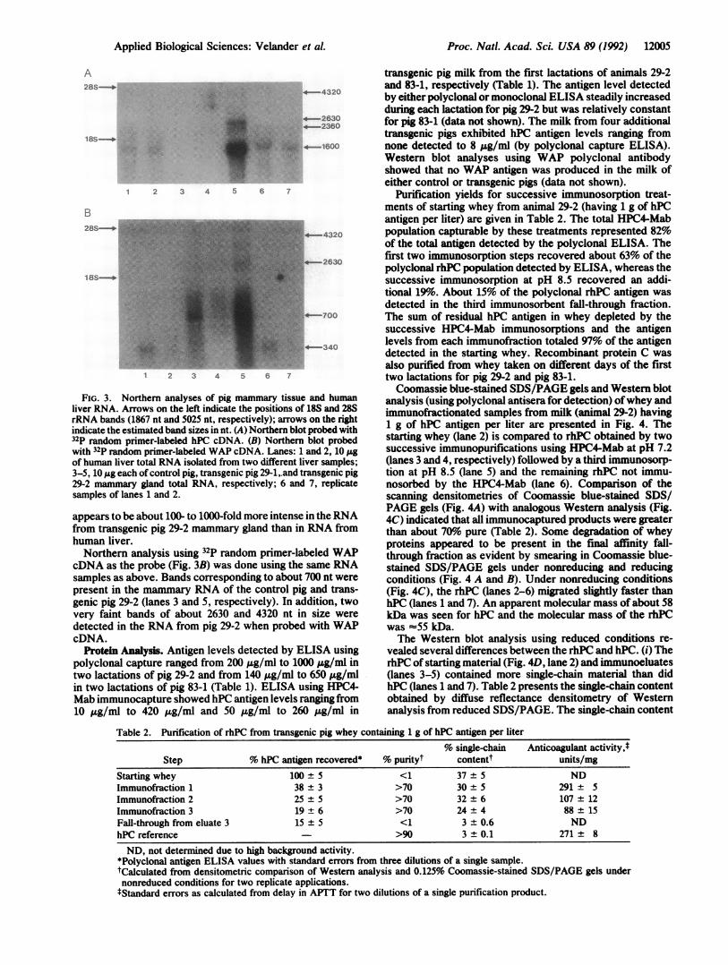
FIG. 3. Northern analyses of pig mammary tissue and humanliver RNA. Arrows on the left indicate the positions of 18S and 28SrRNA bands (1867 nt and 5025 nt, respectively); arrows on the rightindicate the estimated band sizes in nt. (A) Northern blot probed with32P random primer-labeled hPC cDNA. (B) Northern blot probedwith 32P random primer-labeled WAP cDNA. Lanes: 1 and 2, 10 ,ugof human liver total RNA isolated from two different liver samples;3-5, 10 pg each of control pig, transgenic pig 29-1,.and transgenic pig29-2 mammary gland total RNA, respectively; 6 and 7, replicatesamples of lanes 1 and 2.
appears to be about 100- to 1000-fold more intense in theRNAfrom transgenic pig 29-2 mammary gland than in RNA fromhuman liver.Northern analysis using 32P random primer-labeled WAP
cDNA as the probe (Fig. 3B) was done using the same RNAsamples as above. Bands corresponding to about 700 nt werepresent in the mammary RNA of the control pig and trans-genic pig 29-2 (lanes 3 and 5, respectively). In addition, twovery faint bands of about 2630 and 4320 nt in size were
detected in the RNA from pig 29-2 when probed with WAPcDNA.
Protein Analysis. Antigen levels detected by ELISA usingpolyclonal capture ranged from 200 ,ug/ml to 1000 .g/ml intwo lactations of pig 29-2 and from 140 ,ug/ml to 650 jg/mlin two lactations of pig 83-1 (Table 1). ELISA using HPC4-Mab immunocapture showed hPC antigen levels ranging from10 ,ug/ml to 420 ,ug/ml and 50 ug/ml to 260 ,ug/ml in
transgenic pig milk from the first lactations of animals 29-2and 83-1, respectively (Table 1). The antigen level detectedby either polyclonal or monoclonal ELISA steadily increasedduring each lactation for pig 29-2 but was relatively constantfor pig 83-1 (data not shown). The milk from four additionaltransgenic pigs exhibited hPC antigen levels ranging fromnone detected to 8 ;&g/ml (by polyclonal capture ELISA).Western blot analyses using WAP polyclonal antibodyshowed that no WAP antigen was produced in the milk ofeither control or transgenic pigs (data not shown).
Purification yields for successive immunosorption treat-ments of starting whey from animal 29-2 (having 1 g of hPCantigen per liter) are given in Table 2. The total HPC4-Mabpopulation capturable by these treatments represented 82%of the total antigen detected by the polyclonal ELISA. Thefirst two immunosorption steps recovered about 63% of thepolyclonal rhPC population detected by ELISA, whereas thesuccessive immunosorption at pH 8.5 recovered an addi-tional 9%o. About 15% of the polyclonal rhPC antigen wasdetected in the third immunosorbent fall-through fraction.The sum of residual hPC antigen in whey depleted by thesuccessive HPC4-Mab immunosorptions and the antigenlevels from each immunofraction totaled 97% of the antigendetected in the starting whey. Recombinant protein C wasalso purified from whey taken on different days of the firsttwo lactations for pig 29-2 and pig 83-1.Coomassie blue-stained SDS/PAGE gels and Western blot
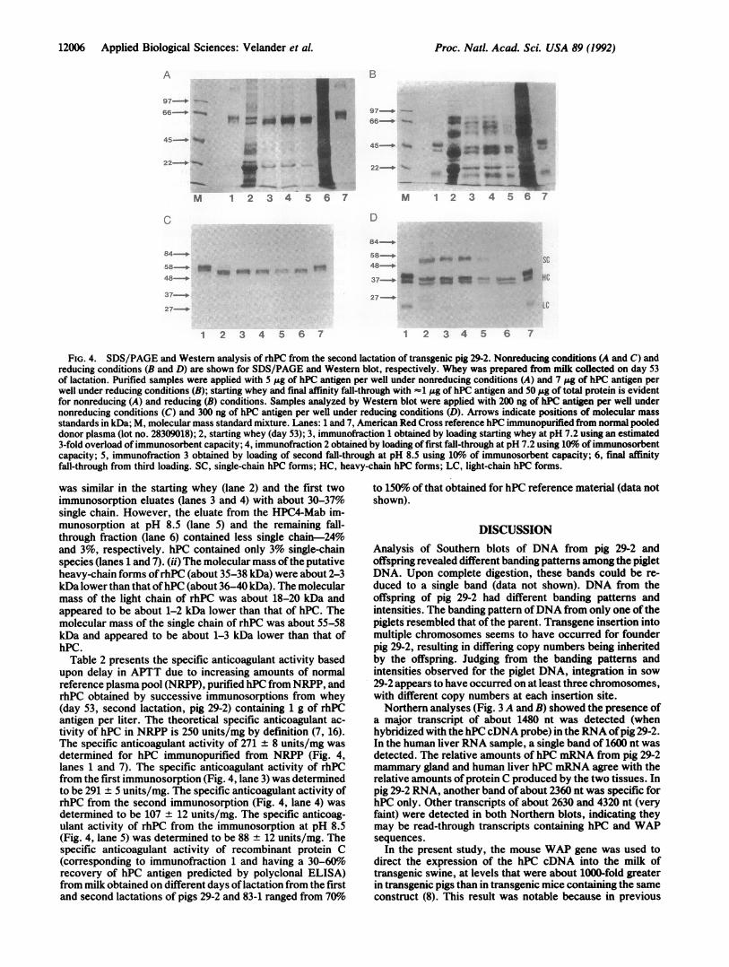
analysis (using polyclonal antisera for detection) ofwhey andimmunofractionated samples from milk (animal 29-2) having1 g of hPC antigen per liter are presented in Fig. 4. Thestarting whey (lane 2) is compared to rhPC obtained by twosuccessive immunopurifications using HPC4-Mab at pH 7.2(lanes 3 and 4, respectively) followed by a third immunosorp-tion at pH 8.5 (lane 5) and the remaining rhPC not immu-nosorbed by the HPC4-Mab (lane 6). Comparison of thescanning densitometries of Coomassie blue-stained SDS/PAGE gels (Fig. 4A) with analogous Western analysis (Fig.4C) indicated that all immunocaptured products were greaterthan about 70%6 pure (Table 2). Some degradation of wheyproteins appeared to be present in the final affinity fall-through fraction as evident by smearing in Coomassie blue-stained SDS/PAGE gels under nonreducing and reducingconditions (Fig. 4 A and B). Under nonreducing conditions(Fig. 4C), the rhPC (lanes 2-6) migrated slightly faster thanhPC (lanes 1 and 7). An apparent molecular mass of about 58kDa was seen for hPC and the molecular mass of the rhPCwas -55 kDa.The Western blot analysis using reduced conditions re-
vealed several differences between the rhPC and hPC. (i) TherhPC of starting material (Fig. 4D, lane 2) and immunoeluates(lanes 3-5) contained more single-chain material than didhPC (lanes 1 and 7). Table 2 presents the single-chain contentobtained by diffuse reflectance densitometry of Westernanalysis from reduced SDS/PAGE. The single-chain content
Table 2. Purification of rhPC from transgenic pig whey containing 1 g of hPC antigen per liter% single-chain Anticoagulant activityj
Step % hPC antigen recovered* % purityt contentt units/mgStartingwhey 100 5 <1 37 5 NDImmunofraction 1 38 + 3 >70 30 ± 5 291 + 5Immunofraction 2 25 ± 5 >70 32 ± 6 107 ± 12Immunofraction 3 19 ± 6 >70 24 ± 4 88 ± 15Fall-through from eluate 3 15 ± 5 <1 3 ± 0.6 NDhPC reference >90 3 ± 0.1 271 ± 8
ND, not determined due to high background activity.*Polyclonal antigen ELISA values with standard errors from three dilutions of a single sample.tCalculated from densitometric comparison of Western analysis and 0.125% Coomassie-stained SDS/PAGE gels undernonreduced conditions for two replicate applications.tStandard errors as calculated from delay in APTT for two dilutions of a single purification product.
Applied Biological Sciences: Velander et al.
12006 Applied Biological Sciences: Velander et al.
A
97_
M 24567w
8 4
37-.2 4 6
1 2 3 4 5 6 7
B
.H
22NM 1 2 3 4 5 6 7
D
58-. _ _ SC
48-_
37_"s"" - _ j ~~~~~~HC27_-
..t. L~~~~~~~~~C
1 2 3 4 5 6 7
FIG. 4. SDS/PAGE and Western analysis of rhPC from the second lactation of transgenic pig 29-2. Nonreducing conditions (A and C) andreducing conditions (B and D) are shown for SDS/PAGE and Western blot, respectively. Whey was prepared from milk collected on day 53of lactation. Purified samples were applied with 5 ,ug of hPC antigen per well under nonreducing conditions (A) and 7 pg of hPC antigen perwell under reducing conditions (B); starting whey and final affinity fall-through with -1 .tg of hPC antigen and 50 j.g of total protein is evidentfor nonreducing (A) and reducing (B) conditions. Samples analyzed by Western blot were applied with 200 ng of hPC antigen per well undernonreducing conditions (C) and 300 ng of hPC antigen per well under reducing conditions (D). Arrows indicate positions of molecular massstandards in kDa; M, molecular mass standard mixture. Lanes: 1 and 7, American Red Cross reference hPC immunopurified from normal pooleddonor plasma (lot no. 28309018); 2, starting whey (day 53); 3, immunofraction 1 obtained by loading starting whey at pH 7.2 using an estimated3-fold overload of immunosorbent capacity; 4, immunofraction 2 obtained by loading of first fall-through at pH 7.2 using 10% of immunosorbentcapacity; 5, immunofraction 3 obtained by loading of second fall-through at pH 8.5 using 1o of immunosorbent capacity; 6, final affinityfall-through from third loading. SC, single-chain hPC forms; HC, heavy-chain hPC forms; LC, light-chain hPC forms.
was similar in the starting whey (lane 2) and the first twoimmunosorption eluates (lanes 3 and 4) with about 30-37%single chain. However, the eluate from the HPC4-Mab im-munosorption at pH 8.5 (lane 5) and the remaining fall-through fraction (lane 6) contained less single chain-24%and 3%, respectively. hPC contained only 3% single-chainspecies (lanes 1 and 7). (ii) The molecular mass ofthe putativeheavy-chain forms ofrhPC (about 35-38 kDa) were about 2-3kDa lower than that ofhPC (about 36-40 kDa). The molecularmass of the light chain of rhPC was about 18-20 kDa andappeared to be about 1-2 kDa lower than that of hPC. Themolecular mass of the single chain of rhPC was about 55-58kDa and appeared to be about 1-3 kDa lower than that ofhPC.
Table 2 presents the specific anticoagulant activity basedupon delay in APTT due to increasing amounts of normalreference plasma pool (NRPP), purified hPC from NRPP, andrhPC obtained by successive immunosorptions from whey(day 53, second lactation, pig 29-2) containing 1 g of rhPCantigen per liter. The theoretical specific anticoagulant ac-tivity of hPC in NRPP is 250 units/mg by definition (7, 16).The specific anticoagulant activity of 271 ± 8 units/mg wasdetermined for hPC immunopurified from NRPP (Fig. 4,lanes 1 and 7). The specific anticoagulant activity of rhPCfrom the first immunosorption (Fig. 4, lane 3) was determinedto be 291 ± 5 units/mg. The specific anticoagulant activity ofrhPC from the second immunosorption (Fig. 4, lane 4) wasdetermined to be 107 ± 12 units/mg. The specific anticoag-ulant activity of rhPC from the immunosorption at pH 8.5(Fig. 4, lane 5) was determined to be 88 ± 12 units/mg. Thespecific anticoagulant activity of recombinant protein C(corresponding to immunofraction 1 and having a 30-60%orecovery of hPC antigen predicted by polyclonal ELISA)from milk obtained on different days of lactation from the firstand second lactations of pigs 29-2 and 83-1 ranged from 70o
to 150% of that obtained for hPC reference material (data notshown).
DISCUSSIONAnalysis of Southern blots of DNA from pig 29-2 andoffspring revealed different banding patterns among the pigletDNA. Upon complete digestion, these bands could be re-duced to a single band (data not shown). DNA from theoffspring of pig 29-2 had different banding patterns andintensities. The banding pattern ofDNA from only one ofthepiglets resembled that ofthe parent. Transgene insertion intomultiple chromosomes seems to have occurred for founderpig 29-2, resulting in differing copy numbers being inheritedby the offspring. Judging from the banding patterns andintensities observed for the piglet DNA, integration in sow29-2 appears to have occurred on at least three chromosomes,with different copy numbers at each insertion site.Northern analyses (Fig. 3 A and B) showed the presence of
a major transcript of about 1480 nt was detected (whenhybridized with the hPCcDNA probe) in the RNA ofpig 29-2.In the human liver RNA sample, a single band of 1600 nt wasdetected. The relative amounts of hPC mRNA from pig 29-2mammary gland and human liver hPC mRNA agree with therelative amounts of protein C produced by the two tissues. Inpig 29-2 RNA, another band of about 2360 nt was specific forhPC only. Other transcripts of about 2630 and 4320 nt (veryfaint) were detected in both Northern blots, indicating theymay be read-through transcripts containing hPC and WAPsequences.
In the present study, the mouse WAP gene was used todirect the expression of the hPC cDNA into the milk oftransgenic swine, at levels that were about 1000-fold greaterin transgenic pigs than in transgenic mice containing the sameconstruct (8). This result was notable because in previous
Proc. Natl. Acad Sci. USA 89 (1992)

Proc. Natl. Acad. Sci. USA 89 (1992) 12007
studies high-level production ofheterologous proteins in milkwas achieved only in transgenic animals carrying eithergenomic (2, 17) or minigene constructs (3). Our results showthat a cDNA may be used to obtain high-level expression ofheterologous proteins in the milk of transgenic pigs.The highest expression levels of rhPC in the milk of
transgenic pigs using the cDNA of hPC were similar to theexpression levels reported for the WAP using the intactgenomic mouse WAP gene in transgenic pigs (2). A constantlevel of WAP of about 1 g/liter was found in milk oftransgenic pigs over a 26-day lactation (17); rhPC (polyclonalpopulation) increased about 2-fold during the first 26 days and>5-fold during the entire 55-day lactation ofpig 29-2, whereasthe rhPC in the milk of pig 83-1 varied about 2.5-fold. SinceWAP and rhPC transgenic pigs contained about 10-20 copiesofthe transgene, differences in regulation may have been dueto the location of integration as well as the differencesbetween the WAP and WAP-hPC gene structures.The molecular mass of the single, heavy, and light chains
of rhPC appeared to be similar to, but slightly lower than,those for hPC. This may be due to differences in carbohy-drate content and structure. The rhPC contained a signifi-cantly larger population of single-chain material, which mayindicate a rate limitation in posttranslational removal of thedipeptide Lys-Arg at positions 156 to 157 (7).The presence of different rhPC populations was also evi-
dent from the differences in antigen content detected bypolyclonal and HPC4-Mab ELISAs and the presence ofdifferent immunofractions obtained using the HPC4-Mab.The HPC4-Mab binds the activation peptide ofhPC at pH 7.3and thus provides a measure of the presentation of a domainessential for conversion of zymogen hPC to active serineprotease form (15). Each of the immunofractions possesseddifferent anticoagulant activities, but activity did not corre-late well with single-chain rhPC content. The most activefraction represented about 38% of the hPC antigen andcontained 30% single-chain material. This suggests that asignificant portion of the single-chain material contained inthis fraction may be biologically active or that some het-erodimeric forms are hyperactive. The lower activities of thesecond and third fractions may be a result of nonnativeconformations or insufficient y-carboxylation.The anticoagulant activity of protein C is dependent upon
proper y-carboxylation ofthe membrane binding domain thatoccurs in the light chain (18). To determine whether y-car-boxylation had occurred properly in rhPC, its anticoagulantactivity was assayed in vitro by APTT. This assay simulatescoagulation in vivo by initiating clotting in a mixture con-taining calcium, phospholipid membrane, and the proteinsassociated with hemostasis (16). As much as 38% (or 380,ug/ml) of the porcine rhPC may be sufficiently -carboxy-lated, as judged by the specific activity of immunofraction 1by APTT relative to that of hPC. The transgenic pigs studiedhere had milk letdown about every hour and hence themaximum rhPC secretion rate occurred at about 1000 .ug/mlper hr. The amount ofactive rhPC secreted by the pigs (about380 I&g/ml per hr) is significantly higher than the secretionrates of about 1 ,ug/ml per hr produced by human kidney 293
cell lines (7). These results provide evidence of gla formationnot previously known to occur at high levels in mammarytissue.
The technical assistance of Rodolfo Canseco, Barry Williams,James Knight, Janet Young, Tulin Morcol, Arthur Degener, AmySparks, and Julianna Toth was greatly appreciated. The genomicWAP-hPC cDNA fusion gene WAPPC-1 was designed by LotharHennighausen and cloned in the Laboratory of Biochemistry andMetabolism, National Institutes of Health, Building 10, Room9N113, Bethesda, MD 20892. We are grateful to Dr. Charles Esmon,Oklahoma Medical Research Foundation, Oklahoma City, OK73104, for providing the HPC4-Mab. This research was partiallysupported by the Research Division of Virginia Polytechnic Instituteand State University, The American Red Cross, and NationalScience Foundation Grant BCS-9011098-01 to W.H.V. and W.N.D.
1. Clark, A. J., Bessos, H., Bishop, J. O., Brown, P., Harris, S.,Lathe, R., McClenaghan, M., Prowse, C., Simons, J. P.,Whitelaw, C. B. A. & Wilmut, I. (1989) BiolTechnology 7,487-492.
2. Wall, R. J., Pursel, V. G., Shamay, A., McKnight, R. A.,Pittius, C. W. & Henninghausen, L. (1991) Proc. Nati. Acad.Sci. USA 88, 1696-1700.
3. Wright, G., Carver, A., Cottom, D., Reeves, D., Scott, A.,Simons, P., Wilmut, I., Garner, I. & Colman, A. (1991)BiolTechnology 9, 830-834.
4. Ebert, K. M., Selgrath, J. P., DiTullio, P., Denman, J., Smith,T. E., Memon, M. A., Schindler, J. E., Monastersky, G. M.,Vitale, J. A. & Gordon, K. (1991) BiolTechnology 9, 835-838.
5. Pittius, C. W., Hennighausen, L., Lee, E., Westphal, H.,Nicols, E., Vitale, J. & Gordon, K. (1988) Proc. Natl. Acad.Sci. USA 85, 5874-5878.
6. Esmon, C. T. (1987) Science 235, 1348-1352.7. Grinnell, B. W., Walls, J. D., Gerlitz, B., Berg, D. T.,
McClure, D. B., Ehrlich, H., Bang, N. U. & Yan, S. B. (1990)in Protein C and Related Anticoagulants, eds. Bruley, D. F. &Drohan, W. N. (The Portfolio, The Woodlands, TX), pp.29-63.
8. Velander, W. H., Page, R. L., Morcol, T., Russell, C. G.,Canseco, R., Drohan, W. N., Gwazdauskas, F. C., Wilkins,T. D. & Johnson, J. L. (1991) Ann. N.Y. Acad. Sci. 665,391-403.
9. Wall, R., Pursel, V., Hammer, R. & Brinster, R. (1985) Biol.Reprod. 32, 645-651.
10. Brinster, R. L., Chen, H. Y., Drumbeater, N. E., Yagle,M. K. & Palmiter, R. D. (1985) Proc. Natl. Acad. Sci. USA 82,4438 4442.
11. Campbell, S. M., Rosen, J. M., Hennighausen, L., Strech-Jurk, U. & Sippel, A. E. (1984) Nucleic Acids Res. 12, 8685-8697.
12. Marmur, J. (1961) J. Mol. Biol. 3, 208-218.13. Saiki, R. K., Walsh, P. S., Levenson, C. H. & Erlich, H. A.
(1989) Proc. Nall. Acad. Sci. USA 86, 6230-6234.14. Puissant, C. & Houdebine, L.-M. (1990) BioTechniques 8,
148-149.15. Stems, D. J., Kurosawa, S., Sims, P. J., Esmon, N. L. &
Esmon, C. T. (1988) J. Biol. Chem. 263, 826-832.16. Vinazzer, H. & Pangraz, U. (1987) Thromb. Res. 46, 1-8.17. Shamay, A., Solinas, S., Pursel, V. G., McKnight, R. A.,
Alexander, L., Beattie, C., Hennighausen, L. & Wall, R. J.(1991) J. Anim. Sci. 69, 4552-4562.
18. Zhang, L. & Castellino, F. J. (1990) Biochemistry 29, 10828-10834.
Applied Biological Sciences: Velander et aL


![Biochemical Changes in Cancer - jcam.com.tr · dana gelir.[11] Bu proteinin bulunduğu idrarın karakteristik özel-liği idrarın 45 – 60oC’de ısıtılması ile bulutumsu bir](https://img.pdfslide.us/doc/110x75/5e3cc51fd55ab853f360037a/biochemical-changes-in-cancer-jcamcomtr-dana-gelir11-bu-proteinin-bulunduu.jpg)