Embed Size (px)
Citation preview
European Journal of Clinical Investigation (1998) 28, 983±988
High-energy phosphates metabolism and recovery inreperfused ischaemic hearts
M. Samaja*, S. Allibardi², R. De Jonge³ & S. L. Chierchia²
*Department of Biomedical Science and Technology, University of Milan; ²Scienti®c Institute San Raffaele, Milan,
Italy; ³Thoraxcenter, Erasmus University Rotterdam, The Netherlands
Abstract Background The aim of this study was to assess how coronary ¯ow, oxygen supply and
energy demand affect myocardial ATP, phosphocreatine and their metabolites during
oxygen shortage and recovery.
Methods Isolated rat hearts were exposed for 20 min to either low-¯ow ischaemia or
hypoxaemia at the same oxygen supply, followed by return to baseline conditions
(20 min). Seventy-three hearts were divided into four groups: ischaemic or hypoxaemic,
spontaneously beating or paced to increase energy demand.
Results During O2 shortage, myocardial performance was less in ischaemic, spontaneously
beating hearts (SpIs), than in the other groups (14 6 1% of baseline vs. 25±48%).
Consequently, the tissue levels of ATP, total adenylates and phosphocreatine were main-
tained in SpIs, in contrast to marked decreases in the other groups. Upon re¯ow, the
recovery of performance and of myocardial ATP was 94 6 5% in SpIs (P�NS vs. baseline)
compared with 64±85% (P <0´05 vs. baseline) in the other groups. The degree of recovery
was positively related to the ischaemic contents of ATP (P� 0´03) and adenylates
(P� 0´001), but not to that of phosphocreatine (P�NS).
Conclusion The maintenance of the ATP pool under low oxygen supply conditions is
essential for good recovery. The most important factors that determine the ATP pool size are
the energy demand, which increases the formation of diffusible ATP catabolites, and the
coronary ¯ow, which removes these catabolites, rather than the oxygen supply per se.
Keywords ATP, energy demand, hypoxia, myocardial metabolism, phosphocreatine, purine
metabolism.
Eur J Clin Invest 1998: 28 (12): 983±988
Introduction
Although the only way to recover ischaemic hearts, re¯ow
is often associated with damage. Only part of this damage,
however, is due to the free radicals generated concomi-
tantly with oxygen readmission [1]. The biochemical
mechanisms underlying damage are still elusive, but high-
energy phosphates are probably involved, because damage
is limited if the tissue ATP level is maintained during
ischaemia [2±4]. The role of ongoing glycolysis is contro-
versial, as it appears detrimental during no-¯ow ischaemia
but bene®cial during low-¯ow ischaemia [5]. However, the
implications of glycolysis, especially in relation to ischaemic
preconditioning, are described elsewhere [6].
Assessing the effects of energy demand and coronary
¯ow on the metabolism of total adenine nucleotides and
purines (TANP) may help to clarify the above issue. We
have recently shown that a situation of low coronary ¯ow,
by depressing the washout of lactate, can strongly down-
regulate myocardial performance [7,8]. In addition,
coronary ¯ow appears crucial in determining recovery
after ischaemia as it helps to wash out diffusible TANP,
i.e. inosine, hypoxanthine, xanthine and urate, thereby
reducing tissue ATP level [9]. Although it has already
been hypothesized that residual ¯ow increases the washout
of TANP formed during O2 shortage [10,11], the relation-
ship between reperfusion injury, tissue TANP, coronary
¯ow and energy demand has not yet been established.
In this work, we verify the existence of such link by
testing the hypothesis that dysfunction measured during
re¯ow depends on factors which depend on both the energy
demand and the residual coronary ¯ow during ischaemia.
High energy demand enhances formation of ATP-derived
Q 1998 Blackwell Science Ltd
Correspondence: Dr M. Samaja, Dipartimento di Scienze e
Tecnologie Biomediche, LITA, via F.Cervi 93, 20090 Segrate
Milan, Italy. E-mail [email protected]
Received 19 September 1997; accepted 17 July 1998
984 M. Samaja et al.
membrane-diffusible substances, whereas high residual cor-
onary ¯ow promotes de®nitive loss of these substances for
the contractile system. By comparing various experimental
conditions characterized by the same O2 supply yet different
energy demand and coronary ¯ow levels during ischaemia,
we show that the maintenance of the ATP level during O2
shortage protects hearts. In addition, we show that both high
energy demand and high coronary ¯ow can determine the
size of the ATP pool during the recovery phase.
Materials and methods
Heart perfusion
Ad libitum-fed male Sprague±Dawley rats (250±280 g)
were anaesthetized by i.p. heparinized sodium thiopental
(10 mg 100 gÿ1 b.w.); hearts were excised and perfused at
378C with a medium containing (in mmol Lÿ1) NaCl
(115´6), KCl (4´7) KH2PO4 (1´2), EDTA (0´5), Na2SO4
(1´2), NaHCO3 (28´5), CaCl2 (2´5), MgCl2 (1´2) and
glucose (16´6), pH 7´40 6 0´02 at PCO2�43 mmHg. A
roller pump (Gilson, France) delivered the medium at
preselected ¯ows to a ®lter (8 mm pore size, 47 mm dia-
meter, Nuclepore Pleasanton, CA, USA), a membrane
oxygenator, ¯owed with gas at PO2 of either 670 or 67
mmHg, a preheater and the aortic cannula. The venous
return was collected from the pulmonary artery for
measurement of venous PO2 (YSI model 5300 Oxygen
Monitor, Yellow Springs, OH, USA) and O2 uptake (VO2).
A saline-®lled balloon in the left ventricle was connected to a
pressure transducer (Harvard Apparatus model 52-9966,
Natick, MA, USA) for measurement of end-diastolic pres-
sure (EDP), left-ventricle developed pressure (LVDP) and
heart rate (HR). Hearts from the paced groups were stimu-
lated throughout at 330 minÿ1 by electrodes (Harvard,
South Natick, MA, USA: square wave stimulator, 5 ms
pulse duration, 10 V pulse amplitude) placed on the aortic
cannula and on the apex of the ventricle. To account for
different HRs in the various groups, myocardial
performance is expressed as LVDP ´ HR.
Experimental protocol
Hearts were stabilized for 30 min at ¯ow� 15 mL minÿ1,
PaO2� 670 mmHg, O2 supply� 14´1 mmol minÿ1. The
balloon volume was adjusted to achieve an EDP of approxi-
mately 10 mmHg and was then kept constant throughout.
Hearts were assigned to one of the following groups: SpIs
(spontaneously beating hearts exposed to low-¯ow ische-
mia and then reperfused at a ¯ow rate of 15 mL minÿ1;
n� 9); SpHy (spontaneously beating hearts exposed to
hypoxaemia and then reoxygenated at PaO2� 670 mmHg;
n� 8); PxIs (paced hearts exposed to low-¯ow ischaemia
and then reperfused at a ¯ow rate of 15 mL minÿ1; n� 9);
PxHy (paced hearts exposed to hypoxaemia and then
reoxygenated at PaO2�670 mmHg; n� 8). Recovery was
quantitated as the ratio (performance during re¯ow or
reoxygenation)/(performance during baseline). Low-¯ow
ischaemia and hypoxaemia were maintained for 20 min
either by reducing ¯ow to 1´5 mL minÿ1 or by switching
to the low-PO2 gas. The two conditions were thus matched
for O2 supply (10% of baseline), duration (20 min) and
temperature (378C). Hearts were freeze-clamped at the end
of re¯ow or reoxygenation (20 min) to determine tissue
content of high-energy phosphates and their metabolites.
In parallel experiments, additional hearts were freeze-
clamped after the periods of baseline (seven spontaneously
beating and six placed), low-¯ow ischaemia (7/6) and
hypoxaemia (8/5).
High performance liquid chromatography
Hearts were freeze-clamped with aluminium clamps and
then cooled in liquid nitrogen; tissue was extracted with 0´5
mol Lÿ1 perchloric acid, neutralized and analysed for ATP,
ADP, AMP, adenosine, inosine-50-monophosphate, ino-
sine, hypoxanthine, xanthine, urate, creatine and phospho-
creatine (Pcr) by high-performance liquid chromatography
as previously described [12]. Using this technique, hypox-
anthine coelutes with xanthine and inosine with urate. The
level of TANPs was calculated as the sum of all the above
substances except creatine and Pcr. The diffusible sub-
stances include adenosine, inosine, hypoxanthine, xanthine
and urate.
Q 1998 Blackwell Science Ltd, European Journal of Clinical Investigation, 28, 983±988
Figure 1 Developed and end-diastolic pressures (mmHg). The
O2 supply was reduced at t�0±20 min by decreasing either
coronary ¯ow or PO2 to 10% of baseline. The vertical bars
represent SE. Two-way ANOVA for both, P <0´0001. SpIs (B),
spontaneously beating hearts exposed to low-¯ow ischaemia and
re¯ow; SpHy (X), spontaneously beating hearts exposed to
hypoxaemia and reoxygenation; PxIs (A), paced hearts exposed
to low-¯ow ischaemia and re¯ow; PxHy (W), paced hearts
exposed to hypoxaemia and reoxygenation. *Signi®cant
difference vs. SpIs.
High-energy phosphates in reperfused ischaemic hearts 985
Q 1998 Blackwell Science Ltd, European Journal of Clinical Investigation, 28, 983±988
Statistics
Data are expressed as means 6 SE. To detect interactions
between the various groups and three consecutive perfu-
sion conditions (baseline, O2 shortage, and recovery), we
used to analysis of variance (ANOVA) procedures (StatView,
Abacus Concepts, Berkeley, CA, USA), depending on the
variable being tested: two-way, repeated-measures ANOVA
for variables available for each heart under the three
conditions; and factorial, six-groups ANOVA for variables
such as, for example, the myocardial metabolites content.
In both cases, if the ANOVA test was signi®cant (P <0´05),
hearts were compared with the Bonferroni/Dunnett
multiple comparison procedure.
Results
General
Hearts did not stop contracting during O2 shortage and
recovery; thus, their performance could be monitored
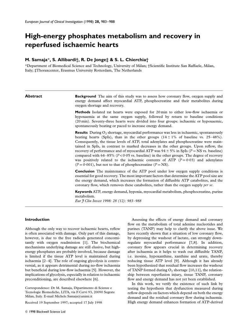
continuously (Fig. 1). Figures 1 and 2 show that, although
almost complete in SpIs, functional recovery upon re¯ow
or reoxygenation was impaired in the other groups. Figures
3 and 4 report the tissue metabolite levels measured at the
end of the various phases. As observed for myocardial
performance, the recovery of ATP and TANP was almost
complete in SpIs, in contrast to progressive impairment in
the other groups. However, both Pcr and the sum Pcr�
creatine were maintained in all the groups. Electron micro-
scopy did not reveal signs of irreversible injury in any group
(not shown).
Effects of pacing at full O2 supply (baseline)
LVDP was lower in paced than in spontaneously beating
hearts, with higher VO2 (Table 1). However, HR was higher
in paced hearts, so that LVDP ´ HR was the same in the
two groups. No differences were observed for EDP or most
metabolic parameters, with the exception of inosine-50-
monophosphate, which was higher, and adenosine, which
was lower in paced than in spontaneously beating hearts.
Low-¯ow ischaemia and re¯ow in spontaneously
beating hearts (SpIs)
As expected myocardial contractility decreased abruptly at
the onset of low-¯ow ischaemia. In addition, HR decreased
to 178 6 12 minÿ1 (P < 0´0001 with respect to baseline).
However, there were no signs of diastolic contracture, and
the level of tissue metabolites was preserved throughout the
ischaemic period. At the end of the re¯ow, none of the
examined parameters, including HR (264 6 15 minÿ1),
was signi®cantly different from baseline. This indicates
near complete functional and metabolic recovery in
SpIs.
Figure 2 Performance recovery at end of re¯ow or reoxygena-
tion. Data are expressed as per cent of baseline values. Two-way
ANOVA, P <0´0001 and P�0´08 for developed pressure ´ heart
rate and VO2 respectively. Same conventions as in Fig. 1.
Figure 3 Myocardial content of adenine
nucleotides and purines in spontaneously
contracting (left) and paced (right)
hearts. The bars on the top represent
IMP on a different scale range. One-way
ANOVA, P <0´0001 for all substances.#Signi®cant difference (P <0´05) in the
toal content of adenine nucleotides and
purines compared with spontaneously
beating hearts exposed to low-¯ow
ischaemia and re¯ow (SpIs). *Signi®cant
difference (P <0´05) compared with
spontaneously beating hearts exposed to
low-¯ow ischaemia and re¯ow (SpIs).
986 M. Samaja et al.
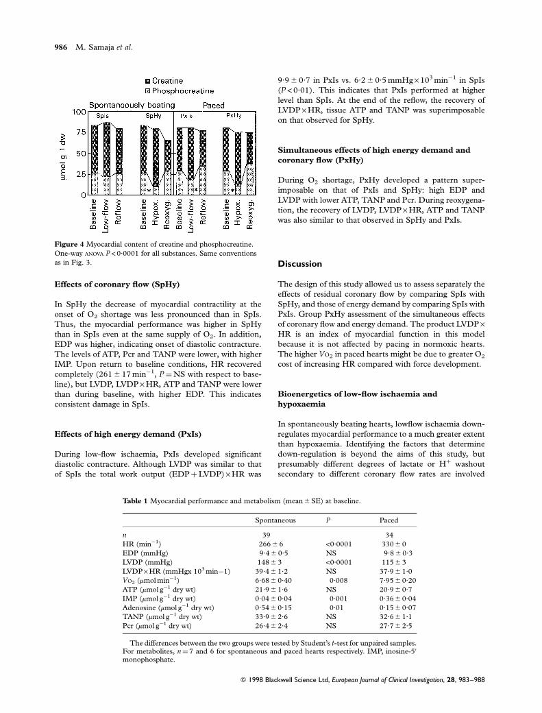
Effects of coronary ¯ow (SpHy)
In SpHy the decrease of myocardial contractility at the
onset of O2 shortage was less pronounced than in SpIs.
Thus, the myocardial performance was higher in SpHy
than in SpIs even at the same supply of O2. In addition,
EDP was higher, indicating onset of diastolic contracture.
The levels of ATP, Pcr and TANP were lower, with higher
IMP. Upon return to baseline conditions, HR recovered
completely (261 6 17 minÿ1, P�NS with respect to base-
line), but LVDP, LVDP ´ HR, ATP and TANP were lower
than during baseline, with higher EDP. This indicates
consistent damage in SpIs.
Effects of high energy demand (PxIs)
During low-¯ow ischaemia, PxIs developed signi®cant
diastolic contracture. Although LVDP was similar to that
of SpIs the total work output (EDP�LVDP) ´ HR was
9´9 6 0´7 in PxIs vs. 6´2 6 0´5 mmHg ´ 103 minÿ1 in SpIs
(P <0´01). This indicates that PxIs performed at higher
level than SpIs. At the end of the re¯ow, the recovery of
LVDP ´ HR, tissue ATP and TANP was superimposable
on that observed for SpHy.
Simultaneous effects of high energy demand and
coronary ¯ow (PxHy)
During O2 shortage, PxHy developed a pattern super-
imposable on that of PxIs and SpHy: high EDP and
LVDP with lower ATP, TANP and Pcr. During reoxygena-
tion, the recovery of LVDP, LVDP ´ HR, ATP and TANP
was also similar to that observed in SpHy and PxIs.
Discussion
The design of this study allowed us to assess separately the
effects of residual coronary ¯ow by comparing SpIs with
SpHy, and those of energy demand by comparing SpIs with
PxIs. Group PxHy assessment of the simultaneous effects
of coronary ¯ow and energy demand. The product LVDP ´HR is an index of myocardial function in this model
because it is not affected by pacing in normoxic hearts.
The higher VO2 in paced hearts might be due to greater O2
cost of increasing HR compared with force development.
Bioenergetics of low-¯ow ischaemia and
hypoxaemia
In spontaneously beating hearts, low¯ow ischaemia down-
regulates myocardial performance to a much greater extent
than hypoxaemia. Identifying the factors that determine
down-regulation is beyond the aims of this study, but
presumably different degrees of lactate or H� washout
secondary to different coronary ¯ow rates are involved
Q 1998 Blackwell Science Ltd, European Journal of Clinical Investigation, 28, 983±988
Figure 4 Myocardial content of creatine and phosphocreatine.
One-way ANOVA P < 0´0001 for all substances. Same conventions
as in Fig. 3.
Table 1 Myocardial performance and metabolism (mean 6 SE) at baseline.
Spontaneous P Paced
n 39 34
HR (minÿ1) 266 6 6 <0´0001 330 6 0
EDP (mmHg) 9´4 6 0´5 NS 9´8 6 0´3
LVDP (mmHg) 148 6 3 <0´0001 115 6 3
LVDP ´ HR (mmHgx 103 minÿ1) 39´4 6 1´2 NS 37´9 6 1´0
VO2 (mmol minÿ1) 6´68 6 0´40 0´008 7´95 6 0´20
ATP (mmol gÿ1 dry wt) 21´9 6 1´6 NS 20´9 6 0´7
IMP (mmol gÿ1 dry wt) 0´04 6 0´04 0´001 0´36 6 0´04
Adenosine (mmol gÿ1 dry wt) 0´54 6 0´15 0´01 0´15 6 0´07
TANP (mmol gÿ1 dry wt) 33´9 6 2´6 NS 32´6 6 1´1
Pcr (mmol gÿ1 dry wt) 26´4 6 2´4 NS 27´7 6 2´5
The differences between the two groups were tested by Student's t-test for unpaired samples.For metabolites, n�7 and 6 for spontaneous and paced hearts respectively. IMP, inosine-50
monophosphate.
High-energy phosphates in reperfused ischaemic hearts 987
Q 1998 Blackwell Science Ltd, European Journal of Clinical Investigation, 28, 983±988
[7]. By reducing contractility and by maintaining the levels
of ATP, TANP and Pcr near preischaemic values, SpIs
achieve a favourable perfusion±contraction matching
between energy supply and demand, which is compatible
with short-term hibernation [13]. This allows greater
recovery in these hearts. Both high ¯ow and pacing
unmatch the perfusion±contraction matching by increas-
ing energy demand. This results in a fall in high-energy
phosphates, increased ATP catabolism, increased washout
of diffusible substances and impaired recovery following
return to baseline conditions.
The fact that oxidative metabolism (VO2) during O2
shortage was the same in all groups stresses the increasing
importance of glycolytic ATP, which meets the energy
requirements under O2 shortage conditions. Presumably,
endogenous glycogen is the most important fuel source to
sustain the higher contractility in SpHy [8]. We did not
measure lactate production in PxIs and PxHy but the data
reported in [7] show that pacing increases the rate of
glycolysis in ischaemic hearts only, whereas this rate is
unaffected in hypoxaemic hearts. Perhaps, in PxIs and
SpHy, the glycolytic rate is already near maximum. In
fact, the values of lactate production during hypoxaemia
(9´2±10´8 mmol minÿ1 gÿ1 wet wt or 71±83 mmol minÿ1
gÿ1 dry wt) are close to the values of 10±14 mmol minÿ1 gÿ1
wet wt [14] and 60±70mmol minÿ1 gÿ1 dry wt [15] reported
for contracting anoxic isolated rate hearts.
Although its role in heart muscle is not yet fully clear,
IMP is considered a good index of metabolic derangement
during O2 shortage. In addition, the lowest IMP level
during O2 shortage (SpIs) was associated with the greatest
down-regulation of myocardial performance during ischae-
mia. Photoaf®nity labelling studies in rat have revealed the
presence of IMP binding sites on the myosin complex [16].
In addition, IMP inhibits actin-activated Mg2�-myosin
ATPase activity [17]. Finally, loss of performance in rat
medial gastrocnemius muscle is associated with increases in
muscle IMP content [18]. In the normoxic heart, AMP is
mainly deaminated to IMP because AMP deaminase activ-
ity is much larger than that of 50-nucleotidase. However,
during O2 shortage, increases in Pi inhibit AMP deaminase,
resulting in the release of adenosine [19,20].
Diffusible substances and myocardial dysfunction
Loss of diffusible substances is crucial to determine
recovery in this model. In oxygenated tissues, ATP dephos-
phorylation is tightly coupled with ADP phosphorylation;
thus, the loss of diffusible ATP catabolites is slow and
compensated by purine salvage and de novo synthesis [21].
When energy supply is low relative to demand, ATP break-
down may exceed synthesis, thereby increasing the amount
of diffusible catabolites. This does not occur in SpIs
because of perfusion±contraction matching, but TANP
progressively decreases in the other groups. Thus, both
the excessive ATP breakdown with respect to synthesis and
the high coronary ¯ow ®nally lead to loss of diffusible
substances. TANP loss is not accompanied by parallel
changes in Pcr� creatine because both Pcr and creatine
do not leak across intact membranes [22]. Therefore,
TANP depletion is a metabolic phenomenon that occurs
even in the absence of irreversible membrane damage.
This is con®rmed by lack of ultrastructural alterations
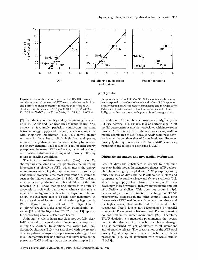
and of enzyme release. The preservation of the ATP pool
during O2 shortage is a major contributor to heart
protection (Fig. 5), in agreement with previous studies
[2,3,23].
Figure 5 Relationship between per cent LVDP ´ HR recovery
and the myocardial contents of ATP, sum of adenine nucleotides
and purines or phosphocreatine, measured at the end of O2
shortage. Best-®t lines are: ATP, y�31´21�3´12x, r2�0´92,
P�0´05; for TANP, y�ÿ23´1�3´44x, r2�0´98, P�0´005; for
phosphocreatine, r2�0´50, P�NS. SpIs, spontaneously beating
hearts exposed to low-¯ow ischaemia and re¯ow; SpHy, sponta-
neously beating hearts exposed to hypoxaemia and reoxygenation;
PxIs, paced hearts exposed to low-¯ow ischaemia and re¯ow;
PxHy, paced hearts exposed to hypoxaemia and reoxygenation.
988 M. Samaja et al.
Implications of this study
A relationship between functional recovery and mainte-
nance of diffusible substances has emerged. In this model,
low coronary ¯ow and low energy demand maintain diffu-
sible substances homeostasis. In contrast, the O2 supply perse does not appear to be as critical. High coronary ¯ow and
high energy demand increase the amount of diffusible
substances formed from ATP catabolism, result in low
tissue ATP levels and are associated with poor recovery.
The extent of recovery critically depends on both energy
demand and ¯ow during ischaemia, and not on the severity
of O2 shortage.
Acknowledgements
We gratefully thank Professor J. W. deJong for helpful
advice and encouragement.
References
1 Samaja M, Motterlini R, Santoro F, Dell'Antonio G, Corno
A. Oxidative injury in reoxygenated and reperfused hearts.
Free Rad Biol Med 1994;16:255±62.
2 Haas GS, DeBoer LWV, O'Keefe DDO, Bodenhamer RM,
Gef®n GA, Drop LJ et al. Reduction of postischemic myo-
cardial dysfunction by substrate repletion during reperfusion.
Circulation 1984;70:65±74.
3 Rubin BB, Liauw S, Tittley J, Romaschin AD, Walker PM.
Prolonged adenine nucleotide resynthesis and reperfusion
injury in postischemic skeletal muscle. Am J Physiol 1992;
262:H1538±47.
4 Schaefer S, Carr LJ, Prussel E, Ramasamy R. Effects of gly-
cogen depletion on ischemic injury in isolated rat hearts:
insights into preconditioning. Am J Physiol 1995;268:H935±
44.
5 de Jonge R, Bradamante S, Speleman L, de Jong JW. Carbo-
hydrates and purines in underperfused hearts protected by
ischemic preconditioning. J Mol Cell Cardiol 1998;30:699±
708.
6 de Jong JW, de Jonge R, Marchesani A, Janssen M, Brada-
mante S. Controversies in preconditioning. Cardiovasc DrugTher 1996;10:767±73.
7 Samaja M, Casalini S, Allibardi S, Corno A, Chierchia S.
Regulation of bioenergetics in O2-limited isolated rat hearts.
J Appl Physiol 1994;77:2530±6.
8 Merati G, Allibardi S, Monti LD, de Jong JW, Samaja M.
Dynamics of myocardial adaptation to low-¯ow ischemia and
hypoxemia. Am J Physiol 1996;271:2300±5.
9 Samaja M, Motterlini R, Allibardi S, Casalini S, Merati G,
Corno A, et al. Myocardial metabolism and function in
acutely ischemic and hypoxemic isolated rat hearts. J Mol CellCardiol 1995;27:1213±18.
10 Bak MI, Ingwall JS. Acidosis during ischemia promotes
adenosite triphosphate resynthesis in postischemic rat heart.
J Clin Invest 1994;93:40±49.
11 Soussi B, Lagerwall K, Idstrom JP, Schersten T. Purine
metabolic pathways in rat hindlimb perfusion model during
ischemia and reperfusion. Am J Physiol 1993;265:H1074±81.
12 Motterlini R, Samaja M, Tarantola M, Micheletti R, Bianchi
G. Functional and metabolic effects of propionyl-L-cartinine
in the isolated perfused hypertrophied rat heart. Mol CellBiochem 1992;116:139±45.
13 Ross J. Myocardial perfusion±contraction matching. Implica-
tions for coronary heart disease and hibernation. Circulation1991;83:1076±83.
14 Matthews PM, Taylor DJ, Radda GK. Biochemical
mechanisms of acute contracture failure in the hypoxic rat
heart. Cardiovasc Res 1986;20:13±19.
15 Williamson JR. Glycolytic control mechanisms. II. Kinetics of
intermediate changes during the aerobic-anoxic transition in
perfused rat heart. J Biol Chem 1966;241:5026±36.
16 Berden JA, de Haan A, de Haan EJ, van Doorn JE, Hartog
AF, Westra HG. Has IMP a regulatory role during fatiguing
contraction? IMP-binding sites on the myosin complex of rat
muscle. J Physiol 1986;381:85P (Abstract).
17 Westra HG, Berden JA, Pasman WJ. A model for the regula-
tion of actin-activated Mg2�-myosin ATPase activity: inhibi-
tion of the formation of actin±myosin complex by IMP. In:
Sargeant AJ, Kernell D, editor. Neuromuscular fatigue.Amsterdam: Royal Netherlands Academy of Sciences,
Elsevier Biomedical; 1997: p.24±6.
18 de Haan A. High-energy phosphates and fatigue during
repeated dynamic contractions of rat muscle. Exp Physiol1990:75:851±4.
19 Chen W, Hoerter J, GuTetharon M. AMP degradation in the
perfused rat heart during 2-deoxy-D-glucose perfusion and
anoxia. The release of adenosine and inosine. J Mol CellCardiol 1996;28:2163±74.
20 Chen W, GuTetharon M. AMP degradation in the perfused
rat heart during 2-deoxy-D-glucose perfusion and anoxia. I.
The determination of the degradation pathways using an
adenosine deaminase inhibitor. J Mol Cell Cardiol 1996;28:
2175±82.
21 Zimmer HG, Trendelenburg C, Kammermeier H, Gerlach E.
De novo synthesis of myocardial adenine nucleotides in the
rat. Circ Res 1973;32:635±42.
22 Reimer KA, Jennings RB, Hill ML. Total ischemia in dog
hearts, in vitro. 2. High energy phosphate depletion and asso-
ciated defects in energy metabolism, cell volume regulation,
and sarcolemmal integrity. Circ Res 1981;49:901±11.
23 Takeo S, Tanonaka K, Miyake K, Imago M. Adenine
nucleotides metabolites are bene®cial for recovery of cardiac
contractile force after hypoxia. J Mol Cell Cardiol 1988;20:
187±99.
Q 1998 Blackwell Science Ltd, European Journal of Clinical Investigation, 28, 983±988