Embed Size (px)
Citation preview
Timothy G. LaskeDepartments of Surgery and Physiology,
University of Minnesota,Minneapolis, MN 55432
andMedtronic Inc.,
Minneapolis, MN 55455
Henry J. HarlowDepartment of Zoology and Physiology,
University of Wyoming,
Laramie, WY 82071
Jon C. Werder
Mark T. Marshall
Medtronic Inc.,Minneapolis, MN 55432
Paul A. laizzo 1Departments 01 Surgery and Physiology,
University 01 Minnesota,
Minneapolis, MN 55455
IntroductionThe use of implantable recording equipment has been essential
for understanding the physiology and behavior in a spectrum ofanimal species. In many cases, data collected by such devices canbe telemetered to high capacity storage devices, but in other situ-ations this approach is not feasible. Furthermore, biomedical de-vices for human application are now incorporating features formonitoring, requiring increased data management and storage ca-pabilities. Typically, for the needs of large capacity laboratorystudies or home monitoring of specified data, tethered or teleme-tered systems (e.g., Holter monitors) have been commonly em-ployed. Yet, advances in technology have allowed such devices tobecome smaller and to have substantially increased data storagecapabilities. Nevertheless, certain applications still necessitate freestanding and or remote systems with capabilities beyond thoseavailable in current commercial systems. Here we describe thedesign process of such prototype devices.
Although significant advances have occurred in sensor technol-ogy and the need for physiological monitoring of ambulatory pa-tients has grown, there are only a limited number of FDA ap-proved implantable monitoring systems available today. Examples
'To whom correspondence should be addressed.Contributed by the Bioengineering Division for publication in the JOURNAL OF
BIOMECHANICAL ENGINEERING. Manuscript received by the Bioengineering Division
April I, 2005; revision received July 29. 2005. Associate Editor: Igor Efimov.
964 I Vol. 127, NOVEMBER 2005 Copyright @ 2005 by ASME Transactions of the ASME
~
High Capacity Implantable DataRecorders: System Design andExperience in Canines andDenning Black BearsBackground: Implantable medical devices have increasingly large capacities for storingpatient data as a diagnostic aid and to allow patient monitoring. Although these devicescan store a significant amount of data, an increased ability for data storage was requiredfor chronic monitoring in recent physiological ~.tudies. Method of Approach: Novel highcapacity implantable data recorders were designed for use in advanced physiologicalstudies of canines and free-ranging black bears. These hermitically sealed titanium en-cased recorders were chronically implanted and programmed to record intrabody broad-band electrical activity to monitor electrocardiograms and electromyograms, and single-axis acceleration to document relative activities. Results: Change.f in cardiac T-wavemorphology were characterized in the canines over a 6 month period, providing newphysiological data for the design of algorithms and filtering schemes that could be em-ployed to avoid inappropriate implantable defibrillator shocks. Unique characteristics ofbear hibernation physiology were successfully identified in the black bears, including:heart rate, respiratory rate, gross body movement, and shiver. An unanticipated highrejection rate of these devices occurred in the bears, with five of six being externalizedduring the overwintering period. including two devices implanted in the peritoneal cavity.Conclusions: High capacity implantable data recorders were designed and utilized forthe collection of long-term physiological data in both laboratory and extreme field envi-ronments. The devices described were programmable to accommodate the diverse re-search protocols. Additionally, we have described substantial differences in the responseof two species to a common device. Variations in the foreign body response of differentmammals must be identified and taken into consideration when choosing tissue-contacting materials in the application of biomedical technology to physiologicresearch. [DOl: 10.1115/1.2049340]
Keywords: Chronic. Titanium Canisters. Programmable. Subcutaneous, Intraperitoneal.Lithium Batteries, Flash Memory Card, Foreign Body Respon.ve
include the Chronicle Implantable Hemodynamic MonitorTM [I]and the Reveal Insertable Loop RecorderTM [2] (both fromMedtronic, Inc., Minneapolis, MN). In contrast, numerous sys-tems exist for wildlife monitoring via telemetry from companiessuch as Advanced Telemetry Systems, Telonics, and TransomaMedical. We envision continued growth in devices for both appli-cation areas. Specifically, such advances will be enabled as batteryand memory technologies evolve and as improvements occur inthe biostability and size of implantable sensors through the appli-cation of microelectromechanical systems and/or application ofnanotechnology.
The objectives of the present report were to describe our devel-opment of novel implantable data recorders and to provide thereaders with information as to the advantages and limitations oftheir applications in two unique studies. Our specific aim was togenerate a design for an implantable data recording system thatcould be used for both: (1) chronic monitoring of canines withimplanted cardiac leads and (2) chronic monitoring of. free-ranging American black bears with subcutaneous lead implantsduring hibernation in their natural habitat. As such, our study pro-tocols dictated that the implantable recorder systems elicit mini-mal influence on the natural behaviors of the animals; e.g., in thecase of the bears, we needed to allow them full motility in theevent that they chose to move to a secondary denning site duringthe overwintering period. An additional goal was to achieve datacollection rates that allowed for the subsequent characterization of
~
Lead
~ ~......... . .. '" .tJ' l8iIIII
~\
Fig. 1 The system and implant configuration used in the bearstudies. The implantable data recorders were programmed andthe data quality was assessed in each animal at the den sitesprior to final system implantation.
broadband electrical data relative to cardiovascular electrical ac-tivities, respiratory frequencies, and/or skeletal muscle activa-tions. Therefore, we leveraged existing technology from the im-plantable pacing and defibrillation industry to create a newgeneration of implantable devices. These studies were designed tonot only collect physiologic data, but also to determine featuresfor future design iterations.
Materials and Methods
Detailed Description of the System Hardware. The implant-able data recorder (IDR) system consisted of market-releaseddefibrillation leads, the implantable device, a programmer, and aprogramming cable, as shown in Fig. 1. The system was designedto be flexible, lightweight (to allow procedures in remote areas),and easy to use. The development of the system took less than12 months and utilized components from existing implantabledevices.
Implantable pacing and defibrillation systems, with the associ-ated intracardiac and subcutaneous leads, have been successfullyused for decades. [3] The materials required for chronic biocom-patibility and biostability in these applications are well known.These devices are typically placed subcutaneously or submuscu-larly in a surgically created pocket. Pocket complications typicallyinclude: hematoma, wound dehiscence, migration, erosion, pain,and infection. Reported infection rates range from 0% to 19%, butdevice rejection due to a foreign body response does not normallyoccur unless the patient has a rare allergic reaction to one of thematerials employed. [4,5] To leverage past. success and experi-ence, these devices were used as a platform for the developmentof the implantable data recording systems. The systems were de-signed for chronic implantability, while allowing preoperativeprogramming and postexplant data recovery.
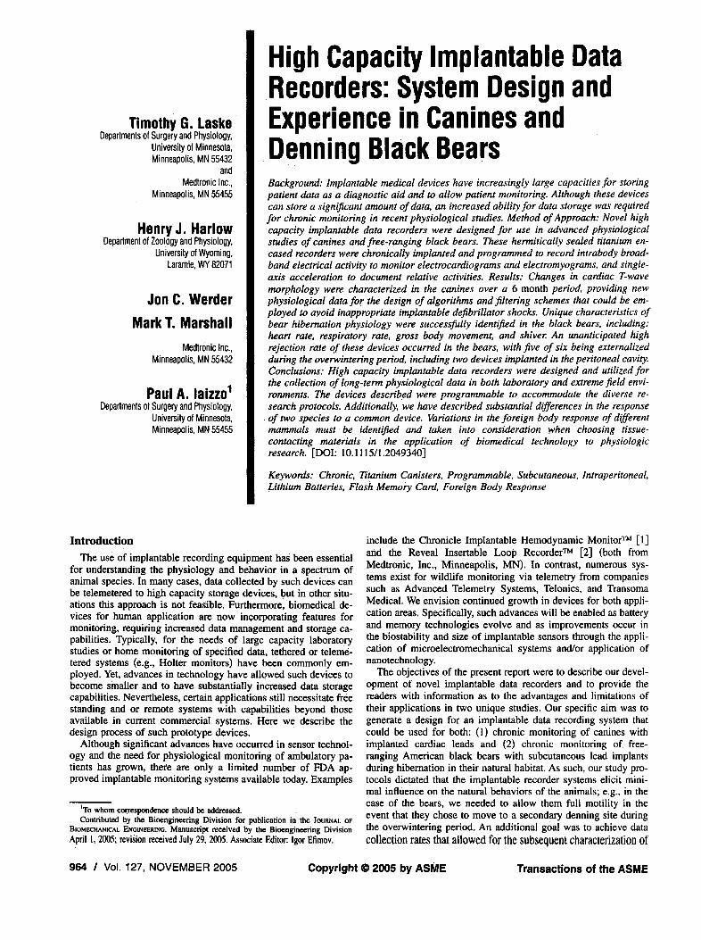
Implantable Device. The IDR was designed to record physi-ologic data over an extended period of time (up to 6 months)without the need for intermittent data retrieval. The implantablecanister was the heart of the system, containing all electronicsnecessary to record and store wave form data (see Fig. 2). In orderfor the system to continue to function in the highly corrosiveenvironment of a living organism, the electronics and powersource needed to be contained in a hermetically sealed enclosure.Specifically, two titanium shield halves, which were seam welded,formed this enclosure. These devices also required glassfeedthroughs, which permitted an electrical connection throughthe enclosure without compromising the hermeticity. A polyure-thane connector block was mechanically attached to each enclo-sure and the interface was sealed using silicone medical adhesive.A laser-etching of the "University of Wyoming" designator and acontact phone number onto the titanium housing, in case of loss(e.g., due to hunting or shed radio-collar) was added to the devicesfor the bear research.
Journal of Biomechanical Engineering NOVEMBER 2005, Vol. 127 / 965
Programmer "\
b Magr:letic Swilch I
~
Fig. 2 Exploded view of the implantable data recorder detail-ing the internal componentry. Within the outer titanium-alloycanister halves the following main components were con-tained: (1) compact-flash memory card (SanDisk@ 96 MbyteCompactFlashTM), (2) microcontroller circuit board with a mi-crocontroller (PIC16F877, Microchip Technology Inc., Chandler,AZ) and an accelerometer, (3) batteries (nominal 3.6 V, two AAtadiran lithium TL-5903, 2.4 AH), (4) a magnetic switch, and (5)a polyurethane connector block.
Electr.onics. Briefly, the electronics consisted of a microcon-troller circuit board, compact-flash memory card, magneticswitch, and batteries. Signals from the lead and programmingcable were routed through the header block and glassfeedthroughs to the circuit board (Figs. 2 and 3).
The primary function of these implantable devices was to col-lect and record sensor signals in a pre-programmed, easy-to-selectmanner. The devices were capable of recording two sensorsignals-broadband electrical potentials (EPs) from the submus-cular lead, and activity from a single-axis accelerometer mountedon the circuit board. Standard operating parameters are listed inTable 1.
As shown in the block diagram of Fig. 4, the generalized cir-cuitry consisted of four major subcomponents:
. Microcontroller (PIC16F877, Microchip Technology Inc.,Chandler, AZ): provides overall management and control ofthe implantable device, including programming, scheduling,wave form conversion, and wave form storage.
. Amplifiers: amplification and filtering for broadband electri-cal .and accelerometer input signals.
. Memory card (96 Mbyte CompactFlashTM, SanDisk Corpo-ration, Sunnyvale, CA): nonvolatile memory for storage ofwave form signals and parameters.
. Power Management: two lithium batteries (TL-5903, 2.4AH, nominal 3.6 V, Port Washington, NY), magnetic switchand circuitry to manage low power modes.
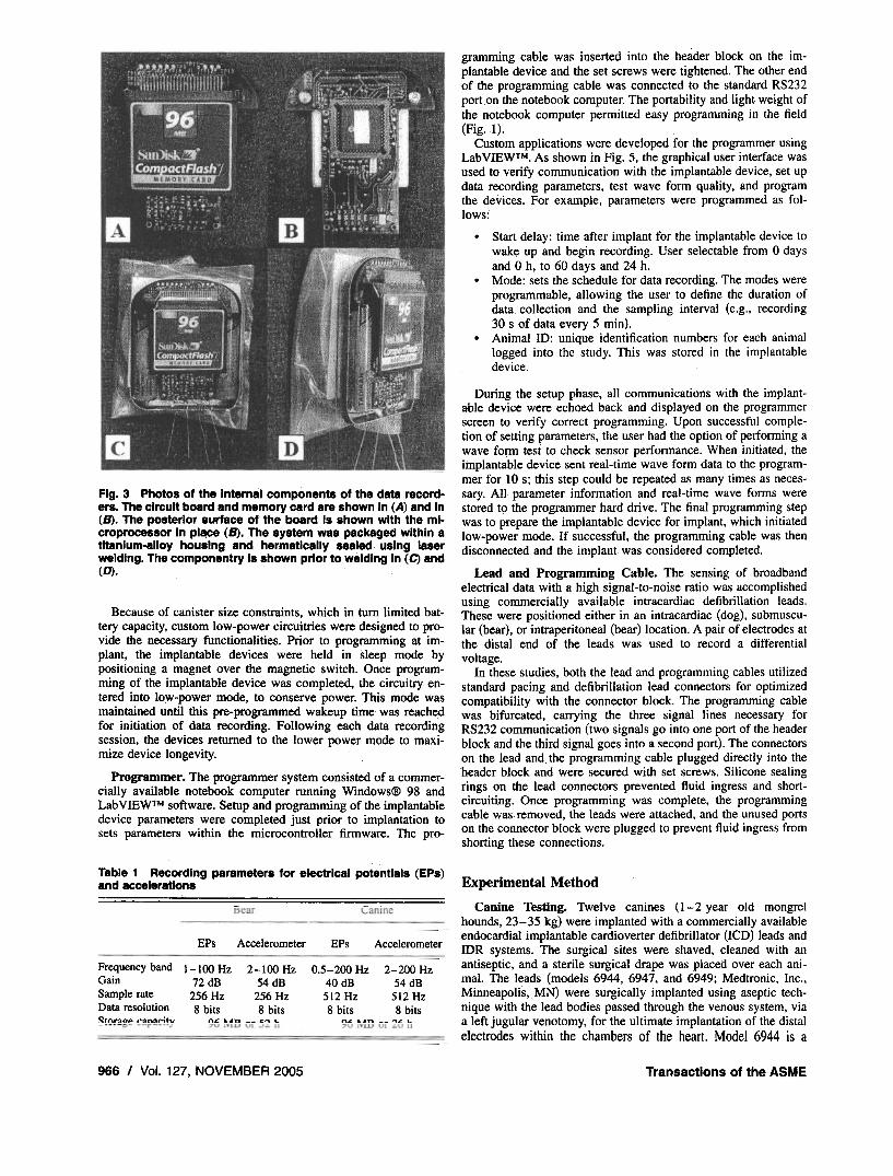
Fig. 3 Photos of the internal components of the data record-ers. The circuit board and memory card are shown in (A) and in(B). The posterior surface of the board is shown with the mi-croprocessor in place (B). The system was packaged within atitanium-alloy housing and hermetically sealed using laserwelding. The componentry is shown prior to welding in (C) and(D).
Because of canister size constraints, which in turn limited bat-tery capacity, custom low-power circuitries were designed to pro-vide the necessary functionalities. Prior to programming at im-plant, the implantable devices were held in sleep mode bypositioning a magnet over the magnetic switch. Once program-ming of the implantable device was completed, the circuitry en-tered into 10',V-power mode; to conserve power. This mode wasmaintained until this pre-programmed wakeup time was reachedfor initiation of data recording. Following each data recordingsession, the devices returned to the lower power mode to max.i-mize device longevity.
Programmer. The programmer system consisted of a commer-cially available notebook computer running Windows@ 98 andLabVIEWTM software. Setup and programming of the implantabledevice parameters were completed just prior to implantation tosets parameters within the microcontroller firmware. The pro-
Table 1 Recording parameters for electrical potentials (EPs)and accelerations
EPs Accelerometer
Frequency band 1-100 Hz 2-100 Hz 0.5-200 Hz 2-200 HzGain 72 dB 54 dB 40 dB 54 dBSample rate 256 Hz 256 Hz 512 Hz 512 HzData resolution 8 bits 8 bits 8 bits 8 bitsC;:tnr~o.. r~n~ritv n.c un -- c,", .. n.. un. -- '"'.. ..
966 I Vol. 127, NOVEMBER 2005 Transactions of the ASME
gramming cable was inserted into the header block on the im-plantable device and the set screws were tightened. The other endof the programming cable was connected to the standard RS232port on the notebook computer. The portability and light weight ofthe notebook computer permitted easy programming in the field(Fig. 1).
Custom applications were developed for the programmer usingLabVIEWTM. As shown in Fig. 5. the graphical user interface wasused to verify communication with the implantable device. set updata recording parameters, test wave form quality, and programthe devices. For example. parameters were programmed as fol-lows:
. Start delay: time after implant for the implantable device towake up and begin recording. User selectable from 0 daysand 0 h, to 60 days and 24 h.
. Mode: sets the schedule for data recording. The modes wereprogrammable, allowing the user to define the duration ofdata collection and the sampling interval (e.g., recording30 s of data every 5 min).
. Animal ID: unique identification numbers for each animallogged into the study. This was stored in the implantabledevice.
During the setup phase, all communications with the implant-able device were echoed back and displayed on the programmerscreen to verify correct programming. Upon successful comple-tion of setting parameters, the user had the option of performing awave form test to check sensor performance. When initiated, theimplantable device sent real-time wave form data to the program-mer for 10 s; this step could be repeated as many times as neces-sary. All parameter information and real-time wave forms werestored t9 the programmer hard drive. The final programming stepwas to prepare the implantable device for implant, which initiatedlow-power mode. If successful, the programming cable was thendisconnected and the implant was considered completed.
Lead and Programming Cable. The sensing of broadbandelectrical data with a high signal-to-noise ratio was accomplishedusing commercially available intracardiac defibrillation leads.These were positioned either in an intracardiac (dog), submuscu-lar (bear), or intraperitoneal (bear) location. A pair of electrodes atthe distal end of the leads was used to record a differentialvoltage.
In these studies, both the lead and programming cables utilizedstandard pacing and defibrillation lead connectors for optimizedcompatibility with the connector block. The programming cablewas bifurcated, carrying the three signal lines necessary forRS232 communication (two signals go into one port of the headerblock and the third signal goes into a second port). The connectorson the lead and the programming cable plugged directly into theheader block and Were secured with set screws. Silicone sealingrings on the lead connectors prevented fluid ingress and short-circuiting. Once programming was complete, the programmingcable was. removed, the leads were attached, and the unused portson the connector block were plugged to prevent fluid ingress fromshorting these connections.
Experimental Method
Canine Testing. Twelve canines (1-2 year old mongrelhounds, 23-35 kg) were implanted with a commercially availableendocardial implantable cardioverter defibrillator (ICD) leads andIDR systems. The surgical sites were shaved, cleaned with anantiseptic, and a sterile surgical drape was placed over each ani-mal. The leads (models 6944, 6947, and 6949; Medtronic, Inc.,Minneapolis, MN) were surgically implanted using aseptic tech-nique with the lead bodies passed through the venous system, viaa left jugular venotomy, for the ultimate implantation of the distalelectrodes within the chambers of the heart. Model 6944 is a
EPs Accelerometer

passive fixation lead that uses a hemispherical electrode in passivecontact with the endocardium and models 6947 and 6949 are ac-tive fixation leads that use a helical electrode screwed into themyocardium for fixation. The IDRs (n= 12) were positioned sub-cutaneously in the left superiolateral thoracic regions, with theleads tunneled superficially from the left jugular vein. The pocketfor the lOR was inferior to the incision site (see Fig. 6). The IORswere programmed to record both electrocardiographic signals andacceleration signals for 30 s every 90 min for the 6 month studyperiod. These parameters were chosen to maximize the utilizationof the battery capacity and flash memory for data collection.
Black Bear Testing. In two different seasons, the IDRs andsensing leads (model 6942; Medtronic, Inc., Minneapolis, MN)were surgically implanted using aseptic techniques in the field.
Fig. 5 Shown is the Interface page employed for programmingthe bear implantable data recorders in the field. This program,written using LabViewTM software, allows the user to set thedesired data collection parameters of each implanted system inthe field setting (e.g., in the case of the bears, in remote re-gions of the Rocky Mountains of Colorado and Wyoming). Thespecific start delay noted on this initial screen was shown at15 days, but in most cases was reprogrammed to 21, 30, or60 days.
Journal of Biomechanical Engineering
Fig. 4 Circuit diagram of the implantable data recorders
The surgical sites were shaved, cleaned with an antiseptic, and asterile surgical drape was placed over each animal. In the firstyear, devices were positioned in a subcutaneous pocket in eitherthe region of the pectoralis muscle or deltoid muscle (to avoidpotential disruption by cubs) with the leads tunneled towards theheart (Fig. 1). The pocket for the IDR was inferior to the incisionsite. The site was closed using a series of single knot stitchesspaced at 6-.8 mm using absorbable sutures (Ethicon Vicryl 2-D,Johnson & Johnson, Piscataway, NJ). Stitches were placed in bothsubdennal and dennal tissues. The sensing electrodes were se-cured with a single suture in a subcutaneous location so to lie overthe left pectoralis muscle near the sternum.
In the second season, the devices were surgically placed in theintraperitoneal cavity with the electrodes. positioned on the lineaalba in the proximity of the diaphragm using a single suture nearthe distal end of the lead. Next, the implanted leads were con-nected to the IDRs, and the signals received were viewed usingthe software interface to verify that adequate broadband electricaldata could be obtained: e.g., we positioned the leads so to obtainelectrocardiogram (ECG) signals and variations in the QRS am-plitude of these signals that correlated with a respiratory cycle (as
lOR
Fig. 6 Implant configuration used in the canine study. lOR= implanted data recorder; ICD = implantable cardioverterdefibrillator.
NOVEMBER 2005, Vol. 127 / 967
Week 1 - Low Actlvltv
Week 1 - High Activity Week 26 - HIQh ActlVltv
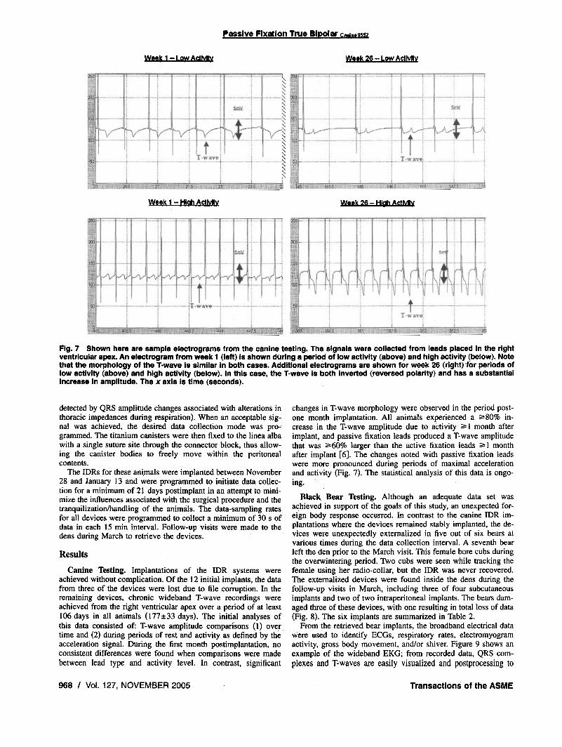
Fig. 7 Shown here are sample electrograms from the canine testing. The signals were collected from leads placed in the rightventricular apex. An electrogram from week 1 (left) is shown during a period of low activity (above) and high activity (below). Notethat the morphology of the T-wave is similar in both cases. Additional electrograms are shown for week 26 (right) for periods oflow activity (above) and high activity (below). In this case, the T-wave is both inverted (reversed polarity) and has a substantialincrease in amplitude. The x axis is time (seconds).
detected by QRS amplitude changes associated with alterations in .
thoracic impedances during respiration). When an acceptable sig-nal was achieved, the desired data collection mode was pro-grammed. The titanium canisters were then fixed to the linea albawith a single suture site through the connector block, thus allow-ing the canister bodies to freely move within the peritonealcontents.
The IDRs for these ani.mals were implanted between November28 and January 13 and were programmed to initiate data collec-tion for a minimum of 21 days postimplant in an attempt to mini-mize the influences associated with the surgical procedure and thetranquilizationlhandling of the animals. The data-sampling ratesfor all devices were programmed to collect a minimum of 30 s ofdata in each 15 min interval. Follow-up visits were made to thedens during March to retrieve the devices.
Results
Canine Testing. Implantations of the IDR systems wereachieved without complication. Of the 12 initial implants. the datafrom three of the devices were lost due to file corruption. In theremaining devices, chronic wideband T-wave recordings wereachieved from the right ventricular apex over a period of at least106 days in all animals (177:t33 days). The initial analyses ofthis data consisted of: T-wave amplitude comparisons (1) overtime and (2) during periods of rest and activity as defined by theacceleration signal. During the first month postimplantation, noconsistent differences were found when comparisons were madebetween lead type and activity level. In contrast, significant
968 I Vol. 127, NOVEMBER 2005 Transactions of the ASME
Passive Fixation True Bipolar C.";,,.9552
Week 26 - Low Actlv~v
changes in T-wave morphology were observed in the period post-one month implantation. All animals experienced a ;;'80% in-crease in the T-wave amplitude due to activity"" I month afterimplant, and passive fixation leads produced a T-wave amplitudethat was ""60% larger than the active fixation leads"" I monthafter implant [6]. The changes noted with passive fixation leadswere more pronounced during periods of maximal accelerationand activity (Fig. 7). The statistical analysis of this data is ongo-ing.
Black. Bear Testing. Although an adequate data set wasachieved in support of the goals of this study, an unexpected for-eign body response occurred. In contrast to the canine IDR im-plantations where the devices remained stably implanted. the de-vices were unexpectedly externalized in five out of six bears atvarious times during the data collection interval. A seventh bearleft the den prior to the March visit. This female bore cubs duringthe overwintering period. Two cubs were seen while tracking thefemale using her radio-collar. but the IDR was never recovered.The externalized devices were found inside the dens during thefollow-up visits in March. including three of four subcutaneousimplants and two of two intraperitoneal implants. The bears dam-aged three of these devices, with one resulting in total loss of data(Fig. 8). The six implants are summarized in Table 2.
From the retrieved bear implants, the broadband electrical datawere used to identify ECGs, respiratory rates, electromyogramactivity, gross body movement, and/or shiver. Figure 9 shows anexample of the wideband EKG; from recorded data, QRS com-plexes and T-waves are easily visualized and postprocessing to

Fig. B Photos of recovered data recorders from two differentimplants: bear 803 shown in panel (A), and bear 801 shown inpanel (B). In both cases these bears bit onto these titaniumcanisters with such force as to leave tooth impression on boththe anterior (left) and posterior surfaces (right).
minimize electromyographic influences was also employed. Therespiratory circle was identified by both the transient changes inheart rates (respiratory sinus arrhythmia) and the modulation ofthe QRS amplitudes, as a result of the variations in intrabodyimpedances associated with chest expansions and lung inflations[7,8]. An expanded view of one such prominent ECG signal isshown in Fig. 10 and an example of an associated accelerationsignal collected is shown in Fig. 11. The heart rates recorded were
Table 2 Summary of the data recorder performance from the bear research. (SubQ=subcutaneous; IP=intraperltoneal). The devices that were externalized are noted In the table.The implant duration is the minimum amount of time that the device was known to remainimplanted (= time of implant to last day of data collection). The devices were externalized at apoint postdata collection and prior to the March den visit.
Start of Externalized?Bear Implant data Implant (implant DataID date collection location duration) collected Comments
19-Dec-99
S-lan-DO
13-lan-DO
801
802
803
804
Journal of Biomechanical Engineering NOVEMBER 2005, Vol. 127 I 969
2010s0
Fig. 9 Subcutaneous electrical data for a 30 s period recordedwhile an American black bear (Ursus Americanus) was over-wintering in a den. The wideband signal is shown in the upperpanel and a filtered signal in the lower panel (third order But-terworth bandpass filter with cutoff frequency of 5-50 Hz). Apronounced respiratory sinus arrhythmia is apparent in this re-cording, which is a transient decrease in vagal tone followinginspiration that results in a temporary increase in heart rate.The amplitude of the ORS complex is modulated by the changein chest impedance due to inspiration and expiration. One res-piratory cycle is shown, with the higher heart rates occurringfollowing inspiration.
remarkably low, with one bear averaging 27.0%7.2 beats/minover a 68 day period (minimum recorded rate=4.6 beats/min).
Discussion
Here we describe two diverse applications of a novel high-capacity implantable data recorder system. For the specialized ap-plications described here, commercially available technologieswere not deemed adequate or practical. Although the IDRs al-lowed for successful completion of both studies, numerous oppor-tunities for improvements in the system design exist.
In the configuration of the systems we describe here, the datastorage capabilities were ultimately limited by the longevity of thelithium batteries. Memory cards with substantially greater storage
Cardiac signal not visible
Good signal quality
Good signal quality
Memory card notreadable~ .. . ".
]-Feb-OO
3] -Ian-DO
4-Feb-DO
Yes (>53 days)
No
9 days
16 days
22 daysYes (>44 days)
Yes (Unknown)

0
0
Fig. 10 Shown here is a single cardiac cycle recorded from asubcutaneous site while an American black bear (Ursus Ameri-canus) was overwintering in a den. The wideband signal isshown in the upper panel and a filtered signal in the lowerpanel (third order Butterworth bandpass filter with cutoff fre-quency of 5-50 Hz). One second of data is shown. Followingan isopotential period, a QRS cardiac wave form can be seen(cardiac depolarization) followed by a T-wave (cardiacrepolarization).
capabilities were available, but the memory could not have beenfilled since the current drain would have remained in the50-65 mA range. Recent advances in removable memory cardshave substantially increased both the memory storage capabilitiesand reduced the current required for writing to memory. For ex-ample, the Toshiba Model TH58NVGlS3A is a single 3.3 V2 Gbit NAND electrically erasable and programmable read-onlymemory that writes to memory at 10 mA. This would allow a 5X improvement in the amount of data that could be stored, but thebattery would remain the limiting factor. Future advances in bat-tery chemistries and/or rechargeable batteries will allow for de-vice downsizing, increased longevity, and/or increased data stor-age capability. In the current design, the devices have to be
Fig. 11 Shown here is an example of the recorded broadband electrical activity and the cor-responding acceleration from a system implanted In the peritoneal cavity of an American blackbear
970 I Vol. 127, NOVEMBER 2005 Transactions of the ASME
opened to access the memory card" whereas future systems couldalso employ wireless data transmission to allow reuse of the de-vices.
Although the minimum data required to complete the caninestudy objectives were recovered from the devices, data corruptiondid occur in three instances. Means of improving data storageintegrity should be employed in future generations of these re-corders. An analysis of the failed devices was performed and asolution that would most likely solve the problem would be tomonitor the battery voltage and shut down the entire device whenit drops below a threshold. This would require slight modificationsto both the hardware and firmware.
In the canine studies, we characterized precise changes in therepolarization patterns (T-waves) of the cardiac electrogram, so tosupport the development of new implantable defibrillators. Thisapplication required precise data collection intervals during each24 h period and we wanted to minimize handling of these studyanimals (including avoiding immobilization of the animal), sincethis in itself can impact the cardiac performance. In addition,sedatives and anesthetics typically used for data retrieval sessionsfor animal trials can also impact cardiac electrical performancesand/or hemodynamics. Unexpected changes were seen chronicallyin the T-wave amplitude and morphology. This finding is impor-tant because improper handling of the T-wave signals (specifi-cally, oversensing) by the sensing circuitry of implantabledefibrillators has been identified as a cause of unnecessary shocks[9]. Hence, these data provide new insights into the normal varia-tions that occur in the signal for use by device designers in futuresensing algorithms.
Although implantable pacemakers and/or defibrillators havebeen used for decades with nominal complications relative to thematerials and configurations used for the implanted canisters andleads [4,5], here we describe the unanticipated rejection of suchimplantable canisters by free-ranging black bears during overwin-tering. This rejection was attributed to a foreign body responseversus removal by the study animals since it typically occurredlate in the study period (>44 days in all cases) and in at least oneinstance was verified to have occurred through an adjacent nonin-cision site. The animals may have assisted the final externalizationas evidenced by the bite marks. This damage could also haveoccurred after the devices were naturally marsupialized. In allcases, substantial healing had already occurred when the animalswere examined in March.
The foreign body responses exhibited by the black bears, to ourknowledge, is unique compared to that which has been reported
1.0
1.0
for other large mammals. It should be noted that subsequent to ourfield studies, it was recently reported that a similar foreign bodyresponse was observed for subcutaneous implants placed in blackbear cubs by Echols [10]. It is intriguing that this rejection re-sponse and subsequent healing occurred during hibernation inthese adult bears, a time of reduced metabolic activity and hypo-thermia. [II] Although we observed a high rejection of the IDRs,these bears also had small intraperitoneal temperature recorders(StowAway@ TidbiTfM temperature loggers, Onset, Bourne, MA)which were, in most cases, not rejected. This could be due to themethod of implantation (freely floating in the peritoneal cavitywith a tether for retrieval) or due to the tissue contacting material(the devices were dipped in ethyl vinyl acetate). Although thetemperature loggers were successfully used in the peritoneal cav-ity, we recommend either avoiding subcutaneous implantation ofdevices in these animals or further work in screening materials forbiocompatibility, possibly through allergic screening. Work is on-going by our group to better understand and characterize this
I unique behavior, with the hope that it may have application topatients in which healing is impaired.
The utilization of these specially constructed implantable datarecorders provided valuable information in the two situations de-scribed. Chronic variations in canine T-wave morphologies fol-lowing the implantations of endocardial defibrillation leads wererecorded and analyzed. Normal physiological variations in the car-diac repolarization pattern (T-wave) were recorded, providing newdata in support of the design of future arrhythmia discriminationalgorithms. Although complicated by unanticipated foreign bodyresponses, new information was also gained about the physiologyof the hibernating black bear by utilizing such devices. The re-corded signals indicated the elicitation of dramatic respiratory si-nus arrhythmias and dramatically low heart and respiratory ratesduring the overwintering period.
In summary, the data recording systems described here haveproven utility for the collection of chronic physiological data,even in remote and extreme environments. This testing in animalpopulations served to identify issues. relating to the design thatshould be addressed prior to human clinical use. Additionally, wehave described substantial differences in the response of two spe-cies to a common device. Potential variations in the foreign body
Journal of Biomechanical Engineering NOVEMBER 2005, Vol. 127 I 971
responses of different mammals must be considered when choos-ing tissue-contacting materials in the application of biomedicaltechnology to physiologic research.
AcknowledgmentsThe authors would like to thank Randy M. Jensen, David L.
Carlson, and Jinback Hong from Medtronic for invaluable assis-tance with the construction, programming, and preliminary dataanalyses of these implantable data recorders. They would also liketo acknowledge Dr. Tom Lohuis and Mike Hooker for their helpwith the bear field studies; Bear Country USA for allowing themto perform pilot studies; and Monica Mahre for her assistance inpreparing this manuscript.
References[I] Compass-HF Study. Retrieved March 3D, 2005 from http://
wwwp.medtronic.com/NewsroomlNewsReleaseDetails.do?itemld= 111O237750252&lang=en_US.
[2] Reveal Plus: Information for health professionals.. Retrieved March 3D, 2005from http://www.medtronic.comlreveaUrevealplus.html.
[3] laizzo, P. A., 2005, "Emerging Cardiac Devices and Technologies," The Hand-book of Cardiac Ana/omy, Physiology and Devices, P. A. laizzo, ed., Humana~--- ~L_- ~,Press, Chap. 23.
[4] Byrd, C. L., 2000, "Management of Implant Complications," Clinical CardiacPacing and Defibrillation, 2nd ed., K. A. Ellenbogen, G. N. Kay, and B. L.Wilkoff, eds., WB. Saunders Co., Philadelphia, PA, pp. 669-694.
[5] Byrd, C. L., and Wilkoff, B. L., 2000, "Techniques and Devices for Extractionof Pacemaker and Implantable Cardioverter-Defibrillator Leads," Clinical Car-diac Pacing and Defibrillation, 2nd ed., K. A. Ellenbogen, G. N. Kay, and B.L. Wilkoff, eds., W.B. Saunders Co., Philadelphia, PA, pp. 695-709.
[6] Marshall, M. T., et aI., 2003, "ICD System T- Wave Changes Impact of Leadand Time," PACE, 26, p. 1100.
[7] Moody, G. B., Mark, R. G., Zoccola, A., and Mantero, S., 1985, "Derivation ofRespiratory Signals from Multi-Lead ECGs," Compu!. Cardiol., 12, pp. 113-116. .
[8] Hirsch, J. A., and Bishop, B., 1981, "Respiratory Sinus Arrhythmia in Hu-mans: How Breathing Pattern Modulates Heart Rate," Am. J. Physiol., 241,pp. H62(}"H629.
[9] Korte, T., KOditz, H., Niehaus, M., Paul, T., and Tebbenjohanns, J., 2004,"High Incidence of Appropriate and Inappropriate TCD Therapies in Childrenand Adolescents With Implantable Cardioverter Defibrillator," Pacing Clin.Electrnphysiol., 27(7), pp. 924-932.
[10] Echols, K. N., Vaughan, M. R., and Moll, H. D., 2004, "Evaluation of Subcu-taneous Tmplants for Monitoring American Black Bear Cub Survival," Ursus,IS, pp. 172-180.
[11] Harlow, H. J., Lohuis, T., Beck, T. D., and laizzo, P. A., 2001, "MuscleStrength in Overwintering Bears," Nature (London), 409, p. 997.