Embed Size (px)
Citation preview

Immunology 1998 93 281–288
High-affinity immunoglobulin E receptor (FceRI)-bearing eosinophils, mast cells,macrophages and Langerhans’ cells in allergen-induced late-phase cutaneous reactions
in atopic subjects
S. YING, L. T. BARATA, Q. MENG, J. A. GRANT,* J. BARKANS, S. R. DURHAM & A. B. KAYAllergy and Clinical Immunology, Imperial College School of Medicine, National Heart & Lung Institute, London, UK
SUMMARY
We have used in situ hybridization (ISH ) and immunohistochemistry (IHC) to investigate thekinetics of the expression for FceRI mRNA (a-, b- and c-chains), the a-chain protein product, aswell as the phenotype of the mRNA- or protein-positive cells in allergen-induced late-phase skinreactions in atopic subjects. Compared with diluent controls, there were significant increases inthe total numbers of mRNA+ cells for the a-, b-and c-chains for FceRI at all time-points (6, 24and 48 hr) after allergen challenge (P<0·01). By double IHC/ISH significant increases in a-, b-and c-chain mRNA+ macrophages, eosinophils, mast cells and CD1a+ cells were also observedafter allergen challenge (P<0·05). The distribution of FceRI subunit (a-, b-, or c-chain) mRNA+co-localization was CD68+ macrophages (42–47%), EG2+ eosinophils (33–39%), tryptase+ mastcells (5–11%) and CD1a+ Langerhans’ cells (2–4%). Using single IHC, significant increases inthe total number of FceRI protein+ cells (P<0·01) were observed 24 and 48 hr after allergenchallenge. Double IHC showed that the distribution of FceRI+ cells was tryptase+ mast cells(33%), CD68+ macrophages (36%), EG2+ eosinophils (20%), CD1a+ Langerhans’ cells (4%) andunidentified cells (7%), at the 24-hr allergen-challenged sites. These observations suggest that thecutaneous late-phase reaction in man is associated with up-regulation of FceRI on eosinophils,macrophages, mast cells and Langerhans’ cells.
INTRODUCTION we have studied first, the kinetics of expression of mRNA forthe a-, b- and c-chains of FceRI of the various cell-types
Injection of allergen into the dermis of sensitized atopicinvolved using a combination of in situ hybridization (ISH )
subjects is associated with an immediate weal and flare reactionand immunohistochemistry (IHC); second, the differences in
which is followed within hours by an oedematous, red andthe total number of FceRI protein+ cells after allergen chal-
slightly indurated late-phase response (LPR).1,2 The LPR islenge in the skin of atopic subjects; and third, the phenotype
dependent on the interaction of allergen with cells bearingof the various FceRI protein+ cells using double IHC.
surface-bound, allergen-specific immunoglobulin E (IgE).3,4High-affinity receptors for IgE (FceRI ) are traditionallyassociated with mast cells and basophils.5,6 More recently MATERIALS AND METHODShowever, FceRI has also been described on other cell types
Human subjectsincluding Langerhans’ cells, peripheral blood monocytes andAtopic subjects (n=12) were recruited from the Allergy Cliniceosinophils.7–11 Interaction of allergen with surface-bound IgEand staff of the Royal Brompton Hospital, London, UK.on these cells may result in the release of inflammatoryInclusion criteria were followed as previously described.16mediators.10,12,13 In addition, IgE-mediated allergen uptake byPatients taking any oral antihistamine or corticosteroidantigen-presenting cells may facilitate subsequent presentationmedication in the 2 weeks before the study began were notof allergen to allergen-specific T cells.14,15 For these reasons,included.
Received 14 August 1997; revised 30 October 1997; accepted Study design and processing of specimens30 October 1997. The study was approved by the Royal Brompton Hospital
Present address: *Division of Allergy and Immunology, University Ethics Committee and was performed with all subjects’ writtenof Texas Medical Branch, Galveston, TX 77 555-0762, USA. informed consent. Lyophilized extracts (Aquagen SQ) of either
Timothy grass pollen or house dust mite extract (ALK) wereCorrespondence: Professor A. B. Kay, Allergy & Clinicalreconstituted at 1500 biological units (BU )/ml in isotonicImmunology, Imperial College School of Medicine, National Heart &
Lung Institute, Dovehouse Street, London, SW3 6LY, UK. sterile saline diluent. Thirty biological units of either allergen
© 1998 Blackwell Science Ltd 281

S. Ying et al.282
extract (0·02 ml ) were injected intradermally into three sites with some modifications. Briefly, after blocking endogenousperoxidase in 3% hydrogen peroxide and pretreatment in 1%on the extensor aspect of the forearms of each subject. A
fourth site was injected with a similar volume of diluent. The bovine serum albumin, mouse anti-human phenotype (CD68,EG2, tryptase, CD1a, or CD3) monoclonal antibodies togethersize of the late response was measured at 6 hr, 24 hr and 48 hr
by evaluating skin induration.16 Biopsies were obtained at with rabbit anti-human FceRI a-subunit (997) were usedsimultaneously for the first layer. The second layer consistedeach time-point and were processed as previously described.16of a biotinylated goat anti-mouse antibody (Dako) together
In situ hybridization with a swine anti-rabbit antibody (alkaline phosphatase-The cDNA fragments encoding FceRI a [base pairs (bp) conjugated) (Dako). The third layer was streptavidin horse-25–936 ], b (bp 43–802) and c (bp 55–475) subunits were radish peroxidase (Amersham). Fast Blue and 3-amino-kindly provided by Dr J.-P. Kinet (Molecular Allergy and 9-ethylcarbazole (AEC; Vector Laboratories, Peterborough,Immunology Section, National Institute of Allergy and UK); these were sequentially used for the development of theInfectious Diseases, Rockville, MD).17–19 These cDNA frag- FceRI a-subunit and cell phenotypes. After development,ments of FceR subunits were inserted into the appropriate FceRI a-subunit-bearing cells stained blue and phenotypesvectors (pGEM) (Promega, Southampton, UK) and linearized stained red. Double-positive cells stained for both colours.to produce antisense and sense riboprobes. The 35S-labelled After developing, the sections were counterstained with methylriboprobes were prepared with SP6 or T7 RNA polymerases green. Whole sections were counted blindly by three indepen-(Promega) to generate anti-sense or sense probes, respectively. dent investigators, at high power magnification (×1000), withISH of the sections of skin biopsies were performed as an eyepiece graticule. The co-localization of 997 staining withdescribed previously.16 For each subunit of FceRI (the a-, b-, the tryptase, CD68, EG2 and CD1a phenotypic markersor c-chain), at least two sections from each biopsy were and the percentages of cells of each phenotype coexpressinghybridized, from which whole sections (10–19 fields per sec- FceRI a-subunit were then calculated.tion, each field=0·202 mm2) were counted blind in a codedrandom order by the two observers. The results are expressed Statistical analysisin the numbers of mRNA+ cells per mm2. Data were analysed using a statistical package (Minitab
Release 7, Minitab Inc., State College, PA). Variability ofSequential immunohistochemistry and in situ hybridization parameters studied was analysed with Friedman’s test, fol-To identify the cell types expressing mRNA for FceRI subunits, lowed by two-by-two comparisons between time points usingfrozen sections were first stained by the alkaline phosphatase– the Wilcoxon signed rank test. Correlation coefficients wereanti-alkaline phosphatase (APAAP) technique using mono- obtained by Spearman’s method with correction for tiedclonal antibodies against macrophages (CD68; Dako Ltd, values. For all tests, P-values less than 0·05 were consideredHigh Wycombe, UK), mast cells (anti-tryptase; Dako), eosino- significant.phils (EG2, Sanbio BV, Amsterdam, the Netherlands),Langerhans’ cells (anti-CD1a; Ortho Diagnostics, Bucks, UK)
RESULTSand T cells (CD3; Becton Dickinson, Mountain View, CA).After developing with Fast Red (Sigma, Poole, UK), FceRI Late-phase response and cell infiltrationa-, b-and c-chain-specific ISH were performed as mentioned
All subjects gave a cutaneous LPR after injection of theabove, incorporating the same negative controls. Using thisallergen but not after the diluent saline control (P<0·002).technique, the cellular markers stained red whereas cellsThe peak of the LPR was reached 24 hr after challenge [meanhybridizing with the antisense riboprobes for FceRI subunitssize (mm)±SEM=79·3±5·4], although there were no signifi-were identified by a dense collection of overlying silver grains.20cant differences between this and the 6 hr (70·5±4·7) or 48 hrThe results are expressed as the numbers of single-positive(71·6±5·8) time-points.(cell marker+ or mRNA+) and double-positive cells per mm2.
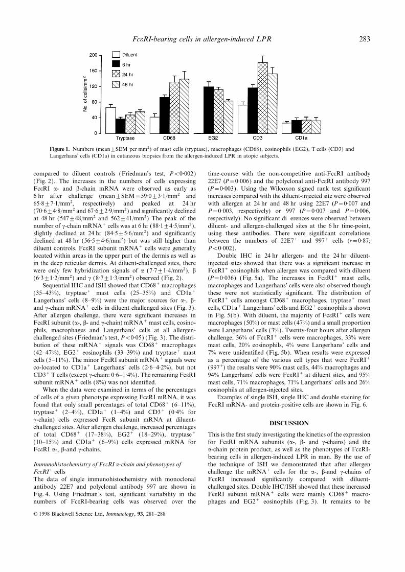
Using the technique of immunohistochemistry we con-firmed that after allergen challenge the numbers of infiltratingSingle immunohistochemistry for FceRIEG2+ eosinophils, CD3+ total T cells and CD68+ macro-FceRI-bearing cells were detected with the murine monoclonalphages were significantly elevated at 6, 24 and 48 hr time-antibodies 22E7 (a kind gift of Drs R. Chizzonite and J. P.points compared with 24 hr diluent challenge sites (Fig. 1)Kochan, Hoffman La Roche Inc., Nutley, NJ) directed against(Friedman’s test, P<0·05). The highest infiltration of thesethe a-chain of FceRI,21 using the APAAP method.1,2 Isotype-cell types was observed at 6, 24 and 48 hr, respectively, whichmatched monoclonal antibodies of irrelevant specificity andare consistent with our previous studies.1,2,16 After allergenomission of primary layer antibodies were included as negativechallenge, the number of tryptase+ mast cells was reducedcontrols. Positive cells stained red after development with Fastcompared with diluent controls, suggesting degranulation ofRed (Sigma).mast cells. There was no significant change in the number ofIHC was also performed using a rabbit polyclonal antibodyCD1a+ Langerhans’ cells at any time-points studied (Fig. 1).to the human FceRI a-subunit (997, a kind gift of Dr J.-P.
Kinet)22 by APAAP method. The positive cells within thewhole sections (see above) were counted and the results are Expression of FceRI mRNA and phenotypes of FceRI mRNA+expressed in the numbers of FceRI+ cells per mm2. cells
Using the technique of ISH, significant increases in theDouble immunohistochemistryTo examine the phenotype of FceRI a-subunit-bearing cells, numbers of cells expressing a-, b- and c-chain mRNA for
FceRI were observed at all time-points after challengecryostat sections were studied by double IHC as described23
© 1998 Blackwell Science Ltd, Immunology, 93, 281–288
FceRI-bearing cells in allergen-induced LPR 283
Figure 1. Numbers (mean±SEM per mm2) of mast cells (tryptase), macrophages (CD68), eosinophils (EG2), T cells (CD3) andLangerhans’ cells (CD1a) in cutaneous biopsies from the allergen-induced LPR in atopic subjects.
compared to diluent controls (Friedman’s test, P<0·002) time-course with the non-competitive anti-FceRI antibody22E7 (P=0·006) and the polyclonal anti-FceRI antibody 997(Fig. 2). The increases in the numbers of cells expressing
FceRI a- and b-chain mRNA were observed as early as (P=0·003). Using the Wilcoxon signed rank test significantincreases compared with the diluent-injected site were observed6 hr after challenge (mean±SEM=59·0±3·1/mm2 and
65·8±7·1/mm2, respectively) and peaked at 24 hr with allergen at 24 hr and 48 hr using 22E7 (P=0·007 andP=0·003, respectively) or 997 (P=0·007 and P=0·006,(70·6±4·8/mm2 and 67·6±2·9/mm2) and significantly declined
at 48 hr (547±48/mm2 and 562±41/mm2) The peak of the respectively). No significant differences were observed betweendiluent- and allergen-challenged sites at the 6 hr time-point,number of c-chain mRNA+ cells was at 6 hr (88·1±4·5/mm2),
slightly declined at 24 hr (84·5±5·6/mm2) and significantly using these antibodies. There were significant correlationsbetween the numbers of 22E7+ and 997+ cells (r=0·87;declined at 48 hr (56·5±4·6/mm2) but was still higher than
diluent controls. FceRI subunit mRNA+ cells were generally P<0·002).Double IHC in 24 hr allergen- and the 24 hr diluent-located within areas in the upper part of the dermis as well as
in the deep reticular dermis. At diluent-challenged sites, there injected sites showed that there was a significant increase inFceRI+ eosinophils when allergen was compared with diluentwere only few hybridization signals of a (7·7±1·4/mm2), b
(6·3±1·2/mm2) and c (8·7±1·3/mm2) observed (Fig. 2). (P=0·036) (Fig. 5a). The increases in FceRI+ mast cells,macrophages and Langerhans’ cells were also observed thoughSequential IHC and ISH showed that CD68+ macrophages
(35–43%), tryptase+ mast cells (25–35%) and CD1a+ these were not statistically significant. The distribution ofFceRI+ cells amongst CD68+ macrophages, tryptase+ mastLangerhans’ cells (8–9%) were the major sources for a-, b-
and c-chain mRNA+ cells in diluent challenged sites (Fig. 3). cells, CD1a+ Langerhans’ cells and EG2+ eosinophils is shownin Fig. 5(b). With diluent, the majority of FceRI+ cells wereAfter allergen challenge, there were significant increases in
FceRI subunit (a-, b- and c-chain) mRNA+ mast cells, eosino- macrophages (50%) or mast cells (47%) and a small proportionwere Langerhans’ cells (3%). Twenty-four hours after allergenphils, macrophages and Langerhans’ cells at all allergen-
challenged sites (Friedman’s test, P<0·05) (Fig. 3). The distri- challenge, 36% of FceRI+ cells were macrophages, 33% weremast cells, 20% eosinophils, 4% were Langerhans’ cells andbution of these mRNA+ signals was CD68+ macrophages
(42–47%), EG2+ eosinophils (33–39%) and tryptase+ mast 7% were unidentified (Fig. 5b). When results were expressedas a percentage of the various cell types that were FceRI+cells (5–11%). The minor FceRI subunit mRNA+ signals were
co-located to CD1a+ Langerhans’ cells (2·6–4·2%), but not (997+) the results were 90% mast cells, 44% macrophages and94% Langerhans’ cells were FceRI+ at diluent sites, and 95%CD3+ T cells (except c-chain: 0·6–1·4%). The remaining FceRI
subunit mRNA+ cells (8%) was not identified. mast cells, 71% macrophages, 71% Langerhans’ cells and 26%eosinophils at allergen-injected sites.When the data were examined in terms of the percentages
of cells of a given phenotype expressing FceRI mRNA, it was Examples of single ISH, single IHC and double staining forFceRI mRNA- and protein-positive cells are shown in Fig. 6.found that only small percentages of total CD68+ (6–11%),
tryptase+ (2–4%), CD1a+ (1–4%) and CD3+ (0·4% forc-chain) cells expressed FceR subunit mRNA at diluent-
DISCUSSIONchallenged sites. After allergen challenge, increased percentagesof total CD68+ (17–38%), EG2+ (18–29%), tryptase+ This is the first study investigating the kinetics of the expression
for FceRI mRNA subunits (a-, b- and c-chains) and the(10–15%) and CD1a+ (6–9%) cells expressed mRNA forFceRI a-, b-and c-chains. a-chain protein product, as well as the phenotypes of FceRI-
bearing cells in allergen-induced LPR in man. By the use ofthe technique of ISH we demonstrated that after allergenImmunohistochemistry of FceRI a-chain and phenotypes of
FceRI+ cells challenge the mRNA+ cells for the a-, b-and c-chains ofFceRI increased significantly compared with diluent-The data of single immunohistochemistry with monoclonal
antibody 22E7 and polyclonal antibody 997 are shown in challenged sites. Double IHC/ISH showed that these increasedFceRI subunit mRNA+ cells were mainly CD68+ macro-Fig. 4. Using Friedman’s test, significant variability in the
numbers of FceRI-bearing cells was observed over the phages and EG2+ eosinophils (Fig. 3). It remains to be
© 1998 Blackwell Science Ltd, Immunology, 93, 281–288
S. Ying et al.284
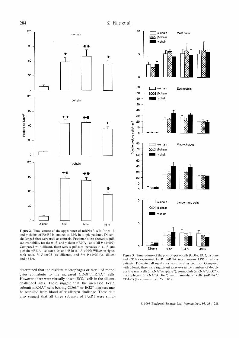
Figure 2. Time–course of the appearance of mRNA+ cells for a-, b-and c-chains of FceRI in cutaneous LPR in atopic patients. Diluent-challenged sites were used as controls. Friedman’s test showed signifi-cant variability for the a-, b- and c-chain mRNA+ cells (all P<0·002).Compared with diluent, there were significant increases in a-, b- andc-chain mRNA+ cells at 6, 24 and 48 hr (all P<0·02; Wilcoxon signedrank test). *: P<0·05 (vs. diluent), and **: P<0·05 (vs. diluent Figure 3. Time–course of the phenotypes of cells (CD68, EG2, tryptaseand 48 hr). and CD1a) expressing FceRI mRNA in cutaneous LPR in atopic
patients. Diluent-challenged sites were used as controls. Comparedwith diluent, there were significant increases in the numbers of double
determined that the resident macrophages or recruited mono- positive mast cells (mRNA+/tryptase+), eosinophils (mRNA+/EG2+),cytes contribute to the increased CD68+/mRNA+ cells. macrophages (mRNA+/CD68+) and Langerhans’ cells (mRNA+/However, there were virtually absent EG2+ cells in the diluent- CD1a+) (Friedman’s test, P<0·05).challenged sites. These suggest that the increased FceRIsubunit mRNA+ cells bearing CD68+ or EG2+ markers maybe recruited from blood after allergen challenge. These dataalso suggest that all three subunits of FceRI were simul-
© 1998 Blackwell Science Ltd, Immunology, 93, 281–288
FceRI-bearing cells in allergen-induced LPR 285
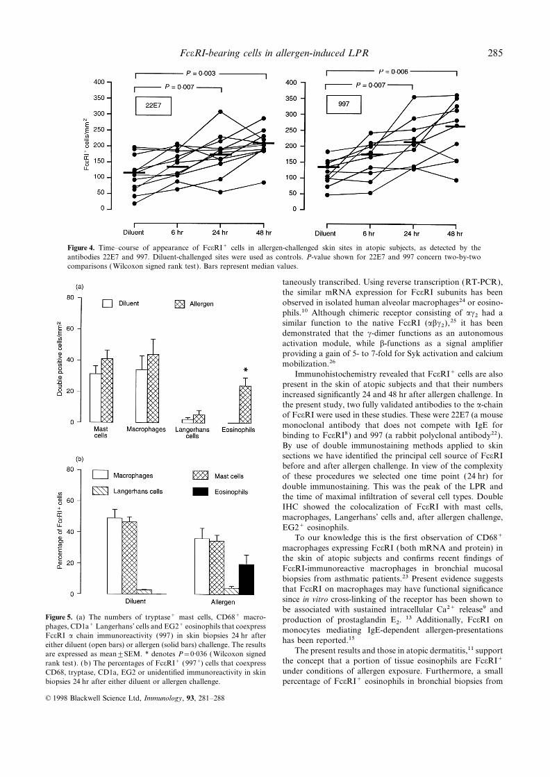
Figure 4. Time–course of appearance of FceRI+ cells in allergen-challenged skin sites in atopic subjects, as detected by theantibodies 22E7 and 997. Diluent-challenged sites were used as controls. P-value shown for 22E7 and 997 concern two-by-twocomparisons ( Wilcoxon signed rank test). Bars represent median values.
taneously transcribed. Using reverse transcription (RT-PCR),the similar mRNA expression for FceRI subunits has beenobserved in isolated human alveolar macrophages24 or eosino-phils.10 Although chimeric receptor consisting of ac2 had asimilar function to the native FceRI (abc2),25 it has beendemonstrated that the c-dimer functions as an autonomousactivation module, while b-functions as a signal amplifierproviding a gain of 5- to 7-fold for Syk activation and calciummobilization.26
Immunohistochemistry revealed that FceRI+ cells are alsopresent in the skin of atopic subjects and that their numbersincreased significantly 24 and 48 hr after allergen challenge. Inthe present study, two fully validated antibodies to the a-chainof FceRI were used in these studies. These were 22E7 (a mousemonoclonal antibody that does not compete with IgE forbinding to FceRI8) and 997 (a rabbit polyclonal antibody22).By use of double immunostaining methods applied to skinsections we have identified the principal cell source of FceRIbefore and after allergen challenge. In view of the complexityof these procedures we selected one time point (24 hr) fordouble immunostaining. This was the peak of the LPR andthe time of maximal infiltration of several cell types. DoubleIHC showed the colocalization of FceRI with mast cells,macrophages, Langerhans’ cells and, after allergen challenge,EG2+ eosinophils.
To our knowledge this is the first observation of CD68+macrophages expressing FceRI (both mRNA and protein) inthe skin of atopic subjects and confirms recent findings ofFceRI-immunoreactive macrophages in bronchial mucosalbiopsies from asthmatic patients.23 Present evidence suggeststhat FceRI on macrophages may have functional significancesince in vitro cross-linking of the receptor has been shown tobe associated with sustained intracellular Ca2+ release9 and
Figure 5. (a) The numbers of tryptase+ mast cells, CD68+ macro- production of prostaglandin E2. 13 Additionally, FceRI onphages, CD1a+ Langerhans’ cells and EG2+ eosinophils that coexpress monocytes mediating IgE-dependent allergen-presentationsFceRI a chain immunoreactivity (997) in skin biopsies 24 hr after
has been reported.15either diluent (open bars) or allergen (solid bars) challenge. The resultsThe present results and those in atopic dermatitis,11 supportare expressed as mean±SEM. * denotes P=0·036 ( Wilcoxon signed
the concept that a portion of tissue eosinophils are FceRI+rank test). (b) The percentages of FceRI+ (997+) cells that coexpressunder conditions of allergen exposure. Furthermore, a smallCD68, tryptase, CD1a, EG2 or unidentified immunoreactivity in skin
biopsies 24 hr after either diluent or allergen challenge. percentage of FceRI+ eosinophils in bronchial biopsies from
© 1998 Blackwell Science Ltd, Immunology, 93, 281–288

S. Ying et al.286
Figure 6. (a–j) Cryostat sections of skin biopsies from an atopic subject after either diluent or allergen intradermal challenge. (aand b) showing single ISH with FceRI a-chain anti-sense riboprobe in skin biopsy 24 hr after allergen challenge (a, bright field;B, dark field). (c and d) Single immunohistochemistry performed with the antibody 22E7 (c, FceRI+ cells in the skin, 24 hr afterdiluent challenge; d, FceRI+ cells, 24 hr after allergen challenge). (e–g) Double IHC and ISH with FceRI a-chain riboprobes. (e)CD68+/mRNA+, (f ) EG2+/mRNA+, (g) Tryptase+/mRNA+ double-positive cells (arrows) and single mRNA+ cells (arrowheads).(h–j) Showing double IHC performed with the antibody 997 in skin biopsies 24 hr after allergen challenge. (h) A tryptase+/FceRI+double-positive mast cell (arrow) and a single tryptase+/FceRI− cell (arrowhead) (i) EG2+/FceRI+ double-positive eosinophils(arrows) and FceRI a-chain single stained cell (arrowhead) and ( j) CD68+/FceRI+ double-positive macrophage (arrow).
© 1998 Blackwell Science Ltd, Immunology, 93, 281–288
FceRI-bearing cells in allergen-induced LPR 287
asthmatic subjects at baseline (unchallenged) as well as in and allergen uptake and presentation, since monocytes,Langerhans’ cells and eosinophils also express CD23.42,43nasal mucosa of atopic rhinitis were reported.23,27 However,
In summary, we have investigated the kinetics of expressionusing flow cytometry and monoclonal antibodies against theof mRNA and protein for FceRI in allergen-induced cutaneousFceRI a-chain, eosinophils from the peripheral blood of atopiclate-phase reaction. FceRI+ mast cells, macrophages andsubjects had negligible specific mean fluorescence.28,29 TheLangerhans’ cells are present in the skin of atopic subjectsmechanism involved in allergen-induced conversion of eosino-and that intradermal allergen challenge is associated withphils to FceRI+ cells is unknown but may be dependent onup-regulation of the expression for FceRI mRNA and proteininteractions with critical cytokines such as interleukin-3 (IL-3),product in the dermis. These results suggest that these FceRI-IL-4, IL-5, granulocyte-macrophage (GM-CSF) or IL-9·30 Abearing cells may be relevant to the pathophysiology of allergicrecent report has indeed shown that IL-4 can up-regulatetissue reactions in terms of mediator release and possiblyFceRI a-chain mRNA in human peripheral blood eosino-antigen presentation.phils.28 The significance of eosinophils expressing FceRI
remains to be determined. Eosinophils have been shown toACKNOWLEDGMENTSact as antigen-presenting cells in vitro.31 High levels of eosino-
phil-peroxidase can be detected in the supernatant of cultures The authors are grateful to Dr J.-P. Kinet for the kind gift of antibodyof eosinophils from patients with hypereosinophilic syndromes 997 and the cDNA for the FceRI subunits and to Dr R. Chizzoniteincubated with the anti-FceRI a-chain antibody.10 Such mech- and Dr J. P. Kochan for the kind gift of antibody 22E7. This workanisms might contribute to the marked degranulation of was supported by grants from the Medical Research Council and the
Wellcome Trust.eosinophils often observed in allergic inflammatory tissuereactions.1,32,33 Recently a positive correlation between thenumber of FceRI+ (997+) eosinophils (but not total eosino- REFERENCESphils) and the size of the 24 hr late-phase reaction had been
1. F A.J. & K A.B. (1988) The relationship between CD4+observed in allergen-induced cutaneous LPR.34 These obser- lymphocytes, activated eosinophils and the magnitude of thevations support a role for eosinophils in allergic tissue reactions allergen-induced late phase skin reaction in man. J Immunolwhich involves the high-affinity IgE receptor. On the other 141, 4158.hand, Kaneko et al. could not induce peripheral blood eosino- 2. G M., F A.J., V V. & K A.B. (1991) Eosinophil
activation and T lymphocyte activation in allergen-induced latephils isolated from atopic asthmatic patients to degranulate inphase reactions and classical delayed-type hypersensitivity in man.the presence of IgE.35 This stresses the need to obtain FceRI-J Immunol 147, 816.bearing eosinophils in free suspension in order to ascertain
3. D J., H F.E., C R., S K.J.,whether these cells can undergo IgE-dependent exocytosis.G J. & B J. (1973) Late cutaneous allergicOur present findings confirm previous studies of FceRI+responses in isolated IgE-dependent reactions. J Allergy Clin
Langerhans’ cells in the dermis36 and show that there was a Immunol 52, 38.moderate increase in the numbers of dermal FceRI-bearing 4. S G.O., G G.J., J R.E. & S A.L.CD1a+ Langerhans’ cells after allergen challenge (Fig. 3 and (1976) The late phase of the immediate wheal and flare skin
reaction: its dependence upon IgE antibodies. J Clin Invest 58, 408.Fig. 5). Again, it is not clear whether this represents increased5. K J.R.A., I C. & M H. (1974) Theexpression of the receptor by resident Langerhans’ cells or
interaction of IgE with rat basophilic leukemia cells. I. Evidencerecruitment of FceRI+ cells from blood, especially since allfor specific binding of IgE. J Exp Med 139, 600.dermal CD1a+ cells were located around blood vessels. Dermal
6. F A. (1980) The presence of two kinds of receptors for IgECD1a+ cells are extremely potent antigen-presenting cells37,38on rat mast cells. J Immunol 125, 981.
and may be relevant to the mechanism of the LPR, by 7. B T., L S H., W A. et al. (1992) Humaninteracting with allergen-specific T cells. epidermal Langerhans cells express the high affinity receptor for
We did not detect FceRI- (mRNA or protein) positive immunoglobulin E (FceRI). J Exp Med 175, 1285.T lymphocytes in the dermis after diluent or allergen challenge 8. W B., R A., K O. et al. (1992) Epidermal
Langerhans cells from normal human skin bind monomeric IgEalthough there were negligible c-chain mRNA co-localized tovia FceRI. J Exp Med 175, 1353.CD3+ cells. This confirms a previous report39 and suggests
9. M D., F E., R B. et al. (1994) Expressionthat although murine T-cell clones can express mRNA forof functional high affinity immunoglobulin E receptors (FceRI)FceRI a-, b-and c-chains, and bind IgE after incubation withon monocytes of atopic individuals. J Exp Med 179, 745.
IL-9 in vitro,39 T cells present in foci of allergic inflammation 10. S G A., L B., O K. et al. (1994) High-in vivo may not express sufficient numbers of FceRI on the affinity IgE receptor on eosinophils is involved in defence againstcell membrane to be detected by ISH and IHC. parasites. Nature 367, 183.
About 8% of the FceRI+ cells 24 hr post-challenge were 11. T Y., T M., M Y. et al. (1995) Highaffinity IgE receptor (FceRI) expression on eosinophils infiltratingunidentified. These may have been basophils, which havethe lesions and mite patch tested sites in atopic dermatitis. Archbeen detected in skin windows after allergen challenge.40Dermatol Res 287, 712.Confirmation or otherwise will require availability of a
12. R J.A., H M., M W., B M.K., Bbasophil-specific marker for immunohistochemical studies inJ.R. & A P.W. (1986) IgE-dependent release of leukotrienetissues.41C4 from alveolar macrophages. Nature 297, 329.
Although several IgE-dependent effects in allergic 13. T M., T Y., A S., Y H. & R C. (1995)inflammation may be mediated through interaction with High affinity IgE receptor-mediated prostaglandin E2 productionFceRI,+ this does not exclude a role for the low-affinity by monocytes in atopic dermatitis. Int Arch Allergy Immunol
108, 247.IgE receptor (FceRII, CD23) in terms of mediator release
© 1998 Blackwell Science Ltd, Immunology, 93, 281–288

S. Ying et al.288
14. M G.C., R F.C., B G.J, G G.C., FceRI a-chain messenger RNA in eosinophils. J Allergy ClinImmunol 96, 1161.B P.L. & B-K C.A.F.M. (1990)
Allergen presentation by epidermal Langerhans’ cells from patients 29. S B.S., K O.M., G J.A. & K A.B. (1997) Expressionof high affinity IgE receptors (FceRI) on peripheral blood baso-with atopic dermatitis is mediated by IgE. Immunology 69, 335.
15. M D., E C., R B. et al. (1995) The high phils, monocytes and eosinophils in atopic and non-atopicsubjects: relationship to total serum immunoglobulin E (IgE)affinity IgE receptor (FceRI) mediates IgE-dependent allergen
presentation. J Immunol 154, 6285. concentrations. J Allergy Clin Immunol 99, 699.30. W A.J., M R. & K A.B. (1996) Eosinophils:16. Y S., T-B L., M Q., H M. & K
A.B. (1995) The kinetics of allergen-induced transcription of biology and role in disease. Adv Immunol 60, 151.31. W P.F., R T.H., B T., E A., W D.T.W.messenger RNA for monocyte chemotactic protein-3 and
RANTES in the skin of human atopic subjects: relationship to & F R.W. (1993) Accessory cell function of human eosino-phils. HLA-DR-dependent, MHC-restricted antigen-presentationeosinophil, T cell, and macrophage recruitment. J Exp Med 181,
2153. and IL-1a expression. J Immunol 150, 2554.32. B R., P U., P D. et al. (1989) Major basic17. K J., P L., H J., K K. & K J.P. (1988)
Isolation of the gene coding for the alpha subunit of the human protein and eosinophil-derived neurotoxin concentrations in nasal-lavage fluid after antigen challenge: effect of systemic corticostero-high affinity receptor. Nucleic Acid Res 16, 3584.
18. K H., Z L., B A.T., MG D.W.J. & ids and relationship to eosinophil influx. J Allergy Clin Immunol84, 338.K J.P. (1992) The gene and cDNA for the human high affinity
immunoglobulin E receptor b chain and expression of the complete 33. B D.H., G G.J., C A.J. et al. (1991) Evidence ofongoing mast cell and eosinophil degranulation in symptomatichuman receptor. J Biol Chem 267, 1272.
19. K H., T H. & K J.P. (1990) Characterization asthma airway. J Allergy Clin Immunol 88, 637.34. B L.T., Y S., G J.A. et al. (1997) Allergen-inducedand expression of the gene for the human Fc receptor c subunit.
J Biol Chem 265, 6448. recruitment of FceRI+ eosinophils in human atopic skin. EurJ Immunol 27, 1236.20. Y S., M Q., B L.T. et al. (1996) Human eosinophils
express messenger RNA encoding RANTES and store and release 35. K M., S M.C., G G.J. & K H. (1995)Allergen-specific IgG1 and IgG3 through FccRII inducebiologically active RANTES protein. Eur J Immunol 26, 70
21. R F., H J., M M. et al. (1991) High affinity eosinophil degranulation. J Clin Invest 95, 2813.36. O B., R K., W B. et al. (1994)human IgE receptor (FceRI). Analysis of functional domains of
the a-subunit with monoclonal antibodies. J Biol Chem 266, 11 245. Immunomorphologic characterization of FceRI-bearing cellswithin the human dermis. J Invest Dermatol 102, 315.22. L O., S S., W-B J., R M.W.
& K J.P. (1995) Glycosylation of human truncated FceRI a 37. S-M C., M S., H A.S. & B
S.M. (1994) Antigen-presenting capacity in normal human dermischain is necessary for efficient folding in the endoplasmicreticulum. J Biol Chem 270, 8249. is mainly subserved by CD1a+ cells. Br J Dermatol 131, 15.
38. M L., G-R A. & C K.C. (1993)23. H M., G J.A., T-B L. et al. (1996) Highaffinity IgE receptor (FceRI) bearing cells in bronchial biopsies Heterogeneous populations of class II MHC+ in human dermal
cell suspensions. Identification of a small subset responsible forfrom atopic and non-atopic asthma. Am J Resp Crit Care Med153, 1931. potent dermal antigen-presenting cell activity with features
analogous to Langerhans cells. J Immunol 151, 4067.24. O K., K M., U K., M R., K
T. & T H. (1996) Expression of high-affinity IgE receptor 39. L J., K A., S J. & R J.C. (1995)IL-9 induces expression of granzymes and high-affinity IgE(FceRI) on human alveolar macrophages from atopic and non-
atopic patients. Int Arch Allergy Immunol 111, 55. receptor in murine T helper clones. J Immunol 154, 5061.40. C E.N., H A.F., S N.A., K-S25. R B., B G., K-F H. et al. (1996) Functional
contributions of the FceRIa and FceRIc subunit domains in A, N P.S. & L L.M. (1985) The cutaneous latephase response to allergen: mediator release and inflammatoryFceRI-mediated signaling in mast cells. J Immunol 156, 4876.
26. L S., C C., S A.M. & K J.P. (1996) The cell infiltration. J Clin Invest 83, 1519.41. K C.L., C S.S. & S L.B. (1995) IdentificationFceRIb subunit functions as an amplifier of FceRIc-mediated cell
activation signals. Cell 85, 985. and partial characterization of a unique marker for humanbasophils. J Immunol 154, 6548.27. R K., D S.R., O’B F. et al. (1997)
Enhanced expression of high affinity IgE receptor (FceRI) a chain 42. D G., S U., M D. et al. (1991) Expression,structure, and function of the CD23 antigen. Adv Immunol 49, 149.in human allergen-induced rhinitis with co-localization to mast
cells, macrophages, eosinophils, and dendritic cells. J Allergy Clin 43. D G., S M., W C.Y., F S. &L M. (1992) The low-affinity receptor for IgE. ImmunolImmunol 100, 78.
28. T N., K A., T Y. et al. (1995) IL-4 upregulates Rev 125, 77.
© 1998 Blackwell Science Ltd, Immunology, 93, 281–288